2013 traducción VR1genmol.blog.unq.edu.ar/wp-content/uploads/sites/33/2014...pylS gene encodes a...
34
1 Víctor Romanowski, 2013 traducción síntesis de proteínas (1) “Dogma central de la biología molecular” DNA RNA transcripción inversa transcripción y replicación de RNA replicación de DNA virus a DNA retrovirus y retroelementos virus a RNA transcripción traducción proteína plegamiento (folding), procesamiento, direccionamiento (targeting) procesamiento de RNA splicing, edición, modificación de nucleótidos y de extremos 5´y3´ Víctor Romanowski reparación recombinación
Transcript of 2013 traducción VR1genmol.blog.unq.edu.ar/wp-content/uploads/sites/33/2014...pylS gene encodes a...

1
Víctor Romanowski, 2013
traducción
síntesis de proteínas (1)
“Dogma central de la biología molecular”
DNA
RNA
transcripcióninversa
transcripción y replicación de RNA
replicación de DNA virus a DNA
retrovirus y retroelementos
virus a RNA
transcripción
traducción
proteínaplegamiento (folding), procesamiento, direccionamiento (targeting)
procesamiento de RNAsplicing, edición, modificación de nucleótidos y de extremos 5´y 3´
Víctor Romanowski
reparación recombinación

2

3
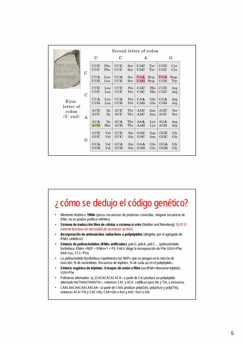
Código genético = diccionariopara traducir el lenguaje de ácidos nucleicos
al lenguaje de proteínas
• secuencia de tres nucleótidos = codón• lectura no superpuesta de codones• sin espacios ni signos de puntuación
3 nucleótidos = 1 codón = 1 aminoácido
El código genéticopuede ser leído en diferentes fases
o marcos (reading frames)
RNA
dsDNA

4
El código genético puede ser leído en diferentes fases o marcos(una secuencia nucleotídica puede ser traducida teóricamente a una
secuencia de aminoácidos, que depende de la fase de lectura)
La inserción o deleción de nucleótidoscambia el marco de lectura
5
¿cómo se dedujo el código genético?• Momento histórico: 1960s (pocas secuencias de proteínas conocidas, ninguna secuencia de
DNA, no se podían purificar mRNAs)• Sistema de traducción libre de células o sistema in vitro (Matthei and Nirenberg): OJO! El
sistema funciona sin necesidad de reconocer un AUG.• Incorporación de aminoácidos radiactivos a polipéptidos (dirigidos por el agregado de
RNAs sintéticos)• Síntesis de polinucleótidos (RNAs artificiales): poli-U, poli-A, poli-C… (polinucleótido
fosforilasa: RNAn +NDP = RNAn+1 + Pi). Poli-U dirige la incorporación de Phe (UUU=Phe, AAA=Lys, CCC=Pro)
• La polinucleótido fosoforilasa copolimeriza los NDPs que se pongan en la mezcla de reacción: % de nucleótidos, frecuencia de tripletes, % de cada aa en el polipéptido…
• Síntesis orgánica de tripletes. Ensayos de unión a filtro (aa-tRNA+ribosoma+triplete). UUU=Phe
• Polímeros alternados: ej. (CACACACACACA– a partir de CA) produce un polipéptido alternado HisThrHisThrHisThr--, entonces CAC y ACA codifican para His y Thr, o viceversa.
• CAACAACAACAACAACAA– (a partir de CAA) produce poly(Gln), poly(Asn) y poly(Thr), entonces ACA=Thr y CAC=His; CAA=Gln o Asn y AAC=Asn o Gln
6
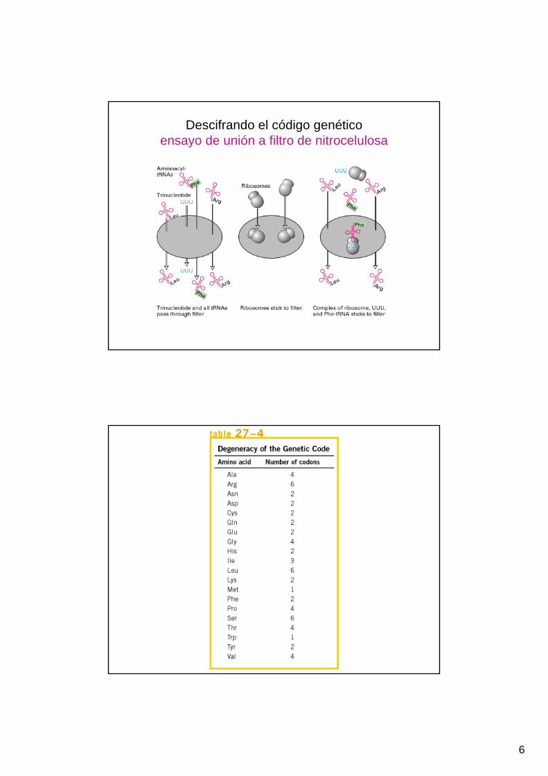
Descifrando el código genéticoensayo de unión a filtro de nitrocelulosa

7
una secuencia de aminoácidos (N→C)
varias secuencias de nucleótidos alternativas (5’→3’)
tRNA

8
Estructura de tRNAs

9
Prestar atención a
las polaridades
de los diferentes RNAs !
hay 61 codonespara aminoácidos
no hay 61 tRNAs !

10
tRNA
mRNA
un tRNA puede reconocer más de un codón

11
Activación del aminoácido
aminoacil-tRNA-sintetasa

12
Las aminoacil-tRNA sintetasas activanaminoácidos uniéndolos a tRNAs
Cada molécula de tRNAes reconocida por unaaminoacil-tRNA sintetasaespecífica
La traducción es un procesode decodificación en dos etapas
etapa 1 = aminoacil-tRNA-sintetasaetapa 2 = reconocimiento codón-anticodón
13
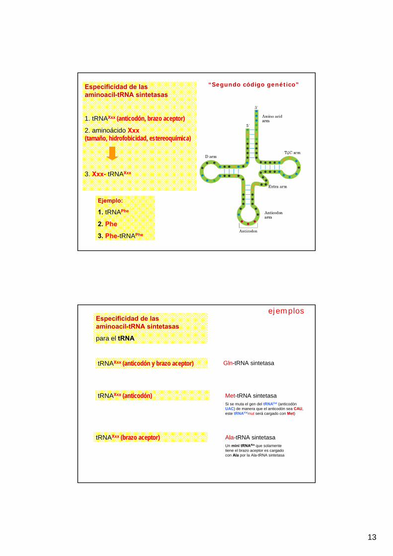
Especificidad de las aminoacil-tRNA sintetasas
1. tRNAXxx (anticodón, brazo aceptor)
2. aminoácido Xxx(tamaño, hidrofobicidad, estereoquímica)
3. Xxx- tRNAXxx
Ejemplo:
1. tRNAPhe
2. Phe
3. Phe-tRNAPhe
“Segundo código genético”
tRNAXxx (anticodón y brazo aceptor)
tRNAXxx (brazo aceptor)
tRNAXxx (anticodón)
Especificidad de las aminoacil-tRNA sintetasas
para el tRNA
Gln-tRNA sintetasa
Met-tRNA sintetasaSi se muta el gen del tRNAVal (anticodón UAC) de manera que el anticodón sea CAU, este tRNAValmut será cargado con Met)
Ala-tRNA sintetasaUn mini tRNAAla que solamente tiene el brazo aceptor es cargado con Ala por la Ala-tRNA sintetasa
ejemplos

14
tRNAAla Especificidad de la alanil - tRNA sintetasa
1. tRNAAla
2. aminoácido Ala
3. Ala - tRNAAla
Especificidad de las aminoacil-tRNA sintetasas
Para el aminoácido (tamaño, hidrofobicidad, estereoquímica)
Edición (proofreading) o corrección de errores
Tyr-tRNA sintetasa
Sitio activo de acilación muy específico, sin edición: Tyr
Thr-tRNA sintetasa
Sitio de acilación excluye aa de mayor tamañoSitio de acilación excluye aa de mayor hidrofobicidad (Val>Thr)Edición: Sitio de hidrólisis hidroliza aa-tRNA de menor tamaño (Ser<Thr)
Ile-tRNA sintetasa
Sitio de acilación no excluye aa de menor tamaño (Val)Edición: Sitio de hidrólisis hidroliza Val-AMP de menor tamaño (Val<Ile)Ile-AMP no entra en el sitio de hidrólisis; sigue hacia la formación de Ile-tRNA
La edición (proofreading) de las aminoacil tRNA sintetasasaumenta la fidelidad de la síntesis de proteínas
ejemplos
15
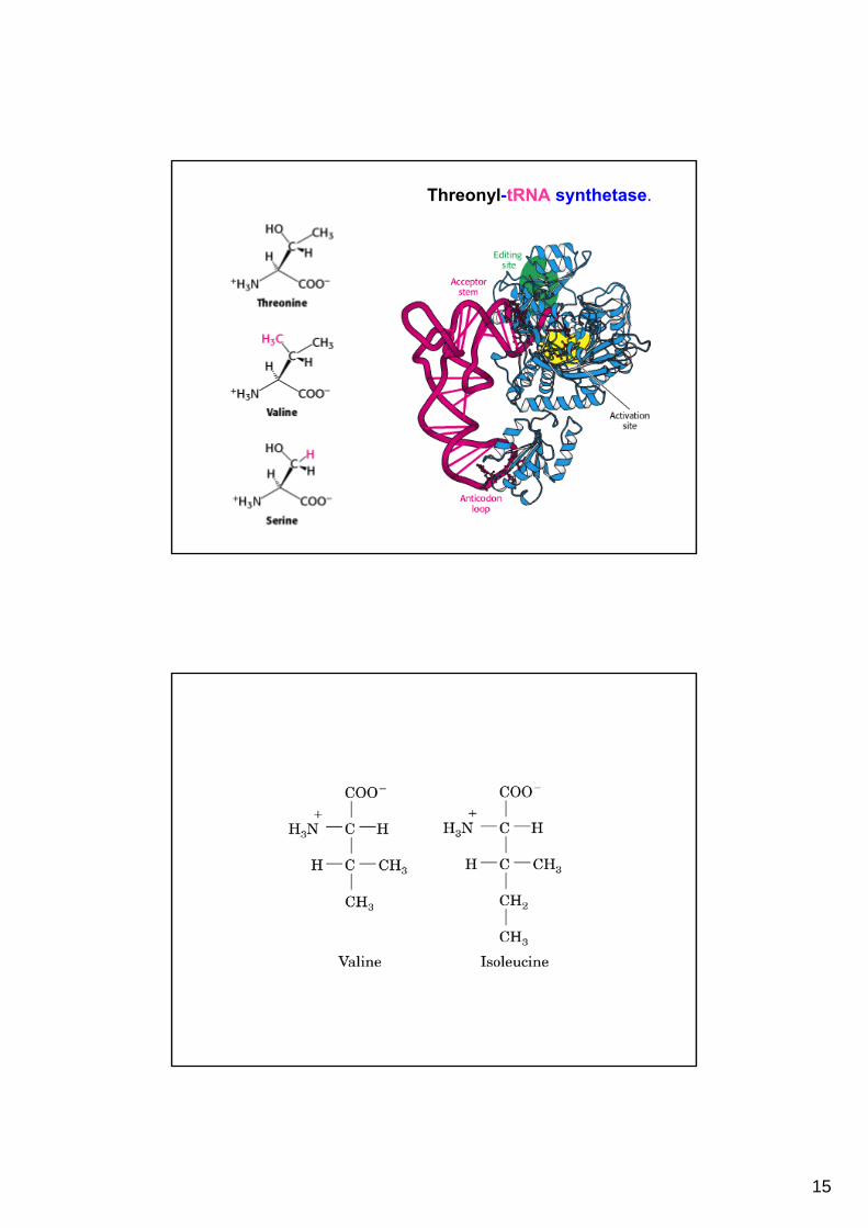
Threonyl-tRNA synthetase.

16
Código casi universal Código genético en mitocondrias
Más de 20 aminoácidos
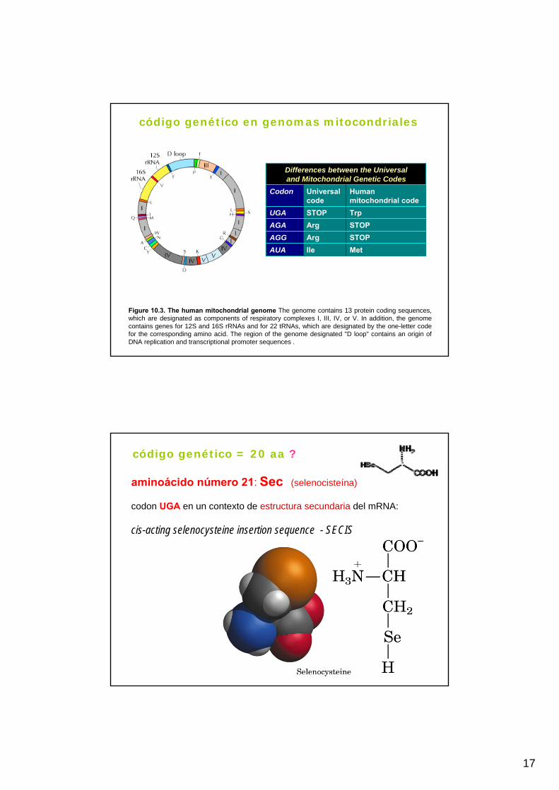
17
Figure 10.3. The human mitochondrial genome The genome contains 13 protein coding sequences, which are designated as components of respiratory complexes I, III, IV, or V. In addition, the genomecontains genes for 12S and 16S rRNAs and for 22 tRNAs, which are designated by the one-letter codefor the corresponding amino acid. The region of the genome designated "D loop" contains an origin ofDNA replication and transcriptional promoter sequences .
MetIleAUASTOPArgAGGSTOPArgAGATrpSTOPUGA
Human mitochondrial code
Universal code
Codon
Differences between the Universaland Mitochondrial Genetic Codes
código genético en genomas mitocondriales
aminoácido número 21: Sec (selenocisteína)
codon UGA en un contexto de estructura secundaria del mRNA:
cis-acting selenocysteine insertion sequence - SECIS
código genético = 20 aa ?

18
aminoácido número 22: Pyl Pirrolisina
(codon UAG en un contextode estructura secundaria del mRNA)
N6-[(2R,3R)-3-methyl-3,4-dihydro-2H-pyrrol-2-ylcarbonyl]-L-lysine
lysine—tRNA Pyl ligase
ATP + L-lysine + tRNA Pyl = AMP + PPi + L-lysyl-tRNAPyl
Science 24 May 2002: Vol. 296. no. 5572, pp. 1459 – 1462 Pyrrolysine Encoded by UAG in Archaea: Charging of a UAG-Decoding Specialized tRNAGayathri Srinivasan, Carey M. James, Joseph A. Krzycki* Pyrrolysine is a lysine derivative encoded by the UAG codon in methylamine methyltransferase genes of Methanosarcina barkeri. Near a methyltransferase gene cluster is the pylT gene, which encodes an unusual transfer RNA (tRNA) with a CUA anticodon. The adjacent pylS gene encodes a class II aminoacyl-tRNA synthetase that charges the pylT-derived tRNA with lysine but is not closely related to known lysyl-tRNA synthetases. Homologs of pylS and pylT are found in a Gram-positive bacterium. Charging a tRNACUA with lysine is a likely first step in translating UAG amber codons as pyrrolysine in certain methanogens. Our results indicate that pyrrolysine is the 22nd genetically encoded natural amino acid. Department of Microbiology, Ohio State University, Columbus, OH 43210, USA. e-mail: [email protected]
Traducción en etapas:iniciación, elongación, terminación
Activación de aminoácidos Plegamiento
19
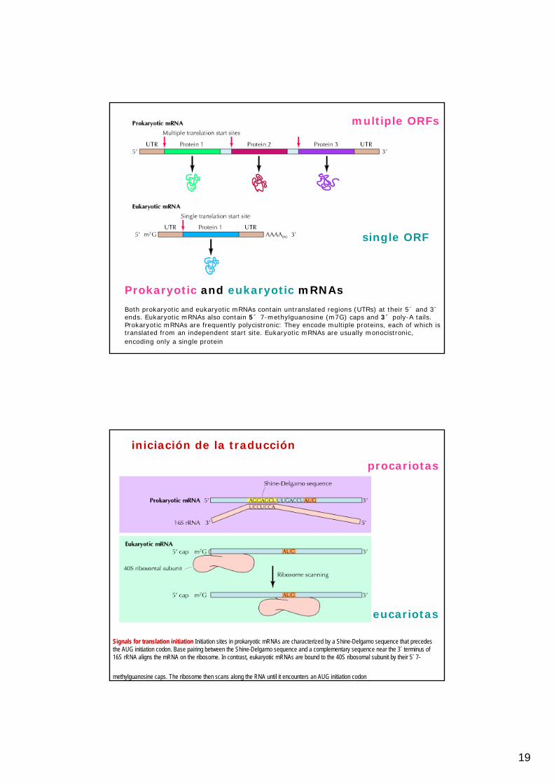
Prokaryotic and eukaryotic mRNAs
Both prokaryotic and eukaryotic mRNAs contain untranslated regions (UTRs) at their 5´ and 3´ends. Eukaryotic mRNAs also contain 5´ 7-methylguanosine (m7G) caps and 3´ poly-A tails. Prokaryotic mRNAs are frequently polycistronic: They encode multiple proteins, each of which istranslated from an independent start site. Eukaryotic mRNAs are usually monocistronic, encoding only a single protein
multiple ORFs
single ORF
Signals for translation initiation Initiation sites in prokaryotic mRNAs are characterized by a Shine-Delgarno sequence that precedes the AUG initiation codon. Base pairing between the Shine-Delgarno sequence and a complementary sequence near the 3´ terminus of 16S rRNA aligns the mRNA on the ribosome. In contrast, eukaryotic mRNAs are bound to the 40S ribosomal subunit by their 5´ 7-
methylguanosine caps. The ribosome then scans along the RNA until it encounters an AUG initiation codon
iniciación de la traducción
procariotas
eucariotas
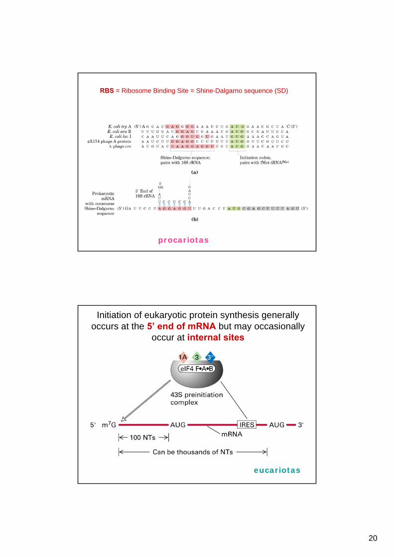
20
RBS = Ribosome Binding Site = Shine-Dalgarno sequence (SD)
procariotas
Initiation of eukaryotic protein synthesis generally occurs at the 5’ end of mRNA but may occasionally
occur at internal sites
eucariotas
21
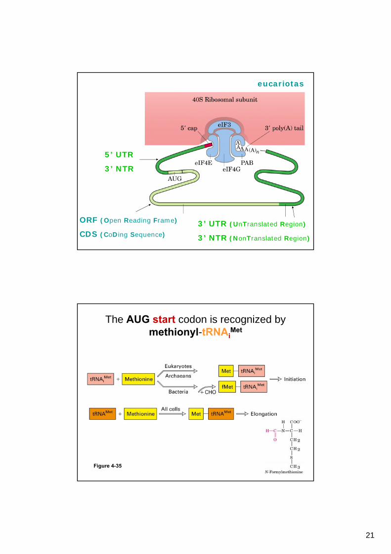
eucariotas
ORF (Open Reading Frame)
CDS (CoDing Sequence)
3’ UTR (UnTranslated Region)
3’ NTR (NonTranslated Region)
5’ UTR
3’ NTR
The AUG start codon is recognized by methionyl-tRNAi
Met
Figure 4-35

22
transcripción y traducciónen el mismo o en diferentes compartimientos
procariotas eucariotas
coupled transcription and translation

23
eRF-1, eRF-3RF-1, RF-2, RF-3Termination
eEF-1α, eEF-1βγ, eEF-2EF-Tu, EF-Ts, EF-GElongation
eIF-1, eIF-1A, eIF-2, eIF-2B, eIF-3, eIF-4A, eIF-4B, eIF-4E, eIF-4G, eIF-5
IF-1, IF-2, IF-3Initiation
EukaryotesProkaryotesRole
Translation Factors
Initiation of translation in bacteriaThree initiation factors (IF-1, IF-2, andIF-3) first bind to the 30S ribosomalsubunit. This step is followed by bindingof the mRNA and the initiator N-formylmethionyl (fMet) tRNA, which isrecognized by IF-2 bound to GTP. IF-3 isthen released, and a 50S subunit bindsto the complex, triggering the hydrolysisof bound GTP, followed by the release ofIF-1 and IF-2 bound to GDP
24
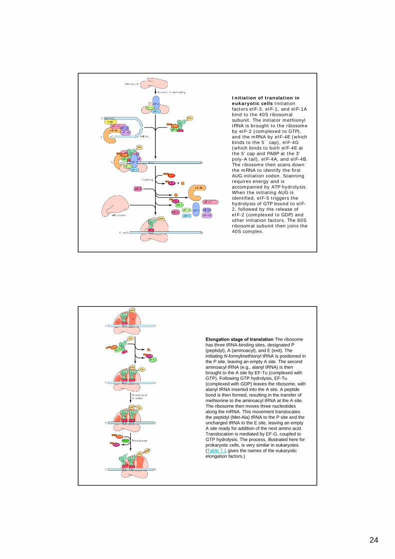
Initiation of translation in eukaryotic cells Initiationfactors eIF-3, eIF-1, and eIF-1A bind to the 40S ribosomalsubunit. The initiator methionyltRNA is brought to the ribosomeby eIF-2 (complexed to GTP), and the mRNA by eIF-4E (whichbinds to the 5´ cap), eIF-4G (which binds to both eIF-4E at the 5' cap and PABP at the 3' poly-A tail), eIF-4A, and eIF-4B. The ribosome then scans downthe mRNA to identify the firstAUG initiation codon. Scanningrequires energy and isaccompanied by ATP hydrolysis. When the initiating AUG isidentified, eIF-5 triggers thehydrolysis of GTP bound to eIF-2, followed by the release ofeIF-2 (complexed to GDP) andother initiation factors. The 60S ribosomal subunit then joins the40S complex.
Elongation stage of translation The ribosomehas three tRNA-binding sites, designated P (peptidyl), A (aminoacyl), and E (exit). Theinitiating N-formylmethionyl tRNA is positioned in the P site, leaving an empty A site. The secondaminoacyl tRNA (e.g., alanyl tRNA) is thenbrought to the A site by EF-Tu (complexed withGTP). Following GTP hydrolysis, EF-Tu (complexed with GDP) leaves the ribosome, withalanyl tRNA inserted into the A site. A peptidebond is then formed, resulting in the transfer ofmethionine to the aminoacyl tRNA at the A site. The ribosome then moves three nucleotidesalong the mRNA. This movement translocatesthe peptidyl (Met-Ala) tRNA to the P site and theuncharged tRNA to the E site, leaving an emptyA site ready for addition of the next amino acid. Translocation is mediated by EF-G, coupled toGTP hydrolysis. The process, illustrated here forprokaryotic cells, is very similar in eukaryotes. (Table 7.1 gives the names of the eukaryoticelongation factors.)

25
Image reconstruction of an E. coli ribosome
Transfer RNA-Binding Sites. (A) Three tRNA-binding sites are present on the 70S ribosome. They are called the A (for aminoacyl), P (for peptidyl), and E (for exit) sites. Each tRNA molecule contacts both the 30S and the 50S subunit. (B) The tRNAmolecules in sites A and P are base paired with mRNA

26
Regeneration of EF-Tu/GTP EF-Tu complexed to GTP escorts the aminoacyl tRNA to the ribosome. The boundGTP is hydrolyzed as the correct tRNA is inserted, so EF-Tu complexed to GDP is released. The EF-Tu/GDP complex isinactive and unable to bind another tRNA. In order fortranslation to continue, the active EF-Tu/GTP complexmust be regenerated by another factor, EF-Ts, whichstimulates the exchange of the bound GDP for free GTP.
Termination of translationA termination codon (e.g., UAA) at the A site isrecognized by a releasefactor rather than by a tRNA. The result is therelease of the completedpolypeptide chain, followedby the dissociation of tRNAand mRNA from theribosome.

27
Peptide-Bond Formation. The amino group of the aminoacyl-tRNAattacks the carbonyl group of the ester linkage of the peptidyl-tRNA to form a tetrahedral intermediate. This intermediate collapses to form the peptide bond and release the deacylated tRNA.
distintos antibióticos y toxinas inhiben la traducción
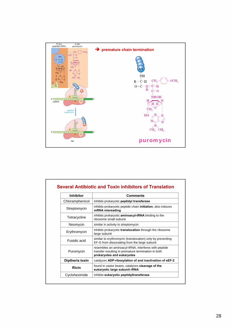
28
premature chain termination
puromycin
Several Antibiotic and Toxin inhibitors of Translation
Inhibitor CommentsChloramphenicol inhibits prokaryotic peptidyl transferase
Streptomycin inhibits prokaryotic peptide chain initiation, also induces mRNA misreading
Tetracycline inhibits prokaryotic aminoacyl-tRNA binding to the ribosome small subunit
Neomycin similar in activity to streptomycin
Erythromycin inhibits prokaryotic translocation through the ribosome large subunit
Fusidic acid similar to erythromycin (translocation) only by preventing EF-G from dissociating from the large subunit
Puromycinresembles an aminoacyl-tRNA, interferes with peptide transfer resulting in premature termination in both prokaryotes and eukaryotes
Diptheria toxin catalyzes ADP-ribosylation of and inactivation of eEF-2
Ricin found in castor beans, catalyzes cleavage of the eukaryotic large subunit rRNA
Cycloheximide inhibits eukaryotic peptidyltransferase
29
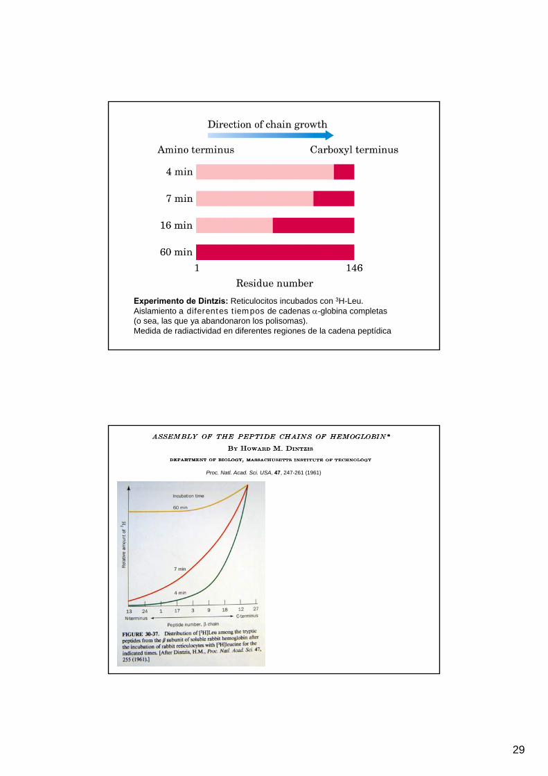
Experimento de Dintzis: Reticulocitos incubados con 3H-Leu.Aislamiento a diferentes tiempos de cadenas α-globina completas (o sea, las que ya abandonaron los polisomas).Medida de radiactividad en diferentes regiones de la cadena peptídica
Proc. Natl. Acad. Sci. USA, 47, 247-261 (1961)

30
polisomas

31
32
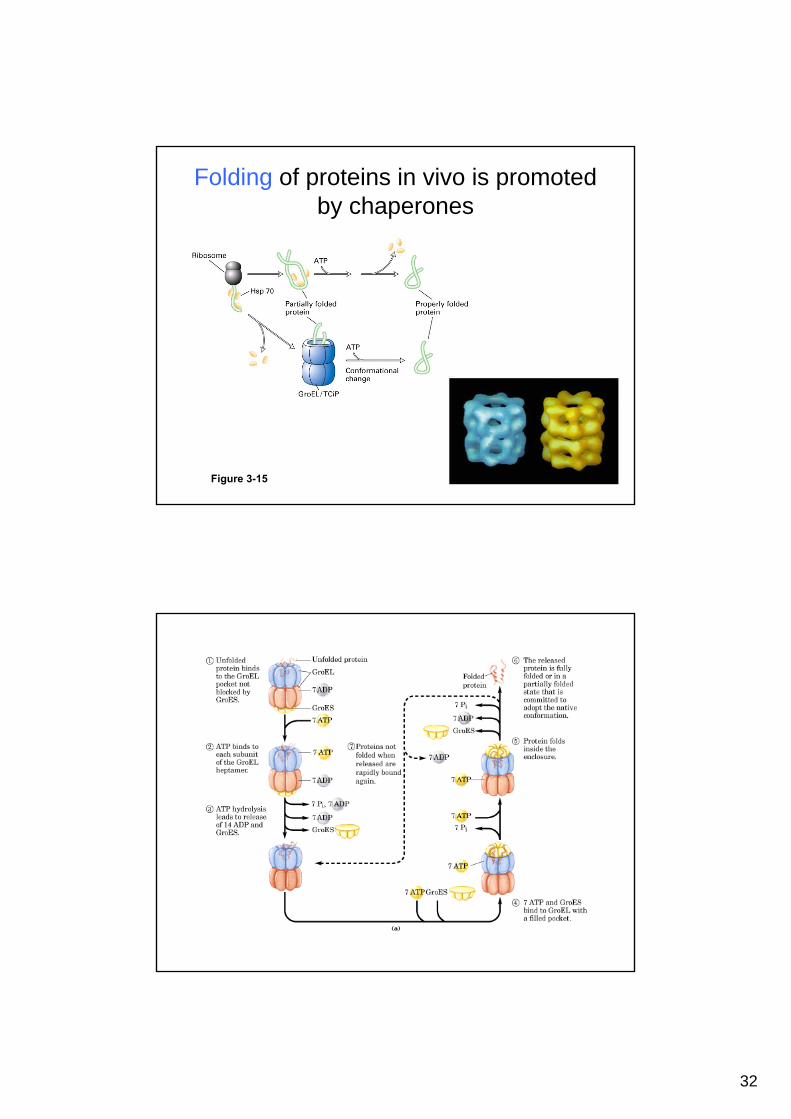
Folding of proteins in vivo is promoted by chaperones
Figure 3-15

33
Aberrantly folded proteins are implicated in slowly developing diseases
Figure 3-19
An amyloid plaque in Alzheimer’s disease is a tangle of protein filaments

34
Encefalitis espongiforme


![MANAGEMENT PERSPECTIVE · molar state (HHS) [1–45]. In 2005, DKA was responsible for approximately 120,000 hospital discharges in the USA, amounting to 7.4 dis-charges per 1000](https://static.fdocuments.es/doc/165x107/605e769b17f482168c6bc95b/management-perspective-molar-state-hhs-1a45-in-2005-dka-was-responsible.jpg)


![Genetica I TUE 2016 [Modo de compatibilidad]I+TUE+... · que incluye el “ cromosoma” y los plásmidos ... Microbiología de los microorganismos. 12os. 12 ... tRNA de formilmetionina](https://static.fdocuments.es/doc/165x107/5a7de83c7f8b9a49588df86a/genetica-i-tue-2016-modo-de-compatibilidad-itueque-incluye-el-cromosoma.jpg)