Activation of Autophagy by Metals in … of Autophagy by Metals in Chlamydomonas reinhardtii Marta...
10
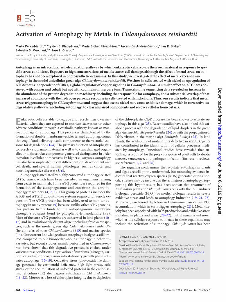
Activation of Autophagy by Metals in Chlamydomonas reinhardtii Marta Pérez-Martín, a Crysten E. Blaby-Haas, b María Esther Pérez-Pérez, a Ascensión Andrés-Garrido, a Ian K. Blaby, b Sabeeha S. Merchant, b,c José L. Crespo a Instituto de Bioquímica Vegetal y Fotosíntesis, Consejo Superior de Investigaciones Científicas (CSIC)-Universidad de Sevilla, Seville, Spain a ; Department of Chemistry and Biochemistry, University of California, Los Angeles, California, USA b ; Institute for Genomics and Proteomics, University of California, Los Angeles, California, USA c Autophagy is an intracellular self-degradation pathway by which eukaryotic cells recycle their own material in response to spe- cific stress conditions. Exposure to high concentrations of metals causes cell damage, although the effect of metal stress on au- tophagy has not been explored in photosynthetic organisms. In this study, we investigated the effect of metal excess on au- tophagy in the model unicellular green alga Chlamydomonas reinhardtii. We show in cells treated with nickel an upregulation of ATG8 that is independent of CRR1, a global regulator of copper signaling in Chlamydomonas. A similar effect on ATG8 was ob- served with copper and cobalt but not with cadmium or mercury ions. Transcriptome sequencing data revealed an increase in the abundance of the protein degradation machinery, including that responsible for autophagy, and a substantial overlap of that increased abundance with the hydrogen peroxide response in cells treated with nickel ions. Thus, our results indicate that metal stress triggers autophagy in Chlamydomonas and suggest that excess nickel may cause oxidative damage, which in turn activates degradative pathways, including autophagy, to clear impaired components and recover cellular homeostasis. E ukaryotic cells are able to degrade and recycle their own ma- terial when they are exposed to nutrient starvation or other adverse conditions through a catabolic pathway known as mac- roautophagy or autophagy. This process is characterized by the formation of double-membrane vesicles termed autophagosomes that engulf and deliver cytosolic components to the vacuole/lyso- some for degradation (1–4). The primary function of autophagy is to recycle cytoplasmic material as well as to clear damaged organ- elles or toxic cellular components generated during stress in order to maintain cellular homeostasis. In higher eukaryotes, autophagy has also been implicated in cell differentiation, development and cell death, and several human pathologies, such as cancer and neurodegenerative diseases (5, 6). Autophagy is mediated by highly conserved autophagy-related (ATG) genes, which have been described in organisms ranging from yeasts to mammals. Some ATG proteins are required for the formation of the autophagosome and constitute the core au- tophagy machinery (4, 7, 8). This group of proteins includes the ATG8 and ATG12 ubiquitin-like systems required for vesicle ex- pansion. The ATG8 protein has been widely used to monitor au- tophagy in many systems (9) because, unlike other ATG proteins, this protein firmly binds to the autophagosome membrane through a covalent bond to phosphatidylethanolamine (PE). Most of the core ATG proteins are conserved in land plants (10– 12) and in evolutionarily distant algae, including freshwater spe- cies, such as the model green alga Chlamydomonas reinhardtii (herein referred to as Chlamydomonas)(13) and marine species (14). Our current knowledge about autophagy in algae is still lim- ited compared to our knowledge about autophagy in other eu- karyotes, but recent studies, mainly performed in Chlamydomo- nas, have shown that this degradative process is elicited under various stress conditions. Deprivation of nutrients (nitrogen, car- bon, or sulfur) or progression into stationary growth phase acti- vates autophagy (15–19). Oxidative stress, photooxidative dam- age generated by carotenoid deficiency, high light stress, cold stress, or the accumulation of unfolded proteins in the endoplas- mic reticulum (ER) also triggers autophagy in Chlamydomonas (19–22). Moreover, a loss of chloroplast integrity due to depletion of the chloroplastic ClpP protease has been shown to activate au- tophagy in this alga (23). Recent studies have also linked this cat- abolic process with the degradation of lipid droplets in the green alga Auxenochlorella protothecoides (24) or with the propagation of DNA viruses in the marine alga Emiliania huxleyi (25). In land plants, the availability of mutant lines defective in key ATG genes has contributed to the identification of cellular processes medi- ated by autophagy. Functional studies have revealed that au- tophagy is required for the proper response of plant cells to abiotic stresses, senescence, and pathogen infection (for recent reviews, see references 1, 2, and 26). The signaling mechanisms that regulate autophagy in plants and algae are still poorly understood, but mounting evidence in- dicates that reactive oxygen species (ROS) generated during spe- cific stresses may be involved in the activation of autophagy. Sup- porting this hypothesis, it has been shown that treatment of Arabidopsis plants or Chlamydomonas cells with the ROS inducer hydrogen peroxide (H 2 O 2 ) or methyl viologen results in severe oxidative stress and leads to autophagy induction (19, 21, 27). Moreover, carotenoid depletion in Chlamydomonas causes ROS accumulation, which in turn triggers autophagy (21). Metal tox- icity has been associated with ROS production and oxidative stress signaling in plants and algae (28–32), but it remains unknown whether the cellular response to metals in these organisms may include the activation of autophagy. Chlamydomonas has been Received 3 May 2015 Accepted 5 July 2015 Accepted manuscript posted online 10 July 2015 Citation Pérez-Martín M, Blaby-Haas CE, Pérez-Pérez ME, Andrés-Garrido A, Blaby IK, Merchant SS, Crespo JL. 2015. Activation of autophagy by metals in Chlamydomonas reinhardtii. Eukaryot Cell 14:964 –973. doi:10.1128/EC.00081-15. Address correspondence to José L. Crespo, [email protected]. Supplemental material for this article may be found at http://dx.doi.org/10.1128 /EC.00081-15. Copyright © 2015, American Society for Microbiology. All Rights Reserved. doi:10.1128/EC.00081-15 964 ec.asm.org September 2015 Volume 14 Number 9 Eukaryotic Cell on June 6, 2019 by guest http://ec.asm.org/ Downloaded from
Transcript of Activation of Autophagy by Metals in … of Autophagy by Metals in Chlamydomonas reinhardtii Marta...
Activation of Autophagy by Metals in Chlamydomonas reinhardtii
Marta Pérez-Martín,a Crysten E. Blaby-Haas,b María Esther Pérez-Pérez,a Ascensión Andrés-Garrido,a Ian K. Blaby,b
Sabeeha S. Merchant,b,c José L. Crespoa
Instituto de Bioquímica Vegetal y Fotosíntesis, Consejo Superior de Investigaciones Científicas (CSIC)-Universidad de Sevilla, Seville, Spaina; Department of Chemistry andBiochemistry, University of California, Los Angeles, California, USAb; Institute for Genomics and Proteomics, University of California, Los Angeles, California, USAc
Autophagy is an intracellular self-degradation pathway by which eukaryotic cells recycle their own material in response to spe-cific stress conditions. Exposure to high concentrations of metals causes cell damage, although the effect of metal stress on au-tophagy has not been explored in photosynthetic organisms. In this study, we investigated the effect of metal excess on au-tophagy in the model unicellular green alga Chlamydomonas reinhardtii. We show in cells treated with nickel an upregulation ofATG8 that is independent of CRR1, a global regulator of copper signaling in Chlamydomonas. A similar effect on ATG8 was ob-served with copper and cobalt but not with cadmium or mercury ions. Transcriptome sequencing data revealed an increase inthe abundance of the protein degradation machinery, including that responsible for autophagy, and a substantial overlap of thatincreased abundance with the hydrogen peroxide response in cells treated with nickel ions. Thus, our results indicate that metalstress triggers autophagy in Chlamydomonas and suggest that excess nickel may cause oxidative damage, which in turn activatesdegradative pathways, including autophagy, to clear impaired components and recover cellular homeostasis.
Eukaryotic cells are able to degrade and recycle their own ma-terial when they are exposed to nutrient starvation or other
adverse conditions through a catabolic pathway known as mac-roautophagy or autophagy. This process is characterized by theformation of double-membrane vesicles termed autophagosomesthat engulf and deliver cytosolic components to the vacuole/lyso-some for degradation (1–4). The primary function of autophagy isto recycle cytoplasmic material as well as to clear damaged organ-elles or toxic cellular components generated during stress in orderto maintain cellular homeostasis. In higher eukaryotes, autophagyhas also been implicated in cell differentiation, development andcell death, and several human pathologies, such as cancer andneurodegenerative diseases (5, 6).
Autophagy is mediated by highly conserved autophagy-related(ATG) genes, which have been described in organisms rangingfrom yeasts to mammals. Some ATG proteins are required for theformation of the autophagosome and constitute the core au-tophagy machinery (4, 7, 8). This group of proteins includes theATG8 and ATG12 ubiquitin-like systems required for vesicle ex-pansion. The ATG8 protein has been widely used to monitor au-tophagy in many systems (9) because, unlike other ATG proteins,this protein firmly binds to the autophagosome membranethrough a covalent bond to phosphatidylethanolamine (PE).Most of the core ATG proteins are conserved in land plants (10–12) and in evolutionarily distant algae, including freshwater spe-cies, such as the model green alga Chlamydomonas reinhardtii(herein referred to as Chlamydomonas) (13) and marine species(14). Our current knowledge about autophagy in algae is still lim-ited compared to our knowledge about autophagy in other eu-karyotes, but recent studies, mainly performed in Chlamydomo-nas, have shown that this degradative process is elicited undervarious stress conditions. Deprivation of nutrients (nitrogen, car-bon, or sulfur) or progression into stationary growth phase acti-vates autophagy (15–19). Oxidative stress, photooxidative dam-age generated by carotenoid deficiency, high light stress, coldstress, or the accumulation of unfolded proteins in the endoplas-mic reticulum (ER) also triggers autophagy in Chlamydomonas(19–22). Moreover, a loss of chloroplast integrity due to depletion
of the chloroplastic ClpP protease has been shown to activate au-tophagy in this alga (23). Recent studies have also linked this cat-abolic process with the degradation of lipid droplets in the greenalga Auxenochlorella protothecoides (24) or with the propagation ofDNA viruses in the marine alga Emiliania huxleyi (25). In landplants, the availability of mutant lines defective in key ATG geneshas contributed to the identification of cellular processes medi-ated by autophagy. Functional studies have revealed that au-tophagy is required for the proper response of plant cells to abioticstresses, senescence, and pathogen infection (for recent reviews,see references 1, 2, and 26).
The signaling mechanisms that regulate autophagy in plantsand algae are still poorly understood, but mounting evidence in-dicates that reactive oxygen species (ROS) generated during spe-cific stresses may be involved in the activation of autophagy. Sup-porting this hypothesis, it has been shown that treatment ofArabidopsis plants or Chlamydomonas cells with the ROS inducerhydrogen peroxide (H2O2) or methyl viologen results in severeoxidative stress and leads to autophagy induction (19, 21, 27).Moreover, carotenoid depletion in Chlamydomonas causes ROSaccumulation, which in turn triggers autophagy (21). Metal tox-icity has been associated with ROS production and oxidative stresssignaling in plants and algae (28–32), but it remains unknownwhether the cellular response to metals in these organisms mayinclude the activation of autophagy. Chlamydomonas has been
Received 3 May 2015 Accepted 5 July 2015
Accepted manuscript posted online 10 July 2015
Citation Pérez-Martín M, Blaby-Haas CE, Pérez-Pérez ME, Andrés-Garrido A, BlabyIK, Merchant SS, Crespo JL. 2015. Activation of autophagy by metals inChlamydomonas reinhardtii. Eukaryot Cell 14:964 –973. doi:10.1128/EC.00081-15.
Address correspondence to José L. Crespo, [email protected].
Supplemental material for this article may be found at http://dx.doi.org/10.1128/EC.00081-15.
Copyright © 2015, American Society for Microbiology. All Rights Reserved.
doi:10.1128/EC.00081-15
964 ec.asm.org September 2015 Volume 14 Number 9Eukaryotic Cell
on June 6, 2019 by guesthttp://ec.asm
.org/D
ownloaded from
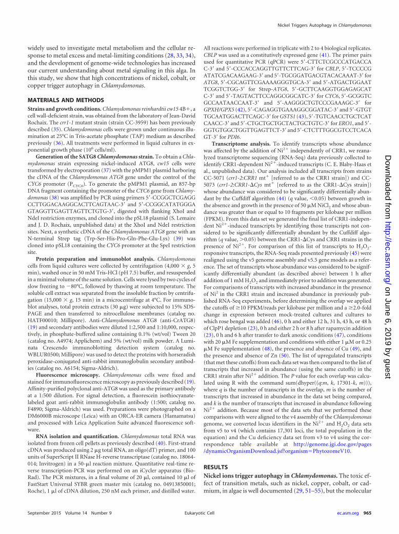
widely used to investigate metal metabolism and the cellular re-sponse to metal excess and metal-limiting conditions (28, 33, 34),and the development of genome-wide technologies has increasedour current understanding about metal signaling in this alga. Inthis study, we show that high concentrations of nickel, cobalt, orcopper trigger autophagy in Chlamydomonas.
MATERIALS AND METHODSStrains and growth conditions. Chlamydomonas reinhardtii cw15 4B�, acell wall-deficient strain, was obtained from the laboratory of Jean-DavidRochaix. The crr1-1 mutant strain (strain CC-3959) has been previouslydescribed (35). Chlamydomonas cells were grown under continuous illu-mination at 25°C in Tris-acetate phosphate (TAP) medium as describedpreviously (36). All treatments were performed in liquid cultures in ex-ponential growth phase (106 cells/ml).
Generation of the SATG8 Chlamydomonas strain. To obtain a Chla-mydomonas strain expressing nickel-induced ATG8, cw15 cells weretransformed by electroporation (37) with the pMPM1 plasmid harboringthe cDNA of the Chlamydomonas ATG8 gene under the control of theCYC6 promoter (PCYC6). To generate the pMPM1 plasmid, an 857-bpDNA fragment containing the promoter of the CYC6 gene from Chlamy-domonas (38) was amplified by PCR using primers 5=-CCGGCTCGAGGCCTTGGACAAGGCACTTCAGTAAC-3= and 5=-CCGGCATATGGGAGTAGGTTGAGTTAGTTCTGTG-3=, digested with flanking XhoI andNdeI restriction enzymes, and cloned into the pSL18 plasmid (S. Lemaireand J. D. Rochaix, unpublished data) at the XhoI and NdeI restrictionsites. Next, a synthetic cDNA of the Chlamydomonas ATG8 gene with anN-terminal Strep tag (Trp-Ser-His-Pro-Gln-Phe-Glu-Lys) (39) wascloned into pSL18 containing the CYC6 promoter at the SpeI restrictionsite.
Protein preparation and immunoblot analysis. Chlamydomonascells from liquid cultures were collected by centrifugation (4,000 � g, 5min), washed once in 50 mM Tris-HCl (pH 7.5) buffer, and resuspendedin a minimal volume of the same solution. Cells were lysed by two cycles ofslow freezing to �80°C, followed by thawing at room temperature. Thesoluble cell extract was separated from the insoluble fraction by centrifu-gation (15,000 � g, 15 min) in a microcentrifuge at 4°C. For immuno-blot analyses, total protein extracts (30 �g) were subjected to 15% SDS-PAGE and then transferred to nitrocellulose membranes (catalog no.HATF00010; Millipore). Anti-Chlamydomonas ATG8 (anti-CrATG8)(19) and secondary antibodies were diluted 1:2,500 and 1:10,000, respec-tively, in phosphate-buffered saline containing 0.1% (wt/vol) Tween 20(catalog no. A4974; Applichem) and 5% (wt/vol) milk powder. A Lumi-nata Crescendo immunoblotting detection system (catalog no.WBLUR0500; Millipore) was used to detect the proteins with horseradishperoxidase-conjugated anti-rabbit immunoglobulin secondary antibod-ies (catalog no. A6154; Sigma-Aldrich).
Fluorescence microscopy. Chlamydomonas cells were fixed andstained for immunofluorescence microscopy as previously described (19).Affinity-purified polyclonal anti-ATG8 was used as the primary antibodyat a 1:500 dilution. For signal detection, a fluorescein isothiocyanate-labeled goat anti-rabbit immunoglobulin antibody (1:500; catalog no.F4890; Sigma-Aldrich) was used. Preparations were photographed on aDM6000B microscope (Leica) with an ORCA-ER camera (Hamamatsu)and processed with Leica Application Suite advanced fluorescence soft-ware.
RNA isolation and quantification. Chlamydomonas total RNA wasisolated from frozen cell pellets as previously described (40). First-strandcDNA was produced using 2 �g total RNA, an oligo(dT) primer, and 100units of SuperScript II RNase H-reverse transcriptase (catalog no. 18064-014; Invitrogen) in a 50-�l reaction mixture. Quantitative real-time re-verse transcription-PCR was performed on an iCycler apparatus (Bio-Rad). The PCR mixtures, in a final volume of 20 �l, contained 10 �l ofFastStart Universal SYBR green master mix (catalog no. 04913850001;Roche), 1 �l of cDNA dilution, 250 nM each primer, and distilled water.
All reactions were performed in triplicate with 2 to 4 biological replicates.CBLP was used as a constitutively expressed gene (41). The primer pairsused for quantitative PCR (qPCR) were 5=-CTTCTCGCCCATGACCAC-3= and 5=-CCCACCAGGTTGTTCTTCAG-3= for CBLP, 5=-TCCCCGATATCGACAAGAAG-3= and 5=-TGCGGATGACGTACACAAAT-3= forATG8, 5=-CGCAGTTCGAAAAGGGTGCA-3= and 5=-ATGACTGGAATTCGGTCTGG-3= for Strep-ATG8, 5=-GCTTCAAGGTGGAGAGCATC-3= and 5=-TAGTACTTCCAGGCGGCATC-3= for CYC6, 5=-GCGGTCGCCAATAACCAAT-3= and 5=-AAGGGCTGTCCCGAAAGC-3= forGPXH/GPX5 (42), 5=-CAGAGGTGAAAGGCGGATAC-3= and 5=-GTGTTGCAATGGACTTCAGC-3= for GSTS1 (43), 5=-TGTCAACCTGCTCATCAACC-3= and 5=-CTGCTGCTGCTACTGCTGTC-3= for ERO1, and 5=-GGTGTGGCTGGTTGAGTTCT-3= and 5=-CTCTTTGGCGTCCTCACAGT-3= for PDI6.
Transcriptome analysis. To identify transcripts whose abundancewas affected by the addition of Ni2� independently of CRR1, we reana-lyzed transcriptome sequencing (RNA-Seq) data previously collected toidentify CRR1-dependent Ni2�-induced transcripts (C. E. Blaby-Haas etal., unpublished data). Our analysis included all transcripts from strainsCC-5071 (crr1-2:CRR1 mt� [referred to as the CRR1 strain]) and CC-5073 (crr1-2:CRR1-�Cys mt� [referred to as the CRR1-�Cys strain])whose abundance was considered to be significantly differentially abun-dant by the Cuffdiff algorithm (44) (q value, �0.05) between growth inthe absence and growth in the presence of 50 �M NiCl2 and whose abun-dance was greater than or equal to 10 fragments per kilobase per million(FPKM). From this data set we generated the final list of CRR1-indepen-dent Ni2�-induced transcripts by identifying those transcripts not con-sidered to be significantly differentially abundant by the Cuffdiff algo-rithm (q value, �0.05) between the CRR1-�Cys and CRR1 strains in thepresence of Ni2�. For comparison of this list of transcripts to H2O2-responsive transcripts, the RNA-Seq reads presented previously (45) wererealigned using the v5 genome assembly and v5.5 gene models as a refer-ence. The set of transcripts whose abundance was considered to be signif-icantly differentially abundant (as described above) between 1 h afteraddition of 1 mM H2O2 and immediately prior to addition was generated.For comparisons of transcripts with increased abundance in the presenceof Ni2 in the CRR1 strain and increased abundance in previously pub-lished RNA-Seq experiments, before determining the overlap we appliedthe cutoffs of �10 FPKM/reads per kilobase per million and a �2.0-foldchange in expression between mock-treated cultures and cultures towhich rose bengal was added (46), 0 h and either 12 h, 31 h, 43 h, or 48 hof ClpP1 depletion (23), 0 h and either 2 h or 8 h after rapamycin addition(23), 0 h and 6 h after transfer to dark anoxic conditions (47), conditionswith 20 �M Fe supplementation and conditions with either 1 �M or 0.25�M Fe supplementation (48), the presence and absence of Cu (49), andthe presence and absence of Zn (50). The list of upregulated transcripts(that met these cutoffs) from each data set was then compared to the list oftranscripts that increased in abundance (using the same cutoffs) in theCRR1 strain after Ni2� addition. The P value for each overlap was calcu-lated using R with the command sum(dhyper((q:m, k, 17301-k, m))),where q is the number of transcripts in the overlap, m is the number oftranscripts that increased in abundance in the data set being compared,and k is the number of transcripts that increased in abundance followingNi2� addition. Because most of the data sets that we performed thesecomparisons with were aligned to the v4 assembly of the Chlamydomonasgenome, we converted locus identifiers in the Ni2� and H2O2 data setsfrom v5 to v4 (which contains 17,301 loci, the total population in theequation) and the Cu deficiency data set from v3 to v4 using the cor-respondence table available at http://genome.jgi.doe.gov/pages/dynamicOrganismDownload.jsf?organismPhytozomeV10.
RESULTSNickel ions trigger autophagy in Chlamydomonas. The toxic ef-fect of transition metals, such as nickel, copper, cobalt, or cad-mium, in algae is well documented (29, 51–55), but the molecular
Nickel Triggers Autophagy in Chlamydomonas
September 2015 Volume 14 Number 9 ec.asm.org 965Eukaryotic Cell
on June 6, 2019 by guesthttp://ec.asm
.org/D
ownloaded from
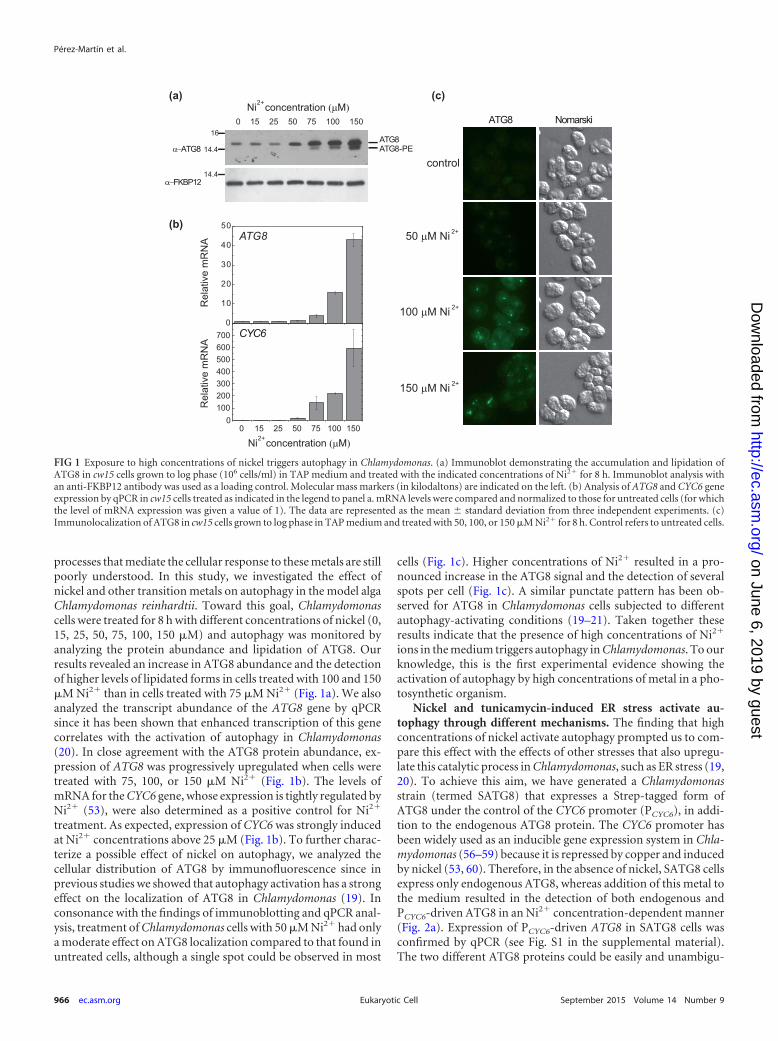
processes that mediate the cellular response to these metals are stillpoorly understood. In this study, we investigated the effect ofnickel and other transition metals on autophagy in the model algaChlamydomonas reinhardtii. Toward this goal, Chlamydomonascells were treated for 8 h with different concentrations of nickel (0,15, 25, 50, 75, 100, 150 �M) and autophagy was monitored byanalyzing the protein abundance and lipidation of ATG8. Ourresults revealed an increase in ATG8 abundance and the detectionof higher levels of lipidated forms in cells treated with 100 and 150�M Ni2� than in cells treated with 75 �M Ni2� (Fig. 1a). We alsoanalyzed the transcript abundance of the ATG8 gene by qPCRsince it has been shown that enhanced transcription of this genecorrelates with the activation of autophagy in Chlamydomonas(20). In close agreement with the ATG8 protein abundance, ex-pression of ATG8 was progressively upregulated when cells weretreated with 75, 100, or 150 �M Ni2� (Fig. 1b). The levels ofmRNA for the CYC6 gene, whose expression is tightly regulated byNi2� (53), were also determined as a positive control for Ni2�
treatment. As expected, expression of CYC6 was strongly inducedat Ni2� concentrations above 25 �M (Fig. 1b). To further charac-terize a possible effect of nickel on autophagy, we analyzed thecellular distribution of ATG8 by immunofluorescence since inprevious studies we showed that autophagy activation has a strongeffect on the localization of ATG8 in Chlamydomonas (19). Inconsonance with the findings of immunoblotting and qPCR anal-ysis, treatment of Chlamydomonas cells with 50 �M Ni2� had onlya moderate effect on ATG8 localization compared to that found inuntreated cells, although a single spot could be observed in most
cells (Fig. 1c). Higher concentrations of Ni2� resulted in a pro-nounced increase in the ATG8 signal and the detection of severalspots per cell (Fig. 1c). A similar punctate pattern has been ob-served for ATG8 in Chlamydomonas cells subjected to differentautophagy-activating conditions (19–21). Taken together theseresults indicate that the presence of high concentrations of Ni2�
ions in the medium triggers autophagy in Chlamydomonas. To ourknowledge, this is the first experimental evidence showing theactivation of autophagy by high concentrations of metal in a pho-tosynthetic organism.
Nickel and tunicamycin-induced ER stress activate au-tophagy through different mechanisms. The finding that highconcentrations of nickel activate autophagy prompted us to com-pare this effect with the effects of other stresses that also upregu-late this catalytic process in Chlamydomonas, such as ER stress (19,20). To achieve this aim, we have generated a Chlamydomonasstrain (termed SATG8) that expresses a Strep-tagged form ofATG8 under the control of the CYC6 promoter (PCYC6), in addi-tion to the endogenous ATG8 protein. The CYC6 promoter hasbeen widely used as an inducible gene expression system in Chla-mydomonas (56–59) because it is repressed by copper and inducedby nickel (53, 60). Therefore, in the absence of nickel, SATG8 cellsexpress only endogenous ATG8, whereas addition of this metal tothe medium resulted in the detection of both endogenous andPCYC6-driven ATG8 in an Ni2� concentration-dependent manner(Fig. 2a). Expression of PCYC6-driven ATG8 in SATG8 cells wasconfirmed by qPCR (see Fig. S1 in the supplemental material).The two different ATG8 proteins could be easily and unambigu-
(b)
0 15 25 50 75 100 150
α−ATG8
Ni concentration 2+ (μM)(a)
Rel
ativ
e m
RN
A
0
10
20
30
40
50ATG8
CYC6
00 15 25 50 75 100 150
Ni concentration 2+ (μM)
Rel
ativ
e m
RN
A
(c)
100200300400500600700
α−FKBP12
ATG8ATG8-PE
ATG8 Nomarski
control
50 μM Ni 2+
100 μM Ni 2+
150 μM Ni 2+
16
14.4
14.4
FIG 1 Exposure to high concentrations of nickel triggers autophagy in Chlamydomonas. (a) Immunoblot demonstrating the accumulation and lipidation ofATG8 in cw15 cells grown to log phase (106 cells/ml) in TAP medium and treated with the indicated concentrations of Ni2� for 8 h. Immunoblot analysis withan anti-FKBP12 antibody was used as a loading control. Molecular mass markers (in kilodaltons) are indicated on the left. (b) Analysis of ATG8 and CYC6 geneexpression by qPCR in cw15 cells treated as indicated in the legend to panel a. mRNA levels were compared and normalized to those for untreated cells (for whichthe level of mRNA expression was given a value of 1). The data are represented as the mean standard deviation from three independent experiments. (c)Immunolocalization of ATG8 in cw15 cells grown to log phase in TAP medium and treated with 50, 100, or 150 �M Ni2� for 8 h. Control refers to untreated cells.
Pérez-Martín et al.
966 ec.asm.org September 2015 Volume 14 Number 9Eukaryotic Cell
on June 6, 2019 by guesthttp://ec.asm
.org/D
ownloaded from
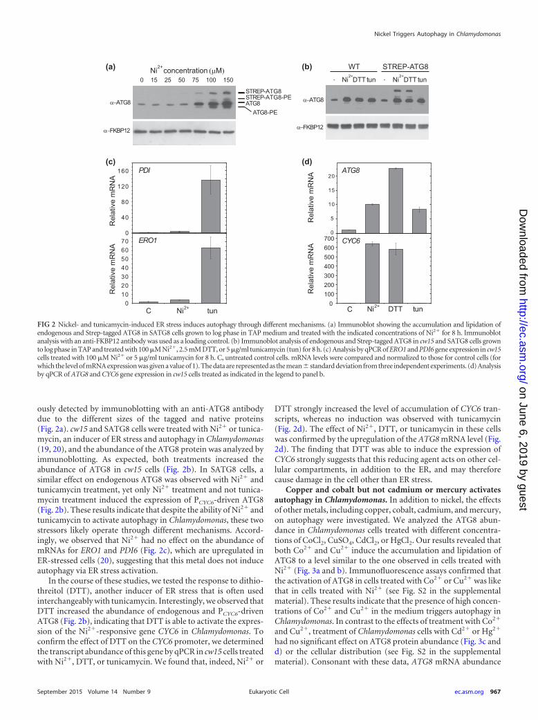
ously detected by immunoblotting with an anti-ATG8 antibodydue to the different sizes of the tagged and native proteins(Fig. 2a). cw15 and SATG8 cells were treated with Ni2� or tunica-mycin, an inducer of ER stress and autophagy in Chlamydomonas(19, 20), and the abundance of the ATG8 protein was analyzed byimmunoblotting. As expected, both treatments increased theabundance of ATG8 in cw15 cells (Fig. 2b). In SATG8 cells, asimilar effect on endogenous ATG8 was observed with Ni2� andtunicamycin treatment, yet only Ni2� treatment and not tunica-mycin treatment induced the expression of PCYC6-driven ATG8(Fig. 2b). These results indicate that despite the ability of Ni2� andtunicamycin to activate autophagy in Chlamydomonas, these twostressors likely operate through different mechanisms. Accord-ingly, we observed that Ni2� had no effect on the abundance ofmRNAs for ERO1 and PDI6 (Fig. 2c), which are upregulated inER-stressed cells (20), suggesting that this metal does not induceautophagy via ER stress activation.
In the course of these studies, we tested the response to dithio-threitol (DTT), another inducer of ER stress that is often usedinterchangeably with tunicamycin. Interestingly, we observed thatDTT increased the abundance of endogenous and PCYC6-drivenATG8 (Fig. 2b), indicating that DTT is able to activate the expres-sion of the Ni2�-responsive gene CYC6 in Chlamydomonas. Toconfirm the effect of DTT on the CYC6 promoter, we determinedthe transcript abundance of this gene by qPCR in cw15 cells treatedwith Ni2�, DTT, or tunicamycin. We found that, indeed, Ni2� or
DTT strongly increased the level of accumulation of CYC6 tran-scripts, whereas no induction was observed with tunicamycin(Fig. 2d). The effect of Ni2�, DTT, or tunicamycin in these cellswas confirmed by the upregulation of the ATG8 mRNA level (Fig.2d). The finding that DTT was able to induce the expression ofCYC6 strongly suggests that this reducing agent acts on other cel-lular compartments, in addition to the ER, and may thereforecause damage in the cell other than ER stress.
Copper and cobalt but not cadmium or mercury activatesautophagy in Chlamydomonas. In addition to nickel, the effectsof other metals, including copper, cobalt, cadmium, and mercury,on autophagy were investigated. We analyzed the ATG8 abun-dance in Chlamydomonas cells treated with different concentra-tions of CoCl2, CuSO4, CdCl2, or HgCl2. Our results revealed thatboth Co2� and Cu2� induce the accumulation and lipidation ofATG8 to a level similar to the one observed in cells treated withNi2� (Fig. 3a and b). Immunofluorescence assays confirmed thatthe activation of ATG8 in cells treated with Co2� or Cu2� was likethat in cells treated with Ni2� (see Fig. S2 in the supplementalmaterial). These results indicate that the presence of high concen-trations of Co2� and Cu2� in the medium triggers autophagy inChlamydomonas. In contrast to the effects of treatment with Co2�
and Cu2�, treatment of Chlamydomonas cells with Cd2� or Hg2�
had no significant effect on ATG8 protein abundance (Fig. 3c andd) or the cellular distribution (see Fig. S2 in the supplementalmaterial). Consonant with these data, ATG8 mRNA abundance
0 15 25 50 75 100 150
α−ATG8
STREP-ATG8
Ni concentration 2+ (μM)
α−ATG8
Ni2+
DTT DTT Ni2+
tuntun --
STREP-ATG8 WT (a) (b)
(c) (d)
Rel
ativ
e m
RN
A R
elat
ive
mR
NA
0
40
80
120
160 PDI
010203040506070 ERO1
C tun 2+NiR
elat
ive
mR
NA ATG8
0
5
10
15
20
0R
elat
ive
mR
NA
DTT 2+ tunNi
CYC6
C
100200300400500600700
α−FKBP12
ATG8ATG8-PE
STREP-ATG8-PE
α−FKBP12
FIG 2 Nickel- and tunicamycin-induced ER stress induces autophagy through different mechanisms. (a) Immunoblot showing the accumulation and lipidation ofendogenous and Strep-tagged ATG8 in SATG8 cells grown to log phase in TAP medium and treated with the indicated concentrations of Ni2� for 8 h. Immunoblotanalysis with an anti-FKBP12 antibody was used as a loading control. (b) Immunoblot analysis of endogenous and Strep-tagged ATG8 in cw15 and SATG8 cells grownto log phase in TAP and treated with 100 �M Ni2�, 2.5 mM DTT, or 5 �g/ml tunicamycin (tun) for 8 h. (c) Analysis by qPCR of ERO1 and PDI6 gene expression in cw15cells treated with 100 �M Ni2� or 5 �g/ml tunicamycin for 8 h. C, untreated control cells. mRNA levels were compared and normalized to those for control cells (forwhich the level of mRNA expression was given a value of 1). The data are represented as the mean standard deviation from three independent experiments. (d) Analysisby qPCR of ATG8 and CYC6 gene expression in cw15 cells treated as indicated in the legend to panel b.
Nickel Triggers Autophagy in Chlamydomonas
September 2015 Volume 14 Number 9 ec.asm.org 967Eukaryotic Cell
on June 6, 2019 by guesthttp://ec.asm
.org/D
ownloaded from
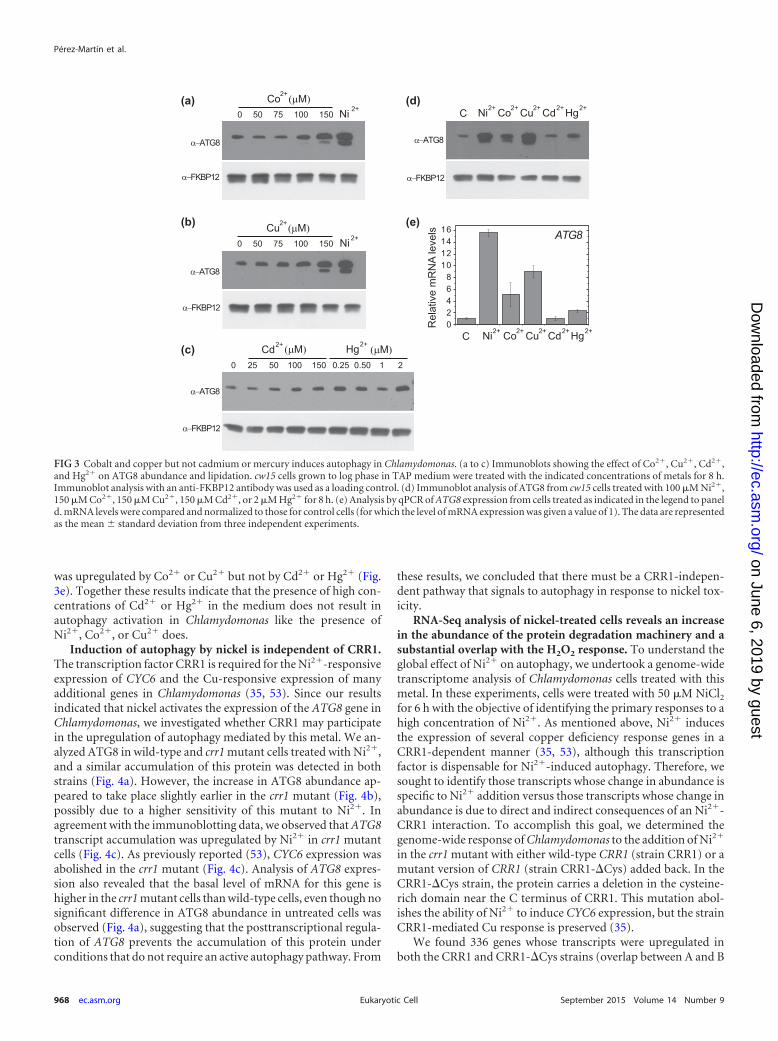
was upregulated by Co2� or Cu2� but not by Cd2� or Hg2� (Fig.3e). Together these results indicate that the presence of high con-centrations of Cd2� or Hg2� in the medium does not result inautophagy activation in Chlamydomonas like the presence ofNi2�, Co2�, or Cu2� does.
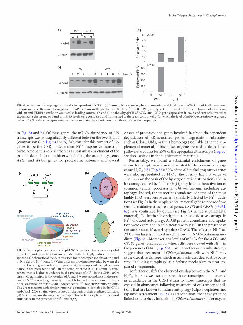
Induction of autophagy by nickel is independent of CRR1.The transcription factor CRR1 is required for the Ni2�-responsiveexpression of CYC6 and the Cu-responsive expression of manyadditional genes in Chlamydomonas (35, 53). Since our resultsindicated that nickel activates the expression of the ATG8 gene inChlamydomonas, we investigated whether CRR1 may participatein the upregulation of autophagy mediated by this metal. We an-alyzed ATG8 in wild-type and crr1 mutant cells treated with Ni2�,and a similar accumulation of this protein was detected in bothstrains (Fig. 4a). However, the increase in ATG8 abundance ap-peared to take place slightly earlier in the crr1 mutant (Fig. 4b),possibly due to a higher sensitivity of this mutant to Ni2�. Inagreement with the immunoblotting data, we observed that ATG8transcript accumulation was upregulated by Ni2� in crr1 mutantcells (Fig. 4c). As previously reported (53), CYC6 expression wasabolished in the crr1 mutant (Fig. 4c). Analysis of ATG8 expres-sion also revealed that the basal level of mRNA for this gene ishigher in the crr1 mutant cells than wild-type cells, even though nosignificant difference in ATG8 abundance in untreated cells wasobserved (Fig. 4a), suggesting that the posttranscriptional regula-tion of ATG8 prevents the accumulation of this protein underconditions that do not require an active autophagy pathway. From
these results, we concluded that there must be a CRR1-indepen-dent pathway that signals to autophagy in response to nickel tox-icity.
RNA-Seq analysis of nickel-treated cells reveals an increasein the abundance of the protein degradation machinery and asubstantial overlap with the H2O2 response. To understand theglobal effect of Ni2� on autophagy, we undertook a genome-widetranscriptome analysis of Chlamydomonas cells treated with thismetal. In these experiments, cells were treated with 50 �M NiCl2for 6 h with the objective of identifying the primary responses to ahigh concentration of Ni2�. As mentioned above, Ni2� inducesthe expression of several copper deficiency response genes in aCRR1-dependent manner (35, 53), although this transcriptionfactor is dispensable for Ni2�-induced autophagy. Therefore, wesought to identify those transcripts whose change in abundance isspecific to Ni2� addition versus those transcripts whose change inabundance is due to direct and indirect consequences of an Ni2�-CRR1 interaction. To accomplish this goal, we determined thegenome-wide response of Chlamydomonas to the addition of Ni2�
in the crr1 mutant with either wild-type CRR1 (strain CRR1) or amutant version of CRR1 (strain CRR1-�Cys) added back. In theCRR1-�Cys strain, the protein carries a deletion in the cysteine-rich domain near the C terminus of CRR1. This mutation abol-ishes the ability of Ni2� to induce CYC6 expression, but the strainCRR1-mediated Cu response is preserved (35).
We found 336 genes whose transcripts were upregulated inboth the CRR1 and CRR1-�Cys strains (overlap between A and B
0 50 75 100 150
α−ATG8
Co 2+(μM)Ni 2+
0 25 50 100 150 0.25 0.50 1 2
Cd 2+ (μM)Hg 2+(μM)
α−ATG8
Ni2+ Co 2+ Cu 2+ Cd 2+ Hg 2+C
α−ATG8
Cu 2+(μM)0 50 75 100 150 Ni 2+
α−ATG8
02468
10121416
Rel
ativ
e m
RN
A le
vels
ATG8(b)
(a)
(c)
(d)
(e)
Ni2+ Co 2+ Cu 2+ Cd 2+ Hg 2+C
α−FKBP12 α−FKBP12
α−FKBP12
α−FKBP12
FIG 3 Cobalt and copper but not cadmium or mercury induces autophagy in Chlamydomonas. (a to c) Immunoblots showing the effect of Co2�, Cu2�, Cd2�,and Hg2� on ATG8 abundance and lipidation. cw15 cells grown to log phase in TAP medium were treated with the indicated concentrations of metals for 8 h.Immunoblot analysis with an anti-FKBP12 antibody was used as a loading control. (d) Immunoblot analysis of ATG8 from cw15 cells treated with 100 �M Ni2�,150 �M Co2�, 150 �M Cu2�, 150 �M Cd2�, or 2 �M Hg2� for 8 h. (e) Analysis by qPCR of ATG8 expression from cells treated as indicated in the legend to paneld. mRNA levels were compared and normalized to those for control cells (for which the level of mRNA expression was given a value of 1). The data are representedas the mean standard deviation from three independent experiments.
Pérez-Martín et al.
968 ec.asm.org September 2015 Volume 14 Number 9Eukaryotic Cell
on June 6, 2019 by guesthttp://ec.asm
.org/D
ownloaded from
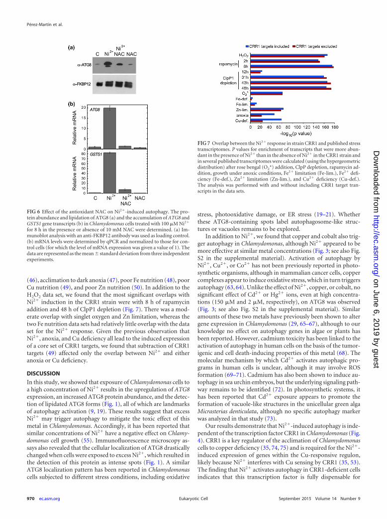
in Fig. 5a and b). Of these genes, the mRNA abundance of 275transcripts was not significantly different between the two strains(comparison C in Fig. 5a and b). We consider this core set of 275genes to be the CRR1-independent Ni2�-responsive transcrip-tome. Among this core set there is a substantial enrichment of theprotein degradation machinery, including the autophagy genesATG3 and ATG8, genes for proteasome subunits and several
classes of proteases, and genes involved in ubiquitin-dependentdegradation of ER-associated protein degradation substrates,such as Cdc48, Ufd1, or Otu1 homologs (see Table S1 in the sup-plemental material). This subset of genes related to degradativepathways accounts for 25% of the upregulated transcripts (Fig. 5c;see also Table S1 in the supplemental material).
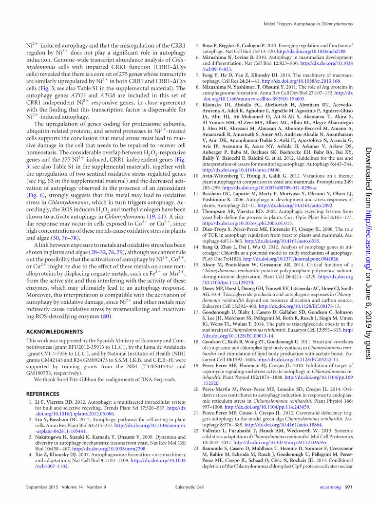
Remarkably, we found a substantial enrichment of geneswhose transcripts were also upregulated by the presence of exog-enous H2O2 (45) (Fig. 5d): 80% of the 275 nickel-responsive geneswere also upregulated by H2O2 (the overlap has a P value of1.5e�227 on the basis of the hypergeometric distribution). Cellu-lar damage caused by Ni2� or H2O2 may lead to the activation ofcommon cellular processes in Chlamydomonas, including au-tophagy. Indeed, the transcript abundance of some of the mosthighly H2O2-responsive genes is similarly affected by Ni2� addi-tion (see Fig. S3 in the supplemental material); the response of twosentinel oxidative stress-related genes, GSTS1 and GPXH (43, 61,62), was confirmed by qPCR (see Fig. S3 in the supplementalmaterial). To further investigate a role of oxidative damage inNi2�-induced autophagy, ATG8 protein abundance and lipida-tion were examined in cells treated with Ni2� in the presence ofthe antioxidant N-acetyl cysteine (NAC). The effect of Ni2� onATG8 was largely reduced in cells grown in NAC-containing me-dium (Fig. 6a). Moreover, the levels of mRNA for the ATG8 andGSTS1 genes remained low when cells were treated with Ni2� inthe presence of NAC (Fig. 6b). Taken together our results stronglysuggest that treatment of Chlamydomonas cells with Ni2� maycause oxidative damage, which in turn activates degradative path-ways, including autophagy, as a defense mechanism to clear im-paired components.
To further qualify the observed overlap between the Ni2� andH2O2 data sets, we also compared those transcripts that increasedin abundance in the CRR1 strain to those transcripts that in-creased in abundance following treatment of cells under condi-tions that are known to induce autophagy (ClpP1 depletion andrapamycin treatment [19, 23]) and conditions that have yet to belinked to autophagy induction in Chlamydomonas: singlet oxygen
(a) (c)
(b)
0 2 4 6 8
α−ATG8
crr1WT 2 4 6 8 time (h)
Ni 2+C Ni 2+C
crr1WT
α−ATG8
0
10
20
30
40
50
Rel
ativ
e m
RN
A
ATG8
Rel
ativ
e m
RN
A
CYC6
Ni 2+C Ni 2+C
crr1WT
0
100
200
300
400
α−FKBP12
α−FKBP12
FIG 4 Activation of autophagy by nickel is independent of CRR1. (a) Immunoblots showing the accumulation and lipidation of ATG8 in cw15 cells comparedto those in crr1 cells grown to log phase in TAP medium and treated with 100 �M Ni2� for 8 h. WT, wild type; C, untreated control cells. Immunoblot analysiswith an anti-FKBP12 antibody was used as loading control. (b and c) Analysis by qPCR of ATG8 and CYC6 gene expression in cw15 and crr1 cells treated asexplained in the legend to panel a. mRNA levels were compared and normalized to those for control cells (for which the level of mRNA expression was given avalue of 1). The data are represented as the mean standard deviation from three independent experiments.
FIG 5 Transcriptomic analysis of 50 �M Ni2�-treated cultures reveals a globalimpact on protein metabolism and overlap with the H2O2-induced stress re-sponse. (a) Schematic of the data sets used for the comparison shown in panelb. Ni refers to Ni2� ions. (b) Venn diagram showing the overlap between thedifferent sets of genes indicated in panel a. A, transcripts with a higher abun-dance in the presence of Ni2� in the complemented (CRR1) strain; B, tran-scripts with a higher abundance in the presence of Ni2� in the CRR1-�Cysstrain; C, transcripts in the overlap of A and B whose abundance in the pres-ence of Ni2� was not significantly different between the two strains. (c) Func-tional classification of the CRR1-independent Ni2�-responsive transcriptome.The 275 transcripts with similar transcript abundances identified in the CRR1and CRR1-�Cys strains were clustered on the basis of their predicted function.(d) Venn diagram showing the overlap between transcripts with increasedabundance in the presence of Ni2� and H2O2.
Nickel Triggers Autophagy in Chlamydomonas
September 2015 Volume 14 Number 9 ec.asm.org 969Eukaryotic Cell
on June 6, 2019 by guesthttp://ec.asm
.org/D
ownloaded from
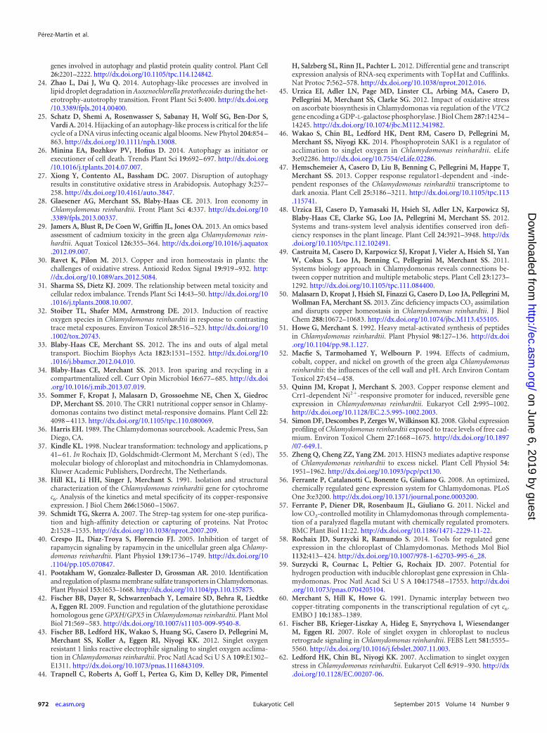
(46), acclimation to dark anoxia (47), poor Fe nutrition (48), poorCu nutrition (49), and poor Zn nutrition (50). In addition to theH2O2 data set, we found that the most significant overlaps withNi2� induction in the CRR1 strain were with 8 h of rapamycinaddition and 48 h of ClpP1 depletion (Fig. 7). There was a mod-erate overlap with singlet oxygen and Zn limitation, whereas thetwo Fe nutrition data sets had relatively little overlap with the dataset for the Ni2� response. Given the previous observation thatNi2�, anoxia, and Cu deficiency all lead to the induced expressionof a core set of CRR1 targets, we found that subtraction of CRR1targets (49) affected only the overlap between Ni2� and eitheranoxia or Cu deficiency.
DISCUSSION
In this study, we showed that exposure of Chlamydomonas cells toa high concentration of Ni2� results in the upregulation of ATG8expression, an increased ATG8 protein abundance, and the detec-tion of lipidated ATG8 forms (Fig. 1), all of which are landmarksof autophagy activation (9, 19). These results suggest that excessNi2� may trigger autophagy to mitigate the toxic effect of thismetal in Chlamydomonas. Accordingly, it has been reported thatsimilar concentrations of Ni2� have a negative effect on Chlamy-domonas cell growth (55). Immunofluorescence microscopy as-says also revealed that the cellular localization of ATG8 drasticallychanged when cells were exposed to excess Ni2�, which resulted inthe detection of this protein as intense spots (Fig. 1). A similarATG8 localization pattern has been reported in Chlamydomonascells subjected to different stress conditions, including oxidative
stress, photooxidative damage, or ER stress (19–21). Whetherthese ATG8-containing spots label autophagosome-like struc-tures or vacuoles remains to be explored.
In addition to Ni2�, we found that copper and cobalt also trig-ger autophagy in Chlamydomonas, although Ni2� appeared to bemore effective at similar metal concentrations (Fig. 3; see also Fig.S2 in the supplemental material). Activation of autophagy byNi2�, Cu2�, or Co2� has not been previously reported in photo-synthetic organisms, although in mammalian cancer cells, coppercomplexes appear to induce oxidative stress, which in turn triggersautophagy (63, 64). Unlike the effect of Ni2�, copper, or cobalt, nosignificant effect of Cd2� or Hg2� ions, even at high concentra-tions (150 �M and 2 �M, respectively), on ATG8 was observed(Fig. 3; see also Fig. S2 in the supplemental material). Similaramounts of these two metals have previously been shown to altergene expression in Chlamydomonas (29, 65–67), although to ourknowledge no effect on autophagy genes in algae or plants hasbeen reported. However, cadmium toxicity has been linked to theactivation of autophagy in human cells on the basis of the tumor-igenic and cell death-inducing properties of this metal (68). Themolecular mechanism by which Cd2� activates autophagic pro-grams in human cells is unclear, although it may involve ROSformation (69–71). Cadmium has also been shown to induce au-tophagy in sea urchin embryos, but the underlying signaling path-way remains to be identified (72). In photosynthetic systems, ithas been reported that Cd2� exposure appears to promote theformation of vacuole-like structures in the unicellular green algaMicrasterias denticulata, although no specific autophagy markerwas analyzed in that study (73).
Our results demonstrate that Ni2�-induced autophagy is inde-pendent of the transcription factor CRR1 in Chlamydomonas (Fig.4). CRR1 is a key regulator of the acclimation of Chlamydomonascells to copper deficiency (35, 74, 75) and is required for the Ni2�-induced expression of genes within the Cu-responsive regulon,likely because Ni2� interferes with Cu sensing by CRR1 (35, 53).The finding that Ni2� activates autophagy in CRR1-deficient cellsindicates that this transcription factor is fully dispensable for
FIG 7 Overlap between the Ni2� response in strain CRR1 and published stresstranscriptomes. P values for enrichment of transcripts that were more abun-dant in the presence of Ni2� than in the absence of Ni2� in the CRR1 strain andin several published transcriptomes were calculated (using the hypergeometricdistribution) after rose bengal (O2*) addition, ClpP depletion, rapamycin ad-dition, growth under anoxic conditions, Fe2� limitation (Fe-lim.), Fe2� defi-ciency (Fe-def.), Zn2� limitation (Zn-lim.), and Cu2� deficiency (Cu-def.).The analysis was performed with and without including CRR1 target tran-scripts in the data sets.
FIG 6 Effect of the antioxidant NAC on Ni2�-induced autophagy. The pro-tein abundance and lipidation of ATG8 (a) and the accumulation of ATG8 andGSTS1 gene transcripts (b) in Chlamydomonas cells treated with 100 �M Ni2�
for 8 h in the presence or absence of 10 mM NAC were determined. (a) Im-munoblot analysis with an anti-FKBP12 antibody was used as loading control.(b) mRNA levels were determined by qPCR and normalized to those for con-trol cells (for which the level of mRNA expression was given a value of 1). Thedata are represented as the mean standard deviation from three independentexperiments.
Pérez-Martín et al.
970 ec.asm.org September 2015 Volume 14 Number 9Eukaryotic Cell
on June 6, 2019 by guesthttp://ec.asm
.org/D
ownloaded from
Ni2�-induced autophagy and that the misregulation of the CRR1regulon by Ni2� does not play a significant role in autophagyinduction. Genome-wide transcript abundance analysis of Chla-mydomonas cells with impaired CRR1 function (CRR1-�Cyscells) revealed that there is a core set of 275 genes whose transcriptsare similarly upregulated by Ni2� in both CRR1 and CRR1-�Cyscells (Fig. 5; see also Table S1 in the supplemental material). Theautophagy genes ATG3 and ATG8 are included in this set ofCRR1-independent Ni2�-responsive genes, in close agreementwith the finding that this transcription factor is dispensable forNi2�-induced autophagy.
The upregulation of genes coding for proteasome subunits,ubiquitin-related proteins, and several proteases in Ni2�-treatedcells supports the conclusion that metal stress must lead to mas-sive damage in the cell that needs to be repaired to recover cellhomeostasis. The considerable overlap between H2O2-responsivegenes and the 275 Ni2�-induced, CRR1-independent genes (Fig.5; see also Table S1 in the supplemental material), together withthe upregulation of two sentinel oxidative stress-regulated genes(see Fig. S3 in the supplemental material) and the decreased acti-vation of autophagy observed in the presence of an antioxidant(Fig. 6), strongly suggests that this metal may lead to oxidativestress in Chlamydomonas, which in turn triggers autophagy. Ac-cordingly, the ROS inducers H2O2 and methyl viologen have beenshown to activate autophagy in Chlamydomonas (19, 21). A sim-ilar response may occur in cells exposed to Co2� or Cu2�, sincehigh concentrations of these metals cause oxidative stress in plantsand algae (30, 76–78).
A link between exposure to metals and oxidative stress has beenshown in plants and algae (28–32, 76, 79), although we cannot ruleout the possibility that the activation of autophagy by Ni2�, Co2�,or Cu2� might be due to the effect of these metals on some met-alloproteins by displacing cognate metals, such as Fe2� or Mn2�,from the active site and thus interfering with the activity of theseenzymes, which may ultimately lead to an autophagy response.Moreover, this interpretation is compatible with the activation ofautophagy by oxidative damage, since Ni2� and other metals mayindirectly cause oxidative stress by mismetallating and inactivat-ing ROS-detoxifying enzymes (80).
ACKNOWLEDGMENTS
This work was supported by the Spanish Ministry of Economy and Com-petitiveness (grant BFU2012-35913 to J.L.C.), by the Junta de Andalucía(grant CVI-�7336 to J.L.C.), and by National Institutes of Health (NIH)grants GM42143 and R24 GM092473 to S.S.M. I.K.B. and C.E.B.-H. weresupported by training grants from the NIH (T32ES015457 andGM100753, respectively).
We thank Sorel Fitz-Gibbon for realignments of RNA-Seq reads.
REFERENCES1. Li F, Vierstra RD. 2012. Autophagy: a multifaceted intracellular system
for bulk and selective recycling. Trends Plant Sci 17:526 –537. http://dx.doi.org/10.1016/j.tplants.2012.05.006.
2. Liu Y, Bassham DC. 2012. Autophagy: pathways for self-eating in plantcells. Annu Rev Plant Biol 63:215–237. http://dx.doi.org/10.1146/annurev-arplant-042811-105441.
3. Nakatogawa H, Suzuki K, Kamada Y, Ohsumi Y. 2009. Dynamics anddiversity in autophagy mechanisms: lessons from yeast. Nat Rev Mol CellBiol 10:458 – 467. http://dx.doi.org/10.1038/nrm2708.
4. Xie Z, Klionsky DJ. 2007. Autophagosome formation: core machineryand adaptations. Nat Cell Biol 9:1102–1109. http://dx.doi.org/10.1038/ncb1007-1102.
5. Boya P, Reggiori F, Codogno P. 2013. Emerging regulation and functions ofautophagy. Nat Cell Biol 15:713–720. http://dx.doi.org/10.1038/ncb2788.
6. Mizushima N, Levine B. 2010. Autophagy in mammalian developmentand differentiation. Nat Cell Biol 12:823– 830. http://dx.doi.org/10.1038/ncb0910-823.
7. Feng Y, He D, Yao Z, Klionsky DJ. 2014. The machinery of macroau-tophagy. Cell Res 24:24 – 41. http://dx.doi.org/10.1038/cr.2013.168.
8. Mizushima N, Yoshimori T, Ohsumi Y. 2011. The role of Atg proteins inautophagosome formation. Annu Rev Cell Dev Biol 27:107–132. http://dx.doi.org/10.1146/annurev-cellbio-092910-154005.
9. Klionsky DJ, Abdalla FC, Abeliovich H, Abraham RT, Acevedo-Arozena A, Adeli K, Agholme L, Agnello M, Agostinis P, Aguirre-GhisoJA, Ahn HJ, Ait-Mohamed O, Ait-Si-Ali S, Akematsu T, Akira S,Al-Younes HM, Al-Zeer MA, Albert ML, Albin RL, Alegre-AbarrateguiJ, Aleo MF, Alirezaei M, Almasan A, Almonte-Becerril M, Amano A,Amaravadi R, Amarnath S, Amer AO, Andrieu-Abadie N, AnantharamV, Ann DK, Anoopkumar-Dukie S, Aoki H, Apostolova N, Arancia G,Aris JP, Asanuma K, Asare NY, Ashida H, Askanas V, Askew DS,Auberger P, Baba M, Backues SK, Baehrecke EH, Bahr BA, Bai XY,Bailly Y, Baiocchi R, Baldini G, et al. 2012. Guidelines for the use andinterpretation of assays for monitoring autophagy. Autophagy 8:445–544.http://dx.doi.org/10.4161/auto.19496.
10. Avin-Wittenberg T, Honig A, Galili G. 2012. Variations on a theme:plant autophagy in comparison to yeast and mammals. Protoplasma 249:285–299. http://dx.doi.org/10.1007/s00709-011-0296-z.
11. Bassham DC, Laporte M, Marty F, Moriyasu Y, Ohsumi Y, Olsen LJ,Yoshimoto K. 2006. Autophagy in development and stress responses ofplants. Autophagy 2:2–11. http://dx.doi.org/10.4161/auto.2092.
12. Thompson AR, Vierstra RD. 2005. Autophagic recycling: lessons fromyeast help define the process in plants. Curr Opin Plant Biol 8:165–173.http://dx.doi.org/10.1016/j.pbi.2005.01.013.
13. Diaz-Troya S, Perez-Perez ME, Florencio FJ, Crespo JL. 2008. The roleof TOR in autophagy regulation from yeast to plants and mammals. Au-tophagy 4:851– 865. http://dx.doi.org/10.4161/auto.6555.
14. Jiang Q, Zhao L, Dai J, Wu Q. 2012. Analysis of autophagy genes in mi-croalgae: Chlorella as a potential model to study mechanism of autophagy.PLoS One 7:e41826. http://dx.doi.org/10.1371/journal.pone.0041826.
15. Aksoy M, Pootakham W, Grossman AR. 2014. Critical function of aChlamydomonas reinhardtii putative polyphosphate polymerase subunitduring nutrient deprivation. Plant Cell 26:4214 – 4229. http://dx.doi.org/10.1105/tpc.114.129270.
16. Davey MP, Horst I, Duong GH, Tomsett EV, Litvinenko AC, Howe CJ, SmithAG. 2014. Triacylglyceride production and autophagous responses in Chlamy-domonas reinhardtii depend on resource allocation and carbon source.Eukaryot Cell 13:392– 400. http://dx.doi.org/10.1128/EC.00178-13.
17. Goodenough U, Blaby I, Casero D, Gallaher SD, Goodson C, JohnsonS, Lee JH, Merchant SS, Pellegrini M, Roth R, Rusch J, Singh M, UmenJG, Weiss TL, Wulan T. 2014. The path to triacylglyceride obesity in thesta6 strain of Chlamydomonas reinhardtii. Eukaryot Cell 13:591– 613. http://dx.doi.org/10.1128/EC.00013-14.
18. Goodson C, Roth R, Wang ZT, Goodenough U. 2011. Structural correlatesof cytoplasmic and chloroplast lipid body synthesis in Chlamydomonas rein-hardtii and stimulation of lipid body production with acetate boost. Eu-karyot Cell 10:1592–1606. http://dx.doi.org/10.1128/EC.05242-11.
19. Perez-Perez ME, Florencio FJ, Crespo JL. 2010. Inhibition of target ofrapamycin signaling and stress activate autophagy in Chlamydomonas re-inhardtii. Plant Physiol 152:1874 –1888. http://dx.doi.org/10.1104/pp.109.152520.
20. Perez-Martin M, Perez-Perez ME, Lemaire SD, Crespo JL. 2014. Oxi-dative stress contributes to autophagy induction in response to endoplas-mic reticulum stress in Chlamydomonas reinhardtii. Plant Physiol 166:997–1008. http://dx.doi.org/10.1104/pp.114.243659.
21. Perez-Perez ME, Couso I, Crespo JL. 2012. Carotenoid deficiency trig-gers autophagy in the model green alga Chlamydomonas reinhardtii. Au-tophagy 8:376 –388. http://dx.doi.org/10.4161/auto.18864.
22. Valledor L, Furuhashi T, Hanak AM, Weckwerth W. 2013. Systemiccold stress adaptation of Chlamydomonas reinhardtii. Mol Cell Proteomics12:2032–2047. http://dx.doi.org/10.1074/mcp.M112.026765.
23. Ramundo S, Casero D, Muhlhaus T, Hemme D, Sommer F, CrevecoeurM, Rahire M, Schroda M, Rusch J, Goodenough U, Pellegrini M, Perez-Perez ME, Crespo JL, Schaad O, Civic N, Rochaix JD. 2014. Conditionaldepletion of the Chlamydomonas chloroplast ClpP protease activates nuclear
Nickel Triggers Autophagy in Chlamydomonas
September 2015 Volume 14 Number 9 ec.asm.org 971Eukaryotic Cell
on June 6, 2019 by guesthttp://ec.asm
.org/D
ownloaded from
genes involved in autophagy and plastid protein quality control. Plant Cell26:2201–2222. http://dx.doi.org/10.1105/tpc.114.124842.
24. Zhao L, Dai J, Wu Q. 2014. Autophagy-like processes are involved inlipid droplet degradation in Auxenochlorella protothecoides during the het-erotrophy-autotrophy transition. Front Plant Sci 5:400. http://dx.doi.org/10.3389/fpls.2014.00400.
25. Schatz D, Shemi A, Rosenwasser S, Sabanay H, Wolf SG, Ben-Dor S,Vardi A. 2014. Hijacking of an autophagy-like process is critical for the lifecycle of a DNA virus infecting oceanic algal blooms. New Phytol 204:854 –863. http://dx.doi.org/10.1111/nph.13008.
26. Minina EA, Bozhkov PV, Hofius D. 2014. Autophagy as initiator orexecutioner of cell death. Trends Plant Sci 19:692– 697. http://dx.doi.org/10.1016/j.tplants.2014.07.007.
27. Xiong Y, Contento AL, Bassham DC. 2007. Disruption of autophagyresults in constitutive oxidative stress in Arabidopsis. Autophagy 3:257–258. http://dx.doi.org/10.4161/auto.3847.
28. Glaesener AG, Merchant SS, Blaby-Haas CE. 2013. Iron economy inChlamydomonas reinhardtii. Front Plant Sci 4:337. http://dx.doi.org/10.3389/fpls.2013.00337.
29. Jamers A, Blust R, De Coen W, Griffin JL, Jones OA. 2013. An omics basedassessment of cadmium toxicity in the green alga Chlamydomonas rein-hardtii. Aquat Toxicol 126:355–364. http://dx.doi.org/10.1016/j.aquatox.2012.09.007.
30. Ravet K, Pilon M. 2013. Copper and iron homeostasis in plants: thechallenges of oxidative stress. Antioxid Redox Signal 19:919 –932. http://dx.doi.org/10.1089/ars.2012.5084.
31. Sharma SS, Dietz KJ. 2009. The relationship between metal toxicity andcellular redox imbalance. Trends Plant Sci 14:43–50. http://dx.doi.org/10.1016/j.tplants.2008.10.007.
32. Stoiber TL, Shafer MM, Armstrong DE. 2013. Induction of reactiveoxygen species in Chlamydomonas reinhardtii in response to contrastingtrace metal exposures. Environ Toxicol 28:516 –523. http://dx.doi.org/10.1002/tox.20743.
33. Blaby-Haas CE, Merchant SS. 2012. The ins and outs of algal metaltransport. Biochim Biophys Acta 1823:1531–1552. http://dx.doi.org/10.1016/j.bbamcr.2012.04.010.
34. Blaby-Haas CE, Merchant SS. 2013. Iron sparing and recycling in acompartmentalized cell. Curr Opin Microbiol 16:677– 685. http://dx.doi.org/10.1016/j.mib.2013.07.019.
35. Sommer F, Kropat J, Malasarn D, Grossoehme NE, Chen X, GiedrocDP, Merchant SS. 2010. The CRR1 nutritional copper sensor in Chlamy-domonas contains two distinct metal-responsive domains. Plant Cell 22:4098 – 4113. http://dx.doi.org/10.1105/tpc.110.080069.
36. Harris EH. 1989. The Chlamydomonas sourcebook. Academic Press, SanDiego, CA.
37. Kindle KL. 1998. Nuclear transformation: technology and applications, p41– 61. In Rochaix JD, Goldschmidt-Clermont M, Merchant S (ed), Themolecular biology of chloroplast and mitochondria in Chlamydomonas.Kluwer Academic Publishers, Dordrecht, The Netherlands.
38. Hill KL, Li HH, Singer J, Merchant S. 1991. Isolation and structuralcharacterization of the Chlamydomonas reinhardtii gene for cytochromec6. Analysis of the kinetics and metal specificity of its copper-responsiveexpression. J Biol Chem 266:15060 –15067.
39. Schmidt TG, Skerra A. 2007. The Strep-tag system for one-step purifica-tion and high-affinity detection or capturing of proteins. Nat Protoc2:1528 –1535. http://dx.doi.org/10.1038/nprot.2007.209.
40. Crespo JL, Diaz-Troya S, Florencio FJ. 2005. Inhibition of target ofrapamycin signaling by rapamycin in the unicellular green alga Chlamy-domonas reinhardtii. Plant Physiol 139:1736 –1749. http://dx.doi.org/10.1104/pp.105.070847.
41. Pootakham W, Gonzalez-Ballester D, Grossman AR. 2010. Identificationand regulation of plasma membrane sulfate transporters in Chlamydomonas.Plant Physiol 153:1653–1668. http://dx.doi.org/10.1104/pp.110.157875.
42. Fischer BB, Dayer R, Schwarzenbach Y, Lemaire SD, Behra R, LiedtkeA, Eggen RI. 2009. Function and regulation of the glutathione peroxidasehomologous gene GPXH/GPX5 in Chlamydomonas reinhardtii. Plant MolBiol 71:569 –583. http://dx.doi.org/10.1007/s11103-009-9540-8.
43. Fischer BB, Ledford HK, Wakao S, Huang SG, Casero D, Pellegrini M,Merchant SS, Koller A, Eggen RI, Niyogi KK. 2012. Singlet oxygenresistant 1 links reactive electrophile signaling to singlet oxygen acclima-tion in Chlamydomonas reinhardtii. Proc Natl Acad Sci U S A 109:E1302–E1311. http://dx.doi.org/10.1073/pnas.1116843109.
44. Trapnell C, Roberts A, Goff L, Pertea G, Kim D, Kelley DR, Pimentel
H, Salzberg SL, Rinn JL, Pachter L. 2012. Differential gene and transcriptexpression analysis of RNA-seq experiments with TopHat and Cufflinks.Nat Protoc 7:562–578. http://dx.doi.org/10.1038/nprot.2012.016.
45. Urzica EI, Adler LN, Page MD, Linster CL, Arbing MA, Casero D,Pellegrini M, Merchant SS, Clarke SG. 2012. Impact of oxidative stresson ascorbate biosynthesis in Chlamydomonas via regulation of the VTC2gene encoding a GDP-L-galactose phosphorylase. J Biol Chem 287:14234 –14245. http://dx.doi.org/10.1074/jbc.M112.341982.
46. Wakao S, Chin BL, Ledford HK, Dent RM, Casero D, Pellegrini M,Merchant SS, Niyogi KK. 2014. Phosphoprotein SAK1 is a regulator ofacclimation to singlet oxygen in Chlamydomonas reinhardtii. eLife3:e02286. http://dx.doi.org/10.7554/eLife.02286.
47. Hemschemeier A, Casero D, Liu B, Benning C, Pellegrini M, Happe T,Merchant SS. 2013. Copper response regulator1-dependent and -inde-pendent responses of the Chlamydomonas reinhardtii transcriptome todark anoxia. Plant Cell 25:3186 –3211. http://dx.doi.org/10.1105/tpc.113.115741.
48. Urzica EI, Casero D, Yamasaki H, Hsieh SI, Adler LN, Karpowicz SJ,Blaby-Haas CE, Clarke SG, Loo JA, Pellegrini M, Merchant SS. 2012.Systems and trans-system level analysis identifies conserved iron defi-ciency responses in the plant lineage. Plant Cell 24:3921–3948. http://dx.doi.org/10.1105/tpc.112.102491.
49. Castruita M, Casero D, Karpowicz SJ, Kropat J, Vieler A, Hsieh SI, YanW, Cokus S, Loo JA, Benning C, Pellegrini M, Merchant SS. 2011.Systems biology approach in Chlamydomonas reveals connections be-tween copper nutrition and multiple metabolic steps. Plant Cell 23:1273–1292. http://dx.doi.org/10.1105/tpc.111.084400.
50. Malasarn D, Kropat J, Hsieh SI, Finazzi G, Casero D, Loo JA, Pellegrini M,Wollman FA, Merchant SS. 2013. Zinc deficiency impacts CO2 assimilationand disrupts copper homeostasis in Chlamydomonas reinhardtii. J BiolChem 288:10672–10683. http://dx.doi.org/10.1074/jbc.M113.455105.
51. Howe G, Merchant S. 1992. Heavy metal-activated synthesis of peptidesin Chlamydomonas reinhardtii. Plant Physiol 98:127–136. http://dx.doi.org/10.1104/pp.98.1.127.
52. Macfie S, Tarmohamed Y, Welbourn P. 1994. Effects of cadmium,cobalt, copper, and nickel on growth of the green alga Chlamydomonasreinhardtii: the influences of the cell wall and pH. Arch Environ ContamToxicol 27:454 – 458.
53. Quinn JM, Kropat J, Merchant S. 2003. Copper response element andCrr1-dependent Ni2�-responsive promoter for induced, reversible geneexpression in Chlamydomonas reinhardtii. Eukaryot Cell 2:995–1002.http://dx.doi.org/10.1128/EC.2.5.995-1002.2003.
54. Simon DF, Descombes P, Zerges W, Wilkinson KJ. 2008. Global expressionprofiling of Chlamydomonas reinhardtii exposed to trace levels of free cad-mium. Environ Toxicol Chem 27:1668 –1675. http://dx.doi.org/10.1897/07-649.1.
55. Zheng Q, Cheng ZZ, Yang ZM. 2013. HISN3 mediates adaptive responseof Chlamydomonas reinhardtii to excess nickel. Plant Cell Physiol 54:1951–1962. http://dx.doi.org/10.1093/pcp/pct130.
56. Ferrante P, Catalanotti C, Bonente G, Giuliano G. 2008. An optimized,chemically regulated gene expression system for Chlamydomonas. PLoSOne 3:e3200. http://dx.doi.org/10.1371/journal.pone.0003200.
57. Ferrante P, Diener DR, Rosenbaum JL, Giuliano G. 2011. Nickel andlow CO2-controlled motility in Chlamydomonas through complementa-tion of a paralyzed flagella mutant with chemically regulated promoters.BMC Plant Biol 11:22. http://dx.doi.org/10.1186/1471-2229-11-22.
58. Rochaix JD, Surzycki R, Ramundo S. 2014. Tools for regulated geneexpression in the chloroplast of Chlamydomonas. Methods Mol Biol1132:413– 424. http://dx.doi.org/10.1007/978-1-62703-995-6_28.
59. Surzycki R, Cournac L, Peltier G, Rochaix JD. 2007. Potential forhydrogen production with inducible chloroplast gene expression in Chla-mydomonas. Proc Natl Acad Sci U S A 104:17548 –17553. http://dx.doi.org/10.1073/pnas.0704205104.
60. Merchant S, Hill K, Howe G. 1991. Dynamic interplay between twocopper-titrating components in the transcriptional regulation of cyt c6.EMBO J 10:1383–1389.
61. Fischer BB, Krieger-Liszkay A, Hideg E, Snyrychova I, WiesendangerM, Eggen RI. 2007. Role of singlet oxygen in chloroplast to nucleusretrograde signaling in Chlamydomonas reinhardtii. FEBS Lett 581:5555–5560. http://dx.doi.org/10.1016/j.febslet.2007.11.003.
62. Ledford HK, Chin BL, Niyogi KK. 2007. Acclimation to singlet oxygenstress in Chlamydomonas reinhardtii. Eukaryot Cell 6:919 –930. http://dx.doi.org/10.1128/EC.00207-06.
Pérez-Martín et al.
972 ec.asm.org September 2015 Volume 14 Number 9Eukaryotic Cell
on June 6, 2019 by guesthttp://ec.asm
.org/D
ownloaded from
63. Trejo-Solis C, Jimenez-Farfan D, Rodriguez-Enriquez S, Fernandez-Valverde F, Cruz-Salgado A, Ruiz-Azuara L, Sotelo J. 2012. Coppercompound induces autophagy and apoptosis of glioma cells by reactiveoxygen species and JNK activation. BMC Cancer 12:156. http://dx.doi.org/10.1186/1471-2407-12-156.
64. Zhong W, Zhu H, Sheng F, Tian Y, Zhou J, Chen Y, Li S, Lin J. 2014.Activation of the MAPK11/12/13/14 (p38 MAPK) pathway regulates thetranscription of autophagy genes in response to oxidative stress inducedby a novel copper complex in HeLa cells. Autophagy 10:1285–1300. http://dx.doi.org/10.4161/auto.28789.
65. Gillet S, Decottignies P, Chardonnet S, Le Marechal P. 2006. Cadmiumresponse and redoxin targets in Chlamydomonas reinhardtii: a proteomicapproach. Photosynth Res 89:201–211. http://dx.doi.org/10.1007/s11120-006-9108-2.
66. Lemaire S, Keryer E, Stein M, Schepens II, Issakidis-Bourguet E, Ge-rard-Hirne C, Miginiac-Maslow M, Jacquot JP. 1999. Heavy-metal reg-ulation of thioredoxin gene expression in Chlamydomonas reinhardtii.Plant Physiol 120:773–778. http://dx.doi.org/10.1104/pp.120.3.773.
67. Quinn JM, Eriksson M, Moseley JL, Merchant S. 2002. Oxygen defi-ciency responsive gene expression in Chlamydomonas reinhardtii througha copper-sensing signal transduction pathway. Plant Physiol 128:463–471. http://dx.doi.org/10.1104/pp.010694.
68. Chiarelli R, Roccheri MC. 2012. Heavy metals and metalloids as au-tophagy inducing agents: focus on cadmium and arsenic. Cells 1:597– 616.http://dx.doi.org/10.3390/cells1030597.
69. Son YO, Wang X, Hitron JA, Zhang Z, Cheng S, Budhraja A, Ding S,Lee JC, Shi X. 2011. Cadmium induces autophagy through ROS-dependent activation of the LKB1-AMPK signaling in skin epidermal cells.Toxicol Appl Pharmacol 255:287–296. http://dx.doi.org/10.1016/j.taap.2011.06.024.
70. Wang SH, Shih YL, Kuo TC, Ko WC, Shih CM. 2009. Cadmium toxicitytoward autophagy through ROS-activated GSK-3beta in mesangial cells.Toxicol Sci 108:124 –131. http://dx.doi.org/10.1093/toxsci/kfn266.
71. Yang LY, Wu KH, Chiu WT, Wang SH, Shih CM. 2009. The cadmium-induced death of mesangial cells results in nephrotoxicity. Autophagy5:571–572. http://dx.doi.org/10.4161/auto.5.4.8311.
72. Chiarelli R, Agnello M, Roccheri MC. 2011. Sea urchin embryos as amodel system for studying autophagy induced by cadmium stress. Au-tophagy 7:1028 –1034. http://dx.doi.org/10.4161/auto.7.9.16450.
73. Andosch A, Affenzeller MJ, Lutz C, Lutz-Meindl U. 2012. A fresh-water green alga under cadmium stress: ameliorating calcium effectson ultrastructure and photosynthesis in the unicellular model Micras-terias. J Plant Physiol 169:1489 –1500. http://dx.doi.org/10.1016/j.jplph.2012.06.002.
74. Eriksson M, Moseley JL, Tottey S, Del Campo JA, Quinn J, Kim Y,Merchant S. 2004. Genetic dissection of nutritional copper signaling inChlamydomonas distinguishes regulatory and target genes. Genetics 168:795– 807. http://dx.doi.org/10.1534/genetics.104.030460.
75. Kropat J, Tottey S, Birkenbihl RP, Depege N, Huijser P, Merchant S.2005. A regulator of nutritional copper signaling in Chlamydomonas is anSBP domain protein that recognizes the GTAC core of copper responseelement. Proc Natl Acad Sci U S A 102:18730 –18735. http://dx.doi.org/10.1073/pnas.0507693102.
76. Jamers A, Van der Ven K, Moens L, Robbens J, Potters G, Guisez Y,Blust R, De Coen W. 2006. Effect of copper exposure on gene expressionprofiles in Chlamydomonas reinhardtii based on microarray analysis.Aquat Toxicol 80:249 –260. http://dx.doi.org/10.1016/j.aquatox.2006.09.002.
77. Merchant SS, Allen MD, Kropat J, Moseley JL, Long JC, Tottey S,Terauchi AM. 2006. Between a rock and a hard place: trace elementnutrition in Chlamydomonas. Biochim Biophys Acta 1763:578 –594. http://dx.doi.org/10.1016/j.bbamcr.2006.04.007.
78. Tan YF, O’Toole N, Taylor NL, Millar AH. 2010. Divalent metal ions inplant mitochondria and their role in interactions with proteins and oxi-dative stress-induced damage to respiratory function. Plant Physiol 152:747–761. http://dx.doi.org/10.1104/pp.109.147942.
79. Rodrigo-Moreno A, Poschenrieder C, Shabala S. 2013. Transition met-als: a double edge sward in ROS generation and signaling. Plant SignalBehav 8:e23425. http://dx.doi.org/10.4161/psb.23425.
80. Imlay JA. 2014. The mismetallation of enzymes during oxidative stress. JBiol Chem 289:28121–28128. http://dx.doi.org/10.1074/jbc.R114.588814.
Nickel Triggers Autophagy in Chlamydomonas
September 2015 Volume 14 Number 9 ec.asm.org 973Eukaryotic Cell
on June 6, 2019 by guesthttp://ec.asm
.org/D
ownloaded from