Cuestionario Epigenética: Epigenetic mechanisms in ... · 1 and type 2 diabetes include...
14
Cuestionario Epigenética: • Lea y discuta de forma grupal el artículo científico Epigenetic mechanisms in diabetic complications and metabolic memory. En grupos de hasta cuatro integrantes responda las siguientes preguntas: 1) Explique brevemente qué es el fenómeno de “memoria metabólica” y qué impacto genera en la salud de pacientes diabéticos. 2) ¿Por qué es de interés considerar el estudio de los epigenomas en el desarrollo de la diabetes mellitus y la “memoria metabólica”?. 3) El mecanismo de metilación del ADN es de importancia en el desarrollo de la nefropatía diabética. Describa a qué se denomina metiloma. 4) Describa que son las HMEs y cómo determinan los estados de hetero/eucromatina. 5) Niveles elevados de glucemia generan nefropatía y complicaciones vasculares en pacientes diabéticos. Comente de qué manera ocurren estas alteraciones considerando únicamente el rol de la proteína SET7. 6) Mencione la importancia del estudio epigenético en pacientes diabéticos y los nuevos desafíos que debe enfrentar este análisis.
Transcript of Cuestionario Epigenética: Epigenetic mechanisms in ... · 1 and type 2 diabetes include...
Cuestionario Epigenética:
• Lea y discuta de forma grupal el artículo científico Epigenetic mechanisms in
diabetic complications and metabolic memory.
En grupos de hasta cuatro integrantes responda las siguientes preguntas:
1) Explique brevemente qué es el fenómeno de “memoria metabólica” y qué impacto
genera en la salud de pacientes diabéticos.
2) ¿Por qué es de interés considerar el estudio de los epigenomas en el desarrollo de la
diabetes mellitus y la “memoria metabólica”?.
3) El mecanismo de metilación del ADN es de importancia en el desarrollo de la
nefropatía diabética. Describa a qué se denomina metiloma.
4) Describa que son las HMEs y cómo determinan los estados de hetero/eucromatina.
5) Niveles elevados de glucemia generan nefropatía y complicaciones vasculares en
pacientes diabéticos. Comente de qué manera ocurren estas alteraciones considerando
únicamente el rol de la proteína SET7.
6) Mencione la importancia del estudio epigenético en pacientes diabéticos y los nuevos
desafíos que debe enfrentar este análisis.
REVIEW
Epigenetic mechanisms in diabetic complications
and metabolic memory
Marpadga A. Reddy & Erli Zhang & Rama Natarajan
Received: 16 August 2014 /Accepted: 6 November 2014 /Published online: 7 December 2014# Springer-Verlag Berlin Heidelberg 2014
Abstract The incidence of diabetes and its associated micro-
and macrovascular complications is greatly increasing world-
wide. The most prevalent vascular complications of both type
1 and type 2 diabetes include nephropathy, retinopathy, neu-
ropathy and cardiovascular diseases. Evidence suggests that
both genetic and environmental factors are involved in these
pathologies. Clinical trials have underscored the beneficial
effects of intensive glycaemic control for preventing the pro-
gression of complications. Accumulating evidence suggests a
key role for epigenetic mechanisms such as DNA methyla-
tion, histone post-translational modifications in chromatin,
and non-coding RNAs in the complex interplay between
genes and the environment. Factors associated with the pa-
thology of diabetic complications, including hyperglycaemia,
growth factors, oxidant stress and inflammatory factors can
lead to dysregulation of these epigenetic mechanisms to alter
the expression of pathological genes in target cells such as
endothelial, vascular smooth muscle, retinal and cardiac cells,
without changes in the underlying DNA sequence. Further-
more, long-term persistence of these alterations to the epige-
nome may be a key mechanism underlying the phenomenon
of ‘metabolic memory’ and sustained vascular dysfunction
despite attainment of glycaemic control. Current therapies
for most diabetic complications have not been fully effica-
cious, and hence a study of epigenetic mechanisms that may
be involved is clearly warranted as they can not only shed
novel new insights into the pathology of diabetic complica-
tions, but also lead to the identification of much needed new
drug targets. In this review, we highlight the emerging role of
epigenetics and epigenomics in the vascular complications of
diabetes and metabolic memory.
Keywords Diabetic complications . DNAmethylation .
Epigenetics . Epigenomics . Histone modifications .
Metabolic memory . Non-coding RNA . Review
Abbreviations
Ang II Angiotensin II
ARB Angiotensin II type1 receptor blocker
CBP CREB-binding protein
ChIP Chromatin immunoprecipitation
CKD Chronic kidney disease
DNAme DNA methylation
ECM Extracellular matrix
EDIC Epidemiology of Diabetes Interventions and
Complications
HAT Histone acetyltransferase
HME Histone-modifying enzymes
HMT Histone methyltransferase
Kac Lysine acetylation
KDM Lysine demethylase
Kme Lysine methylation
lncRNA Long non-coding RNA
miRNA MicroRNA
ncRNA Non-coding RNA
NF-κB Nuclear factor-κB
PTM Post-translational modifications
REC Retinal endothelial cell
SET7 SET domain-containing
(lysine methyltransferase) 7
SNP Single-nucleotide polymorphism
VSMC Vascular smooth muscle cell
M. A. Reddy : E. Zhang : R. Natarajan (*)
Department of Diabetes and Metabolic Diseases Research, Beckman
Research Institute of City of Hope, 1500 East Duarte Road,
Duarte, CA 91010, USA
e-mail: [email protected]
E. Zhang
State Key Laboratory of Cardiovascular Disease, Fuwai Hospital,
Chinese Academy of Medical Sciences and Peking Union Medical
College, Beijing, China
Diabetologia (2015) 58:443–455
DOI 10.1007/s00125-014-3462-y

Introduction
The increased incidence of diabetes worldwide has greatly
augmented the risk for numerous associated complications
that lead to reduced quality of life and increased mortality. A
high percentage of patients with type 1 and type 2 diabetes
develop microvascular complications such as diabetic retinop-
athy, nephropathy and neuropathy, as well as life-threatening
macrovascular diseases such as atherosclerosis, stroke and
hypertension [1–6]. Several common biochemical mecha-
nisms and genetic factors have been implicated in the pathol-
ogy of diabetes and its complications. They have a profound
impact on cellular transcription programs in target organs,
leading to aberrant expression of growth promoting, proin-
flammatory, pro-apoptotic, and pro-fibrotic genes [6–9].
However, it is increasingly evident that, because of the influ-
ences of gene–environment interactions, epigenetic mecha-
nisms may also play a key role. Epigenetics refers to heritable
changes in gene expression and ensuing phenotypes that occur
without changes in the DNA sequence. Epigenetic changes
include DNA cytosine methylation, histone post-translational
modifications (PTMs) in chromatin, and non-coding RNAs
(ncRNAs), all of which can affect gene expression individu-
ally or co-operatively and modulate disease states [8]. Current
therapies for most diabetic complications have not been fully
efficacious, and hence a study of epigenetic mechanisms is
clearly warranted as they can offer novel and valuable insights
into the pathophysiology of diabetic complications, and can
also identify much needed new drug targets. Recent advances
in human genome sequencing have yielded unprecedented
information about chromatin states and the epigenome under
normal and disease states [10, 11] which can be exploited to
enhance our understanding of the molecular mechanisms in-
volved in diabetic complications.
In this review, we highlight the role of epigenetic mecha-
nisms in vascular complications of diabetes and metabolic
memory, their potential use for the development of new bio-
markers and therapeutic targets, and recent technological ad-
vances that have helped accelerate the field.
Biochemical mechanisms and factors involved in diabetic
vascular complications
Diabetes and diabetogenic agents such as high glucose,
AGEs, angiotensin II (Ang II), TGF-β and oxidised lipids
have adverse effects in major target cells involved in vascular
dysfunction, including endothelial cells, vascular smooth
muscle cells (VSMCs), monocytes and retinal, neural and
renal cells [1–8, 12].Multiple signalling pathways and kinases
activated by these agents lead to the activation of key tran-
scription factors, such as nuclear factor-κB (NF-κB) and
Smads, leading to increased expression of growth factors,
inflammatory chemokines and cytokines involved in endothe-
lial dysfunction, vascular cell growth, macrophage infiltration,
fibrosis, inflammation and organ dysfunction [3–9, 13–17].
Whereas many of these mechanisms are common to most
vascular complications of diabetes, the specific genes regulat-
ed, phenotype and pathology can vary depending on the target
cell and organ. For example, levels of pro-fibrotic TGF-β are
increased in most renal cells in diabetes, which mediates
glomerular hypertrophy, accumulation of extracellular matrix
(ECM) proteins and podocyte apoptosis, leading to diabetic
nephropathy [2, 8, 12, 17]. Endothelial dysfunction and in-
flammation caused by macrophage infiltration is seen in most
diabetic complications [6]. In diabetic retinopathy, there is
retinal cell apoptosis and angiogenesis, while diabetic cardiac
disease is manifested as myopathy and hypertrophy [6].
Crosstalk between the actions of various diabetogenic fac-
tors can amplify and perpetuate the expression of pathological
genes associated with the progression of complications
(Fig. 1). Recently, microRNA (miRNA)- and long ncRNA
(lncRNA)-mediated mechanisms have also been implicated
[8] (Fig. 1). Further understanding of these novel epigenetic
mechanisms could help improve the efficacy of currently
available therapies for overt and irreversible complications,
and could also be used in the clinical management of meta-
bolic memory implicated in the continued development of the
diabetic complications despite glycaemic control.
Metabolic memory: a unique phenomenon
Hyperglycaemia is a major pathological factor involved in
diabetic complications. Although it can be controlled through
medication, dietary modifications and exercise, many patients
continue to experience numerous life-threatening complica-
tions. This could be due to a memory of prior exposure of
target cells to high glucose, leading to persistence of its
harmful effects long after attainment of glycaemic control
[15, 18] (Fig. 1). This memory phenomenon has been ob-
served in experimental models as well as in clinical trials such
as the Diabetes Control and Complications Trial (DCCT) and
the follow-up observational Epidemiology of Diabetes Inter-
ventions and Complications (EDIC) study. The results of the
DCCT indicated that patients with type 1 diabetes placed on
intensive glycaemic control had a much lower incidence or
severity of various complications, including nephropathy and
neuropathy, relative to those on standard/conventional therapy
[19]. After the DCCT, both groups were placed on intensive
therapy and were followed over the long term in the EDIC
study phase. Despite attainment of similar levels of HbA1c in
both groups during the EDIC study, patients in the original
DCCT intensive treatment group had significantly lower risks
of developing microvascular and macrovascular complica-
tions relative to the original DCCT conventional treatment
444 Diabetologia (2015) 58:443–455

group [20, 21], a phenomenon termed ‘metabolic memory’.
Other trials with type 2 diabetic patients have also found that
the benefits of intensive glycaemic control lasted long after
cessation of intervention, suggesting a ‘legacy effect’ [22].
Metabolic memory has been demonstrated in several ex-
perimental models. Cell culture studies have implicated met-
abolic memory in the persistently aberrant expression of fi-
brotic, antioxidant and inflammatory genes in VSMCs [18,
23–25], endothelial cells [26, 27] and retinal endothelial cells
(RECs) [28–31], despite glucose normalisation. Furthermore,
metabolic memory has also been studied in animal models of
complications such as atherosclerosis [27], diabetic nephrop-
athy and diabetic retinopathy [28–33]. Thus, both in vivo and
in vitro studies demonstrate the beneficial effects of good
glycaemic control and, conversely, that the deleterious effects
of prior hyperglycaemic exposure, on target organs last even
after subsequent glycaemic control. Metabolic memory re-
mains a major impediment to the effective management of
diabetic complications, and, in recent years, epigenetic mech-
anisms have been implicated.
Epigenetics and the epigenome: rationale for study
in diabetic complications
Epigenetic control of gene regulation plays an important role
in development, cell identity, stable inheritance of gene ex-
pression patterns in differentiated cells, genomic imprinting,
Intracellular signalling,
kinases, oxidative stress
Gene expression
Chromatin remodelling
lncRNAmiRNA
Macrovascular Microvascular
Atherosclerosis RetinopathyNephropathy Neuropathy(MI, stroke) (Blindness)(Renal failure) (Amputation)
Meta
bolic
me
mo
ry
DyslipidaemiaHyperglycaemia
AGEsTGF-β NEFAoxLDL
RAGETBR CD36SR
Humoral
Ang II
AT1R
Mechanical
Shear stress
Haemodynamic Metabolic
Inflammation/proliferation/differentiation/angiogenesis
Hypertrophy/apoptosis/ECM accumulation/remodelling
Transcription factors
(NF-κB, Smads, Zeb1, etc.)
Epigenetics
(Histone PTMs, DNAme)
Diabetic complications
DiabetesFig. 1 Signalling and epigenetic
networks mediating the
pathogenesis of diabetic
complications and metabolic
memory. Diabetes and its
consequent metabolic disorders
can upregulate several growth
factors and lipids that can act via
their receptors and other
mechanisms to trigger multiple
signalling pathways, transcription
factors (TFs) and crosstalk with
epigenetic networks. These
events can lead to chromatin
remodelling and changes in the
transcriptional regulation of key
pathological genes in cells from
target tissues relevant to various
complications of diabetes.
Persistence of such epigenetic
aberrations (including histone
PTMs, DNAme and ncRNAs)
may lead to metabolic memory,
which is implicated in an
increased risk for developing
diabetic complications even after
normalisation of hyperglycaemia.
AT1R, Ang II type 1 receptor; MI,
myocardial infarction; oxLDL,
Oxidized-LDL; RAGE, Receptor
for AGEs; SR, scavenger
receptors; TBR, TGF-β receptor
Diabetologia (2015) 58:443–455 445
X chromosome inactivation, stem cell plasticity, differential
disease susceptibility between monozygotic twins, and cellu-
lar responses to environmental signals [34, 35].
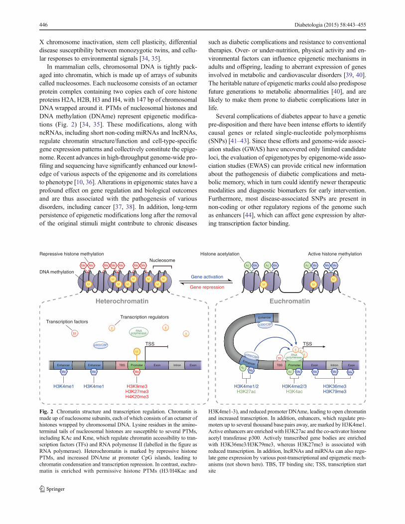
In mammalian cells, chromosomal DNA is tightly pack-
aged into chromatin, which is made up of arrays of subunits
called nucleosomes. Each nucleosome consists of an octamer
protein complex containing two copies each of core histone
proteins H2A, H2B, H3 and H4, with 147 bp of chromosomal
DNAwrapped around it. PTMs of nucleosomal histones and
DNA methylation (DNAme) represent epigenetic modifica-
tions (Fig. 2) [34, 35]. These modifications, along with
ncRNAs, including short non-coding miRNAs and lncRNAs,
regulate chromatin structure/function and cell-type-specific
gene expression patterns and collectively constitute the epige-
nome. Recent advances in high-throughput genome-wide pro-
filing and sequencing have significantly enhanced our knowl-
edge of various aspects of the epigenome and its correlations
to phenotype [10, 36]. Alterations in epigenomic states have a
profound effect on gene regulation and biological outcomes
and are thus associated with the pathogenesis of various
disorders, including cancer [37, 38]. In addition, long-term
persistence of epigenetic modifications long after the removal
of the original stimuli might contribute to chronic diseases
such as diabetic complications and resistance to conventional
therapies. Over- or under-nutrition, physical activity and en-
vironmental factors can influence epigenetic mechanisms in
adults and offspring, leading to aberrant expression of genes
involved in metabolic and cardiovascular disorders [39, 40].
The heritable nature of epigenetic marks could also predispose
future generations to metabolic abnormalities [40], and are
likely to make them prone to diabetic complications later in
life.
Several complications of diabetes appear to have a genetic
pre-disposition and there have been intense efforts to identify
causal genes or related single-nucleotide polymorphisms
(SNPs) [41–43]. Since these efforts and genome-wide associ-
ation studies (GWAS) have uncovered only limited candidate
loci, the evaluation of epigenotypes by epigenome-wide asso-
ciation studies (EWAS) can provide critical new information
about the pathogenesis of diabetic complications and meta-
bolic memory, which in turn could identify newer therapeutic
modalities and diagnostic biomarkers for early intervention.
Furthermore, most disease-associated SNPs are present in
non-coding or other regulatory regions of the genome such
as enhancers [44], which can affect gene expression by alter-
ing transcription factor binding.
M
TBS Promoter Exon Intron
p300/CBP
M M M M
M M M
Me MeMeMe Me MeMe
M
p300/CBP
RNApolymerase
T T
T
T
T
TF
Ac MeMe Me Me Me
Ac
p300/CBP TFRNA
polymerase
T
M
Ac Me Ac Me Ac Me MeMe
M M
Enhancer Enhancer TBS Promoter Exon Intron Exon
MeMeMe
Exon
Heterochromatin Euchromatin
DNA methylation
Repressive histone methylation
Nucleosome
TSS TSS
Histone acetylation
Gene activation
Gene repression
Active histone methylation
Transcription factorsTranscription regulators
H3K4me1 H3K4me1 H3K9me3H3K27me3H4K20me3
H3K4me1/2H3K27ac
H3K4me2/3H3K4ac
H3K36me3H3K79me3
Enhancer
Enhancer
Fig. 2 Chromatin structure and transcription regulation. Chromatin is
made up of nucleosome subunits, each of which consists of an octamer of
histones wrapped by chromosomal DNA. Lysine residues in the amino-
terminal tails of nucleosomal histones are susceptible to several PTMs,
including KAc and Kme, which regulate chromatin accessibility to tran-
scription factors (TFs) and RNA polymerase II (labelled in the figure as
RNA polymerase). Heterochromatin is marked by repressive histone
PTMs, and increased DNAme at promoter CpG islands, leading to
chromatin condensation and transcription repression. In contrast, euchro-
matin is enriched with permissive histone PTMs (H3/H4Kac and
H3K4me1-3), and reduced promoter DNAme, leading to open chromatin
and increased transcription. In addition, enhancers, which regulate pro-
moters up to several thousand base pairs away, are marked by H3K4me1.
Active enhancers are enriched with H3K27ac and the co-activator histone
acetyl transferase p300. Actively transcribed gene bodies are enriched
with H3K36me3/H3K79me3, whereas H3K27me3 is associated with
reduced transcription. In addition, lncRNAs and miRNAs can also regu-
late gene expression by various post-transcriptional and epigenetic mech-
anisms (not shown here). TBS, TF binding site; TSS, transcription start
site
446 Diabetologia (2015) 58:443–455

DNAme and its role in diabetic complications
DNAme, the most well established epigenetic mark,
occurs at 5′ cytosines of CpG dinucleotides, although
non-CpG methylation can also occur. Both DNA methyl
transferase (DNMT)3A and DNMT3B mediate de novo
DNAme, whereas DNMT1 acts as a maintenance meth-
yltransferase [35, 45]. Emerging evidence also shows
the occurrence of cytosine 5-hydroxymethylation enzy-
matically mediated by oxidases (the Tet proteins) and
active DNA demethylation, suggesting that DNAme is
quite dynamic [35]. In general, DNAme at promoter
regions leads to gene repression, whereas at gene bodies
it might regulate transcription elongation and alternative
splicing. DNAme is recognised by methyl binding pro-
teins that can recruit transcriptional co-repressors and
other proteins via protein–protein interactions to alter
gene expression [35].
The role of DNAme has been studied in the
transgenerational inheritance of metabolic diseases, suggest-
ing that environment and diet may influence epigenetic mod-
ifications that predispose individuals to diabetes [46]. Aber-
rant DNAme has also been reported in the reduced expression
of genes involved in diabetes and metabolism, and DNAme
variations have also been noted near diabetes susceptibility
genes and enhancers [15, 47].
Genomic DNA from diabetic patients with nephropa-
thy relative to those without displayed differential meth-
ylation at several genes, including UNC13B, which had
previously been linked to diabetic nephropathy [48]. In
another study, a number of regions with differential
DNAme were identified in saliva samples of patients
with end-stage renal disease relative to chronic kidney
disease (CKD) alone [49]. Interestingly, DNAme was
found to play a role in a model of renal fibrosis and
TGF-β actions [50]. A recent study implicated DNAme
in crosstalk between podocytes and proximal tubules in
diabetic nephropathy that was associated with increased
expression of claudin-1 in podocytes and reduced expres-
sion of sirtuin 1 in tubules [51]. Of note, an examination
of DNAme profiles in microdissected tubuli obtained
from patients with CKD and diabetic nephropathy vs
controls revealed significant differences in their
genome-wide DNAme (methylomes), with key differen-
tially methylated genes being related to fibrosis [52],
indicating direct connections between epigenetics and
human diabetic nephropathy. High glucose treatment in-
duced alterations in DNAme at key genes involved in
dysfunction of endothelial cells and neuronal cells [53,
54].. Additional studies in cell culture and in relevant
tissues of animal models and humans at various stages of
disease are needed to specifically link abnormalities in
DNAme patterns with diabetic complications.
Histone PTMs and their role in diabetic complications
and inflammation
Covalent PTMs of nucleosomal histone proteins in chromatin
also regulate gene expression via epigenetic mechanisms [34,
36]. Several histone PTMs (mostly at exposed amino-terminal
tails) have been identified, including lysine acetylation (Kac)
and lysine methylation (Kme). In general, histone Kac (such
as H3K9ac, H3K14ac, H4K5ac) at gene promoters correlates
with transcriptional activation, whereas its removal is associ-
ated with gene repression. Histone Kme can be associated
with either gene activation or repression depending on the
amino acid residue modified and the extent of methylation, i.e.
mono (Kme1), di (Kme2) or tri (Kme3) methylation.
H3K4me1/2/3 and H3K36me2/3 are generally associated
with transcriptionally active genome regions, whereas
H3K9me3, H3K27me3 and H4K20me3 are associated with
repressed domains [34]. Genome-wide profiling of histone
PTMs has been instrumental in demonstrating that distinct
patterns of specific histone modifications can distinguish key
regulatory regions, including promoters, enhancers, gene bod-
ies and repetitive elements [10, 36] (Fig. 2). Transcriptionally
active gene promoters are enriched with H3K9ac, H3K4me2
and H3K4me3, while gene bodies and transcribed regions are
enrichedwith H3K36me3 andH3K79me3. On the other hand,
inactive or silent gene promoters are enriched with repressive
marks H3K9me3 and H3K27me3 [10, 36]. Enhancers are
typically enriched with H3K4me1 (poised) and H3K27ac
(active) [55] (Fig. 2). Histone Kac is enzymatically mediated
by histone acetyltransferases (HAT) such as p300, CREB-
binding protein (CBP) and Tat-interactive protein 60 kDa
(Tip60), which also act as transcription co-activators. Con-
versely, histone deacetylases (HDAC), including HDAC1–11
and sirtuins, remove acetylation marks and in general act as
co-repressors with some exceptions [34]. Kme is mediated by
histone lysine methyltransferases (HMTs) and removed by
lysine demethylases (KDMs) [36, 56]. Histone modifying
enzymes (HMEs) can also modify lysine residues on non-
histone proteins, including transcription factors [57]. There-
fore, newer nomenclature has been proposed for HMEs based
on their enzymatic activity and the order of discovery [57].
Differential regulation of HME activity, recruitment and
expression under various conditions determines the epige-
nome landscape. They can be recruited to promoters or en-
hancers by binding to specific DNA sequences, or by binding
to pre-existing modifications or via interaction with RNA
polymerase II and transcription factors [58] or via lncRNAs
[59]. Because HMEs use metabolites such as acetyl-CoA
(HATs), S-(5′-adenosyl)-L-methionine (HMTs) and α-
ketoglutarate (KDMs) as cofactors, they might act as meta-
bolic sensors. Therefore, mis-regulation of their functions can
lead to metabolic abnormalities [60]. The role of DNAme in
epigenetic transmission is generally more widely studied than
Diabetologia (2015) 58:443–455 447
that of histone PTMs, although histone modifiers like
Polycomb repressor complexes and histone recycling proteins
have been implicated [61]. Overall, the crosstalk and interplay
between histone PTMs, DNAme and ncRNAs provides an-
other layer of epigenetic regulation to impact gene expression
[62, 63], which, if dysregulated, can result in diabetes and
associated complications.
Changes in histone PTMs in target organs such as pancreas,
liver and adipose tissue can affect the expression of numerous
genes associated with obesity and diabetes [15]. Such epige-
netic changes may also directly or indirectly influence key
genes in target organs affected by vascular complications. Cell
culture and animal models have demonstrated the involve-
ment of histone PTMs in the expression of genes associated
with the pathogenesis of diabetic nephropathy. TGF-β signal-
ling plays an important role in the expression of key fibrotic
and ECM genes and cell cycle inhibitor genes in renal cells,
which contribute to diabetic nephropathy [2, 6, 8, 12]. TGF-β
regulates gene expression mostly through activation of the
transcription factors Smads2/3/4, which can collaborate with
HATs and chromatin remodelling factors [12, 17, 64]. Recent
studies have examined these mechanisms as well as histone
Kac and Kme in rat mesangial cells treated with TGF-β and
high glucose. TGF-β increased H3K9/14ac near Smad and
SP1 binding sites by recruiting the HATs p300 and CBP to the
promoters of the genes encoding plasminogen activator inhib-
itor type 1 (PAI-1) and p21 [65]. Induction of expression of
fibrotic genes in rat mesangial cells by TGF-β was also
associated with enrichment of active Kme marks
(H3K4me1/2/3) and reduced levels of repressive marks
(H3K9me2/me3) at their promoters [66]. Furthermore, SET
domain-containing (lysine methyltransferase) 7 (SET7), a
H3K4 methyltransferase, was found to play a key role in
fibrotic gene expression. High glucose treatment of RMC
also led to similar changes in histone PTMs (H3Kac
and H3K4me) and increased SET7 recruitment at fibrot-
ic and cell cycle gene promoters [65, 66]. Interestingly,
these effects of high glucose were blocked by a TGF-β
antibody, demonstrating that TGF-β acts as a mediator
in high glucose-induced epigenetic effects [65, 66]. To-
gether, these studies support a critical role for epigenetic
mechanisms in TGF-β- and high glucose-induced path-
ological gene expression in mesangial cells which are
relevant to diabetic nephropathy (Fig. 3).
In an in vivo study [67], increases in RNA polymerase II
recruitment and H3K4me2, but decreases in H3K27me3
levels, were associated with the expression of diabetic
nephropathy-related genes in mouse and rat models of diabet-
ic nephropathy, with some differences between the two spe-
cies. In other studies, high glucose increased the expression of
the redox-regulating protein p66Shc by inhibiting promoter
DNAme and increasing H3K9ac in podocytes in vitro and in
experimental diabetic nephropathy [68]. Another report
implicated endoplasmic reticulum stress upstream of SET7-
mediated regulation of monocyte-chemoattractant protein-1
(MCP-1) expression in kidneys from db/db mice [69].
Since chromatin status around expressed genes is likely to
be affected by a code ofmultiple histone PTMs, another recent
study used Matrix chromatin immunoprecipitation (ChIP)
assays to profile several histone PTMs in vivo in mice glo-
meruli [70]. Relative to db/+ mice, glomeruli from diabetic
db/db mice exhibited increased RNA polymerase II recruit-
ment, enhanced levels of key activation marks and decreased
levels of key repressive marks at the promoters of the genes
encoding PAI-1 and receptor for AGEs (RAGE). These results
suggest that epigenetic histone PTMs regulated by diabetes
in vivo can co-operate to promote permissive chromatin states
around these and other promoters, enhanced access to tran-
scription machinery and gene expression. Interestingly, treat-
ment of db/db mice with losartan, an Ang II type 1 receptor
blocker (ARB), ameliorated key indices of diabetic nephrop-
athy, and reversed key changes in epigenetic enzymes and
H3K9ac enrichment at promoters of genes encoding PAI-1
and RAGE, but did not reverse all the diabetes-induced epi-
genetic changes [70]. Thus, the relative inefficiency of drugs
commonly used for diabetic nephropathy, such as ARBs to
prevent progression to renal failure, in many patients could be
due to the incomplete reversal of diabetic nephropathy-
associated epigenetic changes [71].
Changes in histone PTMs at key retinal genes have also
been demonstrated in RECs treated with high glucose and
tissues from animal models of diabetic retinopathy. Inhibition
of superoxide dismutase (SOD2), and concomitant increase in
oxidant stress in RECs are key events in diabetic retinopathy.
High glucose-induced downregulation of Sod2 mRNA was
accompanied by enrichment of the repressive histone mark
H4K20me3 and corresponding HMT SUV420H2 at the Sod2
promoter [31], as well as reduced levels of activation marks
H3K4me1/2, and increased occupancy of lysine-specific
demethylase 1, which erases H3K4me1/2 [29]. Upregulation
of matrix metalloproteinase gene Mmp9, which encodes an-
other enzyme implicated in diabetic retinopathy, was associ-
ated with reduced promoter H3K9me2 and increased H3K9ac
levels, along with increased recruitment of NF-κB in RECs
from diabetic rats [30]. Furthermore, thioredoxin-interacting
protein, a pro-oxidant factor induced by high glucose and
RAGE ligands, upregulates inflammatory genes in RECs by
inhibiting repressive H3K9me3 and increasing active H3K9ac
at their promoters [72]. Mass spectrometry studies demon-
strated that hyperglycaemia causes acetylation of retinal his-
tones, which was associated with increases in proinflammato-
ry proteins [73]. The HAT p300 was implicated in endothelial
fibronectin expression related to diabetic retinopathy [74], and
in gene expression relevant to diabetic cardiac hypertrophy
[75]. With respect to diabetic neuropathy, several biological
mechanisms have been studied, although the role of
448 Diabetologia (2015) 58:443–455
epigenetics has only recently been suggested [5, 6]. Epigenet-
ic mechanisms have also been evaluated in the impaired
wound healing associated with diabetes [76].
Chronic inflammation is a hallmark of the majority of the
vascular complications of diabetes, with increased macro-
phage infiltration and inflammatory gene expression being
observed in the kidney, blood vessels, eyes and other target
organs. High glucose-mediated activation of NF-κB is a major
mechanism of inflammatory gene expression in vascular cells
and monocytes, and several studies have demonstrated the
involvement of epigenetic modifications and histone PTMs
in these events [15]. In monocytes, high glucose treatment
increased the recruitment of co-activator HATs CBP and p300,
and augmented the levels of active marks H3Kac and H4Kac
at inflammatory gene promoters to enhance chromatin relax-
ation and gene expression [77]. Notably, profiling approaches
with ChIP linked to microarrays (ChIP-on-chip) revealed
differential enrichment of H3K4me2 (active) and H3K9me2
(repressive) marks at gene bodies of several genes in high
glucose-treated THP-1 monocytes [78]. Similar changes in
H3K9/14ac, H3K4me2 and H3K9me2 at key genes were also
observed in blood monocytes obtained from diabetic patients,
demonstrating direct relevance to diabetes [77, 78]. Further-
more, ChIP-on-chip epigenome profiling of blood lympho-
cytes from type 1 diabetic patients vs healthy controls dem-
onstrated significant variations in the repressive H3K9me2
mark at a subset of genes associated with type 1 diabetes,
inflammation and autoimmunity [79]. Key variations in
monocyte H3K9ac were also observed at two HLA genes
with SNPs that are closely linked to type 1 diabetes
[80], suggesting a crosstalk between epigenetic and ge-
netic variations—a concept being increasingly investi-
gated. Other studies noted that SET7 was required for
the maximal activation of a subset of NF-κB-inducible
inflammatory genes in monocytes [81], besides its role
in regulating fibrotic genes in the kidney.
AcM Me
Me
Ac
M
TF
Me
Me
M
Ac Me Ac Me Ac Me MeMe
M M
Diabetic nephropathy
Gene activationPersistent epigenetic changes
?
Ang II
miR-200b/c
Zeb1
?
DNAme H3K9ac
p300/CBP
TGF-β
Diabetes
HG
Inflammation
ECM accumulation
H3K4me1/2
SET7 ? miR-192
H3K9me2/3
TGF-β, cytokines, RAGE, PAI-1, MCP-1, Coll1α2, claudin-1, miRNAs, lncRNAs
TF
Active histone methylation
Repressive histone methylation
Lysine acetylation
DNA methylation
Transcription factor
Me
tab
olic
me
mo
ry
Fig. 3 Epigenetic mechanisms in diabetic nephropathy signal transduc-
tion events downstream of high glucose (HG), AGEs and growth factors
(TGF-β and Ang II) alter the expression/recruitment of epigenetic factors
that mediate or remove histone PTMs and DNA methylation leading to
chromatin remodelling. This alters promoter/enhancer access to transcrip-
tion factors (TFs) such as Smads, SP1 and NF-κB, which are involved in
the expression of genes mediating the pathogenesis of diabetic nephrop-
athy. Recent studies also reveal regulatory roles for several miRNAs in
diabetic nephropathy, including those that promote fibrotic gene expres-
sion in renal cells by targeting transcription repressors (Zeb1/2). Certain
lncRNAs have also been shown to modulate fibrotic genes. Inhibitors of
TGF-β, Ang II type 1 receptor signalling or miRNAs can block some but
not all the events involved in the pathogenesis of diabetic nephropathy,
suggesting the need for novel combination therapeutic approaches.
Coll1α2, collagen, type I, α2; RAGE, receptor for AGEs
Diabetologia (2015) 58:443–455 449
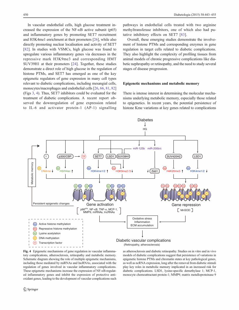
In vascular endothelial cells, high glucose treatment in-
creased the expression of the NF-κB active subunit (p65)
and inflammatory genes by promoting SET7 recruitment
and H3K4me1 enrichment at their promoters [26], while also
directly promoting nuclear localisation and activity of SET7
[82]. In studies with VSMCs, high glucose was found to
upregulate various inflammatory genes via decreases in the
repressive mark H3K9me3 and corresponding HMT
SUV39H1 at their promoters [24]. Together, these studies
demonstrate a direct role of high glucose in the regulation of
histone PTMs, and SET7 has emerged as one of the key
epigenetic regulators of gene expression in many cell types
relevant to diabetic complications, including mesangial cells,
monocytes/macrophages and endothelial cells [26, 66, 81, 82]
(Figs 3, 4). Thus, SET7 inhibitors could be evaluated for the
treatment of diabetic complications. A recent report ob-
served the downregulation of gene expression related
to IL-6 and activator protein-1 (AP-1) signalling
pathways in endothelial cells treated with two arginine
methyltransferase inhibitors, one of which also had pu-
tative inhibitory effects on SET7 [83].
Overall, these emerging studies demonstrate the involve-
ment of histone PTMs and corresponding enzymes in gene
regulation in target cells related to diabetic complications.
They also highlight the complexity of profiling tissues from
animal models of chronic progressive complications like dia-
betic nephropathy or retinopathy, and the need to study several
stages of disease progression.
Epigenetic mechanisms and metabolic memory
There is intense interest in determining the molecular mecha-
nisms underlying metabolic memory, especially those related
to epigenetics. In recent years, the potential persistence of
histone Kme variations at key genes related to complications
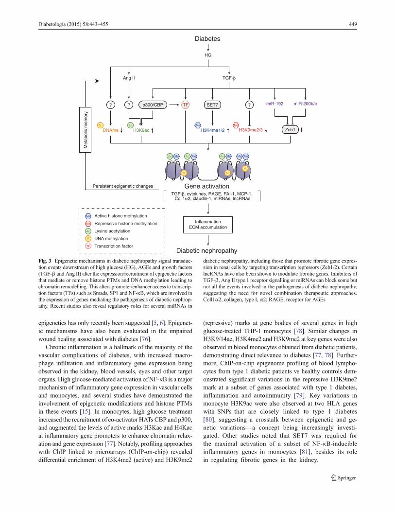
Me
Ac
M
TF
Me
Me MeAcM Me Me
M M M M
M M M
Me MeMeMe Me MeMe
MM
Ac Me Ac Me Ac Me MeMe
M M
H3K4me1/2
LSD1SET7 SUV39H1
H3K9me2 /3
miR-125b
Zeb1
miR-200b/c
HG
(Retinopathy, atherosclerosis)
p300/CBP?
H3/H4acDNAme
Oxidative stress
Inflammation
ECM accumulation
H3K4me1/2H4K20me3
MnSOD
LSD1SUV420H2
Meta
bolic
mem
ory
Persistent epigenetic changes
p66shc, NF-κB, TNF-α, MCP-1, MMP9, miRNAs, lncRNAs
TF
Active histone methylation
Repressive histone methylation
Lysine acetylation
DNA methylation
Transcription factorDiabetic vascular complications
Gene activation Gene repression
Diabetes
Fig. 4 Epigenetic mechanisms of gene regulation in vascular inflamma-
tory complications, atherosclerosis, retinopathy and metabolic memory.
Schematic diagram showing the role of multiple epigenetic mechanisms,
including those mediated by miRNAs and lncRNAs, associated with the
regulation of genes involved in vascular inflammatory complications.
These epigenetic mechanisms increase the expression of NF-κB-regulat-
ed inflammatory genes and inhibit the expression of protective anti-
oxidant genes, leading to the development of vascular complications such
as atherosclerosis and diabetic retinopathy. Studies on in vitro and in vivo
models of diabetic complications suggest that persistence of variations in
epigenetic histone PTMs and chromatin states at key pathological genes,
as well as ncRNA expression, long after the removal from diabetic stimuli
play key roles in metabolic memory implicated in an increased risk for
diabetic complications. LSD1, lysine-specific demethylase 1; MCP-1,
monocyte chemoattractant protein-1; MMP9, matrix metalloproteinase 9
450 Diabetologia (2015) 58:443–455

has been evaluated as a mechanism for their chronic mis-
regulation and metabolic memory. In one study, enhanced
inflammatory gene expression and migration in VSMCs ob-
tained from db/db mice relative to control db/+ mice despite
being cultured in vitro for several passages was associated
with decreased enrichment of the repressive mark H3K9me3
and the corresponding H3K9me3 methyltransferase
SUV39H1 at inflammatory gene promoters. The expression
levels of SUV39H1 were also downregulated in parallel, due
in part to the upregulation of miR-125b (which targets
SUV39H1) in db/db mice, indicating crosstalk between these
epigenetic layers (ncRNAs and chromatin) in diabetes [24,
84]. Similarly, sustained changes in H3K4me1 and SET7
were implicated in the prolonged upregulation of p65 in
endothelial cells previously cultured in high glucose for short
time periods [26]. In a rat model of diabetic retinopathy and
metabolic memory, sustained downregulation of Sod2 was
attributed to persistent promoter enrichments of the repressive
mark H4K20me3 and decreases in the activation mark
H3K4me2 [29, 31]. Together, these reports strongly suggest
that alterations in epigenetic histone PTMs might establish a
metabolic memory of diabetic complications (Figs 1, 2, 3, 4).
Further studies are needed to understand how high glucose
and diabetes provoke these epigenetic events, and how they
can be reversed to prevent the progression of complications
despite glycaemic control.
Importantly, investigations with appropriate human diabet-
ic individuals can help extrapolate these observations to clin-
ical metabolic memory and glycaemic variations. In the first
report of epigenome profiling of patients with type 1 diabetes
experiencing metabolic memory [85], several histone PTMs
were compared in white blood cells collected from a case
group of EDIC study participants selected from the former
DCCT conventional treatment group experiencing progres-
sion of nephropathy and retinopathy vs a control group from
the former DCCT intensive treatment group not showing
progression. Results showed significant enrichment in
H3K9ac at promoters of key inflammatory genes and genes
related to diabetic complications in monocytes from cases
compared with controls. Monocyte H3K9ac was significantly
associated with the mean HbA1c during long-term periods of
DCCT and EDIC. These novel findings suggest a potential
epigenetic explanation for metabolic memory in humans [85].
Further studies with bigger cohorts of patients will help pro-
vide additional support for the role of various epigenetic
marks in the metabolic memory phenomenon.
ncRNAs: miRNAs and lncRNAs in diabetic complications
Emerging data from whole transcriptome sequencing (RNA-
seq) have revealed that the majority of the genome is tran-
scribed into RNA, much of which is non-coding (i.e. unlike
mRNAwhich codes for protein) [86]. The role of ncRNAs in
diabetic complications has elicited great interest because they
are also epigenetic regulators that can modulate the expression
of genes via various transcriptional and post-transcriptional
mechanisms and thus fine-tune the actions of diabetogenic
stimuli. ncRNAs include not only small ncRNAs such as
miRNAs (about 22 nucleotides long), but also lncRNAs,
which are >200 nucleotides, up to 100 kb, in length. Recent
evidence demonstrates that miRNAs and lncRNAs can also
regulate the expression of genes and modulate the actions of
growth factors and inflammatory factors related to diabetic
complications [8]. These reports have been described in sev-
eral reviews [8, 87–91] and are only briefly discussed here.
Numerous recent reports have demonstrated abnormal ex-
pression of various miRNAs in renal, vascular and retinal cells
under diabetic conditions, and in vivo models of related
diabetic complications [8, 87–91]. Notably, the functional
relevance of these miRNAs has been highlighted by the fact
they target key genes associated with the progression of, or
protection against, these complications. In particular, the role
of miRNAs in diabetic nephropathy has been extensively
studied, including in the actions of TGF-β related to fibrosis
and other key renal outcomes in vitro and in vivo [8, 87–90].
In diabetic retinopathy, several miRNAs have been reported to
modulate the disease by targeting factors associated with
angiogenesis, inflammation, and oxidant stress in RECs and
in diabetic retinas [88, 89]. Reports have also implicated
various miRNAs in the aberrant expression of genes associ-
ated with diabetic cardiomyopathy [88, 91]. In addition, ef-
fective in vivo targeting of miRNAs has now been demon-
strated thanks to advances in nucleotide chemistry and the
design of nuclease-resistant anti-miRNAs, which suggest fu-
ture translational potential of miRNA-based therapies for hu-
man diabetic complications [8]. Importantly, since miRNAs
are stable in biological fluids such as urine and serum [8], they
are being assessed in samples from various clinical cohorts as
valuable biomarkers for the early detection of diabetic com-
plications, for which there is a major unmet clinical need. It is
clear that research in the field of miRNAs and diabetic com-
plications will continue at a rapid pace.
LncRNAs are long transcripts similar to mRNAs, but lack
protein-coding (translation) potential [63, 92]. Most lncRNAs
are expressed at much lower levels than protein-coding genes
and exhibit tissue-specific expression. They can affect gene
expression by various epigenetic mechanisms, including act-
ing as scaffolds to bring protein complexes together, recruiting
chromatin-modifying complexes to key gene loci, acting as
sponges of miRNAs and as host genes for miRNA [59, 63]. A
few reports have now demonstrated roles for lncRNAs in
diabetic complications, especially diabetic nephropathy [8].
Key miRNAs were induced by TGF-β, together with their
host gene, RP23, an ncRNA, in mesangial cells [93]. One
lncRNA, plasmacytoma variant translocation 1, was identified
Diabetologia (2015) 58:443–455 451

as potential locus for end-stage renal disease, and implicated
in the pathogenesis of diabetic nephropathy [94, 95]. In
VSMCs, Ang II induced several lncRNAs, of which one novel
lncRNAwas a host gene of miR-221/222, and could regulate
VSMC proliferation [96]. Furthermore, lncRNAE330013P06
(also known as MIR143HG) was recently reported to be
induced in macrophages under diabetic conditions in mice
and human cells [97]. Interestingly, this lncRNA could mod-
ulate the expression of macrophage inflammatory genes and
foam cell formation [97], thus implicating lncRNAs in vascu-
lar complications such as atherosclerosis. Research into the
role of lncRNAs in human disease and approaches to harness
their therapeutic potential are still in their infancy. Clearly, this
emerging field is expected to show rapid growth in the up-
coming years, aided by technological advances, including the
CRISPR-Cas9 system for genome editing [98].
Epigenomic approaches: applications in diabetic
complications research
Epigenetic studies in human disease have been greatly accel-
erated as a result of advances in whole-genome and epige-
nome profiling technologies as well as bioinformatics and
genomic data analysis platforms [99, 100]. DNAme is
analysed using bisulfite conversion of genomic DNA, immu-
noprecipitation of methylated DNA, followed by
hybridisation to arrays or next-generation sequencing to ob-
tain genome-wide distribution of DNAme [100]. Histone
PTMs are analysed using ChIP assays in which DNA in
cross-linked chromatin is immunoprecipitated with antibodies
against specific histone modifications, transcription factors or
other chromatin factors. ChIP-enriched DNA is analysed by
PCR to identify candidate enriched genome locations or by
hybridisation to microarrays (ChIP-on-chip) or next-
generation sequencing (ChIP-Seq) for genome-wide
localisation analyses [99]. Formaldehyde Assisted Isolation
of Regulatory Elements (FAIRE)-Seq detects accessible chro-
matin and regulatory elements based on differential cross-
linking efficiency of nucleosome enriched and depleted re-
gions [101]. Recently, in order to determine whether obesity
alters chromatin accessibility, FAIRE-seq was used for the
first time to examine chromatin variations in mouse livers
induced by a high-fat diet [102]. Such genomic approaches
can be used to evaluate variations in chromatin accessibility
associated with diabetic complications.
RNA-Seq has revolutionised transcriptome analysis in di-
verse cell types and disease conditions and led to the genome-
wide detection of known and novel transcripts, including
lncRNAs [92, 103]. Integration of transcriptome (RNA-seq),
DNA-methylome and ChIP-seq data can yield comprehensive
information about the epigenomic state and its outcomes on
gene expression under pathophysiological conditions [99].
The Encyclopedia of DNA Elements (ENCODE) project
completed high-quality whole-genome functional annotations
of the human and mouse genomes [10, 104]. These publicly
available data, including from NIH Roadmap Epigenomics
consortium (www.roadmapepigenomics.org), are valuable
reference tools to accelerate and catalyse new research into
the epigenomics of human disease.
As discussed earlier, these high-throughput approaches are
already being implemented in diabetic complications research.
They have been complemented with systems biology and
systems genetics efforts to effectively identify new players
in and drug targets for diabetic complications [105]. There are
also ongoing efforts to systematically profile epigenetic marks
in tissues, cells and archived genomic DNA from various
clinical trials. The major challenge, however, is expected
to be in the analysis of the ensuing large datasets, the
complexity of bioinformatics/biostatistics and in silico
modelling. If these hurdles can be overcome, these
efforts are likely to yield novel insights into epigenome
variations linked with diabetic complications.
Summary
Increasing evidence shows that, besides the well-described
biochemical mechanisms, epigenetic mechanisms might also
participate by fine-tuning gene expression to modulate the
aetiology of diabetic complications. Persistence of epigenetic
modifications triggered by diabetic stimuli could be one of the
key mechanisms underlying metabolic memory. However, the
involvement of many epigenetic factors and mechanisms in-
volved in the regulation of the modifications by upstream
signal transduction pathways remains unknown. However,
this is a rapidly expanding and dynamic field and it is likely
that other epigenetic factors related to diabetic complications
will soon be uncovered. Epigenomics may also aid in deter-
mining the functional roles of complications-associated genet-
ic variants. It would be worthwhile to assess whether lifestyle
modifications such as exercise and healthy diets can reduce
diabetic complications by altering epigenetic marks. A recent
study showed the beneficial effects of exercise on epigenetic
marks related to diabetes [106]. Because epigenetic changes
are potentially reversible in nature, combination therapies with
epigenetic drugs (epidrugs) [38] and antagomirs (miRNA
inhibitors) [8] could be considered to complement the current
treatments for complications. However, there are also key
challenges. Since epigenetic patterns are cell specific, data
from heterogeneous tissue samples and biopsies could be
difficult to interpret. Furthermore, apart from hyperglycaemia,
other factors associated with diabetes, including insulin resis-
tance, obesity, dyslipidaemia, environment, lifestyles and ge-
netics, can work independently or co-operatively to also pro-
mote epigenetic changes in various affected target tissues.
452 Diabetologia (2015) 58:443–455

Because inflammation is closely associated with most diabetic
complications, epigenetic variations could be examined non-
invasively in inflammatory cells like blood monocytes and
lymphocytes. Overall, it is anticipated that further research in
the field of epigenetics could lead to the identification of much
needed new biomarkers and drug targets for the early detec-
tion and treatment of the debilitating vascular complications
of diabetes.
Funding The authors are supported by funding fromNational Institutes
of Health (R01 DK081705, R01 DK058191, R01 DK065073, R01
HL106089) (to RN) the Juvenile Diabetes Research Foundation (to
RN) and China Scholarship Council (to EZ).
Duality of interest The authors declare that there is no duality of
interest associated with this manuscript.
Contribution statement MAR, EZ and RN were responsible for the
conception and design of the manuscript, drafting the manuscript, revis-
ing it critically for intellectual content and approving the final version.
References
1. Beckman JA, Creager MA, Libby P (2002) Diabetes and athero-
sclerosis: epidemiology, pathophysiology, and management. JAMA
287:2570–2581
2. Ziyadeh FN, Sharma K (2003) Overview: combating diabetic ne-
phropathy. J Am Soc Nephrol 14:1355–1357
3. Fong DS, Aiello L, Gardner TW et al (2003) Diabetic retinopathy.
Diabetes Care 26:226–229
4. Natarajan R, Nadler JL (2004) Lipid inflammatory mediators
in diabetic vascular disease. Arterioscler Thromb Vasc Biol
24:1542–1548
5. Vincent AM, Calabek B, Roberts L, Feldman EL (2013) Biology of
diabetic neuropathy. Handb Clin Neurol 115:591–606
6. Forbes JM, Cooper ME (2013) Mechanisms of diabetic complica-
tions. Physiol Rev 93:137–188
7. Brownlee M (2001) Biochemistry and molecular cell biology of
diabetic complications. Nature 414:813–820
8. Kato M, Natarajan R (2014) Diabetic nephropathy—emerging epi-
genetic mechanisms. Nat Rev Nephrol 10:517–530
9. Woroniecka KI, Park AS, Mohtat D, Thomas DB, Pullman JM,
Susztak K (2011) Transcriptome analysis of human diabetic kidney
disease. Diabetes 60:2354–2369
10. Dunham I, Kundaje A, Aldred SF et al (2012) An integrated
encyclopedia of DNA elements in the human genome. Nature
489:57–74
11. Human Epigenome Task Force (2008) Moving AHEAD with an
international human epigenome project. Nature 454:711–715
12. Kanwar YS, Sun L, Xie P, Liu FY, Chen S (2011) A glimpse
of various pathogenetic mechanisms of diabetic nephropathy.
Annu Rev Pathol 6:395–423
13. Geraldes P, KingGL (2010) Activation of protein kinase C isoforms
and its impact on diabetic complications. Circ Res 106:1319–1331
14. Ramasamy R, Yan SF, Schmidt AM (2011) Receptor for AGE
(RAGE): signaling mechanisms in the pathogenesis of diabetes
and its complications. Ann N YAcad Sci 1243:88–102
15. Reddy MA, Natarajan R (2011) Epigenetic mechanisms in diabetic
vascular complications. Cardiovasc Res 90:421–429
16. Averill MM, Bornfeldt KE (2009) Lipids versus glucose in inflam-
mation and the pathogenesis of macrovascular disease in diabetes.
Curr Diabetes Rep 9:18–25
17. Sanchez AP, Sharma K (2009) Transcription factors in the patho-
genesis of diabetic nephropathy. Expert Rev Mol Med 11:e13
18. Villeneuve LM, Natarajan R (2010) The role of epigenetics in the
pathology of diabetic complications. Am J Physiol Ren Physiol 299:
F14–F25
19. Writing Team for the DCCT/EDIC Research Group (2002) Effect of
intensive therapy on the microvascular complications of type 1
diabetes mellitus. JAMA 287:2563–2569
20. EDIC study (2003) Sustained effect of intensive treatment of type 1
diabetes mellitus on development and progression of diabetic ne-
phropathy: the Epidemiology of Diabetes Interventions and
Complications (EDIC) study. JAMA 290:2159–2167
21. Nathan DM, Cleary PA, Backlund JYet al (2005) Intensive diabetes
treatment and cardiovascular disease in patients with type 1 diabe-
tes. N Engl J Med 353:2643–2653
22. Chalmers J, Cooper ME (2008) UKPDS and the legacy effect.
N Engl J Med 359:1618–1620
23. Li SL, Reddy MA, Cai Q et al (2006) Enhanced proatherogenic
responses inmacrophages and vascular smooth muscle cells derived
from diabetic db/db mice. Diabetes 55:2611–2619
24. Villeneuve LM, Reddy MA, Lanting LL, Wang M, Meng L,
Natarajan R (2008) Epigenetic histone H3 lysine 9 methylation in
metabolic memory and inflammatory phenotype of vascular smooth
muscle cells in diabetes. Proc Natl Acad Sci U S A 105:9047–9052
25. Reddy MA, Jin W, Villeneuve L et al (2012) Pro-inflammatory role
of microRNA-200 in vascular smooth muscle cells from diabetic
mice. Arterioscler Thromb Vasc Biol 32:721–729
26. El-Osta A, Brasacchio D, Yao D et al (2008) Transient high glucose
causes persistent epigenetic changes and altered gene expression
during subsequent normoglycemia. J Exp Med 205:2409–2417
27. Brasacchio D, Okabe J, Tikellis C et al (2009) Hyperglycaemia
induces a dynamic cooperativity of histone methylase and
demethylase enzymes associated with gene-activating epigenetic
marks that coexist on the lysine tail. Diabetes 58:1229–1236
28. Kowluru RA (2003) Effect of reinstitution of good glycaemic
control on retinal oxidative stress and nitrative stress in diabetic
rats. Diabetes 52:818–823
29. Zhong Q, Kowluru RA (2013) Epigenetic modification of
Sod2 in the development of diabetic retinopathy and in the
metabolic memory: role of histone methylation. Invest
Ophthalmol Vis Sci 54:244–250
30. ZhongQ,KowluruRA (2013) Regulation ofmatrixmetalloproteinase-
9 by epigenetic modifications and the development of diabetic
retinopathy. Diabetes 62:2559–2568
31. Zhong Q, Kowluru RA (2011) Epigenetic changes in mitochondrial
superoxide dismutase in the retina and the development of diabetic
retinopathy. Diabetes 60:1304–1313
32. Engerman RL, Kern TS (1987) Progression of incipient diabetic
retinopathy during good glycaemic control. Diabetes 36:
808–812
33. Kowluru RA, Abbas SN, Odenbach S (2004) Reversal of
hyperglycaemia and diabetic nephropathy: effect of reinstitution
of good metabolic control on oxidative stress in the kidney of
diabetic rats. J Diabetes Complications 18:282–288
34. Kouzarides T (2007) Chromatin modifications and their function.
Cell 128:693–705
35. Jones PA (2012) Functions of DNA methylation: islands, start sites,
gene bodies and beyond. Nat Rev Genet 13:484–492
36. Zhou VW, Goren A, Bernstein BE (2011) Charting histone modifi-
cations and the functional organization of mammalian genomes. Nat
Rev Genet 12:7–18
37. Portela A, Esteller M (2010) Epigenetic modifications and human
disease. Nat Biotechnol 28:1057–1068
Diabetologia (2015) 58:443–455 453

38. Baylin SB, Jones PA (2011) A decade of exploring the
cancer epigenome—biological and translational implications.
Nat Rev Cancer 11:726–734
39. Ling C, Groop L (2009) Epigenetics: a molecular link between
environmental factors and type 2 diabetes. Diabetes 58:2718–2725
40. Wang J, Wu Z, Li D et al (2012) Nutrition, epigenetics, and
metabolic syndrome. Antioxid Redox Signal 17:282–301
41. Sandholm N, Salem RM, McKnight AJ et al (2012) New sus-
ceptibility loci associated with kidney disease in type 1 diabetes.
PLoS Genet 8:e1002921
42. Luo J, Zhao L, Chen AY et al (2013) TCF7L2 variation and
proliferative diabetic retinopathy. Diabetes 62:2613–2617
43. McKnight AJ, McKay GJ, Maxwell AP (2014) Genetic and epige-
netic risk factors for diabetic kidney disease. Adv Chron Kidney Dis
21:287–296
44. Maurano MT, Humbert R, Rynes E et al (2012) Systematic locali-
zation of common disease-associated variation in regulatory DNA.
Science 337:1190–1195
45. Chen ZX, Riggs AD (2011) DNAmethylation and demethylation in
mammals. J Biol Chem 286:18347–18353
46. Jirtle RL, Skinner MK (2007) Environmental epigenomics and
disease susceptibility. Nat Rev Genet 8:253–262
47. Simmons R (2011) Epigenetics and maternal nutrition: nature v.
nurture. Proc Nutr Soc 70:73–81
48. Bell CG, Teschendorff AE, Rakyan VK, Maxwell AP, Beck S,
Savage DA (2010) Genome-wide DNA methylation analysis for
diabetic nephropathy in type 1 diabetes mellitus. BMCMed Gen 3:
33–42
49. Sapienza C, Lee J, Powell J et al (2011) DNA methylation
profiling identifies epigenetic differences between diabetes pa-
tients with ESRD and diabetes patients without nephropathy.
Epigenetics Off J DNA Methylation Soc 6:20–28
50. Bechtel W, McGoohan S, Zeisberg EM et al (2010) Methylation
determines fibroblast activation and fibrogenesis in the kidney.
Nat Med 16:544–550
51. Hasegawa K, Wakino S, Simic P et al (2013) Renal tubular
Sirt1 attenuates diabetic albuminuria by epigenetically sup-
pressing Claudin-1 overexpression in podocytes. Nat Med
19:1496–1504
52. Ko YA, Mohtat D, Suzuki M et al (2013) Cytosine methylation
changes in enhancer regions of core pro-fibrotic genes characterize
kidney fibrosis development. Genome Biol 14:R108
53. Pirola L, Balcerczyk A, Tothill RW et al (2011) Genome-wide
analysis distinguishes hyperglycaemia regulated epigenetic signa-
tures of primary vascular cells. Genome Res 21:1601–1615
54. Kim ES, Isoda F, Kurland I, Mobbs CV (2013) Glucose-induced
metabolic memory in Schwann cells: prevention by PPAR agonists.
Endocrinology 154:3054–3066
55. Jin F, Li Y, Ren B, Natarajan R (2011) Enhancers: multi-
dimensional signal integrators. Transcription 2:226–230
56. Klose RJ, Zhang Y (2007) Regulation of histone methylation by
demethylimination and demethylation. Nat Rev Mol Cell Biol 8:
307–318
57. Allis CD, Berger SL, Cote J et al (2007) New nomenclature for
chromatin-modifying enzymes. Cell 131:633–636
58. Smith E, Shilatifard A (2010) The chromatin signaling pathway:
diverse mechanisms of recruitment of histone-modifying enzymes
and varied biological outcomes. Mol Cell 40:689–701
59. Rinn JL, Chang HY (2012) Genome regulation by long noncoding
RNAs. Annu Rev Biochem 81:145–166
60. Sassone-Corsi P (2013) Physiology. When metabolism and epige-
netics converge. Science 339:148–150
61. Abmayr SM, Workman JL (2012) Holding on through DNA repli-
cation: histone modification or modifier? Cell 150:875–877
62. Cedar H, Bergman Y (2009) Linking DNAmethylation and histone
modification: patterns and paradigms. Nat Rev Genet 10:295–304
63. Guttman M, Rinn JL (2012) Modular regulatory principles of large
non-coding RNAs. Nature 482:339–346
64. Das F, Ghosh-Choudhury N, Venkatesan B, Li X, Mahimainathan
L, Choudhury GG (2008) Akt kinase targets association of CBP
with SMAD 3 to regulate TGFbeta-induced expression of plasmin-
ogen activator inhibitor-1. J Cell Physiol 214:513–527
65. Yuan H, Reddy MA, Sun G et al (2013) Involvement of p300/CBP
and epigenetic histone acetylation in TGF-beta1-mediated gene
transcription in mesangial cells. Am J Physiol Ren Physiol 304:
F601–F613
66. Sun G, ReddyMA, Yuan H, Lanting L, KatoM, Natarajan R (2010)
Epigenetic histone methylation modulates fibrotic gene expression.
J Am Soc Nephrol 21:2069–2080
67. Komers R, Mar D, Denisenko O, Xu B, Oyama TT, Bomsztyk K
(2013) Epigenetic changes in renal genes dysregulated inmouse and
rat models of type 1 diabetes. Lab Investig J Tech Methods Pathol
93:543–552
68. Bock F, Shahzad K, Wang H et al (2013) Activated protein C
ameliorates diabetic nephropathy by epigenetically inhibiting the
redox enzyme p66Shc. Proc Natl Acad Sci U S A 110:648–653
69. Chen J, Guo Y, Zeng W et al (2014) ER stress triggers MCP-1
expression through SET7/9-induced histone methylation in the
kidneys of db/db mice. Am J Physiol Ren Physiol 306:F916–F925
70. Reddy MA, Sumanth P, Lanting L et al (2014) Losartan reverses
permissive epigenetic changes in renal glomeruli of diabetic db/db
mice. Kidney Int 85:362–373
71. Ruggenenti P, Cravedi P, Remuzzi G (2010) The RAAS in
the pathogenesis and treatment of diabetic nephropathy.
Nat Rev Nephrol 6:319–330
72. Perrone L, Devi TS, Hosoya K, Terasaki T, Singh LP (2009)
Thioredoxin interacting protein (TXNIP) induces inflammation
through chromatin modification in retinal capillary endothelial cells
under diabetic conditions. J Cell Physiol 221:262–272
73. Kadiyala CS, Zheng L, Du Y et al (2012) Acetylation of retinal
histones in diabetes increases inflammatory proteins: effects of
minocycline and manipulation of histone acetyltransferase (HAT)
and histone deacetylase (HDAC). J Biol Chem 287:25869–25880
74. Kaur H, Chen S, Xin X, Chiu J, Khan ZA, Chakrabarti S (2006)
Diabetes-induced extracellular matrix protein expression is mediat-
ed by transcription coactivator p300. Diabetes 55:3104–3111
75. Feng B, Chen S, Chiu J, George B, Chakrabarti S (2008) Regulation
of cardiomyocyte hypertrophy in diabetes at the transcriptional
level. Am J Physiol Endocrinol Metab 294:E1119–E1126
76. Rafehi H, El-Osta A, Karagiannis TC (2011) Genetic and epigenetic
events in diabetic wound healing. Int Wound J 8:12–21
77. Miao F, Gonzalo IG, Lanting L, Natarajan R (2004) In vivo chro-
matin remodeling events leading to inflammatory gene transcription
under diabetic conditions. J Biol Chem 279:18091–18097
78. Miao F, Wu X, Zhang L, Yuan YC, Riggs AD, Natarajan R (2007)
Genome-wide analysis of histone lysine methylation variations
caused by diabetic conditions in human monocytes. J Biol Chem
282:13854–13863
79. Miao F, Smith DD, Zhang L, Min A, Feng W, Natarajan R (2008)
Lymphocytes from patients with type 1 diabetes display a distinct
profile of chromatin histone H3 lysine 9 dimethylation: an epige-
netic study in diabetes. Diabetes 57:3189–3198
80. Miao F, Chen Z, Zhang L et al (2012) Profiles of epigenetic histone
post-translational modifications at type 1 diabetes susceptible genes.
J Biol Chem 287:16335–16345
81. Li Y, ReddyMA,Miao F et al (2008) Role of the histoneH3 lysine 4
methyltransferase, SET7/9, in the regulation of NF-κB-dependent
inflammatory genes. Relevance to diabetes and inflammation.
J Biol Chem 283:26771–26781
82. Okabe J, Orlowski C, Balcerczyk A et al (2012) Distinguishing
hyperglycaemic changes by Set7 in vascular endothelial cells.
Circ Res 110:1067–1076
454 Diabetologia (2015) 58:443–455
83. Okabe J, Fernandez AZ, ZiemannM,Keating ST, BalcerczykA, El-
Osta A (2014) Endothelial transcriptome in response to pharmaco-
logical methyltransferase inhibition. ChemMedChem 9:1755–1762
84. Villeneuve LM, Kato M, Reddy MA, Wang M, Lanting L,
Natarajan R (2010) Enhanced levels ofmicroRNA-125b in vascular
smooth muscle cells of diabetic db/db mice lead to increased
inflammatory gene expression by targeting the histone methyltrans-
ferase Suv39h1. Diabetes 59:2904–2915
85. Miao F, Chen Z, Genuth S et al (2014) Evaluating the role of
epigenetic histone modifications in the metabolic memory of type
1 diabetes. Diabetes 63:1748–1762
86. Guttman M, Amit I, Garber M et al (2009) Chromatin signature
reveals over a thousand highly conserved large non-coding RNAs in
mammals. Nature 458:223–227
87. Alvarez ML, Distefano JK (2013) The role of non-coding RNAs in
diabetic nephropathy: potential applications as biomarkers for dis-
ease development and progression. Diabetes Res Clin Pract 99:1–11
88. Kato M, Castro NE, Natarajan R (2013) MicroRNAs: po-
tential mediators and biomarkers of diabetic complications.
Free Radic Biol Med 64:85–94
89. Kantharidis P, Wang B, Carew RM, Lan HY (2011) Diabetes
complications: the microRNA perspective. Diabetes 60:1832–1837
90. Badal SS, Danesh FR (2014) MicroRNAs and their applications in
kidney diseases. Pediatr Nephrol. doi:10.1007/s00467-014-2867-7
91. Zhou Q, Lv D, Chen P et al (2014) MicroRNAs in diabetic cardio-
myopathy and clinical perspectives. Front Genet 5:185
92. Cabili MN, Trapnell C, Goff L et al (2011) Integrative annotation of
human large intergenic noncoding RNAs reveals global properties
and specific subclasses. Genes Dev 25:1915–1927
93. Kato M, Putta S, Wang M et al (2009) TGF-β activates Akt kinase
through a microRNA-dependent amplifying circuit targeting PTEN.
Nat Cell Biol 11:881–889
94. Alvarez ML, DiStefano JK (2011) Functional characterization of
the plasmacytoma variant translocation 1 gene (PVT1) in diabetic
nephropathy. PLoS One 6:e18671
95. Hanson RL, Craig DW, Millis MP et al (2007) Identification of
PVT1 as a candidate gene for end-stage renal disease in type 2
diabetes using a pooling-based genome-wide single nucleotide
polymorphism association study. Diabetes 56:975–983
96. Leung A, Trac C, Jin W et al (2013) Novel long noncoding RNAs
Are regulated by angiotensin II in vascular smooth muscle cells.
Circ Res 113:266–278
97. Reddy MA, Chen Z, Park JT et al (2014) Regulation of inflamma-
tory phenotype in macrophages by a diabetes-induced long non-
coding RNA. Diabetes. doi:10.2337/db14-0298
98. Mali P, Aach J, Stranges PB et al (2013) CAS9 transcriptional
activators for target specificity screening and paired nickases for
cooperative genome engineering. Nat Biotechnol 31:833–838
99. Hawkins RD, HonGC, Ren B (2010) Next-generation genomics: an
integrative approach. Nat Rev Genet 11:476–486
100. Laird PW (2010) Principles and challenges of genome-wide DNA
methylation analysis. Nat Rev Genet 11:191–203
101. Simon JM, Giresi PG, Davis IJ, Lieb JD (2012) Using
formaldehyde-assisted isolation of regulatory elements (FAIRE) to
isolate active regulatory DNA. Nat Protoc 7:256–267
102. Leung A, Parks BW, Du J et al (2014) Open chromatin profiling in
mice livers reveals unique chromatin variations induced by high fat
diet. J Biol Chem 289:23557–23567
103. Mortazavi A, Williams BA, McCue K, Schaeffer L, Wold B (2008)
Mapping and quantifying mammalian transcriptomes by RNA-Seq.
Nat Methods 5:621–628
104. Mouse ENCODE Consortium, Stamatoyannopoulos JA, Snyder M
et al (2012) An encyclopedia of mouse DNA elements (Mouse
ENCODE). Genome Biol 13:418
105. Hodgin JB, Nair V, Zhang H et al (2013) Identification of cross-
species shared transcriptional networks of diabetic nephropathy in
human and mouse glomeruli. Diabetes 62:299–308
106. Ronn T, Ling C (2013) Effect of exercise on DNA methylation and
metabolism in human adipose tissue and skeletal muscle.
Epigenomics 5:603–605
Diabetologia (2015) 58:443–455 455