Cultivo in vitro con colágeno y fibroblastos humanos de un ...
9
Resumen: Objetivos. El presente trabajo tiene por objetivo obtener, median- te cultivo in vitro, láminas de tejido oral en las que se pueda identificar las estructuras de una mucosa oral completa. La aplicación clínica del presente estudio permitiría, en determinados casos, la sustitución del empleo de injer- tos libres de piel o autólogos de mucosa oral por esta técnica. Material y Méto- do. A partir de pequeñas biopsias de mucosa oral se hicieron cultivos prima- rios de queratinocitos. A partir de estos cultivos primarios se realizaron culti- vos secundarios sobre una submucosa artificial constituida por colágeno y fibro- blastos humanos. Se analizaron histológicamente sus características in vitro, y ulteriormente se procedió a la realización de injertos en ratones atímicos para conocer su comportamiento in vivo. Resultados. Los cultivos primarios fueron confluentes en un plazo mínimo de 10 días y máximo de 12 días, perio- do similar al observado para la confluencia de los cultivos secundarios. El tiem- po transcurrido desde la toma de la muestra hasta la obtención de una muco- sa artificial completa osciló entre los 20 y los 22 días, mostrando las caracte- rísticas histológicas de una mucosa normal. Tras 17 días de injerto en rato- nes inmunoincompetentes, sin ningún tipo de contingencia clínica, la carac- terización histológica e inmunohistoquímica (citoqueratinas 13 y 19, coláge- no IV y laminina) confirmó la similitud de la mucosa in vitro con la mucosa oral sana. Conclusión. Es posible mediante técnicas de cultivo in vitro la obtención de un equivalente de mucosa oral completa con colágeno y fibroblastos. Si bien esta mucosa muestra un importante grado de retracción, su manejo clí- nico es muy favorable. Palabras clave: Cultivo in vitro; Colágeno; Fibroblastos; Mucosa Oral. Recibido: 15.11.07 Aceptado: 28.01.09 *Premio “Beca de Investigación Básica, Dr. Gómez Iglesias” Abstract: Objectives. The objective of this study was to obtain, by in vitro culture, sheets of oral tissue in which complete oral mucosa structures can be identified. Clinical application of the findings of this study will allow the replacement of free skin grafts or autologous oral mucosa grafts by this technique in certain cases. Material and Method. Primary keratinocyte cultures were prepared from small biopsy samples of oral mucosa. Secondary cultures were prepared from these primary cultures on an artificial submucosa constituted by collagen and human fibroblasts. The cell cultures were analyzed histologically in vitro and then used for graft implants in athymic mice to study their behavior in vivo. Results. The primary cultures were confluent within a minimum period of 10 days and maximum of 12 days, which is similar to the period that the secondary cultures required to reach confluence. The time from sampling to achieving a complete artificial mucosa ranged from 20 to 22 days. The artificial mucosa showed histologic characteristics of a normal mucosa. After 17 days of graft implantation in immunoincompetent mice without any clinical contingency, histologic and immunohistochemical characterization (cytokeratins 19 and 13, collagen IV, and laminin) confirmed the similarity of the mucosa in vitro to healthy oral mucosa. Conclusion. A complete oral mucosa equivalent can be prepared with collagen and fibroblasts using in vitro culture techniques. Although this mucosa shows considerable retraction, its clinical handling is very favorable. Key words: In vitro cultive; Collagen; Fibroblasts; Oral mucose. Cultivo in vitro con colágeno y fibroblastos humanos de un equivalente de mucosa oral de espesor total* In vitro culture with collagen and human fibroblasts of a full-thickness oral mucosa equivalent* S. González Mendez 1 , L.M. Junquera Gutiérrez 2 , I. Peña González 3 , V. García Díaz 4 , L. Gallego López 3 , E. García Pérez 4 , A. Meana Infiesta 5 Artículo Científico 1 Especialista en Cirugía Oral y Maxilofacial. Práctica Privada. 2 Profesor Titular Vinculado de Cirugía Oral y Maxilofacial. Universidad de Oviedo. Hospital Universitario Central de Asturias. España 3 Médico Residente. Servicio de Cirugía Oral y Maxilofacial. Hospital Universitario Central de Asturias. España 4 Bióloga. Banco de Tejidos. 5 Coordinador del Banco de Tejidos. Centro Comunitario de Sangre y Tejidos del Principado de Asturias. Oviedo. España Correspondencia: L.M. Junquera Universidad de Oviedo. Escuela de Estomatología Catedrático José Serrano s/n 33009 Oviedo. España E-mail: [email protected] Rev Esp Cir Oral y Maxilofac 2009;31,2 (marzo-abril):98-106 © 2009 ergon
Transcript of Cultivo in vitro con colágeno y fibroblastos humanos de un ...
Resumen: Objetivos. El presente trabajo tiene por objetivo obtener, median-te cultivo in vitro, láminas de tejido oral en las que se pueda identificar lasestructuras de una mucosa oral completa. La aplicación clínica del presenteestudio permitiría, en determinados casos, la sustitución del empleo de injer-tos libres de piel o autólogos de mucosa oral por esta técnica. Material y Méto-do. A partir de pequeñas biopsias de mucosa oral se hicieron cultivos prima-rios de queratinocitos. A partir de estos cultivos primarios se realizaron culti-vos secundarios sobre una submucosa artificial constituida por colágeno y fibro-blastos humanos. Se analizaron histológicamente sus características in vitro, yulteriormente se procedió a la realización de injertos en ratones atímicospara conocer su comportamiento in vivo. Resultados. Los cultivos primariosfueron confluentes en un plazo mínimo de 10 días y máximo de 12 días, perio-do similar al observado para la confluencia de los cultivos secundarios. El tiem-po transcurrido desde la toma de la muestra hasta la obtención de una muco-sa artificial completa osciló entre los 20 y los 22 días, mostrando las caracte-rísticas histológicas de una mucosa normal. Tras 17 días de injerto en rato-nes inmunoincompetentes, sin ningún tipo de contingencia clínica, la carac-terización histológica e inmunohistoquímica (citoqueratinas 13 y 19, coláge-no IV y laminina) confirmó la similitud de la mucosa in vitro con la mucosa oralsana. Conclusión. Es posible mediante técnicas de cultivo in vitro la obtenciónde un equivalente de mucosa oral completa con colágeno y fibroblastos. Sibien esta mucosa muestra un importante grado de retracción, su manejo clí-nico es muy favorable.
Palabras clave: Cultivo in vitro; Colágeno; Fibroblastos; Mucosa Oral.
Recibido: 15.11.07Aceptado: 28.01.09
*Premio “Beca de Investigación Básica, Dr. Gómez Iglesias”
Abstract: Objectives. The objective of this study was to obtain,by in vitro culture, sheets of oral tissue in which complete oral mucosastructures can be identified. Clinical application of the findings ofthis study will allow the replacement of free skin grafts or autologousoral mucosa grafts by this technique in certain cases.Material and Method. Primary keratinocyte cultures were preparedfrom small biopsy samples of oral mucosa. Secondary cultures wereprepared from these primary cultures on an artificial submucosaconstituted by collagen and human fibroblasts. The cell cultureswere analyzed histologically in vitro and then used for graft implantsin athymic mice to study their behavior in vivo.Results. The primary cultures were confluent within a minimumperiod of 10 days and maximum of 12 days, which is similar to theperiod that the secondary cultures required to reach confluence.The time from sampling to achieving a complete artificial mucosaranged from 20 to 22 days. The artificial mucosa showed histologiccharacteristics of a normal mucosa. After 17 days of graftimplantation in immunoincompetent mice without any clinicalcontingency, histologic and immunohistochemical characterization(cytokeratins 19 and 13, collagen IV, and laminin) confirmed thesimilarity of the mucosa in vitro to healthy oral mucosa.Conclusion. A complete oral mucosa equivalent can be preparedwith collagen and fibroblasts using in vitro culture techniques.Although this mucosa shows considerable retraction, its clinicalhandling is very favorable.
Key words: In vitro cultive; Collagen; Fibroblasts; Oral mucose.
Cultivo in vitro con colágeno y fibroblastos humanosde un equivalente de mucosa oral de espesor total*
In vitro culture with collagen and human fibroblasts of a full-thickness oralmucosa equivalent*
S. González Mendez1, L.M. Junquera Gutiérrez2, I. Peña González3, V. García Díaz4,L. Gallego López3, E. García Pérez4, A. Meana Infiesta5
Artículo Científico
1 Especialista en Cirugía Oral y Maxilofacial. Práctica Privada.2 Profesor Titular Vinculado de Cirugía Oral y Maxilofacial. Universidad de Oviedo. Hospital Universitario
Central de Asturias. España3 Médico Residente. Servicio de Cirugía Oral y Maxilofacial. Hospital Universitario Central de Asturias. España4 Bióloga. Banco de Tejidos.5 Coordinador del Banco de Tejidos.Centro Comunitario de Sangre y Tejidos del Principado de Asturias. Oviedo. España
Correspondencia:L.M. JunqueraUniversidad de Oviedo. Escuela de EstomatologíaCatedrático José Serrano s/n33009 Oviedo. EspañaE-mail: [email protected]
Rev Esp Cir Oral y Maxilofac 2009;31,2 (marzo-abril):98-106 © 2009 ergon
CO 31-2 4/5/09 15:44 Página 98
Introducción
En 1975 Rheinwald y Green, describieron un método para cul-tivar láminas de tejido epitelial in vitro utilizando como células ceba-doras una capa de fibroblastos de ratón (células 3T3) letalmenteirradiados, en un medio que contenía suero fetal-bovino y factoresde crecimiento celular. Consiguieron la primera línea de querati-nocitos a partir de un teratoma de ratón.1 Dos años más tarde publi-caron la técnica para la obtención de queratinocitos humanos cul-tivados in vitro.2 El epitelio humano autólogo obtenido mediantecultivo ha sido usado con éxito desde 1980 como cobertura per-manente de grandes defectos cutáneos.3-6 Es interesante remarcarque el epitelio trasplantado conserva las características del sitiodonante. Actualmente esta técnica constituye una reconocida formade tratamiento en grandes quemados.5
En 1990 De Luca y cols.4 utilizaron clínicamente mucosa oralobtenida mediante cultivo de queratinocitos procedentes del pala-dar, para el tratamiento de defectos gingivales de origen perio-dontal, demostrando la posibilidad de obtener grandes cantidadesde epitelio cultivado capaces de mantener las propiedades de lazona donante, a partir de una biopsia de 1-3 mm2. Ragoebar y cols.,7
utilizaron la técnica de cultivo in vitro de mucosa oral para cubrirdefectos causados durante la realización de vestibuloplastias. En sutrabajo, una mitad del área denudada fue cubierta con mucosa obte-nida de manera convencional del paladar como injerto libre, y laotra mitad con mucosa palatina obtenida por cultivo. A los tresmeses de la cirugía, la mucosa que recubría ambas mitades era simi-lar a la mucosa palatina, sin objetivarse reacción contra el injertoobtenido mediante cultivo in vitro. Mediante microscopía de luz yelectrónica, se demostró que ambos injertos formaban una muco-sa totalmente diferenciada, comparable ultraestructuralmente conla del paladar.
Sin embargo, las dificultades para el transporte del cultivo, sufijación al lecho receptor, y su permanencia en el mismo ante lostraumatismos del medio oral, limitan la aplicación clínica de estatécnica, por el exclusivo hecho de la fragilidad inherente a un teji-do exclusivamente epitelial. Diversos autores han desarrollado mode-los cutáneos en cultivo de espesor total, utilizando varios tipos desoporte dérmico,8-12 demostrando un mejor comportamiento clí-nico de los mismos, con respecto al observado en los cultivos epi-teliales aislados. En relación con la mucosa oral, las investigacionesen este sentido, son mas escasas.13,14 El presente trabajo tiene lossiguientes objetivos: 1. Elaborar una mucosa oral completa utili-zando como soporte epitelial un gel de colágeno I y fibroblastoshumanos. 2. Evaluar su comportamiento clínico mediante injertoen ratones inmunoincompetentes. 3. Caracterizar inmunohisto-químicamente su comportamiento tras 17 días de injerto.
Material y método
Cultivo primario de queratinocitos humanos Se obtuvieron tres muestras de la mucosa oral en pacientes inter-
venidos en el Quirófano Ambulatorio del Servicio de Cirugía Oral yMaxilofacial de nuestro Centro, haciendo coincidir la toma de la
Introduction
In 1975, Rheinwald and Green described a method forculturing sheets of epithelial tissue in vitro using a layer oflethally irradiated mouse fibroblasts (3T3 cells) as primercells in a medium containing fetal bovine serum and cellu-lar growth factors. The first line of keratinocytes was obtainedfrom a murine teratoma.1 Two years later these authorsreported the technique for obtaining human keratinocytesby in vitro culture.2 Autologous human epithelium obtainedby culture has been used successfully since 1980 as a per-manent covering for large skin defects.3–6 It is interestingto note that the transplanted epithelium conserves the char-acteristics of the donor site. This technique is currently a rec-ognized form of treatment for major burns.5
In 1990, De Luca et al.4 used oral mucosa obtainedby culturing keratinocytes from the palate in clinical prac-tice for the treatment of gingival defects of periodontal ori-gin. They demonstrated that large amounts of cultured epithe-lium capable of maintaining the properties of the donor zonecould be prepared from a biopsy sample of 1-3 mm2. Ragoe-bar et al.7 used the in vitro culture technique for oral mucosato cover defects that occur during vestibuloplasty. In theirstudy, half of the denuded area was covered with mucosaobtained conventionally from the palate as a free graft, andother half with palatal mucosa obtained by culture. At threemonths of surgery, the mucosa covering both halves wassimilar to the palatal mucosa; no reaction against the graftobtained by culture in vitro was evident. Light microscopyand electron microscopy demonstrated that both graftsformed a fully differentiated mucosa that was ultrastruc-turally consistent with the palate.
However, difficulties of culture transport, fixation tothe receptor bed, and permanence in the receptor bed inresponse to trauma in the oral medium limit the clinicalapplicability of this technique due simply to the intrinsicfragility of exclusively epithelial tissue. Various authors havedeveloped full thickness cutaneous models in culture usingseveral types of skin substrate8-12 that exhibit better clinicalbehavior than isolated epithelial cultures. Fewer investiga-tions of this type have been made in relation to oralmucosa.13,14 The objectives of the present study were: 1. Pre-pare a complete oral mucosa using a gel of collagen I andhuman fibroblasts as the epithelial substrate. 2. Evaluatethe clinical behavior of the mucosa by graft implantation inimmunoincompetent mice. 3. Characterize the behavior ofthe mucosa immunohistochemically 17 days after graftimplantation.
Material and method
Primary culture of human keratinocytes Three samples of oral mucosa were obtained in patients
intervened in the outpatient surgery of the Oral and Max-illofacial Surgery Department of our center during an inter-
Rev Esp Cir Oral y Maxilofac 2009;31,2 (marzo-abril):98-106 © 2009 ergon 99S. González y cols.
CO 31-2 4/5/09 15:44 Página 99
Cultivo in vitro con colágeno y fibroblastos humanos de un equivalente de mucosa oral...100 Rev Esp Cir Oral y Maxilofac 2009;31,2 (marzo-abril):98-106 © 2009 ergon
vention for a benign oral pathology with local anesthesia.The surface area of the samples in every case was less than0.25 cm2. All the patients gave written consent to allow thesample to be collected after receiving documented informa-tion about the nature of the study. The samples wereprocessed within 4 hours of collection. They were dividedmechanically until fragments of the smallest possible sizewere obtained. The resulting material was submitted to enzy-matic digestion in 4 ml of Trypsin/EDTA (T/E) during 30 min-utes at 37ºC and with gentle agitation. The supernatant wascollected and centrifuged for 10 minutes at 1400 rpm, sothat the cells suspended in fluid were deposited at the bot-tom of the test tube. The resulting pellet was diluted in 0.5ml of culture medium to count the cells obtained. The cellsobtained were cultured at a density of 5,000 to 12,000cells/cm2 on cell culture plates in the presence of lethallyirradiated mouse fibroblasts (3T3; European Collection ofAnimal Cell culture 85022108). The medium was changedevery 3 days. After the first change, EGF (Epidermal GrowthFactor, Austral Biologicals) was added to the medium. Thecultures were kept in an oven with a humid atmosphere and5% CO2 at 37ºC.
Preparation of an artificial submucosa consistingof collagen and human fibroblasts
The collagen source used was obtained from dissectedtendons of Wistar rat tail. The tails were kept in 70º alcoholfor 2 hours and then dissected using sterile technique. Oncethe skin was removed, the tendinous bundles of the tail wereseparated carefully and the blood vessels were eliminated toensure that pure collagen was obtained. Once the tendonswere individualized, they were divided into small pieces andwashed twice in distilled water. Later the tendons were driedbetween sterile gauze pads. The material obtained wasweighed and dissolved in sterile acetic acid in a proportionof 0.01% (100 ml of solution per gram of tissue). The solu-tion containing the previously prepared tendinous bundleswas incubated at 4ºC with gentle agitation for at least 48hours. Once the digestion process was complete, the solu-tion was centrifuged for 30 minutes at 30,000 G and theresulting pellet was lyophilized and stored in sterile tubes.The purity of the collagen obtained was analyzed by elec-trophoresis on polyacrylamide gel.
The fibroblasts used were obtained from human fore-skins removed from pediatric patients treated for phimosisat our center, after previously obtaining permission from theirlegal guardians. These samples were fragmented mechani-cally and then digested enzymatically with T/E while beingmechanically agitated. Every 30 minutes the T/E mediumwas replaced with fresh medium. Finally the solution wascentrifuged during 10 minutes at 1,400 rpm to recover thecells from the sample. The cells were seeded at a density of100,000 cells/cm2 in a fibroblast culture medium. When thecells of this culture were confluent, the culture bottle waswashed twice with T/E and then incubated at 37ºC with T/E
muestra con el tratamiento quirúrgico bajo anestesia local, de unapatología oral no maligna. La superficie de las muestras fue en todoslos casos inferior a 0,25 cm2. Todos los pacientes dieron su confor-midad por escrito para la toma de la muestra, previa informacióndocumentada sobre la naturaleza del estudio. El procesamiento delas muestras, se realizó en las 4 horas siguientes a su recogida. Sedividieron mecánicamente hasta obtener fragmentos del menortamaño posible. El material resultante se sometió a un proceso dedigestión enzimática en 4 ml de Tripsina/EDTA (T/E) durante 30minutos a 37º C y bajo agitación suave. Se recogió el sobrena-dante, que fue centrifugado durante 10 minutos a 1400 rpm, demanera que las células suspendidas en el líquido se depositaran enel fondo del tubo de ensayo. El pellet resultante se diluyó en 0,5 mlde medio de cultivo para contar las células obtenidas. Las célulasobtenidas se cultivaron, a una densidad de entre 5.000 y 12.000cel/cm2 en placas de cultivo celular en presencia de fibroblastos deratón (3T3; European Collection of Animal Cell culture 85022108)letalmente irradiados. Este medio se cambió cada 3 días. A partirdel primer cambio se le añadieron al medio EGF (Factor de Creci-miento Epidérmico, Austral Biologicals). Los cultivos se mantuvie-ron en estufa con una atmósfera húmeda con un 5% de CO2 a 37ºC.
Preparación de una submucosa artificial compuestapor colágeno y fibroblastos humanos
Se utilizó como fuente de colágeno, el obtenido a partir de ten-dones disecados de cola de rata Wistar. Éstas se mantuvieron enalcohol de 70º durante 2 horas y posteriormente se procedió a rea-lizar la disección mediante técnica estéril. Una vez retirada la piel,los haces tendinosos de la cola se fueron separando cuidadosamenteeliminando los vasos sanguíneos para asegurar la obtención de colá-geno puro. Una vez individualizados, los tendones se dividieron enpequeñas piezas y se lavaron 2 veces en agua destilada. Posterior-mente se secaron entre gasas estériles. El material así obtenidofue pesado y se disolvió en una proporción de 0,01% de ácido acé-tico estéril (100 ml de solución por gramo de tejido). La soluciónconteniendo los haces tendinosos previamente preparados se incu-bó a 4º C bajo agitación suave un mínimo de 48 horas. Una vezcompletado el proceso de digestión la solución se centrifugó duran-te 30 minutos a 30.000 G y el pellet resultante fue liofilizado y alma-cenado en tubos estériles. La pureza del colágeno obtenido se estu-dió mediante electroforesis en gel de poliacrilamida.
Los fibroblastos utilizados, se obtuvieron a partir de prepucioshumanos de pacientes pediátricos intervenidos de fimosis en nues-tro Centro, previo permiso de sus representantes legales. Estas mues-tras se fragmentaron mecánicamente y posteriormente fueron some-tidas a un proceso de digestión enzimática con T/E bajo agitaciónmecánica. Cada 30 minutos se sustituyó la T/E utilizada por otrafresca y se centrifugaron durante 10 minutos a 1.400 rpm con elfin de recuperar las células obtenidas de la muestra. Se procedió ala siembra celular a una densidad de 100.000 cel/cm2 en un mediode cultivo para fibroblastos. Cuando las células de este cultivo fue-ron confluentes se lavó el frasco de cultivo 2 veces con T/E, tras locual fue incubado a 37º C con T/E hasta que las células se despe-garon del mismo. Se recuperaron las células y se sembraron en otrosfrascos de cultivo a una densidad de entre 5.000 y 10.000 cél/cm2
CO 31-2 4/5/09 15:44 Página 100
Rev Esp Cir Oral y Maxilofac 2009;31,2 (marzo-abril):98-106 © 2009 ergon 101S. González y cols.
(cultivo secundario). Los fibroblastos semantuvieron mediante cultivos sucesi-vos o bien se congelaron en medio decongelación de fibroblastos para poste-riores usos. Para la preparación del gelde colágeno y fibroblastos se utilizaron0,8 ml de solución de colágeno con 0,1ml de NaOH y 0,1 ml de Ham-F12 10X.A esta solución se añadieron los fibro-blastos a una concentración aproxima-da de entre 30.000 a 50.000 cel/ml. Parauna placa de 25 cm2 se utilizaron 5 mlde gel. El material obtenido por cultivosegún la técnica previamente descrita,se estudió histológicamente mediantefijación en formol al 10% y tinción conhematoxilina- eosina.
Experimentación animalSe procedió al injerto de los cultivos
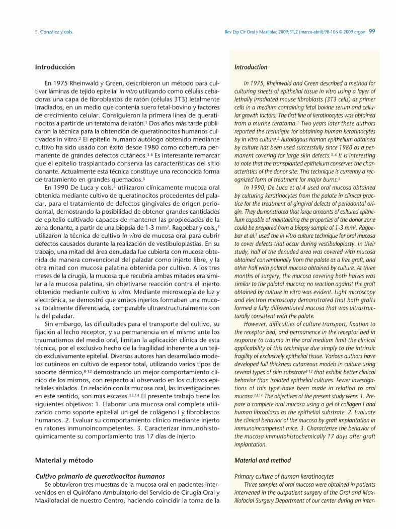
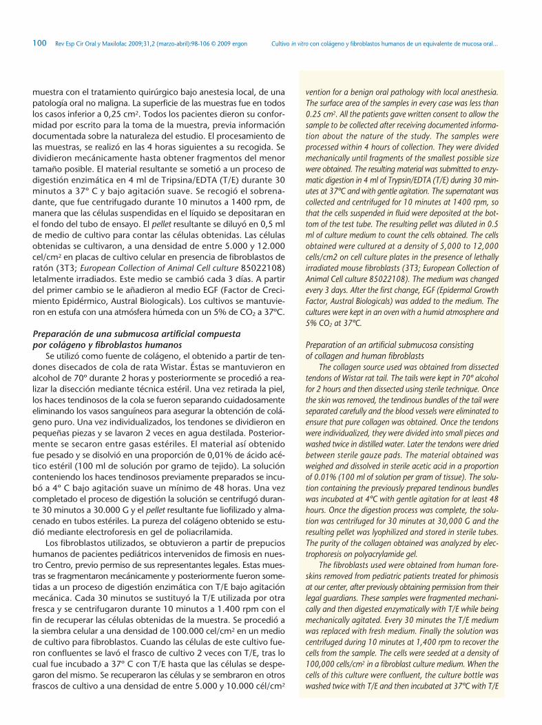
obtenidos en tres ratones atímicos(BALD/ c nu/un ) según la técnica des-crita por Barrandon y cols.15 (Fig. 1). Alos 17 días del injerto, se procedió alsacrificio del animal y biopsia del teji-do injertado para su análisis histológico.El análisis inmunohistoquímico se rea-lizó con kits para citoqueratinas 13 y 19,colágeno IV y laminina. En la figura 2 serecoge de manera esquemática, las prin-cipales etapas del trabajo.
Resultados
Cultivos primariosA los tres o cuatro días del cultivo fue
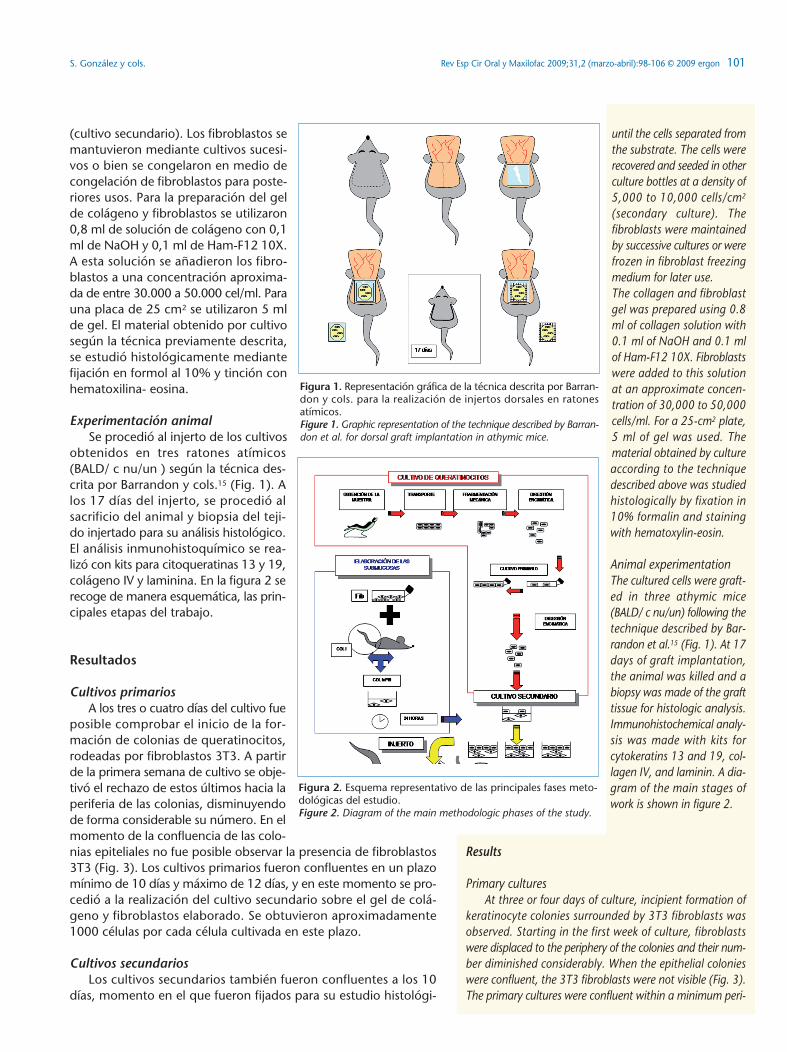
posible comprobar el inicio de la for-mación de colonias de queratinocitos,rodeadas por fibroblastos 3T3. A partirde la primera semana de cultivo se obje-tivó el rechazo de estos últimos hacia laperiferia de las colonias, disminuyendode forma considerable su número. En elmomento de la confluencia de las colo-nias epiteliales no fue posible observar la presencia de fibroblastos3T3 (Fig. 3). Los cultivos primarios fueron confluentes en un plazomínimo de 10 días y máximo de 12 días, y en este momento se pro-cedió a la realización del cultivo secundario sobre el gel de colá-geno y fibroblastos elaborado. Se obtuvieron aproximadamente1000 células por cada célula cultivada en este plazo.
Cultivos secundarios Los cultivos secundarios también fueron confluentes a los 10
días, momento en el que fueron fijados para su estudio histológi-
until the cells separated fromthe substrate. The cells wererecovered and seeded in otherculture bottles at a density of5,000 to 10,000 cells/cm2
(secondary culture). Thefibroblasts were maintainedby successive cultures or werefrozen in fibroblast freezingmedium for later use.The collagen and fibroblastgel was prepared using 0.8ml of collagen solution with0.1 ml of NaOH and 0.1 mlof Ham-F12 10X. Fibroblastswere added to this solutionat an approximate concen-tration of 30,000 to 50,000cells/ml. For a 25-cm2 plate,5 ml of gel was used. Thematerial obtained by cultureaccording to the techniquedescribed above was studiedhistologically by fixation in10% formalin and stainingwith hematoxylin-eosin.
Animal experimentationThe cultured cells were graft-ed in three athymic mice(BALD/ c nu/un) following thetechnique described by Bar-randon et al.15 (Fig. 1). At 17days of graft implantation,the animal was killed and abiopsy was made of the grafttissue for histologic analysis.Immunohistochemical analy-sis was made with kits forcytokeratins 13 and 19, col-lagen IV, and laminin. A dia-gram of the main stages ofwork is shown in figure 2.
Results
Primary culturesAt three or four days of culture, incipient formation of
keratinocyte colonies surrounded by 3T3 fibroblasts wasobserved. Starting in the first week of culture, fibroblastswere displaced to the periphery of the colonies and their num-ber diminished considerably. When the epithelial colonieswere confluent, the 3T3 fibroblasts were not visible (Fig. 3).The primary cultures were confluent within a minimum peri-
Figura 1. Representación gráfica de la técnica descrita por Barran-don y cols. para la realización de injertos dorsales en ratonesatímicos.Figure 1. Graphic representation of the technique described by Barran-don et al. for dorsal graft implantation in athymic mice.
Figura 2. Esquema representativo de las principales fases meto-dológicas del estudio.Figure 2. Diagram of the main methodologic phases of the study.
CO 31-2 4/5/09 15:44 Página 101
od of 10 days and a maxi-mum period of 12 days. Atthis moment the secondaryculture was made on the col-lagen and fibroblast gel pre-pared. The yield was approx-imately 1000 cells per cellcultured in this period.
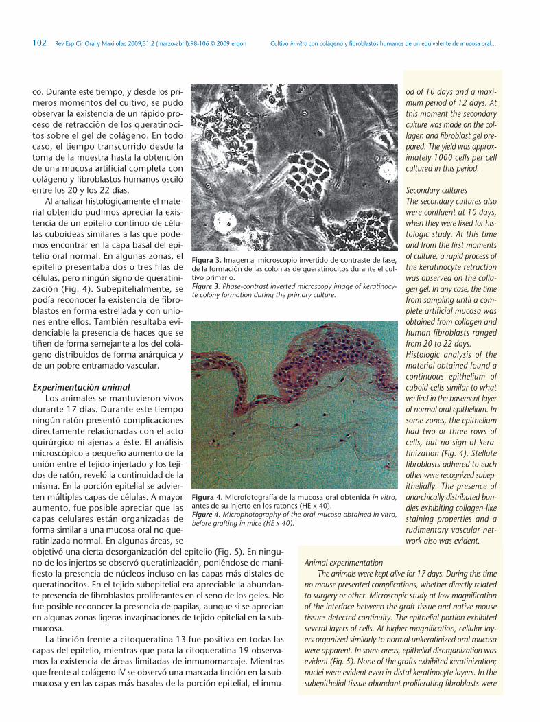
Secondary cultures The secondary cultures alsowere confluent at 10 days,when they were fixed for his-tologic study. At this timeand from the first momentsof culture, a rapid process ofthe keratinocyte retractionwas observed on the colla-gen gel. In any case, the timefrom sampling until a com-plete artificial mucosa wasobtained from collagen andhuman fibroblasts rangedfrom 20 to 22 days.Histologic analysis of thematerial obtained found acontinuous epithelium ofcuboid cells similar to whatwe find in the basement layerof normal oral epithelium. Insome zones, the epitheliumhad two or three rows ofcells, but no sign of kera-tinization (Fig. 4). Stellatefibroblasts adhered to eachother were recognized subep-ithelially. The presence ofanarchically distributed bun-dles exhibiting collagen-likestaining properties and arudimentary vascular net-work also was evident.
Animal experimentationThe animals were kept alive for 17 days. During this time
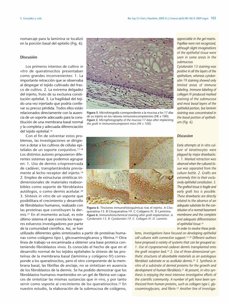
no mouse presented complications, whether directly relatedto surgery or other. Microscopic study at low magnificationof the interface between the graft tissue and native mousetissues detected continuity. The epithelial portion exhibitedseveral layers of cells. At higher magnification, cellular lay-ers organized similarly to normal unkeratinized oral mucosawere apparent. In some areas, epithelial disorganization wasevident (Fig. 5). None of the grafts exhibited keratinization;nuclei were evident even in distal keratinocyte layers. In thesubepithelial tissue abundant proliferating fibroblasts were
co. Durante este tiempo, y desde los pri-meros momentos del cultivo, se pudoobservar la existencia de un rápido pro-ceso de retracción de los queratinoci-tos sobre el gel de colágeno. En todocaso, el tiempo transcurrido desde latoma de la muestra hasta la obtenciónde una mucosa artificial completa concolágeno y fibroblastos humanos oscilóentre los 20 y los 22 días.
Al analizar histológicamente el mate-rial obtenido pudimos apreciar la exis-tencia de un epitelio continuo de célu-las cuboideas similares a las que pode-mos encontrar en la capa basal del epi-telio oral normal. En algunas zonas, elepitelio presentaba dos o tres filas decélulas, pero ningún signo de queratini-zación (Fig. 4). Subepitelialmente, sepodía reconocer la existencia de fibro-blastos en forma estrellada y con unio-nes entre ellos. También resultaba evi-denciable la presencia de haces que setiñen de forma semejante a los del colá-geno distribuidos de forma anárquica yde un pobre entramado vascular.
Experimentación animalLos animales se mantuvieron vivos
durante 17 días. Durante este tiemponingún ratón presentó complicacionesdirectamente relacionadas con el actoquirúrgico ni ajenas a éste. El análisismicroscópico a pequeño aumento de launión entre el tejido injertado y los teji-dos de ratón, reveló la continuidad de lamisma. En la porción epitelial se advier-ten múltiples capas de células. A mayoraumento, fue posible apreciar que lascapas celulares están organizadas deforma similar a una mucosa oral no que-ratinizada normal. En algunas áreas, seobjetivó una cierta desorganización del epitelio (Fig. 5). En ningu-no de los injertos se observó queratinización, poniéndose de mani-fiesto la presencia de núcleos incluso en las capas más distales dequeratinocitos. En el tejido subepitelial era apreciable la abundan-te presencia de fibroblastos proliferantes en el seno de los geles. Nofue posible reconocer la presencia de papilas, aunque si se aprecianen algunas zonas ligeras invaginaciones de tejido epitelial en la sub-mucosa.
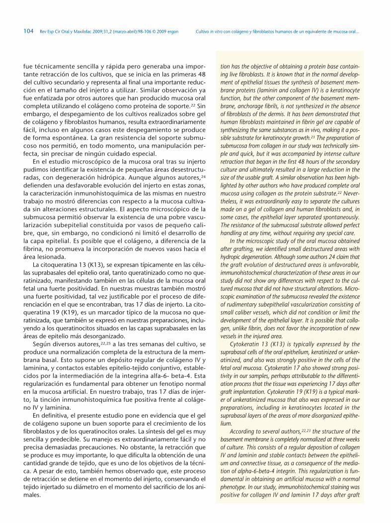
La tinción frente a citoqueratina 13 fue positiva en todas lascapas del epitelio, mientras que para la citoqueratina 19 observa-mos la existencia de áreas limitadas de inmunomarcaje. Mientrasque frente al colágeno IV se observó una marcada tinción en la sub-mucosa y en las capas más basales de la porción epitelial, el inmu-
Figura 3. Imagen al microscopio invertido de contraste de fase,de la formación de las colonias de queratinocitos durante el cul-tivo primario.Figure 3. Phase-contrast inverted microscopy image of keratinocy-te colony formation during the primary culture.
Figura 4. Microfotografía de la mucosa oral obtenida in vitro,antes de su injerto en los ratones (HE x 40).Figure 4. Microphotography of the oral mucosa obtained in vitro,before grafting in mice (HE x 40).
Cultivo in vitro con colágeno y fibroblastos humanos de un equivalente de mucosa oral...102 Rev Esp Cir Oral y Maxilofac 2009;31,2 (marzo-abril):98-106 © 2009 ergon
CO 31-2 4/5/09 15:44 Página 102
Rev Esp Cir Oral y Maxilofac 2009;31,2 (marzo-abril):98-106 © 2009 ergon 103S. González y cols.
nomarcaje para la laminina se localizóen la porción basal del epitelio (Fig. 6).
Discusión
Los primeros intentos de cultivo invitro de queratinocitos presentabancomo grandes inconvenientes: 1. Laimportante retracción que se observabaal despegar el tejido cultivado del fras-co de cultivo. 2. La extrema delgadezdel injerto, fruto de su exclusiva consti-tución epitelial. 3. La fragilidad del teji-do una vez injertado que podría conlle-var su precoz pérdida. Todos ellos estánrelacionados directamente con la ausen-cia de un soporte adecuado para la cons-titución de una membrana basal normaly la completa y adecuada diferenciacióndel tejido epitelial.16
Con el fin de solventar estos pro-blemas, las investigaciones se dirigie-ron a dotar a los cultivos de células epi-teliales de un soporte conjuntivo.17,18
Los distintos autores propusieron dife-rentes sistemas que podemos agruparen: 1. Uso de dermis criopreservadade cadáver, transplantándola previa-mente al lecho receptor del injerto.19
2. Empleo de estructuras sintéticas tri-dimensionales de materiales reabsor-bibles como soporte de fibroblastosautólogos, o como dermis acelular.20
3. Síntesis in vitro de un soporte queposibilitara el crecimiento y desarrollode fibroblastos humanos, realizado conlas proteínas que constituyen la der-mis.21 En el momento actual, es esteúltimo sistema el que concita los mayo-res esfuerzos investigadores por partede la comunidad científica. Así, se hanutilizado diferentes geles sintetizados a partir de proteínas huma-nas como colágeno tipo I, glicosaminoglicanos y fibrina.22 Otralínea de trabajo va encaminada a obtener una base proteica con-teniendo fibroblastos vivos. Es conocido el hecho de que en eldesarrollo normal de los tejidos epiteliales la síntesis de las pro-teínas de la membrana basal (laminina y colágeno IV) corres-ponde a los queratinocitos, pero el otro componente de la mem-brana basal, las fibrillas de anclaje, no se sintetizan en ausenciade los fibroblastos de la dermis. Se ha podido demostrar que losfibroblastos humanos mantenidos en un gel de fibrina son capa-ces de sintetizar las mismas sustancias que in vivo, y por tantoservir como soporte al crecimiento de los queratinocitos.23 Ennuestro estudio, la elaboración de la submucosa de colágeno,
appreciable in the gel matrix.Papillae were not recognized,although slight invaginationsof the epithelial tissue wereseen in some areas in thesubmucosa. Cytokeratin 13 staining waspositive in all the layers of theepithelium, whereas cytoker-atin 19 staining showed onlylimited areas of immunelabeling. Immune labeling ofcollagen IV produced markedstaining of the submucosaand most basal layers of theepithelial portion, but lamininstaining was concentrated inthe basal portion of epitheli-um (Fig. 6).
Discussion
Early attempts at in vitro cul-ture of keratinocytes wereplagued by major drawbacks:1. 1. Marked retraction wasobserved when the cultured tis-sue was separated from theculture bottle. 2. Grafts areextremely thin to their exclu-sively epithelial constitution. 3.The grafted tissue is fragile andearly graft loss is possible.These drawbacks are directlyrelated to the absence of anadequate substrate for the con-stitution of a normal basementmembrane and the completeand adequate differentiationof epithelial tissue.16
In order to resolve these prob-lems, investigations have focused on developing epithelialcell cultures with connective support.17,18 Different authorshave proposed a variety of systems that can be grouped as:1. Use of cryopreserved cadaver dermis transplanted ontothe graft receptor bed.19 2. Use of three-dimensional syn-thetic structures of absorbable materials as an autologousfibroblast substrate or as acellular dermis.20 3. Synthesis invitro of a substrate of dermal proteins for the growth anddevelopment of human fibroblasts.21 At present, in vitro syn-thesis is enjoying the most intensive investigative efforts ofthe scientific community. A number of gels have been syn-thesized from human proteins, such as collagen type I, gly-cosaminoglycans, and fibrin.22 Another line of investiga-
Figura 5. Microfotografía correspondiente a la mucosa a los 17 díasde su injerto en los ratones inmunoincompetentes (HE x 100).Figure 5. Microphotography of the mucosa 17 days after implantingthe graft in immunoincompetent mice (HE x 100).
Figura 6. Tinciones inmunohistoquímicas tras el injerto. A Cito-queratina-13. B Citoqueratina-19. C Colágeno IV. D LamininaFigure 6. Immunohistochemical staining after graft implantation. A.Cytokeratin-13. B. Cytokeratin-19. C. Collagen IV. D. Laminin.
CO 31-2 4/5/09 15:44 Página 103
tion has the objective of obtaining a protein base contain-ing live fibroblasts. It is known that in the normal develop-ment of epithelial tissues the synthesis of basement mem-brane proteins (laminin and collagen IV) is a keratinocytefunction, but the other component of the basement mem-brane, anchorage fibrils, is not synthesized in the absenceof fibroblasts of the dermis. It has been demonstrated thathuman fibroblasts maintained in fibrin gel are capable ofsynthesizing the same substances as in vivo, making it a pos-sible substrate for keratinocyte growth.23 The preparation ofsubmucosa from collagen in our study was technically sim-ple and quick, but it was accompanied by intense cultureretraction that began in the first 48 hours of the secondaryculture and ultimately resulted in a large reduction in thesize of the usable graft. A similar observation has been high-lighted by other authors who have produced complete oralmucosa using collagen as the protein substrate.22 Never-theless, it was extraordinarily easy to separate the culturesmade on a gel of collagen and human fibroblasts and, insome cases, the epithelial layer separated spontaneously.The resistance of the submucosal substrate allowed perfecthandling at any time, without requiring any special care.
In the microscopic study of the oral mucosa obtainedafter grafting, we identified small destructured areas withhydropic degeneration. Although some authors 24 claim thatthe graft evolution of destructured areas is unfavorable,immunohistochemical characterization of these areas in ourstudy did not show any differences with respect to the cul-tured mucosa that did not have structural alterations. Micro-scopic examination of the submucosa revealed the existenceof rudimentary subepithelial vascularization consisting ofsmall caliber vessels, which did not condition or limit thedevelopment of the epithelial layer. It is possible that colla-gen, unlike fibrin, does not favor the incorporation of newvessels in the injured area.
Cytokeratin 13 (K13) is typically expressed by thesuprabasal cells of the oral epithelium, keratinized or unker-atinized, and also was strongly positive in the cells of thefetal oral mucosa. Cytokeratin 17 also showed strong posi-tivity in our samples, perhaps attributable to the differenti-ation process that the tissue was experiencing 17 days aftergraft implantation. Cytokeratin 19 (K19) is a typical mark-er of unkeratinized mucosa that also was expressed in ourpreparations, including in keratinocytes located in thesuprabasal layers of the areas of more disorganized epithe-lium.
According to several authors,22,23 the structure of thebasement membrane is completely normalized at three weeksof culture. This consists of a regular deposition of collagenIV and laminin and stable contacts between the epitheli-um and connective tissue, as a consequence of the media-tion of alpha-6-beta-4 integrin. This regularization is fun-damental in obtaining an artificial mucosa with a normalphenotype. In our study, immunohistochemical staining waspositive for collagen IV and laminin 17 days after graft
fue técnicamente sencilla y rápida pero generaba una impor-tante retracción de los cultivos, que se inicia en las primeras 48del cultivo secundario y representa al final una importante reduc-ción en el tamaño del injerto a utilizar. Similar observación yafue enfatizada por otros autores que han producido mucosa oralcompleta utilizando el colágeno como proteína de soporte.22 Sinembargo, el despegamiento de los cultivos realizados sobre gelde colágeno y fibroblastos humanos, resulta extraordinariamentefácil, incluso en algunos casos este despegamiento se producede forma espontánea. La gran resistencia del soporte submu-coso nos permitió, en todo momento, una manipulación per-fecta, sin precisar de ningún cuidado especial.
En el estudio microscópico de la mucosa oral tras su injertopudimos identificar la existencia de pequeñas áreas desestructu-radas, con degeneración hidrópica. Aunque algunos autores,24
defienden una desfavorable evolución del injerto en estas zonas,la caracterización inmunohistoquímica de las mismas en nuestrotrabajo no mostró diferencias con respecto a la mucosa cultiva-da sin alteraciones estructurales. El aspecto microscópico de lasubmucosa permitió observar la existencia de una pobre vascu-larización subepitelial constituida por vasos de pequeño cali-bre, que, sin embargo, no condicionó ni limitó el desarrollo dela capa epitelial. Es posible que el colágeno, a diferencia de lafibrina, no promueva la incorporación de nuevos vasos hacia elárea lesionada.
La citoqueratina 13 (K13), se expresan típicamente en las célu-las suprabasales del epitelio oral, tanto queratinizado como no que-ratinizado, manifestando también en las células de la mucosa oralfetal una fuerte positividad. En nuestras muestras también mostróuna fuerte positividad, tal vez justificable por el proceso de dife-renciación en el que se encontraban, tras 17 días de injerto. La cito-queratina 19 (K19), es un marcador típico de la mucosa no que-ratinizada, que también se expresó en nuestras preparaciones, inclu-yendo a los queratinocitos situados en las capas suprabasales en lasáreas de epitelio más desorganizado.
Según diversos autores,22,25 a las tres semanas del cultivo, seproduce una normalización completa de la estructura de la mem-brana basal. Esto supone un depósito regular de colágeno IV ylaminina, y contactos estables epitelio-tejido conjuntivo, estable-cidos por la intermediación de la integrina alfa-6- beta-4. Estaregularización es fundamental para obtener un fenotipo normalen la mucosa artificial. En nuestro trabajo, tras 17 días de injer-to, la tinción inmunohistoquímica fue positiva frente al coláge-no IV y laminina.
En definitiva, el presente estudio pone en evidencia que el gelde colágeno supone un buen soporte para el crecimiento de losfibroblastos y de los queratinocitos orales. La síntesis del gel es muysencilla y predecible. Su manejo es extraordinariamente fácil y noprecisa demasiadas precauciones. No obstante, la retracción quese produce es muy importante, lo que dificulta la obtención de unacantidad grande de tejido, que es uno de los objetivos de la técni-ca. A pesar de esto, también hemos observado que, este procesode retracción se detiene en el momento del injerto, conservando eltejido injertado su diámetro en el momento del sacrificio de los ani-males.
Cultivo in vitro con colágeno y fibroblastos humanos de un equivalente de mucosa oral...104 Rev Esp Cir Oral y Maxilofac 2009;31,2 (marzo-abril):98-106 © 2009 ergon
CO 31-2 4/5/09 15:44 Página 104
Rev Esp Cir Oral y Maxilofac 2009;31,2 (marzo-abril):98-106 © 2009 ergon 105S. González y cols.
Agradecimientos
A la Sociedad Española de Cirugía Oral y Maxilofacial e indus-trias Tarma S.A., por el apoyo prestado para la realización de estetrabajo (Beca de Investigación Básica Dr. Gómez Iglesias).
A la Ficyt (Eva García Pérez) y MBA (Verónica García Díaz) porsu apoyo técnico.
Bibliografía
1. Rheinwald J, Green H. Serial cultivation of strain of human epidermal kerati-
nocytes: the formation of keratinizing colonies from single cells. Cell 1975;6:331-
44.
2. Green H, Kehinde O, ThomasJ. Growth of cultured human epidermal cells into
multiple epithelia suitable for grafting. Proc Natl Acad Sci 1979;76:5655-68.
3. O’Connor EN., Mulliken JB., Banks-Schlegel S, Kehinde O. Grafting of burns with
cultured epithelium prepared from autologous epidermal cells. Lancet 1981;
1:75-8.
4. De Luca M., Albanese E, Megna M, y cols. Evidence that human oral epithe-
lium reconstituted in vitro and transplanted onto patients with defects in the
oral mucosa retains the properties of the donor site. Transplantation 1990;50:
454-59.
5. Gallico GC, O’Conor NE., Compton CC, et al. Permanent coverage of large burn
wounds with autologous cultured human epithelium. N Engl J Med 1984; 311:448-
51.
6. Pellegrini G, Bondanza S, Guerra L, De Luca M. Cultivation of human kerati-
nocyte item cells: current and future clinical applications. Med Biol Eng Comp
1998;36:778-90.
7. Ragoebar JM, Tomson AM, Scholma J. Blaauw EH. Use of culture mucosal grafts
to cover defects caused by vestibuloplasty. J Oral Maxillofac Surg 1995;53:872-
78.
8. Lam KP, Cham SY, To WH. y cols. Development and evaluation of a new com-
posite laser skin graft. J Traumatol 1999;47:918-22.
9. Bohnert A, Hormung J, MacKenzie IC, Epithelio- mesenchimal interactions con-
trol basement membrane production and diferentiation in cultured and trans-
planted mouse keratinocytes. Cell Tissue Res 1986;244:413-29.
10. Bell E, Ehrlich NP, Buttle DJ. Aliving tissue formed in vitro and accepted as a full
thickness skin equivalent. Science 1981;211:1052-56.
11. Worst PKM, MacKenzie IC, Fusenig NE. Reformation of organized epidermal
structure by transplantation of suspensions and cultures of epidermal and der-
mal cells. Cell Tissue Res 1982;225:65-9.
12. Heck EL, Bergstresser PR, Baxter CR. Composite skin graft: frozen dermal allo-
grafts support the engraftment and expansion of autólogous epidermia. J Trau-
matol 1985;25:106-11.
13. Odioso LL, Doyle MJ, Quinn KW, Bartel RL, Zimber MP. Developement and
caracterization of an in vitro gingival epithelial model. J Periodontol Res
1995;30:210-9.
14. Moriyama T, Asahina I, Ishii M y cols. Development of composite cultured oral
mucosa utilizing collagen sponge matrix and contracted collagen gel: a preli-
minary study for clinical applications. Tissue Eng 2001;7:415-27.
15. Barrandon Y, Li V, Green H. New techniques for the grafting of cultured human
epidermal cells onto athymic animals. J Investig Med 1988;91:315-18.
16. Fabre JW, Cullen PR. Rejection of cultured keratinocyte allografts in the rat. Trans-
plantation 1989;48:306-15.
implantation. The present study shows that collagen gel was a good
substrate for the growth of fibroblasts and oral keratinocytes.Synthesis of the gel was simple and predictable. Its man-agement was extraordinarily easy and no exceptional pre-cautions were necessary. However, marked retraction occurredand it was difficult to obtain a large amount of tissue, whichwas one of the objectives of the technique. Nonetheless, wealso observed that the retraction process ceased when thegraft is implanted and the grafted tissue conserved its diam-eter when the animals were killed.
Acknowledgements
We would like to thank the Spanish Society of Oral andMaxillofacial Surgery and Industrias Tarma, S.A. for sup-porting our work (Dr. Gómez Iglesias Basic Research Grant),
as well as Ficyt (Eva García Perez) and MBA (VerónicaGarcía Díaz) for their technical support.
CO 31-2 4/5/09 15:44 Página 105
17. Izumi K, Feinberg SE, Iida A, Yoshizawa M. Intraoral grafting o fan
ex vivo produced oral mucosa equivalent: a preliminary report. Int J
Oral Maxillofac Surg 2003;32:188-97.
18. Bodner L, Grossman N. Autologous cultured mucosal graft to cover
large intraoral mucosal defects: a clinical study. J Oral Maxillofac Surg
2003;61:169-73.
19. Medalie DA, Erming SA, Tompinks RG, Yarmush ML, Krueger GG, Mor-
gan JR. Evaluation of human skin reconstituted from composite grafts
of cultured keratinocytes and human acellular dermis transplanted to
atimic mice. J Investigat Dermatol 1996;107:121-7.
20. Wright KA, Nadire KB, Busto P, Tubo R, McPherson JM, Wentworth
R. Alternative delivery of keratinocytes using a poliuretane membra-
ne and the implications for its use in the treatment of full-thickness
burn injury. Burns 1998;24:7-21.
22. Ueda M, Ebata K, Kaneda T. In vitro fabrication of bioartificial muco-
sa for reconstitution of oral mucosa: basic research and clinical appli-
cation. Ann Plast Surg 1991;27:540-9.
23. Meana A., Iglesias J, Del Rio M, Larcher F, Madrigal B, Fresno MF. Large
surface of cultureed human epithelium obtained on a dermal matrix
based on live fibroblast-containing fibrin gels. Burns 1998;24:
621-30.
24. Lindberg K, Rheinwald JG. Three distinct keratinocyte subtypes iden-
tified in human oral epithelium by their patterns of keratin expression
in culture and xenografts. Differentiation 1990;45:230-41.
25. Loro LL, Dimba EA, Vintermyr OK, Johannessen AC. Crucial effects
of fibroblasts and keratinocyte growth factor on morphogenesis of
reconstitued human oral epithelium. J Invest Dermatol 2003;121:
1479-86.
Cultivo in vitro con colágeno y fibroblastos humanos de un equivalente de mucosa oral...106 Rev Esp Cir Oral y Maxilofac 2009;31,2 (marzo-abril):98-106 © 2009 ergon
CO 31-2 4/5/09 15:44 Página 106


![Péptidos de colágeno – Soluciones versátiles en salud y ... · ¿Qué son los péptidos de colágeno? [2] Los péptidos de colágeno, son cadenas pequeñas de proteínas, que](https://static.fdocuments.es/doc/165x107/5ac079cc7f8b9ac6688c3499/pptidos-de-colgeno-soluciones-verstiles-en-salud-y-son-los-pptidos-de-colgeno.jpg)