Diagnosis and Treatment of Forefoot Disorders. Section 1 ...
43
CLINICAL PRACTICE GUIDELINE Diagnosis and Treatment of Forefoot Disorders. Section 1: Digital Deformities Clinical Practice Guideline Forefoot Disorders Panel: James L. Thomas, DPM, 1 Edwin L. Blitch, IV, DPM, 2 D. Martin Chaney, DPM, 3 Kris A. Dinucci, DPM, 4 Kimberly Eickmeier, DPM, 5 Laurence G. Rubin, DPM, 6 Mickey D. Stapp, DPM, 7 and John V. Vanore, DPM 8 T his clinical practice guideline (CPG) is based upon consensus of current clinical practice and review of the clinical literature. The guideline was developed by the Clin- ical Practice Guideline Forefoot Disorders Panel of the American College of Foot and Ankle Surgeons. The guide- line and references annotate each node of the corresponding pathways. Introduction to Forefoot Disorders (Pathway 1) Forefoot pain is a common presenting complaint seen by foot and ankle surgeons. Patients often describe their pain in a vague and encompassing manner. The purpose of this clinical practice guideline is to review the varied patholo- gies that comprise the differential diagnosis of forefoot pain, with the exclusion of disorders of the first ray. The pathol- ogies in the differential diagnosis range from acquired or- thopedic deformities (eg, hammertoes, digital deformities) to overuse problems and traumatic injuries. These clinical problems, encountered daily by the foot and ankle surgeon, typically involve the lesser toes and metatarsals and their respective joints. Presented in this document are current practice guidelines for the diagnosis and treatment of ham- mertoe (digital deformities) (Pathway 2); central metatar- salgia (Pathway 3); Morton’s neuroma (Pathway 4); tailor’s bunion (Pathway 5); and trauma (Pathway 6). Digital Deformities (Pathway 2) Digital deformities are among the most common fore- foot pathologies encountered by foot and ankle surgeons. These deformities may be either congenital or acquired, with the incidence of digital deformities greater among females than males in almost all age groups (1). Whereas biomechanical dysfunction is usually discussed as the primary cause of digital deformities, these pathologies also may be caused by a variety of other conditions including neuromuscular and arthritic disorders (2-4). The proper identification of the deforming forces and resultant tendon and capsuloligamentous imbalance is critical in determining the treatment plan. Digital defor- mities may occur as an isolated entity or as a component of other foot and ankle conditions (1, 5). Significant History (Pathway 2, Node 1) Patients presenting with digital deformities may either report varying degrees of pain or be asymptomatic. If pain is present, it may occur dorsally, medially, laterally, at the distal end of the toe, or plantar to the respective metatarsal head. Dorsal pain may be secondary to pres- sure from footwear, whereas pain at the distal end of the toe may be secondary to contracture and the resultant shift of pressure away from the more plantar padded area of the toe. Patients may report a history of deformity since birth or early childhood. More commonly, patients will have first noticed positional changes of the toe during either early adulthood or in later years. Patients may state either that the extent of deformity of the toe seems to have reached an endpoint, or that they are still noticing a Address correspondence to: James L. Thomas, DPM, University of Florida, Department of Orthopaedics and Rehabilitation, 655 West 8th St, Jacksonville, FL 32209. E-mail: [email protected]. 1 Chair, Jacksonville, FL; 2 Charleston, SC; 3 San Antonio, TX; 4 Scotts- dale, AZ; 5 Champaign, IL; 6 Mechanicsville, VA; 7 Augusta, GA; 8 Gadsden, AL. Copyright © 2009 by the American College of Foot and Ankle Surgeons 1067-2516/09/4802-0022$36.00/0 doi:10.1053/j.jfas.2008.12.003 230 THE JOURNAL OF FOOT & ANKLE SURGERY
Transcript of Diagnosis and Treatment of Forefoot Disorders. Section 1 ...

CLINICAL PRACTICE GUIDELINE
Diagnosis and Treatment of ForefootDisorders. Section 1: Digital Deformities
Clinical Practice Guideline Forefoot Disorders Panel: James L. Thomas, DPM,1
Edwin L. Blitch, IV, DPM,2 D. Martin Chaney, DPM,3 Kris A. Dinucci, DPM,4
Kimberly Eickmeier, DPM,5 Laurence G. Rubin, DPM,6 Mickey D. Stapp, DPM,7
and John V. Vanore, DPM8
This clinical practice guideline (CPG) is based uponconsensus of current clinical practice and review of theclinical literature. The guideline was developed by the Clin-ical Practice Guideline Forefoot Disorders Panel of theAmerican College of Foot and Ankle Surgeons. The guide-line and references annotate each node of the correspondingpathways.
Introduction to Forefoot Disorders (Pathway 1)
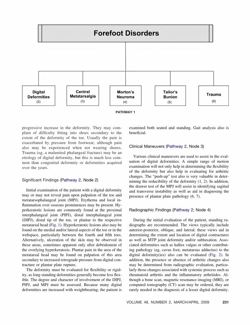
Forefoot pain is a common presenting complaint seen byfoot and ankle surgeons. Patients often describe their pain ina vague and encompassing manner. The purpose of thisclinical practice guideline is to review the varied patholo-gies that comprise the differential diagnosis of forefoot pain,with the exclusion of disorders of the first ray. The pathol-ogies in the differential diagnosis range from acquired or-thopedic deformities (eg, hammertoes, digital deformities)to overuse problems and traumatic injuries. These clinicalproblems, encountered daily by the foot and ankle surgeon,typically involve the lesser toes and metatarsals and theirrespective joints. Presented in this document are currentpractice guidelines for the diagnosis and treatment of ham-mertoe (digital deformities) (Pathway 2); central metatar-salgia (Pathway 3); Morton’s neuroma (Pathway 4); tailor’sbunion (Pathway 5); and trauma (Pathway 6).
Digital Deformities (Pathway 2)
Digital deformities are among the most common fore-foot pathologies encountered by foot and ankle surgeons.These deformities may be either congenital or acquired,with the incidence of digital deformities greater amongfemales than males in almost all age groups (1). Whereasbiomechanical dysfunction is usually discussed as theprimary cause of digital deformities, these pathologiesalso may be caused by a variety of other conditionsincluding neuromuscular and arthritic disorders (2-4).The proper identification of the deforming forces andresultant tendon and capsuloligamentous imbalance iscritical in determining the treatment plan. Digital defor-mities may occur as an isolated entity or as a componentof other foot and ankle conditions (1, 5).
Significant History (Pathway 2, Node 1)
Patients presenting with digital deformities may eitherreport varying degrees of pain or be asymptomatic. Ifpain is present, it may occur dorsally, medially, laterally,at the distal end of the toe, or plantar to the respectivemetatarsal head. Dorsal pain may be secondary to pres-sure from footwear, whereas pain at the distal end of thetoe may be secondary to contracture and the resultantshift of pressure away from the more plantar padded areaof the toe.
Patients may report a history of deformity since birthor early childhood. More commonly, patients will havefirst noticed positional changes of the toe during eitherearly adulthood or in later years. Patients may state eitherthat the extent of deformity of the toe seems to havereached an endpoint, or that they are still noticing a
Address correspondence to: James L. Thomas, DPM, University ofFlorida, Department of Orthopaedics and Rehabilitation, 655 West 8th St,Jacksonville, FL 32209. E-mail: [email protected].
1Chair, Jacksonville, FL; 2Charleston, SC; 3San Antonio, TX; 4Scotts-dale, AZ; 5Champaign, IL; 6Mechanicsville, VA; 7Augusta, GA; 8Gadsden,AL.
Copyright © 2009 by the American College of Foot and Ankle Surgeons1067-2516/09/4802-0022$36.00/0doi:10.1053/j.jfas.2008.12.003
230 THE JOURNAL OF FOOT & ANKLE SURGERY
progressive increase in the deformity. They may com-plain of difficulty fitting into shoes secondary to theextent of the deformity of the toe. Usually the pain isexacerbated by pressure from footwear, although painalso may be experienced when not wearing shores.Trauma (eg, a malunited phalangeal fracture) may be anetiology of digital deformity, but this is much less com-mon than congenital deformity or deformities acquiredover the years.
Significant Findings (Pathway 2, Node 2)
Initial examination of the patient with a digital deformitymay or may not reveal pain upon palpation of the toe andmetatarsophalangeal joint (MPJ). Erythema and local in-flammation over osseous prominences may be present. Hy-perkeratotic lesions are commonly found at the proximalinterphalangeal joint (PIPJ), distal interphalangeal joint(DIPJ), distal tip of the toe, or plantar to the respectivemetatarsal head (Fig. 1). Hyperkeratotic lesions also may befound on the medial and/or lateral aspects of the toe or in thewebspace, particularly between the fourth and fifth toes.Alternatively, ulceration of the skin may be observed inthese areas, sometimes apparent only after debridement ofthe overlying hyperkeratosis. Plantar pain in the area of themetatarsal head may be found on palpation of this areasecondary to increased retrograde pressure from digital con-tracture or plantar plate rupture.
The deformity must be evaluated for flexibility or rigid-ity, as long-standing deformities generally become less flex-ible. The degree and character of involvement of the DIPJ,PIPJ, and MPJ must be assessed. Because many digitaldeformities are increased with weightbearing, the patient is
examined both seated and standing. Gait analysis also isbeneficial.
Clinical Maneuvers (Pathway 2, Node 3)
Various clinical maneuvers are used to assist in the eval-uation of digital deformities. A simple range of motionexamination will not only help in determining the flexibilityof the deformity but also help in evaluating for arthriticchanges. The “push-up” test also is very valuable in deter-mining the reducibility of the deformity (1, 2). In addition,the drawer test of the MPJ will assist in identifying sagittaland transverse instability as well as aid in diagnosing thepresence of plantar plate pathology (6, 7).
Radiographic Findings (Pathway 2; Node 4)
During the initial evaluation of the patient, standing ra-diographs are recommended. The views typically includeanterior-posterior, oblique, and lateral; these views aid indetermining the extent and location of digital contracturesas well as MTP joint deformity and/or subluxation. Asso-ciated deformities such as hallux valgus or other contribut-ing pathology (eg, cavus foot, metatarsus adductus) to thedigital deformity(ies) also can be evaluated (Fig. 2). Inaddition, the presence or absence of arthritic changes alsomay be determined from radiographic evaluation, particu-larly those changes associated with systemic process such asrheumatoid arthritis and the inflammatory arthritides. Al-though a bone scan, magnetic resonance imaging (MRI), orcomputed tomography (CT) scan may be ordered, they arerarely needed in the diagnosis of a lesser digital deformity.
PATHWAY 1
VOLUME 48, NUMBER 2, MARCH/APRIL 2009 231

PATHWAY 2
232 THE JOURNAL OF FOOT & ANKLE SURGERY

FIGURE 1 Digital deformities are associated with a variety of hyperkeratotic lesions, clavi, or ulcerations including (A) dorsally at PIPJ orDIPJ, (B) distal tip of toes, or (C) medial or lateral condylar surfaces at DIPJs or PIPJs, where adjacent toes rub each other.
FIGURE 2 Digital deformities are generally associated with foot pathologies that result in MTP joint instability and digital contractures.Shown here: (A) hallux varus with digital adductus, (B) hallux valgus, (C) rheumatoid arthritis, and (D) pes cavus.
VOLUME 48, NUMBER 2, MARCH/APRIL 2009 233

Diagnosis (Pathway 2, Node 5)
After consideration of the history, examination, radiog-raphy, and clinical maneuver results, diagnosis of the typeand extent of digital deformity can be made. Deformities ofthe lesser toes are defined classically as hammertoe, claw-toe, and mallet toe (Fig. 3). Although these deformities areall very similar to each other, a few minor differences exist.Hammertoe refers to the deformity that consists of an ex-tension contracture at the MPJ, flexion contracture at thePIPJ, and hyperextension at the DIPJ. Clawtoe deformityexhibits an extension contracture at the MPJ and a flexioncontracture at both the PIPJ and DIPJ. A toe whose onlydeformity consists of a flexion contracture at the DIPJ istermed a mallet toe.
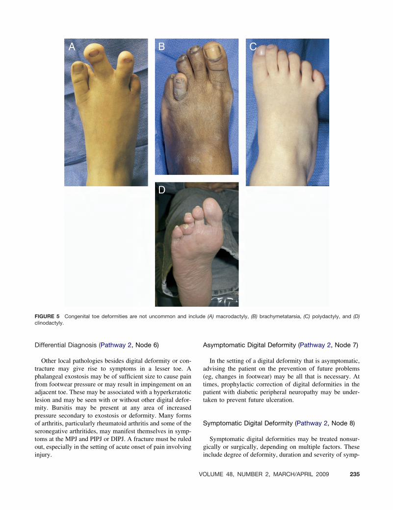
There also are separate and distinct deformities involvingthe second toe and fifth toe. When an extension contractureis combined with medial deviation (subluxation) at the levelof the second MPJ, a “crossover” second toe deformityresults (6, 7). This deformity often is combined with ahallux valgus deformity. Pain in and around the second MPJthat occurs before significant subluxation is seen is referredto as “pre-subluxation syndrome.” Adduction or abductiondigital deformities may involve all lesser MTP joints or, insome cases, divergent digital contractures are seen (Fig. 4).Fifth toe pathology may include deformity in multipleplanes (adductovarus deformity), or significant overlap ofthe fifth toe over the fourth toe may be seen. These toedeformities of the fifth toe may be congenital in nature;other congenital deformities include polydactyly, syndac-tyly, clinodactyly and macrodactyly (Fig. 5).
FIGURE 3 Digital deformities may be described by the combina-tion of joint contractures at the digital segments: (A) Hammertoe:flexion at the PIPJ with extension at the MPJ with a neutral orhyperextended DIPJ, (B) Clawtoe: flexion at both the PIPJ and DIPJcombined with extension at the MPJ, (C) Mallet toe: flexion at theDIPJ.
FIGURE 4 Although digital deformities are typically described as sagittal plane deformities, they may take on multiplanar deviation.Radiographs show: (A) typical hammertoe with sagittal plane instability, (B) divergent toe with abduction of third toe, and (C) adductioncontracture of the lesser toes.
234 THE JOURNAL OF FOOT & ANKLE SURGERY
Differential Diagnosis (Pathway 2, Node 6)
Other local pathologies besides digital deformity or con-tracture may give rise to symptoms in a lesser toe. Aphalangeal exostosis may be of sufficient size to cause painfrom footwear pressure or may result in impingement on anadjacent toe. These may be associated with a hyperkeratoticlesion and may be seen with or without other digital defor-mity. Bursitis may be present at any area of increasedpressure secondary to exostosis or deformity. Many formsof arthritis, particularly rheumatoid arthritis and some of theseronegative arthritides, may manifest themselves in symp-toms at the MPJ and PIPJ or DIPJ. A fracture must be ruledout, especially in the setting of acute onset of pain involvinginjury.
Asymptomatic Digital Deformity (Pathway 2, Node 7)
In the setting of a digital deformity that is asymptomatic,advising the patient on the prevention of future problems(eg, changes in footwear) may be all that is necessary. Attimes, prophylactic correction of digital deformities in thepatient with diabetic peripheral neuropathy may be under-taken to prevent future ulceration.
Symptomatic Digital Deformity (Pathway 2, Node 8)
Symptomatic digital deformities may be treated nonsur-gically or surgically, depending on multiple factors. Theseinclude degree of deformity, duration and severity of symp-
FIGURE 5 Congenital toe deformities are not uncommon and include (A) macrodactyly, (B) brachymetatarsia, (C) polydactyly, and (D)clinodactyly.
VOLUME 48, NUMBER 2, MARCH/APRIL 2009 235

toms, previous treatment, associated medical conditions,and ability to perform work duties comfortably.
Nonsurgical Treatment Options (Pathway 2, Node 9)
Nonsurgical treatment is often the initial treatment choicefor the symptomatic digital deformity. Various paddingtechniques exist, serving to cushion or offload pressurepoints that may involve both the affected toe(s) as well as itsrespective metatarsal head plantarly. Orthotic devices orshoe insole modifications using a metatarsal pad may offerrelief of excessive metatarsal head pressures. Debridementof associated hyperkeratotic lesions usually is effective inhelping to reduce symptoms. If local inflammation or bur-sitis exists, a corticosteroid injection into the affected areamay be beneficial. Taping to reduce and splint flexibledeformities may be performed, especially in the setting ofan early crossover second toe deformity. Last but not least,footwear changes such as a wider and/or deeper toe boxmay be used to accommodate the deformity and decreaseshoe pressure over osseous prominences.
Surgical Treatment Options (Pathway 2, Node 10)
The surgical treatment of digital deformities includes aspectrum of soft tissue and osseous procedures. The degreeand flexibility of the deformity along with any associatedpathology determine the surgical procedure(s) to be per-formed.
When the deformity is manually reducible, tenotomy ortendon lengthening at the level of the MPJ, PIPJ, or DIPJ
may be sufficient for deformity correction; however, thismay require combining with capsular and/or ligamentousrelease (or reefing), especially at the level of the MPJ(8-10). In some cases, phalangeal head resection (partial orcomplete) and/or flexor tendon transfer also may be neces-sary (11).
When the deformity is only manually semi-reducible orrigid, both osseous and soft tissue procedures often areperformed in combination. Osseous procedures of the toeinclude phalangeal head resection (with or without implant)and arthrodesis of the PIPJ and DIPJ (3, 11-24) (Fig. 6).Sometimes metatarsal osteotomy, partial metatarsal headresection, or phalangeal base resection may be required toachieve complete correction of the digital deformity, espe-cially at the level of the MPJ (1, 2, 25-27) (Fig. 7). Softtissue procedures are commonly utilized to augment osse-ous procedures in this patient population; soft tissue proce-dures include all of the aforementioned procedures for flex-ible deformities. Exostectomy also may be beneficial,particularly in addressing hyperkeratotic lesions along themedial or lateral aspects of the toe (27, 28). Partial ampu-tation of the toe may be indicated in some cases, especiallyin conditions involving the fifth toe. In selected cases,complete amputation of a lesser toe may be considered toallow shoe fitting, such as in the coexistence of second toedeformity and hallux valgus deformity in an elderly patient(29).
Correction of associated conditions may be indicated inthe surgical care of some digital deformities. This is espe-cially true in crossover second toe deformity, where halluxvalgus deformity often is seen concurrently and may influ-ence attempts at correction of the second toe deformity (30).
FIGURE 6 (A) This patient had digital deformites that included flexion at both the PIPJ and DIPJ. Note the rather long middle phalanges.(B) The patient underwent PIPJ fusion of the second, third, and fourth toes combined with arthroplasty of the DIPJ with kirschner wirestabilization as well as hallux valgus correction. (C) This is a radiograph at 1 year postsurgery.
236 THE JOURNAL OF FOOT & ANKLE SURGERY

Surgical repair of associated tears of the plantar plate alsohas been advocated (6) (see Section 2. Central Metatarsal-gia, Fig. 8). In addition, correction of other forefoot, mid-foot, or hindfoot conditions contributing to the formation ofdigital deformity may be indicated.
Continued Symptoms (Pathway 5, Node 11)
Treatment of the patient who continues to experiencesymptoms after surgical care of a digital deformity mayrequire a variety of revisional surgical techniques and/or
nonsurgical measures. In some cases, recurrence of theoriginal deformity or migration of phalangeal segmentsoccur as a complication of the original repair. Revisionalsurgery alternatives are similar to the above for originalprocedural selection.
References
1. Kelikian H. Deformities of the lesser toes. In: Hallux Valgus, AlliedDeformities of the Forefoot and Metatarsalgia, pp 282–336, W.B.Saunders Co, Philadelphia, 1965.
2. Cooper PS. Disorders and deformities of the lesser toes. In: Foot andAnkle Disorders, Vol 1, pp 308–358, edited by MS Myerson, W.B.Saunders Co, Philadelphia, 2000.
3. McGlamry ED, Jimenez AL, Green DR. Lesser ray deformities. In:McGlamry’s Comprehensive Textbook of Foot and Ankle Surgery, pp253–371, edited by Alan S. Banks, Michael S. Downey, Dennis E.Martin, Stephen J. Miller. Lippincott Williams & Wilkins, Philadel-phia, 2001.
4. O’Connell PG, Lohmann Siegel K, Kepple TM, Stanhope SJ, GerberLH. Forefoot deformity, pain, and mobility in rheumatoid and nonar-thritic subjects. J Rheumatol 25:1681–1686, 1998.
5. Root ML, Orien WP, Weed JH. Hammertoe deformity. In: Normal andAbnormal Function of the Foot, pp 452–459, edited by ML Root, WPOrien, and JH Weed, Clinical Biomechanics Corporation, Los Ange-les, 1977.
6. Yu GV, Judge MS, Hudson JR, Seidelmann FE. Predislocation syn-drome. Progressive subluxation/dislocation of the lesser metatarsopha-langeal joint. J Am Podiatr Med Assoc 92:182–199, 2002.
7. Stainsby GD. Pathological anatomy and dynamic effect of the dis-placed plantar plate and the importance of the integrity of the plantarplate-deep transverse metatarsal ligament tie-bar. Ann R Coll SurgEngl 79:58–68, 1997.
8. Roven MD. Tenotomy, tenectomy, and capsulotomy for the lessertoes. Clin Podiatry 2:471–475, 1985.
9. Greenberg HH. Plantar digital tenotomy for underlapping and con-tracted toes. A preliminary report. J Am Podiatry Assoc 56:65–66,1966.
10. Barbari SG, Brevig K. Correction of clawtoes by the Girdlestone-Taylor flexor-extensor transfer procedure. Foot Ankle 5:67–73, 1984.
11. O’Kane C, Kilmartin T. Review of proximal interphalangeal jointexcisional arthroplasty for the correction of second hammer toe defor-mity in 100 cases. Foot Ankle Int 26:320–325, 2005.
12. Konkel KF, Menger AG, Retzlaff SA. Hammer toe correction using anabsorbable intramedullary pin. Foot Ankle Int 28:916–920, 2007.
13. Jones S, Hussainy HA, Flowers MJ. Re: Arthrodesis of the toe jointswith an intramedullary cannulated screw for correction of hammertoedeformity. Foot Ankle Int 26:1101, author reply 1101, 2005.
14. Caterini R, Farsetti P, Tarantino U, Potenza V, Ippolito E. Arthrodesisof the toe joints with an intramedullary cannulated screw for correctionof hammertoe deformity. Foot Ankle Int 25:256–261, 2004.
15. Weil LS Jr. Hammertoe arthrodesis using conical reamers and internalpin fixation. J Foot Ankle Surg 38:370–371, 1999.
16. Harmonson JK, Harkless LB. Operative procedures for the correctionof hammertoe, claw toe, and mallet toe: a literature review. ClinPodiatr Med Surg 13:211–220, 1996.
17. Yu GV, Vincent AL, Khoury WE, Schinke TL. Techniques of digitalarthrodesis: revisiting the old and discovering the new. Clin PodiatrMed Surg 21:17–50, 2004.
18. Sgarlato TE, Tafuri SA. Digital implant arthroplasty. Clin Podiatr MedSurg 13:255–262, 1996.
FIGURE 7 (A) Digital deformities may be very complex, as seen inthis 65-year-old female and (B) presurgical radiograph with a cross-over second toe but degree of adduction contracture of all thelesser toes combined with a hallux valgus deformity. (C) Surgicalmanagement included bunionectomy with implant arthroplasty andPIPJ fusion of the second, third, and fourth toes with MPJ releasesincluding a proximal phalangeal base resection of the second toe.(D) Shown here is a postsurgical radiograph.
VOLUME 48, NUMBER 2, MARCH/APRIL 2009 237

19. Kimmel HM, Garrow S. A comparison of end-to-end versus “V”arthrodesis procedures for the correction of digital deformities. ClinPodiatr Med Surg 13:239–250, 1996.
20. Lehman DE, Smith RW. Treatment of symptomatic hammertoe with aproximal interphalangeal joint arthrodesis. Foot Ankle Int 16:535–541,1995.
21. Pichney GA, Derner R, Lauf E. Digital “V” arthrodesis. J Foot AnkleSurg 32:473–479, 1993.
22. Ohm OW II, McDonell M, Vetter WA. Digital arthrodesis: an alternatemethod for correction of hammer toe deformity. J Foot Surg 29:207–211, 1990.
23. Monson DK, Buell TR, Scurran BL. Lesser digital arthrodesis. ClinPodiatr Med Surg 3:347–356, 1986.
24. Lanham RH Jr. Digital implant arthroplasty. Clin Podiatry 1:47–68,1984.
25. Conklin MJ, Smith RW. Treatment of the atypical lesser toe deformitywith basal hemiphalangectomy. Foot Ankle Int 15:585–594, 1994.
26. van Loon PJ, Aries RP, Karthaus RP, Steenaert BJ. Metatarsal headresection in the deformed, symptomatic rheumatic foot. A comparisonof two methods. Acta Orthop Belg 58:11–15, 1992.
27. Mercado OA. Digital surgery. In: An Atlas of Foot Surgery, Vol 1.Forefoot Surgery, pp 45–92, Carolando Press, Oak Park, IL, 1979.
28. Coughlin MJ, Kennedy MP. Operative repair of fourth and fifth toecorns. Foot Ankle Int 24:147–157, 2003.
29. Gallentine JW, DeOrio JK. Removal of the second toe for severehammertoe deformity in elderly patients. Foot Ankle Int 26:353–358,2005.
30. Karlock LG. Second metatarsophalangeal joint fusion: a new tech-nique for crossover hammertoe deformity. A preliminary report. J FootAnkle Surg 42:178–182, 2003.
238 THE JOURNAL OF FOOT & ANKLE SURGERY

Diagnosis and Treatment of ForefootDisorders. Section 2. Central Metatarsalgia
Clinical Practice Guideline Forefoot Disorders Panel: James L. Thomas, DPM,1
Edwin L. Blitch, IV, DPM,2 D. Martin Chaney, DPM,3 Kris A. Dinucci, DPM,4
Kimberly Eickmeier, DPM,5 Laurence G. Rubin, DPM,6 Mickey D. Stapp, DPM,7 andJohn V. Vanore, DPM8
This clinical practice guideline (CPG) is based uponconsensus of current clinical practice and review of theclinical literature. The guideline was developed by the Clin-ical Practice Guideline Forefoot Disorders Panel of theAmerican College of Foot and Ankle Surgeons. The guide-line and references annotate each node of the correspondingpathways.
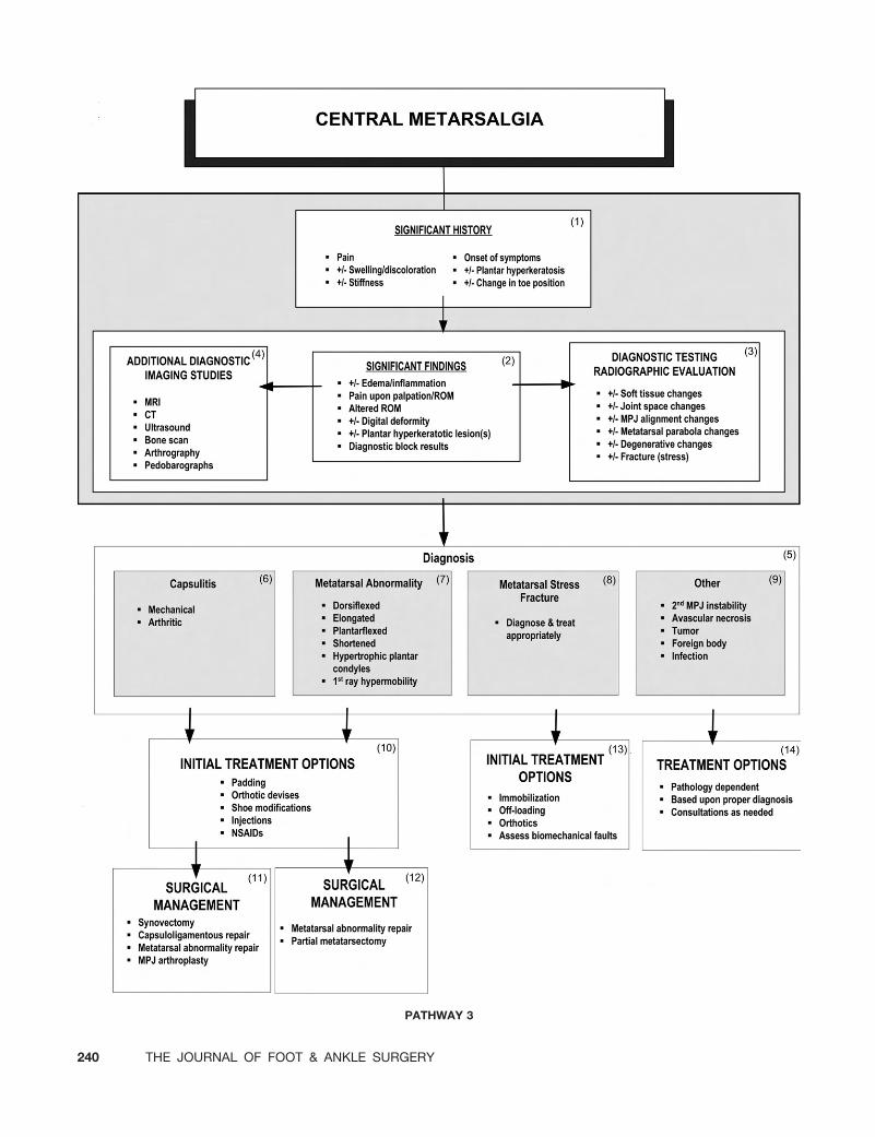
Central Metatarsalgia (Pathway 3)
Central metatarsalgia involves pathology of the second,third, and fourth metatarsals and their respective metatarso-phalangeal joints (MPJs). Metatarsal pathology may be sec-ondary to a variety of problems including trauma, lengthabnormalities, structural deformity, and others. Pathologyof the central MPJs is also secondary to numerous differentetiologies and encompasses both osseous and soft tissueconditions. Osseous changes may be secondary to arthritis,whereas soft tissue conditions can be complex, often leadingto instability of the MPJ and resultant multiplanar deformi-ties. Systemic inflammatory conditions may produce bothosseous and soft tissue abnormalities in the areas of thecentral MPJs.
Significant History (Pathway 3, Node 1)
Patients presenting with complaints related to the second,third, and fourth metatarsals and their respective MPJstypically relate a history of pain in the area of the ball of thefoot, with or without swelling and/or discoloration. Theymay report a history of partial or complete stiffness of theaffected joint(s). Symptoms are usually of gradual onset,
tend to be progressive in nature, and may have been aggra-vated by a recent change in activity or footwear. Frequentlythere is no history of trauma (1, 2). Related complaints mayinclude the development of plantar calluses in the area ofwhere symptoms occur as well as a gradual change inappearance or position of one or more toes (3).
Significant Findings (Pathway 3, Node 2)
Examination of the patient with central metatarsalgia symp-toms may reveal edema and or inflammation in the area of theinvolved metatarsal(s) or MPJs. Pain on palpation of the af-fected metatarsal or joint is typically present (4). Pain involv-ing the MPJ usually is exacerbated upon reaching end range ofmotion with manual testing. Decreased range of motion orcrepitus may indicate arthrosis or other osseous changes at theMPJ. Alternatively chronic hyperextension of the MPJ maypredispose the plantar plate and collateral ligaments to atten-uate and rupture (5). In these joints, manual stress testing of thelesser MPJ may demonstrate instability as evidenced by dorsaltranslocation of the digit at the metatarsal head (6). Typicallypatients with plantar plate rupture have pain with palpationplantarly at the metatarsal head or flexor crease of the affectedMPJ (7).
Change in position or alignment of the toe may or maynot be seen in central metatarsalgia, as patients may presentwith complaints of pain at the MPJ prior to the developmentof associated digital deformities. If digital deformities arepresent, they may be multiplanar, flexible, or nonflexible. Ahyperkeratotic lesion plantar to the affected metatarsal orMPJ may be present and may contribute to the patient’ssymptomatology (Fig. 1). Diagnostic blocks often are ofgreat help in localizing the area of the patient’s symptomsand establishing an accurate diagnosis.
Diagnostic Testing: Radiographic Evaluation(Pathway 3, Node 3)
Radiographic evaluation of the patient with central meta-tarsalgia symptoms should include weightbearing anterior-posterior (AP), lateral, and oblique views. A plantar axial
Address correspondence to: James L. Thomas, DPM, University ofFlorida, Department of Orthopaedics and Rehabilitation, 655 West 8th St,Jacksonville, FL 32209. E-mail: [email protected].
1Chair, Jacksonville, FL; 2Charleston, SC; 3San Antonio, TX; 4Scotts-dale, AZ; 5Champaign, IL; 6Mechanicsville, VA; 7Augusta, GA; 8Gadsden,AL.
Copyright © 2009 by the American College of Foot and Ankle Surgeons1067-2516/09/4802-0023$36.00/0doi:10.1053/j.jfas.2008.12.004
VOLUME 48, NUMBER 2, MARCH/APRIL 2009 239
PATHWAY 3
240 THE JOURNAL OF FOOT & ANKLE SURGERY

view may also be beneficial in evaluating the structure andposition of the central metatarsal heads.
Radiographs are evaluated for both soft tissue and osseouschanges. Soft tissue is investigated for edema, foreign bodies,and other abnormalities. Joint dislocation, subluxation, or ir-regularities of the metatarsal head or phalangeal base may beseen. Alignment of the second, third, and fourth MPJs as wellas any abnormalities of the metatarsal parabola are assessed on
the AP radiograph (Fig. 2). The presence of degenerativeand/or avascular changes may be indicated by erosions, jointspace narrowing, subchondral cysts, osteophyte formation,sclerosis, and alteration in the normal contour of the metatarsalhead. A metatarsal stress fracture may or may not be radio-graphically apparent. Indeed, the first radiographic evidence ofa stress fracture may be reflected by healing bone callus severalweeks after the fracture had occurred (Fig. 3).
AA
FE
B
C
D
FIGURE 1 Forefoot submetatarsal hyperkeratotic lesions vary considerably from (A) a localized discrete one to (B ) a diffuse lesion underan isolated metatarsal or (C) under multiple metatarsals. (D) Biomechanical evaluation with Harris mat or computer force plate analysis mayprovide useful clinical information regarding pressure distribution or loading points. Soft tissue pathology such as ganglia, (E) bursa, and (F)skin pathology such as verruca and porokeratoses must be considered.
VOLUME 48, NUMBER 2, MARCH/APRIL 2009 241

Additional Diagnostic Imaging Studies (Pathway 3,Node 4)
Evidence of pathology at the MPJ joint or metatarsal maybe further substantiated with the use of magnetic resonanceimaging (MRI), computed tomography (CT), diagnostic ul-trasound, radionucleotide scanning, and arthrography.These more advanced imaging techniques may assist in
determining the presence and extent of both soft tissue andosseous damage in this area of the foot.
MRI, CT, and radionucleotide scanning are helpful in de-termining the presence of metatarsal stress fracture or articularpathology such as an arthritide, Freiberg’s infraction, or plantarplate rupture not appreciated radiographically (7). Ultrasoundmay support clinical evidence of soft tissue trauma/edema andmay offer an alternative diagnostic aid for suspected plantar
M1-2 M2-5 M1-2 M2-5
Metatarsal Tangent Angles
Metatarsal ParabolaA
B
FIGURE 2 Metatarsal deformities are oftencorrelated with length relationships to eachother on standard weightbearing radiographs.(A) Parabola have been described as well as (B)metatarsal tangents. Certainly, osseous rela-tionships vary from patient to patient, butweightbearing radiographs have been used asan objective parameter to explain the forefootpathologies discussed in this document. (Meta-tarsal tangents from ACFAS Scoring Scale,2006.)
242 THE JOURNAL OF FOOT & ANKLE SURGERY

plate ruptures (8). The use of intra-articular radio-opaque dyeis beneficial in documenting a plantar plate rupture if extrav-asation of the dye is found to be present (9). Plantar pressurestudies may be helpful in identifying weightbearing anomaliesof the forefoot.
Diagnosis (#3; Node 5)
Establishment of the correct diagnosis may be very chal-lenging as quite a variety of pathologies may be etiologic ofpain in this anatomic location. These include:
— Capsulitis (mechanical, arthritic, or secondary to secondMPJ instability)
— Metatarsal abnormalities (dorsiflexed, elongated, plan-tarflexed, shortened, hypertrophic plantar condyles andfirst ray hypermobility)
— Metatarsal stress fracture— Second MPJ Instability— Other (avascular necrosis, tumor, foreign body, infection)
Capsulitis (Pathway 3, Node 6)
Capsulitis of the central MPJs may be secondary tomechanical or arthritic etiologies (Fig 4). Mechanicalcauses include any condition that results in increased forcesthrough the joint itself as well as overload to the plantarmetatarsal head. This may be associated with tears of theplantar plate or ligament disruption. Arthritic conditions
include any of the inflammatory arthritidies such as rheu-matoid arthritis and many of the seronegative arthritidies.Laboratory testing often is indicated in the establishment ofan arthritic process. In the case of a mechanical etiology ofcapsulitis, treatment includes offloading and management ofany contributing biomechanical abnormality with paddingand/or orthotic therapy. Oral anti-inflammatory medicationas well as local injection of a corticosteroid also may bebeneficial. If the patient fails to respond appropriately tothese measures, surgical treatment may be necessary. Thiswould include synovectomy along with correction of anycontributing pathology (eg, metatarsal abnormality) and re-pair of any capsuloligamentous tears if present (see Node 9).
When arthritis of the MPJ is the cause of capsulitis,attempts should be made to establish an accurate diagnosisof the arthritic process involved. These attempts include thepreviously-mentioned laboratory testing, as well as jointaspiration and rheumatologic consultation/referral if indi-cated. Treatment for an inflammatory arthritic condition ofthe central MPJs includes all of the nonsurgical and surgicalalternatives previously listed for mechanically-inducedcapsulitis. In addition, arthroplasty-type procedures may benecessary to remove painful ostephytes, remove loose bod-ies, perform other procedures such as chondroplasty or jointimplantation. In some cases, metatarsal head resection mayat times also be considered, but this usually is done only inthe presence of significant deformity, such as in the perfor-mance of pan metatarsal head resection in the setting ofrheumatoid arthritis (Fig 5).
FIGURE 3 Metatarsal stress fractures are common.This patient had (A) radiographs taken 2 weeks priorin the emergency room that were negative and nowshow complete fracture with displacement of thesecond metatarsal. (B) A follow-up radiograph 3weeks later shows exuberant bone callus indicativeof the unstable nature of the fracture.
VOLUME 48, NUMBER 2, MARCH/APRIL 2009 243
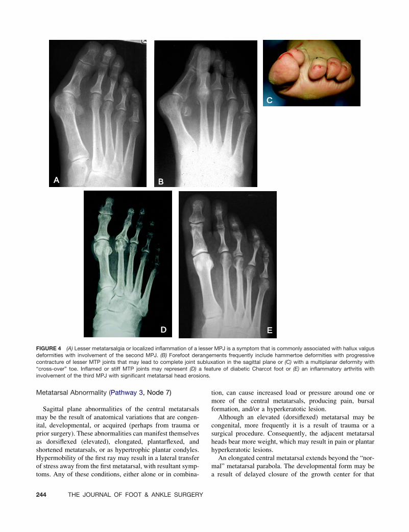
Metatarsal Abnormality (Pathway 3, Node 7)
Sagittal plane abnormalities of the central metatarsalsmay be the result of anatomical variations that are congen-ital, developmental, or acquired (perhaps from trauma orprior surgery). These abnormalities can manifest themselvesas dorsiflexed (elevated), elongated, plantarflexed, andshortened metatarsals, or as hypertrophic plantar condyles.Hypermobility of the first ray may result in a lateral transferof stress away from the first metatarsal, with resultant symp-toms. Any of these conditions, either alone or in combina-
tion, can cause increased load or pressure around one ormore of the central metatarsals, producing pain, bursalformation, and/or a hyperkeratotic lesion.
Although an elevated (dorsiflexed) metatarsal may becongenital, more frequently it is a result of trauma or asurgical procedure. Consequently, the adjacent metatarsalheads bear more weight, which may result in pain or plantarhyperkeratotic lesions.
An elongated central metatarsal extends beyond the “nor-mal” metatarsal parabola. The developmental form may bea result of delayed closure of the growth center for that
AA B
C
D E
FIGURE 4 (A) Lesser metatarsalgia or localized inflammation of a lesser MPJ is a symptom that is commonly associated with hallux valgusdeformities with involvement of the second MPJ. (B) Forefoot derangements frequently include hammertoe deformities with progressivecontracture of lesser MTP joints that may lead to complete joint subluxation in the sagittal plane or (C) with a multiplanar deformity with“cross-over” toe. Inflamed or stiff MTP joints may represent (D) a feature of diabetic Charcot foot or (E) an inflammatory arthritis withinvolvement of the third MPJ with significant metatarsal head erosions.
244 THE JOURNAL OF FOOT & ANKLE SURGERY

particular metatarsal. However, the elongation may be onlyrelative if adjacent metatarsals have been shortened fromeither trauma or surgery. During the gait cycle, particularlyat the push-off phase, the elongated metatarsal tends to bearmore weight for a longer period of time, resulting in symp-toms of increased stress under the involved metatarsal head.
A structurally plantarflexed metatarsal results in a moreplantar location of its respective metatarsal head in compar-ison to the adjacent metatarsals. Congenital plantarflexedmetatarsal is rare, but if present it is commonly associatedwith an anterior cavus foot deformity. The condition of anisolated plantarflexed metatarsal most often exists as a result
of trauma or prior surgery. A clinical plantar prominence ofthe metatarsal head may also be the result of increasedretrograde force from an associated digital deformity withdorsal contraction of the MPJ. This results in increasedweightbearing stress, which may result in pain or a hyper-keratotic lesion beneath the metatarsal.
A shortened metatarsal may be associated with a congen-ital or acquired syndromic condition (10) or iatrogenicallyinduced secondary to a surgical procedure. In addition to thedecreased length of the metatarsal, relative elevation to theadjacent metatarsals results due to the inherent declinationof the metatarsals. This shortening may increase the load or
AA
C
B
D E
FIGURE 5 (A) The MPJs are a target area for rheumatoid arthritis and this patient exhibits severe deformity (B) with dislocation of the first,second, and third MPJs. (C) This patient underwent forefoot arthroplasty with first MPJ fusion and panmetatarsal head resections. (D and E)Shown are the foot and a radiograph at 1 year postsurgery.
VOLUME 48, NUMBER 2, MARCH/APRIL 2009 245

pressure to the adjacent metatarsal heads as they bear moreweight, at times producing pain and/or hyperkeratotic le-sions. Congenital shortening of a metatarsal (brachymeta-tarsia) usually becomes clinically evident between the agesof 4 and 15 years. Brachymetatarsia is relatively rare, witha reported incidence of 0.022% or 1 in 4586, and affectsfemales more commonly than males in an approximate 25:1ratio (11, 12). Several retrospective studies report the fourthray being the most commonly affected (10). A congenitallyshort metatarsal may also result in metatarsalgia secondaryto increased weightbearing forces around the adjacent meta-tarsal(s). An elevated toe can cause footwear difficulties andpainful hyperkeratotic lesions. Physeal abnormalities andother changes may indicate associated syndromic condi-tions. Complaints from patients seeking treatment for acongenitally short fourth metatarsal may be only for cos-metic concerns.
Congenital hypertrophy of the plantar condyles of themetatarsal heads is rare. The condition is most commonlythe result of exostosis formation secondary to an arthriticcondition or a degenerative process. Inflammatory jointdisease with or without bursitis may also be a significantcontributing factor. Patients with atrophy or anterior dis-placement of the plantar fat pad will appear to have thiscondition and may increase weightbearing stress under theinvolved metatarsal head(s). The lateral plantar condyle ismost commonly involved.
Hypermobility of the first ray has been reported to resultin overload of the second metatarsal head with resultantpathology in this area. Treatment of associated first rayhypermobility may be necessary in the treatment of centralmetatarsalgia.
Each of these conditions creates similar symptomatologyin the area of the metatarsal with the deformity or in the area
of adjacent metatarsals. Elongated or plantarflexed metatar-sals as well as metatarsals adjacent to an elevated metatarsalsustain increased weightbearing forces. In addition, meta-tarsals with hypertrophy of the plantar condyles show asimilar pattern of increased forces and possible focal kera-toses at the level of the condyles.
Nonsurgical treatment involves medications, offloading thepainful joint or metatarsal head, and treating any associatedbiomechanical pathology. Treatment includes padding, ortho-ses, and rocker-bottom shoe modifications. Nonsteroidal anti-inflammatory drugs and corticosteroid injections may also beused (13).
Surgical treatment is indicated for those patients who failto respond to nonsurgical care or for patients in whomnonsurgical care would not be considered beneficial.
Dorsiflexed (elevated) metatarsals are surgically treatedby an osteotomy at either the proximal or distal aspect of themetatarsal to plantarflex the metatarsal with the goal ofreestablishing the normal weight bearing pattern of theforefoot. Care must be taken to avoid over- or under-correction of the deformity.
Structurally plantarflexed metatarsals are surgicallytreated by an elevating osteotomy at either the proximal orthe distal aspect of the metatarsal to re-establish the sagittalplane alignment of the bone and a normal metatarsal parab-ola (14) (Fig 6). Care must be taken to prevent commoncomplications of excessive elevation or shortening resultingin transfer lesions or metatarsalgia. Painful dorsal exostosisor prominence secondary to excessive elevation and floatingtoe/lack of toe purchase may also occur (15).
Surgical procedures for a shortened metatarsal includeacute or gradual lengthening of the metatarsal, soft tissuerelease, correction of associated digital deformity, and insome cases syndactyly. Specific techniques used for meta-
FIGURE 6 (A) Management of metatarsalgia asso-ciated with an elongated third metatarsal and a dis-crete keratotic lesion. (B). This patient underwent ashortening third metatarsal osteotomy to reduce lo-calized pressures and a discrete sub-third metatarsalhead lesion.
246 THE JOURNAL OF FOOT & ANKLE SURGERY

tarsal lengthening include metatarsal osteotomy, bone graft-ing, and distraction osteogenesis, (Fig. 7). Potential compli-cations include under- or over-correction, decreased jointmotion, delayed union, nonunion, malunion, or graft failure.Amputation at the MTP joint should be avoided because ofthe risk of subsequent adjacent toe deformity (16).
Surgical treatment of enlarged plantar condyles is per-formed when the symptoms of metatarsalgia are due to theprominence and resultant increased pressure or load causedby this abnormality. Surgical treatment of enlarged plantarcondyles involves remodeling or resection (condylectomy)of the involved metatarsal condyles. If other etiologic fac-tors such as those described previously are determined toexist, consideration should be given to other procedures totreat the coexisting deformity.
Operative treatment of first ray hypermobility may benecessary in some instances. This is usually done via medialcolumn arthrodesis.
Metatarsal Stress Fracture (Pathway 3, Node 8)
Stress fractures of the central metatarsals develop when thebone is subjected repetitively to sub-failure loads (17-19).Stress fractures make up as much as 20% of all sports-relatedinjuries (20). A greater incidence of these injuries amongfemales has been reported (21). Patients with chronic inflam-matory arthropathies, severe osteoporosis, marked joint defor-mity, or receive chronic corticosteroid therapy are at high riskof developing stress fractures (22). Stress fractures of thesecond metatarsal base are most commonly seen in dancers
(23-26). Although the length of the first metatarsal has beenimplicated as a risk factor for weight transference, equivalentrates of fracture in short, average, and long first metatarsalconditions have been reported (27). Numerous studies haveshown that abnormal mechanics of the first ray may transferweight to the adjacent metatarsals and lead to stress transfer-ence and fracture of the adjacent metatarsal (28-30).
The majority of metatarsal stress fractures occur in thesecond and third metatarsals. Radiographic examinationmay be normal for several weeks after the onset of symp-toms. In cases of uncertain diagnosis or in cases where moreaggressive treatment might be indicated (ie, high perfor-mance athletes), a technetium bone scan, MRI, or CT scanmay assist in the early detection of a stress fracture (31).
The primary treatment of a central metatarsal stress frac-ture is immobilization and offloading with the use of awalking boot or surgical shoe along with activity modifica-tion (32, 33). Proper shoes and orthoses are used to controlabnormal biomechanical influences and may relieve symp-toms. Failure to address pre-existing biomechanical abnor-malities that result in metatarsal stress fractures can lead tocomplete fracture or possible recurrence (34, 35). Return toregular activity or sport is allowed once adequate fracturehealing has been accomplished. In the event of malunion,the metatarsal may become elevated, plantar-flexed, and/orshortened and this may increase the load to adjacent meta-tarsals. Surgical treatment of a metatarsal stress fracture israrely necessary. However, surgery may be required incases of complete fracture, failure to heal with usual non-surgical measures, or in cases of malunion (24).
FIGURE 7 (A) Brachymetatarsia most often involves the fourth metatarsal. This patient was surgically treated with distraction osteogenesis.(B) Fixator is in place. (C) Radiography at 1-year postsurgery shows very nice reconstitution of metatarsal length and morphology. (CourtesyAlan Banks, DPM, Atlanta, GA)
VOLUME 48, NUMBER 2, MARCH/APRIL 2009 247

Other (Pathway 3, Node 9)
Other causes of central metatarsalgia-related symptomsalso exist. These include second MTP joint instability, avas-cular necrosis, tumor, foreign body, and infection.
Second MPJ Instability The second MPJ is most fre-quently implicated in MPJ instability. Second MPJ instabil-ity with or without inflammation of the articular and peri-articular structures can lead to multiplanar malalignment.Second MPJ instability has been described as predislocationsyndrome (36), plantar plate dysfunction (37), mono-artic-ular non-traumatic synovitis, MPJ capsulitis and synovitis,metatarsalgia, and crossover second toe deformity. Manyfactors that include both mechanical and inflammatory con-
ditions have been described to contribute to this condition.Possible mechanical causes include trauma to the plantarplate and supporting ligaments and joint capsule; an elon-gated second metatarsal; Freiberg’s infraction; prior surgi-cal intervention; congenital deformities; and the forces pro-duced on the second MTP joint by an adjacent hallux valgusdeformity. Inflammatory conditions such as isolated syno-vitis or systemic inflammatory arthropathies may be caus-ative factors as well.
The confirmation of an unstable joint is determined byclinical and radiographic evaluations (Fig 8). This conditionis characterized by pain and inflammation of the secondMPJ and varying degrees of digital deviation in the trans-verse and sagittal plane. As the condition progresses a
AA
E
G
F
D
B
C
FIGURE 8 (A) Plantar plate rupture leading to digitalinstability and chronic localized pain is the presentingsymptom in this patient. (B) Lateral radiograph showsloss of toe purchase with a (C) positive MPJ stresstest. (D) The MRI image shows discontinuity of theplantar plate and its phalangeal base insertion is con-firmed by (E) an intraoperative photo illustrating rup-ture. (F and G) Postsurgically, this patient shows res-toration of toe purchase, both clinically andradiographically. (Courtesy Craig Camasta, DPM,Douglasville, GA)
248 THE JOURNAL OF FOOT & ANKLE SURGERY

positive dorsal stress test on the digit at the MPJ (drawertest) reveals subluxation and, sometimes in later stages,dorsal dislocation (36). Transverse plane deviation of thedigit may be better appreciated on weightbearing examina-tion and is commonly present with sagittal plane instability.It should be noted that clinical instability at the MPJ is notnecessarily pathognomonic for a plantar plate rupture;rather it may be identified in patients with ligamentouslaxity and chronic plantar plate attenuation (37).
Weightbearing radiographic evaluation reveals the jointalignment changes. MRI and arthrography may be benefi-cial in further delineating the competency of the plantarplate.
Nonsurgical treatment may involve offloading of the sec-ond MPJ, splinting or taping of the second toe, orthoses,rocker-bottom shoe modifications and metatarsal padding.Nonsteroidal anti-inflammatory drugs and corticosteroid in-jections also may be used (13).
Surgical treatment is indicated for those patients whohave failed nonsurgical care or for patients who are notcandidates for nonsurgical treatment. Surgical correction isaimed at restoring the alignment and function of the secondMPJ and any associated digital deformity. Surgical optionsinclude interphalangeal joint arthrodesis, flexor tendontransfer (38), reefing of the plantar-lateral capsule, collateralligament repair, plantar plate repair (37), MPJ arthroplasty,and various metatarsal osteotomies (13).
Avascular Necrosis of the Metatarsal Head A uniquefinding of the lesser MTP joint (typically the second) isosteochondrosis. Commonly referred to as Freiberg’s in-fraction, these cartilage and osseous changes represent anavascular necrosis of the metatarsal head (39-41). Surgicalmanagement of this condition may include: distal metatarsalosteotomy (42-46), implant arthroplasty (47-50), resectionarthroplasty (51) and interpositional grafting (52, 53), andinterpositional soft tissue grafts (54, 55).
Tumor Pain and/or swelling in the forefoot may besecondary to tumors of soft tissue or bone. Timely diagnosisand any indicated consultation and /or referral are impor-tant. Treatment is specific to the type of tumor present.
Foreign Body Symptoms secondary to a foreign body inthe forefoot are not uncommon. A symptomatic retained for-eign body may be old or new and may be associated with asignificant wound (laceration, gunshot or puncture wound) orshow no discernable portal of entry. Excision of the offendingforeign body is the treatment of choice. In the acute settingconfirmation of tetanus prophylaxis is indicated.
Infection Infection of the central metatarsal area maybe secondary to a variety of etiologies including puncturewound, foreign bodies, diabetic ulceration (see DiabeticFoot Disorders: A Clinical Practice Guideline, 2006, sup-plement to Journal of the Foot and Ankle Surgeons), lacer-ation, fungal infections etc. It may involve joint, soft tissue,bone or occur in combination. Identification of the anatomic
structure(s) involved and the causative organism(s) willdirect treatment.
References
1. Brunet JA, Tubin S. Traumatic dislocations of the lesser toes. FootAnkle Int 18:406–411, 1997.
2. Goldner JL, Ward WG. Traumatic horizontal deviation of the secondtoe: mechanism of deformity, diagnosis, and treatment. Bull Hosp JtDis Orthop Inst 47:123–135, 1987.
3. Menz HB, Zammit GV, Munteanu SE. Plantar pressures are higherunder callused regions of the foot in older people. Clin Exp Dermatol32:375–380, 2007.
4. Mendicino RW, Statler TK, Saltrick KR, Catanzariti AR. Predisloca-tion syndrome: a review and retrospective analysis of eight patients. JFoot Ankle Surg 40:214–224, 2001.
5. Johnston RB III, Smith J, Daniels T. The plantar plate of the lessertoes: an anatomical study in human cadavers. Foot Ankle Int 15:276–282, 1994.
6. Thompson FM, Hamilton WG. Problems of the second metatarsopha-langeal joint. Orthopedics 10:83–89, 1987.
7. Mandell GA, Harcke HT. Scintigraphic manifestations of infraction ofthe second metatarsal (Freiberg’s disease). J Nucl Med 28:249–251,1987.
8. Gregg J, Silverstein M, Schneider T, Marks P. Sonographic and MRIevaluation of the plantar plate: a prospective study. Eur Radiol 16:2661–2669, 2006.
9. Blitz NM, Ford LA, Christensen JC. Second metatarsophalangeal jointarthrography: a cadaveric correlation study. J Foot Ankle Surg, 43:231–240, 2004.
10. Schimizzi A, Brage M. Brachymetatarsia. Foot Ankle Clin 9:555–570,ix, 2004.
11. Davidison RS. Metatarsal lengthening. Foot Ankle Clin 6:499–518,2001.
12. Urano Y, Kobayashi A. Bone-lengthening for shortness of the fourthtoe. J Bone Joint Surg Am 60:91–93, 1978.
13. Co AY, Ruch JA, Malay DS. Radiographic analysis of transverseplane digital alignment after surgical repair of the second metatarso-phalangeal joint. J Foot Ankle Surg 45:380–399, 2006.
14. Feibel JB, Tisdel CL, Donley BG. Lesser metatarsal osteotomies. Abiomechanical approach to metatarsalgia. Foot Ankle Clin 6:473–489,2001.
15. Migues A, Slullitel G, Bilbao F, Carrasco M, Solari G. Floating-toedeformity as a complication of the Weil osteotomy. Foot Ankle Int25:609–613, 2004.
16. Ferrandez L, Yubero J, Usabiaga J, Ramos L. Congenital brachymeta-tarsia: three cases. Foot Ankle 14:529–533, 1993.
17. Milgrom C, Finestone A, Sharkey N, Hamel A, Mandes V, Burr D, etal. Metatarsal strains are sufficient to cause fatigue fracture duringcyclic overloading. Foot Ankle Int 23:230–235, 2002.
18. Brukner P, Bradshaw C, Khan KM, White S, Crossley K. Stressfractures: a review of 180 cases. Clin J Sport Med 6:85–89, 1996.
19. Arnd A, Ekenman I, Westblad P, Lundberg A. Effects of fatigue andload variation on metatarsal deformation measured in vivo duringbarefoot walking. J Biomech 35:621–628, 2002.
20. Fredericson M, Jennings F, Beaulieu C, Matheson GO. Stress fracturesin athletes. Top Magn Reson Imaging 17:309–325, 2006.
21. Kaye RA. Insufficiency stress fractures of the foot and ankle inpostmenopausal women. Foot Ankle Int 19:221–224, 1988.
22. Maenpaa H, Lehto MU, Belt EA. Stress fractures of the ankle andforefoot in patients with inflammatory arthritides. Foot Ankle Int23:833–837, 2002.
VOLUME 48, NUMBER 2, MARCH/APRIL 2009 249

23. Hardaker WT Jr. Foot and ankle injuries in classical ballet dancers.Orthop Clin North Am 20:621–627, 1989.
24. Sarimo J, Orava S, Alanen J. Operative treatment of stress fractures ofthe proximal second metatarsal. Scand J Med Sci Sports 17:383–386,2007.
25. Micheli LJ, Sohn RS, Solomon R. Stress fractures of the secondmetatarsal involving Lisfranc’s joint in ballet dancers:a new overuseinjury of the foot. J Bone Joint Surg Am 67:1372–1375, 1985.
26. O’Malley MJ, Hamilton WG, Munyak J, DeFranco MJ. Stress frac-tures at the base of the second metatarsal in ballet dancers. Foot AnkleInt 17:89–94, 1996.
27. Drez D Jr, Young JC, Johnston RD, Parker WD. Metatarsal stressfractures. Am J Sports Med 8:123–125, 1980.
28. Kitaoka HB, Cracchiolo A III. Stress fracture of the lateral metatarsalsfollowing double-stem silicone implant arthroplasty of the halluxmetatarsophalangeal joint. Clin Orthop Relat Res 239:211–216, 1989.
29. Friend G. Sequential metatarsal stress fractures after Keller arthro-plasty with implant. J Foot Surg 20: 227–231, 1981.
30. Zechman JS. Stress fracture of the second metatarsal after Kellerbunionectomy. J Foot Surg 23:63–65, 1984.
31. Santi M, Sartoris, DJ, Resnick D. Diagnostic imaging of tarsal andmetatarsal stress fractures. Part II. Orthop Rev 18: 305–310, 1989.
32. Gehrmann RM, Renard RL. Current concepts review: stress fracturesof the foot. Foot Ankle Int 27:750–757, 2006.
33. Fetzer GB, Wright RW. Metatarsal shaft fractures and fractures of theproximal fifth metatarsal. Clin Sports Med 25:139–150, x, 2006.
34. Kaeding CC, Yu JR, Wright R, Amendola A, Spindler KP. Manage-ment and return to play of stress fractures. Clin J Sport Med 15:442–447, 2005.
35. Heaslet MW, Kanda-Mehtani SL. Return-to-activity levels in 96 ath-letes with stress fractures of the foot, ankle, and leg: a retrospectiveanalysis. J Am Podiatr Med Assoc 97:81–84, 2007.
36. Yu GV, Judge MS, Hudson JR, Seidelmann FE. Predislocation syn-drome. Progressive subluxation/dislocation of the lesser metatarsopha-langeal joint. J Am Podiatr Med Assoc 92:182–199, 2002.
37. Blitz NM, Ford LA, Christensen JC. Plantar plate repair of the secondmetatarsophalangeal joint: technique and tips. J Foot Ankle Surg43:266–270, 2004.
38. Myerson MS, Jung HG. The role of toe flexor-to-extensor transfer incorrecting metatarsophalangeal joint instability of the second toe. FootAnkle Int 26:675–679, 2005.
39. Freiberg AA, Freiberg RA. Core decompression as a novel treatmentfor early Freiberg’s infraction of the second metatarsal head. Ortho-pedics 18:1177–1178, 1995.
40. Binek R, Levinsohn EM, Bersani F, Rubenstein H. Freiberg diseasecomplicating unrelated trauma. Orthopedics 11:753–757, 1988.
41. Scartozzi G, Schram A, Janigian J. Freiberg’s infraction of the secondmetatarsal head with formation of multiple loose bodies. J Foot Surg28:195–199, 1989.
42. Lee SK, Chung MS, Baek GH, Oh JH, Lee YH, Gong HS. Treatmentof Freiberg disease with intra-articular dorsal wedge osteotomy andabsorbable pin fixation. Foot Ankle Int 28:43–48, 2007.
43. Chao KH, Lee CH, Lin LC. Surgery for symptomatic Freiberg’sdisease: extraarticular dorsal closing-wedge osteotomy in 13 patientsfollowed for 2-4 years. Acta Orthop Scand 70:483–486, 1999.
44. Katcherian DA. Treatment of Freiberg’s disease. Orthop Clin NorthAm 25:69–81, 1994.
45. Smith TW, Stanley D, Rowley DI. Treatment of Freiberg’s disease: anew operative technique. J Bone Joint Surg Br 73:129–130, 1991.
46. Kinnard P, Lirette R. Dorsiflexion osteotomy in Freiberg’s disease.Foot Ankle 9:226–231, 1989.
47. Bordelon RL. Silicone implant for Freiberg’s disease. South Med J70:1002–1004, 1977.
48. Miller ML, Lenet MD, Sherman M, Surgical treatment of Freiberg’sinfraction with the use of total joint replacement arthroplasty. J FootSurg 23:35–40, 1984.
49. Scartozzi G, Schram A, Janigian J. Freiberg’s infraction of the secondmetatarsal head with formation of multiple loose bodies. J Foot Surg28:195–199, 1989.
50. Shih AT, Quint RE, Armstrong DG, Nixon BP. Treatment ofFreiberg’s infraction with the titanium hemi-implant. J Am PodiatrMed Assoc 94:590–593, 2004.
51. Young MC, Fornasier VL, Cameron HU. Osteochondral disruption ofthe second metatarsal: a variant of Freiberg’s infraction? Foot Ankle8:103–109, 1987.
52. Beito SB, Lavery LA. Freiberg’s disease and dislocation of the secondmetatarsophalangeal joint: etiology and treatment. Clin Podiatr MedSurg 7:619–631, 1990.
53. el-Tayeby HM. Freiberg’s infraction: a new surgical procedure. J FootAnkle Surg 37:23–27, discussion 79, 1998.
54. Zgonis T, Jolly GP, Kanuck DM, Interpositional free tendon graft forlesser metatarsophalangeal joint arthropathy. J Foot Ankle Surg 44:490–492, 2005.
55. Roukis TS, Landsman AS, Ringstrom JB, Kirschner P, Wuenschel M.Distally based capsule-periosteum interpositional arthroplasty for hal-lux rigidus. Indications, operative technique, and short-term follow-up.J Am Podiatr Med Assoc 93:349–366, 2003.
250 THE JOURNAL OF FOOT & ANKLE SURGERY

Diagnosis and Treatment of ForefootDisorders. Section 3. Morton’sIntermetatarsal Neuroma
Clinical Practice Guideline Forefoot Disorders Panel: James L. Thomas, DPM,1
Edwin L. Blitch, IV, DPM,2 D. Martin Chaney, DPM,3 Kris A. Dinucci, DPM,4
Kimberly Eickmeier, DPM,5 Laurence G. Rubin, DPM,6 Mickey D. Stapp, DPM,7 andJohn V. Vanore, DPM8
This clinical practice guideline (CPG) is based uponconsensus of current clinical practice and review of theclinical literature. The guideline was developed by the Clin-ical Practice Guideline Forefoot Disorders Panel of theAmerican College of Foot and Ankle Surgeons. The guide-line and references annotate each node of the correspondingpathways.
Morton’s Intermetatarsal Neuroma (Pathway 4)
Nerve pathologies are a common cause of forefoot painand include diverse conditions with similar symptoms. Thesymptoms are characteristic of sensory nerve disorders anddiffer from other musculoskeletal conditions.
Morton’s intermetatarsal neuroma is a compression neu-ropathy of the common digital nerve (Fig. 1). It is mostcommonly seen in the third intermetatarsal space, but it alsocan be seen in other intermetatarsal spaces (Fig. 2). Aneuroma may occur in more than one intermetatarsal spaceand may be bilateral. Neuromas are more prevalent in adultsbeginning in the third decade of life, and are more commonin females than males (1-7).
Significant History (Pathway 4, Node 1)
The subjective history reported by the patient is usuallycharacteristic for this entity. The patient may complain ofnumbness and tingling, and/or radiating, burning pain. Thepain often is localized at the plantar aspect of the respectiveintermetatarsal space, but it can radiate into the adjacent
toes. Patients frequently describe a “lump” on the bottom oftheir foot or a feeling of walking on a rolled-up or wrinkledsock. The symptoms may increase with weightbearing andactivity. Closed-toed shoes and especially tight-fitting foot-wear can increase the symptoms. Patients report relief ofsymptoms upon removing or changing their shoes. Theyalso may get relief from massaging the foot and moving thetoes.
Significant Findings (Pathway 4, Node 2)
Objective findings are unique to Morton’s neuroma andcan provide further insight to aid the clinician in the diag-nostic process. Although patients frequently describe numb-ness, a sensory deficit may or may not be present onexamination. The clinical presentation may demonstrate asplaying or divergence of the digits (8). Usually little to noedema or inflammation is seen clinically. Reproduction ofthe pain with palpation to the intermetatarsal space is typ-ical. Care must be taken to press in the intermetatarsal spaceand avoid the metatarsal heads.
Clinical Maneuvers (Pathway 4, Node 3)
Various clinical maneuvers have been described to assistthe clinician in the diagnosis of Morton’s neuroma. Thepatient may demonstrate a Mulder’s sign (9, 10), elicited bysqueezing the forefoot and applying plantar and dorsalpressure. A positive test result consists of a click or pop thatcan be felt or heard; this can be painful to the patient.Symptoms of Morton’s neuroma may be replicated throughthe Gauthier’ test, in which the forefoot is squeezed andmedial to lateral pressure is applied (5). Bratkowski de-scribed a test that involves hyperextending the toes androlling the thumb of the examiner in the area of symptoms.This maneuver may reveal a tender, thickened, longitudinalmass (11). Patients with Morton’s neuroma also may dem-onstrate Tinel’s sign and Valleix phenomenon.
Address correspondence to: James L. Thomas, DPM, University ofFlorida, Department of Orthopaedics and Rehabilitation, 655 West 8th St,Jacksonville, FL 32209. E-mail: [email protected]
1Chair, Jacksonville, FL; 2Charleston, SC; 3San Antonio, TX; 4Scotts-dale, AZ; 5Champaign, IL; 6Mechanicsville, VA; 7Augusta, GA; 8Gadsden,AL.
Copyright © 2009 by the American College of Foot and Ankle Surgeons1067-2516/09/4802-0024$36.00/0doi:10.1053/j.jfas.2008.12.005
VOLUME 48, NUMBER 2, MARCH/APRIL 2009 251

PATHWAY 4
252 THE JOURNAL OF FOOT & ANKLE SURGERY
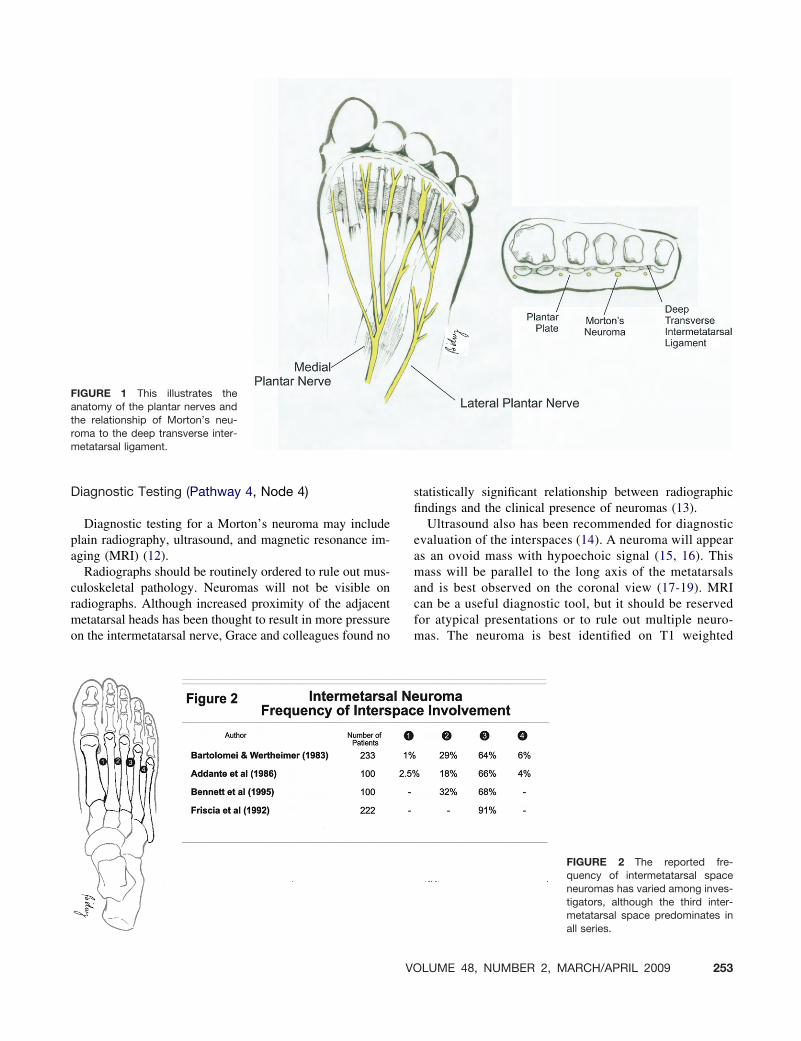
Diagnostic Testing (Pathway 4, Node 4)
Diagnostic testing for a Morton’s neuroma may includeplain radiography, ultrasound, and magnetic resonance im-aging (MRI) (12).
Radiographs should be routinely ordered to rule out mus-culoskeletal pathology. Neuromas will not be visible onradiographs. Although increased proximity of the adjacentmetatarsal heads has been thought to result in more pressureon the intermetatarsal nerve, Grace and colleagues found no
statistically significant relationship between radiographicfindings and the clinical presence of neuromas (13).
Ultrasound also has been recommended for diagnosticevaluation of the interspaces (14). A neuroma will appearas an ovoid mass with hypoechoic signal (15, 16). Thismass will be parallel to the long axis of the metatarsalsand is best observed on the coronal view (17-19). MRIcan be a useful diagnostic tool, but it should be reservedfor atypical presentations or to rule out multiple neuro-mas. The neuroma is best identified on T1 weighted
FIGURE 1 This illustrates theanatomy of the plantar nerves andthe relationship of Morton’s neu-roma to the deep transverse inter-metatarsal ligament.
FIGURE 2 The reported fre-quency of intermetatarsal spaceneuromas has varied among inves-tigators, although the third inter-metatarsal space predominates inall series.
VOLUME 48, NUMBER 2, MARCH/APRIL 2009 253
images. It will be revealed as a well-demarcated masswith low signal intensity (20).
Differential Diagnosis (Pathway 4, Node 5)
The diagnosis of Morton’s neuroma requires a carefulclinical history correlated with the condition’s unique set ofcharacteristics found on examination. Care must be taken torule out other possible etiologies of symptoms in this area ofthe forefoot (19, 21, 22). The differential diagnosis of Mor-ton’s neuroma includes:
● Stress fracture (23)● Neoplasm (eg, rheumatoid nodule) (24-27)● Bursitis (23, 25, 28)● MPJ pathology (27-29)● Metabolic neuropathy● Fibromyalgia and other chronic pain syndromes
Diagnosis (Pathway 4, Node 6)
The diagnosis of Morton’s neuroma is primarily a clinicaldiagnosis that is reached after examination and diagnostictesting have ruled out other possible etiologies of symp-toms.
Initial Treatment Options (Pathway 4, Node 7)
Nonsurgical care of Morton’s neuroma is centered onalleviating pressure and irritation of the nerve. Initially,patients should wear shoes that have a wide toe box to allowthe metatarsals to spread out. High-heel shoes should beavoided.
Metatarsal pads also can be beneficial. These pads, placedproximal to the metatarsal heads, help alleviate pressure onthe nerve and assist in spreading out the metatarsals.
Injection therapy includes a variety of alternative ap-proaches to nonsurgical treatment. A local anesthetic blockcan be used to provide some diagnostic information, but ithas not been shown to be therapeutic (30). Corticosteroidinjection is cited as having an 11% to 47% success rate, withmultiple injections obtaining better results (31-34, 35). Careshould be taken to avoid overusing corticosteroid injections;the literature contains reports of atrophy of the plantar fatpad secondary to cortisone injections, as well as joint sub-luxation (36). Dilute alcohol injections (3-7 injections of4% alcohol administered at 5-10 day intervals) has beenassociated with an 89% success rate, with 82% of patientsachieving complete relief of symptoms (37). Several otherinvestigators have verified the efficacy of sclerosing injec-tions as a nonsurgical treatment alternative (38, 39 40).Another injection modality involves injecting the nerve withvitamin B12 (cyanocobalamin); this has been discussed in
the literature, but the effects observed may have been due tothe preserving agent, benzyl alcohol (41). Phenol also hasbeen reported as a safe and effective injection modality (42).
Surgical Treatment Options (Pathway 4, Node 8)
Excision of the affected portion of the nerve is perhapsthe most common approach to neuroma surgery (1, 7, 19,43). Excision requires identifying the common digital por-tion of the nerve and following the structure to the properdigital branches. Care must be taken to avoid other struc-tures in the area. Various surgical approaches have beenused, the most common of which is a dorsal incision overthe involved intermetatarsal space (44, 45) (Fig. 3). Plantarincisional approaches are most often used in revisionaryprocedures, although they also have been described as aninitial surgical approach (19, 22, 46, 47). Excision may alsobe elected when prior decompression surgery has failed toresolve symptoms (48).
Decompression of the intermetatarsal nerve through theuse of endoscopic and minimally invasive techniques hasbeen reported in recent years (49-51). Open decompressionof the nerve by releasing the deep transverse intermetatarsalligament and performing an external neurolysis has beendescribed (52). In addition, transposition with nerve releasehas been shown to be useful (53, 54).
Cryogenic neuroablation is a minimally invasive proce-dure that applies a temperature of -50°C to -70°C to thenerve. This results in Wallerian degeneration of the axonsand myelin, while leaving the epineurium and perineuriumintact. Preserving these structures helps prevent stump neu-romas during nerve regeneration; this is the greatest advan-tage of cryogenic ablation. There are limitations of thisprocedure. The results are not permanent, and it is not aseffective on larger neuromas or in the presence of thickfibrosis. Several investigators have advocated this technique(55, 56).
Continued Symptoms (Pathway 4, Node 9)
All treatments may have complications, with eitherineffective relief of symptoms or worsening of the con-dition. Careful reassessment in failed surgical manage-ment may reveal tarsal tunnel or other proximal nervepathology. Complications of surgical procedures includeinfection, hematoma, stump neuroma formation, andchronic pain syndromes. Surgical failures may requiremore aggressive surgical intervention including plantarapproach and implantation of the proximal portion ofnerve into muscle (57, 58).
254 THE JOURNAL OF FOOT & ANKLE SURGERY

References
1. Keh RA, Ballew KK, Higgins KR, Odom R, Harkless LB. Long-termfollow-up of Morton’s neuroma. J Foot Surg 31:93–95, 1992.
2. Mann RA, Reynolds JC. Interdigital neuroma: a critical clinical anal-ysis. Foot Ankle 3:238–243, 1983.
3. Bradley N, Miller WA, Evans JP. Plantar neuroma: analysis of resultsfollowing surgical excision in 145 patients. South Med J 69:853–854,1976.
4. Friscia DA, Strom DE, Parr JW. Surgical treatment for primary inter-digital neuroma. Orthopedics 14:669–672, 1992.
5. Gauthier G. Thomas Morton’s disease: a nerve entrapment syndrome.A new surgical technique. Clin Orthop Relat Res 142:90–92, 1979.
6. Karges DE. Plantar excision of primary interdigital neuromas. FootAnkle 9:120–124, 1988.
7. Miller SJ, Nakra A. In: McGlamry’s Comprehensive Textbook of Footand Ankle Surgery, pp 231–252, edited by AS Banks, MS Downey, DEMartin, SJ Miller Lippincott Williams and Wilkins, Philadelphia, 2001.
8. Sullivan JD. Neuroma diagnosis by means X-ray evaluation. J FootAnkle Surg 6:45–46, 1967.
9. Betts LO. Morton’s metatarsalgia neuritis of the fourth digital nerve.Med J Aust 1:514–515, 1940.
10. Mulder JD. The causative mechanism in Morton’s metatarsalgia.J Bone Joint Surg Br 33B:94–95, 1951.
11. Bratkowski B. Differential diagnosis of plantar neuromas: a prelimi-nary report. J Foot Ankle Surg 17:99–102, 1978.
AA B C
D EFIGURE 3 The intermetatarsal neuroma lies (A) below the deep transverse intermetatarsal ligament, which is implicated in its symptom-atology. Surgical dissection generally begins dorsally and involves severing the deep transverse intermetatarsal ligament to visualize theneuroma. (B) Dissection distal isolating the proper digital branches is performed followed by (C) proximal isolation of the common digitalbranches prior to its excision. Histologic examination reveals the nature of this nerve lesion as a traumatic neuroma with distorted orangulated nerve segments and disarray of neural elements (D) 400x and (E) 250x. (Pathology images courtesy of Max Sanders, MD, GadsdenAL).
VOLUME 48, NUMBER 2, MARCH/APRIL 2009 255

12. Alexander IJ, Johnson KA, Parr JW. Morton’s neuroma: a review ofrecent concepts. Orthopedics 10:103–106, 1987.
13. Grace TS, Sunshein K, Jones R, Harkless L. Metatarsus proximus anddigital divergence. Association with intermetatarsal neuromas. J AmPodiatr Med Assoc, 83:406–411, 1993.
14. Kaminsky S, Griffin L, Milsap J, Page D. Is ultrasonography a reliableway to confirm the diagnosis of Morton’s neuroma? Orthopedics20:37–39, 1997.
15. Beggs I. Sonographic appearances of nerve tumors. J Clin Ultrasound27:363–368, 1999.
16. Pollak RA, Bellacosa RA, Dornbluth NC, Strash WW, Devall JM.Sonographic analysis of Morton’s neuroma. J Foot Surg 31:534–537,1992.
17. Kankanala G, Jain AS. The operational characteristics of ultrasonog-raphy for the diagnosis of plantar intermetatarsal neuroma. J FootAnkle Surg 46:213–217, 2007.
18. Redd RA, Peters VJ, Emery SF, Branch HM, Rifkin MD. Mortonneuroma: sonographic evaluation. Radiology 171:415–417, 1989.
19. Hassouna H, Singh D. Morton’s metatarsalgia: pathogenesis, aetiologyand current management. Acta Orthop Belg 71:646–655, 2005.
20. Mendicino SS, Rockett MS. Morton’s neuroma. Update on diagnosisand imaging. Clin Podiatr Med Surg 14:303–311, 1997.
21. Sharp RJ, Wade CM, Hennessy MS, Saxby TS. The role of MRI andultrasound imaging in Morton’s neuroma and the effect of size oflesion on symptoms. J Bone Joint Surg Br 85:999–1005, 2003.
22. Rosenberg GA, Sferra JJ. Morton’s neuroma. Primary and recurrent antheir treatment. Foot Ankle Clin 3:473–484, 1998.
23. Zanetti M, Weishaupt D. MR imaging of the forefoot: Morton neu-roma and differential diagnoses. Semin Musculoskelet Radiol 9:175–186, 2005.
24. Hofbauer PG. Rheumatoid nodule in Morton’s neuroma. A case report.J Am Podiatry Assoc 64:424–426, 1974.
25. Morris MA. Morton’s metatarsalgia. Clin Orthop Relat Res 127:203–207, 1977.
26. Perini L, Del Borrello M, Cipriano R, Cavallo A, Volpe A. Dynamicsonography of the forefoot in Morton’s syndrome: correlation withmagnetic resonance and surgery. Radiol Med (Torino) 111:897–905,2006.
27. Zielaskowski LA, Kruljac SJ, DiStazio JJ, Bastacky S. Multiple neu-romas coexisting with rheumatoid synovitis and a rheumatoid nodule.J Am Podiatr Med Assoc 90:252–255, 2000.
28. Iagnocco A, Coari G, Palombi G, Valesini G. Sonography in the studyof metatarsalgia. J Rheumatol 28:1338–1340, 2001.
29. Vainio K. Morton’s metatarsalgia in rheumatoid arthritis. Clin OrthopRelat Res 142:85–89, 1979.
30. Okafor B, Shergill G, Angel J. Treatment of Morton’s neuroma byneurolysis. Foot Ankle Int 18:284–287, 1997.
31. Bennett GL, Graham CE, Mauldin DM. Morton’s interdigital neu-roma: a comprehensive treatment protocol. Foot Ankle Int 16:760–763, 1995.
32. Greenfield J, Rea J Jr, Ilfeld FW. Morton’s interdigital neuroma.Indications for treatment by local injections versus surgery. ClinOrthop Relat Res 185:142–144, 1984.
33. Rassmussen MR, Kitaoka HB, Pantzer GL. Nonoperative treatment ofplantar interdigital neuroma with single corticosteroid injection. ClinOrthop Relat Res 326:188–193, 1996.
34. Saygi B, Yildirim Y, Saygi EK, Kara H, Esemenli T. Morton neuroma:comparative results of two conservative methods. Foot Ankle Int26:556–559, 2005.
35. Strong G, Thomas PS. Conservative treatment of Morton’s neuroma.Orthop Rev 16:343–345, 1987.
36. Basadonna PT, Rucco V, Gasparini D, Onorato A. Plantar fat pad
atrophy after corticosteroid injection for an interdigital neuroma: acase report. Am J Phys Med Rehabil 78:283–285, 1999.
37. Dockery GL. The treatment of intermetatarsal neuromas with 4%alcohol sclerosing injections. J Foot Ankle Surg 38:403–408, 1999.
38. Fanucci E, Masala S, Fabiano S, Perugia D, Squillaci E, Varrucciu V,et al. Treatment of intermetatarsal Morton’s neuroma with alcoholinjection under US guide: 10-month follow-up. Eur Radiol 14:514–518, 2004.
39. Hyer CF, Mehl LR, Block AJ, Vancourt RB. Treatment of recalcitrantintermetatarsal neuroma with 4% sclerosing alcohol injection: a pilotstudy. J Foot Ankle Surg 44:287–291, 2005.
40. Masala S, Fanucci E, Ronconi P, Sodani G, Taormina P, Romagnoli A,et al. Treatment of intermetatarsal neuromas with alcohol injectionunder US guide. Radiol Med (Torino) 102:370–373, 2001.
41. Steinberg MD. The use of vitamin B-12 in Morton’s neuralgia. J AmPodiatr Med Assoc 45:566–567, 1955.
42. Magnan B, Marangon A, Frigo A, Bartolozzi P. Local phenol injectionin the treatment of interdigital neuritis of the foot (Morton’s neuroma).Chir Organi Mov 90:371–377, 2005.
43. Kitting RW, McGlamry ED. Removal of an intermetatarsal neuroma.J Am Podiatry Assoc 63:274–276, 1973.
44. Dereymaeker G, Schroven I, Steenwerckx A, Stuer P. Results ofexcision of the interdigital nerve in the treatment of Morton’s meta-tarsalgia. Acta Orthop Belg 62:22–25, 1996.
45. Ruuskanen MM, Niinimaki T, Jalovaara P. Results of the surgicaltreatment of Morton’s neuralgia in 58 operated intermetatarsal spacesfollowed over 6 (2-12) years. Arch Orthop Trauma Surg 113:78–80,1994.
46. Wu KK. Morton’s interdigital neuroma: a clinical review of its etiol-ogy, treatment, and results. J Foot Ankle Surg 35:112–119, discussion187-188, 1996.
47. Johnson JE, Johnson KA, Unni KK. Persistent pain after excision of aninterdigital neuroma: results of reoperation. J Bone Joint Surg Am70A:651–657, 1988.
48. Jarde O, Trinquier JL, Pleyber A, Meire P, Vives P. Treatment ofMorton neuroma by neurectomy. Apropos of 43 cases. Rev ChirOrthop Reparatrice Appar Mot 81:142–146, 1995.
49. Barrett SL, Walsh AS. Endoscopic decompression of intermetatarsalnerve entrapment: a retrospective study. J Am Podiatr Med Assoc96:19–23, 2006.
50. Shapiro SL. Endoscopic decompression of the intermetatarsal nervefor Morton’s neuroma. Foot Ankle Clin 9:297–304, 2004.
51. Zelent ME, Kane RM, Neese DJ, Lockner WB. Minimally invasiveMorton’s intermetatarsal neuroma decompression. Foot Ankle Int 28:263–265, 2007.
52. Dellon AL. Treatment of Morton’s neuroma as a nerve compression.The role for neurolysis. J Am Podiatr Med Assoc 82:399–402, 1992.
53. Vito GR, Talarico LM. A modified technique for Morton’s neuroma.Decompression with relocation. J Am Podiatr Med Assoc 93:190–194,2003.
54. Colgrove RC, Huang EY, Barth AH, Greene MA. Interdigital neu-roma: intermuscular neuroma transposition compared with resection.Foot Ankle Int 21:206–211, 2000.
55. Hodor L, Barkal K, Hatch-Fox LD. Cryogenic denervation of theintermetatarsal space neuroma. J Foot Ankle Surg 36:311–314, 1997.
56. Caporusso EF, Fallat LM, Savoy-Moore R. Cryogenic neuroablationfor the treatment of lower extremity neuromas. J Foot Ankle Surg41:286–290, 2002.
57. Wolfort SF, Dellon AL. Treatment of recurrent neuroma of the inter-digital nerve by implantation of the proximal nerve into muscle in thearch of the foot. J Foot Ankle Surg 40:404–410, 2001.
58. Banks AS, Vito GR, Giorgini TL. Recurrent intermetatarsal neuroma.A follow-up study. J Am Podiatr Med Assoc 86:299–306, 1996.
256 THE JOURNAL OF FOOT & ANKLE SURGERY

Diagnosis and Treatment of ForefootDisorders. Section 4. Tailor’s Bunion
Clinical Practice Guideline Forefoot Disorders Panel: James L. Thomas, DPM,1
Edwin L. Blitch, IV, DPM,2 D. Martin Chaney, DPM,3 Kris A. Dinucci, DPM,4
Kimberly Eickmeier, DPM,5 Laurence G. Rubin, DPM,6 Mickey D. Stapp, DPM,7 andJohn V. Vanore, DPM8
This clinical practice guideline (CPG) is based uponconsensus of current clinical practice and review of theclinical literature. The guideline was developed by the Clin-ical Practice Guideline Forefoot Disorders Panel of theAmerican College of Foot and Ankle Surgeons. The guide-line and references annotate each node of the correspondingpathways.
Tailor’s Bunion (Pathway 5)
Tailor’s bunion (also called bunionette) involves defor-mity of the fifth metatarsophalangeal joint (MPJ), much likea bunion that occurs medially. Although tailor’s buniontypically involves deformity with lateral prominence of thefifth metatarsal head, both lateral and plantar clinical pa-thology will be discussed in this document.
Numerous factors can contribute to the development of atailor’s bunion. Structural causes include a prominent lateralcondyle, a plantarflexed fifth metatarsal, a splay foot defor-mity, lateral bowing of the fifth metatarsal, or a combinationof these deformities (1-5). In addition, there may be hyper-trophy of the soft tissues over the lateral aspect of themetatarsal head (6). Other contributing factors may includea varus fifth toe, hallux valgus with abnormal pronation ofthe fifth metatarsal, hindfoot varus, and flatfoot (7).
Tailor’s bunion is seen most commonly in adolescentsand adults. It has been reported that the mean age ofpresentation of tailor’s bunion is 28 years (range, 16–57years) (8), with a female-to-male ratio greater than 2:1 (1).
Significant History (Pathway 5, Node 1)
The patient with a tailor’s bunion may or may not havepain related to the deformity. Patients who have symptomsmay complain that they are exacerbated by footwear, as theprominence of the fifth metatarsal head results in increasedpressure from shoes, leading to inflammation and pain.There also may be a history of localized swelling and/orcallus formation.
Significant Findings (Pathway 5, Node 2)
The clinical examination of a patient with a tailor’sbunion will reveal a lateral or plantar-lateral prominence ofthe fifth metatarsal head (Fig. 1). Tenderness on palpation ofthe lateral and/or plantar-lateral fifth metatarsal head may beassociated with an overlying adventitial bursa or hyperker-atotic lesion. Adduction or adductovarus deformity of thefifth toe may be present.
Radiographic Findings (Pathway 5, Node 3)
Standard weightbearing foot radiographs to evaluate tai-lor’s bunion include anterior-posterior, oblique, and lateralviews. An increase in the fourth and fifth intermetatarsalangle usually is present (Fig. 2). The angle between thefourth and fifth metatarsal has been reported to range from14.4° to 0.6° (average and mean values: 7.1° and 7.2°,respectively) among a standardized asymptomatic patientpopulation (9). Bowing of the fifth metatarsal also may berevealed on radiographs. The lateral deviation angle de-scribes the degree of lateral bowing that usually occurs atthe distal third of the shaft of the fifth metatarsal. The meannormal value of this angle is 2.64° (range: 0°-7°) in patientswithout tailor’s bunion and 8° in patients with this defor-mity (1). Radiographs also may reveal a lateral exostosis ofthe fifth metatarsal head and/or significant adduction (oradductovarus) deformity of the fifth toe.
Address correspondence to: James L. Thomas, DPM, University ofFlorida, Department of Orthopaedics and Rehabilitation, 655 West 8th St,Jacksonville, FL 32209. E-mail: [email protected]
1Chair, Jacksonville, FL; 2Charleston, SC; 3San Antonio, TX; 4Scotts-dale, AZ; 5Champaign, IL; 6Mechanicsville, VA; 7Augusta, GA; 8Gadsden,AL.
Copyright © 2009 by the American College of Foot and Ankle Surgeons1067-2516/09/4802-0025$36.00/0doi:10.1053/j.jfas.2008.12.006
VOLUME 48, NUMBER 2, MARCH/APRIL 2009 257

PATHWAY 5
258 THE JOURNAL OF FOOT & ANKLE SURGERY

A B
FIGURE 1 (A) Tailor’s bunion deformity may beassessed radiographically with a lateral splaying inthe distal fifth metatarsal. (B) Clinically, the patientgenerally presents with symptoms occurring later-ally or plantarlaterally, often with an adduction ofthe fifth toe.
IM 4-5
IM 4-5
4th-5th Intermetatarsal
FIGURE 2 The intermetatarsal angle 4-5may be measured with bisections of thefourth and fifth metatarsal or use of a tan-gent to the medial shaft of the fifth metatar-sal. (From ACFAS Scoring Scale Manual,2006)
VOLUME 48, NUMBER 2, MARCH/APRIL 2009 259

AA
B
FIGURE 3 Patients with a tailor’s bunion may present witha deformity that is due to (A) a symptomatic lateral or (B)plantar keratotic lesion.
AA B
FIGURE 4 Exostectomy for tailor’s bunion hasbeen used, but it is associated with recurrent de-formity and continued adduction of the fifth toe, asseen on these (A) presurgical and (B) postsurgicalradiographs.
260 THE JOURNAL OF FOOT & ANKLE SURGERY

Optional Ancillary Studies (Pathway 5, Node 4)
Ancillary studies rarely are necessary to evaluate a tai-lor’s bunion deformity. When indicated, ancillary studiesmay include magnetic resonance imaging (MRI), computedtomography (CT), and technetium bone scan (10, 11).
Diagnosis (Pathway 5, Node 5)
The diagnosis of tailor’s bunion is predominantly a clin-ical one. However, radiographic findings, in particular, maybe very helpful in the assessment of the exact nature of thedeformity and contributory structural pathology.
Asymptomatic Tailor’s Bunion (Pathway 5, Node 6)
The asymptomatic patient with a tailor’s bunion defor-mity should be provided with patient education addressingthe etiology of the condition and prevention of future symp-toms. In particular, the patient should be given recommen-dations regarding proper footwear.
Symptomatic Tailor’s Bunion (Pathway 5, Node 7)
Nonsurgical treatment of tailor’s bunion deformity iscentered on alleviating pressure and irritation over the fifth
metatarsal head. This may be accomplished by footwearmodifications and/or padding as well as debridement ofassociated hyperkeratotic lesions (Fig. 3). If an inflamedbursa is present, injection therapy may be indicated. Ortho-ses and padded insoles also may be beneficial in offloadingthe symptomatic area or in treating associated hindfootvarus or flatfoot deformity. Anti-inflammatory medicationalso may be used (12).
Surgical treatment is indicated for patients who havefailed nonsurgical care and patients who are not candidatesfor nonsurgical care. The goal of surgical treatment is todecrease the prominence of the fifth metatarsal laterally.Selection of the surgical procedure is based on the physicalevaluation and radiographic assessment. Surgical correctionto alleviate the pain at the bone prominence varies fromexostectomy (Fig. 4) to differing types of osteotomies (3,13-23) (Fig. 5). Resection of the fifth metatarsal head fortreatment of tailor’s bunion generally is indicated for sal-vage conditions or in the presence of unreconstructabledeformities (12, 24) (Fig. 6).
In summary, tailor’s bunion is an inherited, progres-sive deformity that is frequently associated with certainfoot types, aggravated by footwear, and painful whenwearing normal shoes. Although nonsurgical measuresmay be used initially to reduce the symptomatologyassociated with this deformity, surgical repair is oftennecessary.
AA B C
FIGURE 5 (A) Tailor’s bunion deformity generally is addressed with some type of osteotomy. (B) This illustrates a distal type of medialdisplacement osteotomy at 2 weeks postsurgery and (C) at 3 months postsurgery.
VOLUME 48, NUMBER 2, MARCH/APRIL 2009 261

References
1. Fallat LM, Buckholz J. An analysis of the tailor’s bunion by radio-graphic and anatomical display. J Am Podiatry Assoc 70;597–603,1980.
2. Nestor BJ, Kitaoka HB, Ilstrup DM, Berquist TH, Bergmann AD.Radiologic anatomy of the painful bunionette. Foot Ankle 11:6–11,1990.
3. Steinke MS, Boll KL. Hohmann-Thomasen metatarsal osteotomy fortailor’s bunion (bunionette). J Bone Joint Surg Am 71:423–426, 1989.
4. Yancey HA Jr. Congenital lateral bowing of the fifth metatarsal.Report of 2 cases and operative treatment. Clin Orthop Relat Res62:203–205, 1969.
5. Frankel JP, Turf RM, King BA. Tailor’s bunion: clinical evaluationand correction by distal metaphyseal osteotomy with cortical screwfixation. J Foot Surg 28:237–243, 1989.
6. Buchbinder IJ. DRATO procedure for tailor’s bunion. J Foot Surg21:177–180, 1982.
7. Diebold PF, Bejjani FJ. Basal osteotomy of the fifth metatarsal withintermetatarsal pinning: a new approach to tailor’s bunion. Foot Ankle8:40–45, 1987.
8. Steinke MS, Boll KL. Hohmann-Thomasen metatarsal osteotomy fortailor’s bunion (bunionette). J Bone Joint Surg Am 71:423–426, 1989.
9. Thomas JT, Kunkel MW, Lopez R, Sparks D. Radiographic values ofthe adult foot in a standardized population. J Foot Ankle Surg 45:3–12,2006.
10. Fallat LM. Pathology of the fifth ray, including the tailor’s buniondeformity. Clin Podiatr Med Surg 7:689–715, 1990.
11. Karasick D. Preoperative assessment of symptomatic bunionette de-formity: radiologic findings. Am J Roentgenol 164:147–149, 1995.
12. Ajis A, Koti M, Maffulli N. Tailor’s bunion: a review. J Foot AnkleSurg 44:236–245, 2005.
13. Kitaoka HB, Holiday AD Jr. Lateral condylar resection for bunionette.Clin Orthop Relat Res 278:183–192, 1992.
14. Koti M, Maffulli N. Bunionette. J Bone Joint Surg Am. 83:1076–1082, 2001.
15. Roukis TS. The tailor’s bunionette deformity: a field guide to surgicalcorrection. Clin Podiatr Med Surg 22:223–245, vi, 2005.
16. Hatch D, Long oblique distal osteotomy of the fifth metatarsal forcorrection of tailor’s bunion: a retrospective review. J Foot Ankle Surg42:247, 2003.
17. Friend G, Grace K, Stone HA. L-osteotomy with absorbable fixa-
AA
B
C
FIGURE 6 This patientwith an undercorrectedadult clubfoot presentedwith a plantar lateral skin le-sion and pain. Shown arepresurgical (A) dorsoplantarand (B) lateral radiographs.(C) This dorsoplantar radio-graph illustrates fifth meta-tarsal head resection andstabilization of the fifth raywith a kirschner wire.
262 THE JOURNAL OF FOOT & ANKLE SURGERY

tion for correction of tailor’s bunion. J Foot Ankle Surg 32:14 –19,1993.
18. Catanzariti AR, Friedman C, DiStazio J. Oblique osteotomy of the fifthmetatarsal: a five year review. J Foot Surg 27:316–320, 1988.
19. Hansson G. Sliding osteotomy for tailor’s bunion: brief report. J BoneJoint Surg Br 71:324, 1989.
20. London BP, Stern SF, Quist MA, Lee RK, Picklesimer EK Longoblique distal osteotomy of the fifth metatarsal for correction of tailor’sbunion: a retrospective review. J Foot Ankle Surg 42:36–42, 2003.
21. Zvijac JE, Janecki CJ, Freeling RM. Distal oblique osteotomy fortailor’s bunion. Foot Ankle 12:171–175, 1991.
22. Weitzel S, Trnka HJ, Petroutsas J. Transverse medial slide osteotomyfor bunionette deformity: long-term results. Foot Ankle Int 28:794–798, 2007.
23. Diebold PF. Basal osteotomy of the fifth metatarsal for the bunionette.Foot Ankle 12:74–79, 1991.
24. Kitaoka HB, Holiday AD Jr. Metatarsal head resection for bunionette:long-term follow-up. Foot Ankle 11:345–349, 1991.
VOLUME 48, NUMBER 2, MARCH/APRIL 2009 263
Diagnosis and Treatment of ForefootDisorders. Section 5. Trauma
Clinical Practice Guideline Forefoot Disorders Panel: James L. Thomas, DPM,1
Edwin L. Blitch, IV, DPM,2 D. Martin Chaney, DPM,3 Kris A. Dinucci, DPM,4
Kimberly Eickmeier, DPM,5 Laurence G. Rubin, DPM,6 Mickey D. Stapp, DPM,7 andJohn V. Vanore, DPM8
This clinical practice guideline (CPG) is based uponconsensus of current clinical practice and review of theclinical literature. The guideline was developed by the Clin-ical Practice Guideline Forefoot Disorders Panel of theAmerican College of Foot and Ankle Surgeons. The guide-line and references annotate each node of the correspondingpathways.
Trauma (Pathway 6)
Trauma in the forefoot can range from simple, nondis-placed fractures to limb-threatening injuries. Proper evalu-ation and diagnosis is critical to determine the extent ofinjury and appropriate treatment.
Significant History (Pathway 6, Node 1)
Trauma to the toes, lesser metatarsals, and their respec-tive joints involves various mechanisms and injury types (1,2). These include a history of both direct and indirecttrauma. Patients may exhibit symptoms acutely at the timeof trauma or at a later onset. Symptoms include pain,swelling, discoloration, loss of joint motion, and difficultystanding and/or walking. An accurate history of the incitingtraumatic event should be elicited.
Significant Findings (Pathway 6, Node 2)
Clinical examination of the traumatized forefoot mayshow pain upon palpation and motion of affected joints. The
patient may have decreased range of motion, with or with-out tendon dysfunction. Deformity may or may not bepresent. The patient may experience pain with or withoutweightbearing. Soft tissue damage must be evaluated andany neurovascular compromise recognized. Edema is com-mon and often does not allow a shoe to be worn. Ecchy-mosis and/or erythema may be present, depending on theinjury type.
Radiographic Findings (Pathway 5, Node 3)
Radiographs are indicated in most cases of trauma to theforefoot to rule out fracture and/or joint dislocation. Ante-rior-posterior, lateral, and oblique views may be obtainedwith the patient in either a weightbearing or nonweightbear-ing position. In some cases, stress views under anesthesiamay be required to identify the injuries.
Positive Diagnosis for Fracture or Dislocation(Pathway 6, Node 4)
Fractures should be evaluated and treated appropri-ately. Special attention should be directed to restoringarticular congruity and segmental alignment, paying par-ticular attention to maintaining alignment in the sagittalplane. Nondisplaced fractures of the forefoot may requireonly appropriate immobilization (Fig. 1), whereas dis-placed fractures may require closed or open reductiontechniques (3-5) (Figs. 2 and 3). Of special note arefractures of the proximal diaphyseal area of the fifthmetatarsal (Jones fracture) (6-8) (Fig. 4). Although manyfractures of this type may be treated with immobilizationand avoidance of weightbearing, internal fixation may beindicated in some patient populations (eg, high-caliberathletes) (9-26). Significant intra-articular injury of theinterphalangeal or metatarsophalangeal joint may requiresubsequent arthroplasty.
Dislocations of the interphalangeal joints of the lessertoes probably are somewhat more common than disloca-tions of the metatarsophalangeal joint. Traumatic disloca-
Address correspondence to: James L. Thomas, DPM, University ofFlorida, Department of Orthopaedics and Rehabilitation, 655 West 8th St,Jacksonville, FL 32209. E-mail: [email protected]
1Chair, Jacksonville, FL; 2Charleston, SC; 3San Antonio, TX; 4Scotts-dale, AZ; 5Champaign, IL; 6Mechanicsville, VA; 7Augusta, GA; 8Gadsden,AL.
Copyright © 2009 by the American College of Foot and Ankle Surgeons1067-2516/09/4802-0026$36.00/0doi:10.1053/j.jfas.2008.12.007
264 THE JOURNAL OF FOOT & ANKLE SURGERY

PATHWAY 6
VOLUME 48, NUMBER 2, MARCH/APRIL 2009 265

tions most often occur in the dorsal direction or in thetransverse plane. Acute treatment focuses on reduction ofthe joint dislocation, which usually can be accomplished ina closed fashion (Fig. 5). In some cases, soft tissue inter-position may require open reduction. Late repair and bal-ancing of capsuloligamentous tissues rarely is necessary.
Diagnosis and treatment of any concomitant soft tissueinjury (eg, soft tissue wound, tendon injury, compartmentsyndrome) are carried out appropriately. If clinical im-provement is not seen within the expected timeframe,further diagnostic imaging such as technetium bone scan,
magnetic resonance imaging (MRI), computed tomogra-phy (CT), or ultrasound may be indicated to evaluate fornon-union or unrecognized osseous or soft tissue injury.
Negative Diagnosis for Fracture or Dislocation(Pathway 6, Node 5)
Trauma to the forefoot is always associated with a degreeof soft tissue injury (27). This may include a variety of softtissue conditions.
AAB
C D E
FIGURE 1 Fifth metatarsal fractures are not uncommon. This spiral oblique fracture visualized on (A) anteroposterior and (B) obliqueradiographs was treated nonsurgically with immobilization. (C, D, and E) Gradual progression to bony union and good alignment is shownin these radiographs.
266 THE JOURNAL OF FOOT & ANKLE SURGERY

Puncture wounds of the foot are not uncommon andmay or may not be associated with a retained foreignbody (28, 29). Appropriate wound care must be per-formed acutely, along with assurance of updated tetanusprophylaxis (30-42). When seen subacutely, puncturewounds may present with signs and symptoms of infec-tion, necessitating more aggressive incision and drainageas well as indicated laboratory testing. Further diagnosticimaging such as MRI and ultrasound may be indicated toidentify a suspected retained foreign body not revealedon radiographic studies (43-47).
Nail and nail bed injuries range from simple subungualhematoma to open fracture with tissue loss. Approxi-mately one fourth of injuries with subungual hematomasalso have fractures of the distal phalanx (48-50). Nailbedlacerations frequently are associated with subungual he-matomas. Simple nail bed lacerations can be irrigated andsutured with absorbable sutures (51). A nail bed lacera-tion associated with a fracture of the distal phalanx istechnically an open fracture and should be treated ac-cordingly. Degloving injuries involving the nail and dis-tal phalanx can be treated with resection of bone to a
AA
B
C
D E
FIGURE 2 The fifth metatarsal base avulsion fracture is visualized on this (A) radiograph and (B) intraoperative photograph. The patientunderwent open reduction–internal fixation with tension-banding of the fracture, as shown in the (D) intraoperative view and on postsurgical(E) anteroposterior and (F) lateral radiographs.
VOLUME 48, NUMBER 2, MARCH/APRIL 2009 267

proximal level, which allows for adequate soft tissuecoverage (52).
Tendon disruption occurs most commonly with lacer-ation and rarely with closed injury (Fig. 6). The majority
of cases of extensor hallucis longus (especially proximalto the hood apparatus) and flexor hallucis longus dis-ruption are treated with open repair of the tendon (53).The literature is less clear regarding the treatment of
AA B
D
C
FIGURE 3 This patient suffered an injury with fracture of the third and fourth metatarsals. (A) anteroposterior and (B) oblique radiographsshow lateral displacement. The patient underwent open reduction–internal fixation with kirschner wire stabilization illustrated by (C)anteroposterior and (D) lateral postoperative radiographs.
268 THE JOURNAL OF FOOT & ANKLE SURGERY

BBA
C
D
FIGURE 4 The proximal fifth metatarsal fracture or Jones fracture has apoor prognosis compared with the avulsion fracture. This patient under-went open reduction–internal fixation with a axial screw through thetuberosity. (A) Intraoperative radiography and (B) clinical presentationillustrate screw orientation down medullary canal. (C) anteroposterior and(D) oblique radiographs show final screw placement.
BB CA
FIGURE 5 (A) MPJ dislocations occur, and this radiograph shows the displacement. (B) Closed reduction was performed in a Chinesefinger-trap, with gravity reduction providing an (C) anatomic alignment.
VOLUME 48, NUMBER 2, MARCH/APRIL 2009 269

extensor digitorum longus and flexor digitorum longusdisruption.
The attention and care given to the soft tissue envelope isan integral part of the evaluation and management of anyforefoot injury. High-energy and crush injuries should raisethe level of suspicion for compartment syndrome (54, 55).Clinical signs include digital weakness or paralysis, grossedema, tense compartments, parasthesias, mottled skin, andunrelieved pain (51, 56). Compartment pressures of the footabove 30 mm Hg to 35 mm Hg are diagnostic for compart-ment syndrome (57). Surgical decompression is indicated ifcompartment syndrome is suspected from clinical findingsand/or compartment pressures (58-60).
Lacerations, abrasions, and degloving injuries also mayinvolve the forefoot (61). Evaluation for associated neuro-
vascular compromise, tendon injury, and other injuries mustbe performed.
References
1. Galant JM, Spinosa FA. Digital fractures. A comprehensive review.J Am Podiatr Med Assoc 81:593–600, 1991.
2. Mandracchia VJ, Mandi DM, Toney PA, Halligan JB, Nickles WA.Fractures of the forefoot. Clin Podiatr Med Surg 23:283–301, vi,2006.
3. Anderson EG. Fatigue fractures of the foot. Injury 21:275–279, 1990.4. Shereff MJ. Complex fractures of the metatarsals. Orthopedics 13:
875–882, 1990.5. Zenios M, Kim WY, Sampath J, Muddu BN. Functional treatment of
acute metatarsal fractures: a prospective randomised comparison of
DD
BA
C
FIGURE 6 (A) Intraoperative view of patient whosuffered laceration of dorsum of foot with severing ofher extensor tendons. Intraoperative views show (B)transected tendons, (C) subsequent repair, and (D)final wound closure.
270 THE JOURNAL OF FOOT & ANKLE SURGERY

management in a cast versus elasticated support bandage. Injury 36:832–835, 2005.
6. Vogler HW, Westlin N, Mlodzienski AJ, Moller FB. Fifth metatarsalfractures. Biomechanics, classification, and treatment. Clin PodiatrMed Surg 12:725–747, 1995.
7. Herrera-Soto JA, Scherb M, Duffy MF, Albright JC. Fractures of thefifth metatarsal in children and adolescents. J Pediatr Orthop 27:427–431, 2007.
8. O’Shea MK, Spak W, Sant’Anna S, Johnson C. Clinical perspective ofthe treatment of fifth metatarsal fractures. J Am Podiatr Med Assoc85:473–480, 1995.
9. Fetzer GB, Wright RW. Metatarsal shaft fractures and fractures of theproximal fifth metatarsal. Clin Sports Med 25, 139–150, x, 2006.
10. Glasgow MT, Naranja RJ Jr, Glasgow SG, Torg JS. Analysis of failedsurgical management of fractures of the base of the fifth metatarsaldistal to the tuberosity: the Jones fracture. Foot Ankle Int 17:449–457,1996.
11. Hens J, Martens M. Surgical treatment of Jones fractures. Arch OrthopTrauma Surg 109:277–279, 1990.
12. Josefsson PO, Karlsson M, Redlund-Johnell I, Wendeberg B. Closedtreatment of Jones fracture. Good results in 40 cases after 11-26 years.Acta Orthop Scand 65:545–547, 1994.
13. Josefsson PO, Karlsson M, Redlund-Johnell I, Wendeberg B. Jonesfracture. Surgical versus nonsurgical treatment. Clin Orthop Relat Res299:252–255, 1994.
14. Kavanaugh JH, Brower TD, Mann RV. The Jones fracture revisited.J Bone Joint Surg Am 60:776–782, 1978.
15. Larson CM, Almekinders LC, Taft TN, Garrett WE. Intramedullaryscrew fixation of Jones fractures. Analysis of failure. Am J Sports Med30:55–60, 2002.
16. Low K, Noblin JD, Browne JE, Barnthouse CD, Scott AR. Jonesfractures in the elite football player. J Surg Orthop Adv 13:156–160,2004.
17. Mindrebo N, Shelbourne KD, Van Meter CD, Rettig AC. Outpatientpercutaneous screw fixation of the acute Jones fracture. Am J SportsMed 21:720–723, 1993.
18. Mologne TS, Lundeen JM, Clapper MF, O’Brien TJ. Early screwfixation versus casting in the treatment of acute Jones fractures. Am JSports Med 33:970–975, 2005.
19. Munro TG. Fractures of the base of the fifth metatarsal. Can AssocRadiol J 40:260–261, 1989.
20. Nunley JA. Fractures of the base of the fifth metatarsal: the Jonesfracture. Orthop Clin North Am 32:171–180, 2001.
21. Pietropaoli MP, Wnorowski DC, Werner FW, Fortino MD. Intramed-ullary screw fixation of Jones fractures: a biomechanical study. FootAnkle Int 20:560–563, 1999.
22. Porter DA, Duncan M, Meyer SJ. Fifth metatarsal Jones fracturefixation with a 4.5-mm cannulated stainless steel screw in the com-petitive and recreational athlete: a clinical and radiographic evaluation.Am J Sports Med 33:726–733, 2005.
23. Richli WR, Rosenthal DI. Avulsion fracture of the fifth metatarsal:experimental study of pathomechanics. Am J Roentgenol 143:889–891, 1984.
24. Sammarco GJ. The Jones fracture. Instr Course Lect 42:201–205,1993.
25. Fetzer GB, Wright RW. Metatarsal shaft fractures and fractures of theproximal fifth metatarsal. Clin Sports Med 25:139–150, x, 2006.
26. Konkel KF, Menger AG,Retzlaff SA. Nonoperative treatment of fifthmetatarsal fractures in an orthopaedic suburban private multispecialitypractice. Foot Ankle Int 26:704–707, 2005.
27. Myerson MS, McGarvey WC, Henderson MR, Hakim J. Morbidityafter crush injuries to the foot. J Orthop Trauma 8:343–349, 1994.
28. Armstrong DG, Lavery LA, Quebedeaux TL, Walker SC. Surgicalmorbidity and the risk of amputation due to infected puncture wounds
in diabetic versus nondiabetic adults. J Am Podiatr Med Assoc 87:321–326, 1997.
29. Chang HC, Verhoeven W, Chay WM. Rubber foreign bodies inpuncture wounds of the foot in patients wearing rubber-soled shoes.Foot Ankle Int 22:409–414, 2001.
30. Das De S, McAllister TA. Pseudomonas osteomyelitis following punc-ture wounds of the foot in children. Injury 12:334–339, 1981.
31. del Rosario NC, Rickman LS. Klebsiella pneumoniae infection com-plicating a puncture wound of the foot: a case report. Mil Med154:38–39, 1989.
32. Dixon RS, Sydnor CH IV. Puncture wound pseudomonal osteomyeli-tis of the foot. J Foot Ankle Surg 32:434–442, 1993.
33. Edlich RF, Rodeheaver GT, Horowitz JH, Morgan RF. Emergencydepartment management of puncture wounds and needlestick expo-sure. Emerg Med Clin North Am 4:581–593, 1986.
34. Fitzgerald RH Jr, Cowan JD. Puncture wounds of the foot. Orthop ClinNorth Am 6:965–972, 1975.
35. Graham BS, Gregory DW. Pseudomonas aeruginosa causing osteomy-elitis after puncture wounds of the foot. South Med J 77:1228–1230,1984.
36. Green NE, Bruno J III. Pseudomonas infections of the foot afterpuncture wounds. South Med J 73:146–149, 1980.
37. Hamilton WC. Injuries of the ankle and foot. Emerg Med Clin NorthAm 2:361–389, 1984.
38. Inaba AS, Zukin DD, Perro M. An update on the evaluation andmanagement of plantar puncture wounds and Pseudomonas osteomy-elitis. Pediatr Emerg Care 838–44, 1992.
39. Jacobs RF, McCarthy RE, Elser JM. Pseudomonas osteochondritiscomplicating puncture wounds of the foot in children: a 10-yearevaluation. J Infect Dis 160657–661, 1989.
40. Johnson JH. Puncture wounds of the foot. Vet Med Small Anim Clin65:147–152, 1970.
41. Joseph WS, LeFrock JL. Infections complicating puncture wounds ofthe foot. J Foot Surg, 26: S30–S33, 1987.
42. Lavery LA, Harkless LB, Felder-Johnson K, Mundine S. Bacterialpathogens in infected puncture wounds in adults with diabetes. J FootAnkle Surg 33:91–97, 1994.
43. Barber MJ, Sampson SN, Schneider RK, Baszler T, Tucker RL. Use ofmagnetic resonance imaging to diagnose distal sesamoid bone injury ina horse. J Am Vet Med Assoc 229:717–720, 2006.
44. Brunner UH, Blahs U, Kenn RW. The traumatized foot—clinical andradiological study. Orthopade 20:11–21, 1991.
45. Chao KH, Lee CH, Lin LC. Surgery for symptomatic Freiberg’sdisease: extraarticular dorsal closing-wedge osteotomy in 13 patientsfollowed for 2-4 years. Acta Orthop Scand 70:483–486, 1999.
46. Chuckpaiwong B, Cook C, Nunley JA. Stress fractures of the secondmetatarsal base occur in nondancers. Clin Orthop Relat Res 461:197–202, 2007.
47. Cusmano F, Bellelli A, Pedrazzini M, Uccelli M, Ferrozzi F, Devoti D,et al. Spiral CT and MR in injuries of the ankle and the foot. ActaBiomed Ateneo Parmense 71:281–289, 2000.
48. Chudnofsky CR, Sebastian S. Special wounds. Nail bed, plantar punc-ture, and cartilage. Emerg Med Clin North Am 10:801–822, 1992.
49. Farrington GH. Subungual heamatome: an evaluation of treatment.Br J Med 21:742–744, 1964.
50. Tucker DJ, Jules KT, Raymond F. Nailbed injuries with hallucalphalangeal fractures—evaluation and treatment. J Am Podiatr MedAssoc 86:170–173, 1996.
51. Wallace GF, Pachuda NM, Gumann G. Open fractures. In: Fractures ofthe Foot and Ankle, pp 1–41, edited by G Gumann, Elsevier, Phila-delphia, 2004.
52. Adelaar RS. Complications of forefoot and midfoot fractures. ClinOrthop Relat Res 391:26–32, 2001.
VOLUME 48, NUMBER 2, MARCH/APRIL 2009 271

53. Morvan G, Vuillemin-Bodaghi V, Mathieu P, Wybier M, Busson J.Normal and abnormal imaging of the foot’s extensor system. J Radiol88(1 Pt 2):143–155, 2007.
54. Corey SV, Cicchinelli LD, Pitts TE. Vascular decompression. Thecritical element in forefoot crush injury. J Am Podiatr Med Assoc84:289–296, 1994.
55. Jeffers RF, Tan HB, Nicolopoulos C, Kamath R, Giannoudis PV.Prevalence and patterns of foot injuries following motorcycle trauma.J Orthop Trauma 18:87–91, 2004.
56. Manoli A II. Compartment syndromes of the foot: current concepts.Foot Ankle 10:340–344, 1990.
57. Myerson M. Diagnosis and treatment of compartment syndrome of thefoot. Orthopedics 13:711–717, 1990.
58. Goldman FD, Dayton PD, Hanson CJ. Compartment syndrome of thefoot. J Foot Surg 29:37–43, 1990.
59. Manoli A II, Weber TG. Fasciotomy of the foot: an anatomical studywith special reference to release of the calcaneal compartment. FootAnkle 10:267–275, 1990.
60. Myerson M. Acute compartment syndromes of the foot. Bull Hosp JtDis Orthop Inst 47:251–261, 1987.
61. DeCoster TA, Miller RA. Management of traumatic foot wounds.J Am Acad Orthop Surg 2:226–230, 1994.
272 THE JOURNAL OF FOOT & ANKLE SURGERY