hai1
5
REVIEW Autoimmune Hepatitis: Histopathology Stephen A. Geller M.D.* ,† Autoimmune hepatitis (AIH), a chronic hepatic necroin- flammatory disorder, occurs mostly in women. AIH is char- acterized by prominent interface hepatitis and varying degrees of lobular hepatitis. Laboratory studies show ele- vated aminotransferase values, hypergammaglobulinemia, and serologically demonstrable tissue-directed autoantibod- ies. The clinical presentation of autoimmune hepatitis has been reviewed in this edition of Clinical Liver Disease. 1 Liver biopsy is almost always mandated to establish the diagnosis and estimate the prognosis. 2,3 Immune mechanisms contribute to other liver diseases, including acute and chronic hepatitis caused by hepatotropic viruses A, B and C, primary biliary cirrhosis (PBC), primary sclerosing cholangitis (PSC), and drug-induced chronic hepa- titis as well as alcoholic liver disease, Wilson’s disease, and perhaps alpha-1-antitrypsin deficiency. However, the condi- tions principally considered to be autoimmune liver diseases are autoimmune hepatitis (including drug-induced autoim- mune hepatitis), PBC, PSC, and autoimmune cholangitis/ cholangiopathy. AIH should be considered in any patient with unexplained elevated serum aminotransferase values, particularly because a timely diagnosis and appropriate therapy can be of great value in suppressing disease activity. Untreated, AIH univer- sally leads to cirrhosis and its complications, including death, and there is a low but significant incidence of hepatocellular carcinoma. Remission, both clinical and biochemical, is achieved by as many as 85% of patients, and the need for transplantation can be significantly reduced. 4 Establishing the correct diagnosis can be challenging because of the heteroge- neity of the clinical presentation and the absence of a specific diagnostic test. Simple scoring systems 5 rely principally on the clinical history and an evaluation of autoantibodies, but they also include an evaluation of liver biopsy samples. Histopathology Biopsy allows an assessment of the inflammatory activity and the severity of fibrosis, which is almost always seen, often with the initial biopsy. Importantly, neither amino- transferase values nor immunoglobulin G levels reflect the degree of tissue damage. An adequate tissue sample is vital, and cores with a total length of at least 2.5 cm and with at least 10 portal tracts are needed. 6,7 Although the histologi- cal features listed in the modified staging system are gener- ally integral to establishing the diagnosis, considerable variation and other features can be seen. Differential diagnoses include viral hepatitides (in practice the most common consideration), other immune liver disor- ders, drug reactions (in which eosinophils are generally more prominent), alcoholic liver disease (with fat and Mallory hya- line), alpha-1-antitrypsin deficiency and Wilson disease (both of which can be histochemically or biochemically demon- strated), and other diagnoses. In recent years, it has been rec- ognized that the biopsy appearance of chronic hepatitis E can resemble autoimmune hepatitis with a lymphoplasmacytic portal infiltrate but generally with less prominent interface and lobular inflammation. It has long been recognized that biopsy findings for acute hepatitis A can also resemble those for autoimmune hepatitis, but clinical and serological features generally establish the correct diagnosis. In most cases, histo- logical features as well as histochemistry and immunohisto- chemistry make specific identification feasible (Table 1). In autoimmune hepatitis, a low-magnification image strongly suggests the diagnosis because of prominent inter- face and zone 1 lobular hepatitis (Fig. 1); this picture is unusual for the various conditions listed previously. When interface hepatitis is absent or mild, AIH is unlikely, and Abbreviations: AIC, autoimmune cholangitis/cholangiopathy; AIH, autoimmune hepatitis; LKM, liver/kidney microsome; PBC, primary biliary cirrhosis; PSC, primary sclerosing cholangitis. From the *Department of Pathology and Laboratory Medicine, Weill Cornell Medical College, New York, NY and the † Department of Pathology and Laboratory Medicine, David Geffen School of Medicine, University of California Los Angeles, Los Angeles, CA. Potential conflict of interest: Nothing to report. View this article online at wileyonlinelibrary.com V C 2014 by the American Association for the Study of Liver Diseases doi: 10.1002/cld.301 19 Clinical Liver Disease, Vol 3, No 2, February 2014 An Official Learning Resource of AASLD
-
Upload
hernan-del-carpio -
Category
Documents
-
view
214 -
download
0
Transcript of hai1

REVIEW
Autoimmune Hepatitis: Histopathology
Stephen A. Geller M.D.*,†
Autoimmune hepatitis (AIH), a chronic hepatic necroin-flammatory disorder, occurs mostly in women. AIH is char-acterized by prominent interface hepatitis and varyingdegrees of lobular hepatitis. Laboratory studies show ele-vated aminotransferase values, hypergammaglobulinemia,and serologically demonstrable tissue-directed autoantibod-ies. The clinical presentation of autoimmune hepatitis hasbeen reviewed in this edition of Clinical Liver Disease.1
Liver biopsy is almost always mandated to establish thediagnosis and estimate the prognosis.2,3
Immune mechanisms contribute to other liver diseases,including acute and chronic hepatitis caused by hepatotropicviruses A, B and C, primary biliary cirrhosis (PBC), primarysclerosing cholangitis (PSC), and drug-induced chronic hepa-titis as well as alcoholic liver disease, Wilson’s disease, andperhaps alpha-1-antitrypsin deficiency. However, the condi-tions principally considered to be autoimmune liver diseasesare autoimmune hepatitis (including drug-induced autoim-mune hepatitis), PBC, PSC, and autoimmune cholangitis/cholangiopathy.
AIH should be considered in any patient with unexplainedelevated serum aminotransferase values, particularly becausea timely diagnosis and appropriate therapy can be of greatvalue in suppressing disease activity. Untreated, AIH univer-sally leads to cirrhosis and its complications, including death,and there is a low but significant incidence of hepatocellularcarcinoma. Remission, both clinical and biochemical, isachieved by as many as 85% of patients, and the need fortransplantation can be significantly reduced.4 Establishing thecorrect diagnosis can be challenging because of the heteroge-neity of the clinical presentation and the absence of a specificdiagnostic test. Simple scoring systems5 rely principally onthe clinical history and an evaluation of autoantibodies, butthey also include an evaluation of liver biopsy samples.
HistopathologyBiopsy allows an assessment of the inflammatory activity
and the severity of fibrosis, which is almost always seen,often with the initial biopsy. Importantly, neither amino-transferase values nor immunoglobulin G levels reflect thedegree of tissue damage. An adequate tissue sample is vital,and cores with a total length of at least 2.5 cm and with atleast 10 portal tracts are needed.6,7 Although the histologi-cal features listed in the modified staging system are gener-ally integral to establishing the diagnosis, considerablevariation and other features can be seen.
Differential diagnoses include viral hepatitides (in practicethe most common consideration), other immune liver disor-ders, drug reactions (in which eosinophils are generally moreprominent), alcoholic liver disease (with fat and Mallory hya-line), alpha-1-antitrypsin deficiency and Wilson disease (bothof which can be histochemically or biochemically demon-strated), and other diagnoses. In recent years, it has been rec-ognized that the biopsy appearance of chronic hepatitis E can
resemble autoimmune hepatitis with a lymphoplasmacytic
portal infiltrate but generally with less prominent interface
and lobular inflammation. It has long been recognized that
biopsy findings for acute hepatitis A can also resemble those
for autoimmune hepatitis, but clinical and serological features
generally establish the correct diagnosis. In most cases, histo-
logical features as well as histochemistry and immunohisto-
chemistry make specific identification feasible (Table 1).In autoimmune hepatitis, a low-magnification image
strongly suggests the diagnosis because of prominent inter-face and zone 1 lobular hepatitis (Fig. 1); this picture isunusual for the various conditions listed previously. Wheninterface hepatitis is absent or mild, AIH is unlikely, and
Abbreviations: AIC, autoimmune cholangitis/cholangiopathy; AIH, autoimmune hepatitis; LKM, liver/kidney microsome; PBC, primary biliary cirrhosis; PSC,primary sclerosing cholangitis.
From the *Department of Pathology and Laboratory Medicine, Weill Cornell Medical College, New York, NY and the †Department of Pathology and LaboratoryMedicine, David Geffen School of Medicine, University of California Los Angeles, Los Angeles, CA.
Potential conflict of interest: Nothing to report.View this article online at wileyonlinelibrary.comVC 2014 by the American Association for the Study of Liver Diseases
doi: 10.1002/cld.301
19 Clinical Liver Disease, Vol 3, No 2, February 2014 An Official Learning Resource of AASLD
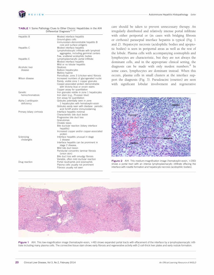
care should be taken to prevent unnecessary therapy. Anirregularly distributed and relatively intense portal infiltratewith either periportal or (in cases with bridging fibrosisor cirrhosis) paraseptal interface hepatitis is typical (Fig. 1and 2). Hepatocyte necrosis (acidophilic bodies and apopto-tic bodies) is seen in periportal areas as well as the rest ofthe lobule. Plasma cells with accompanying eosinophils andlymphocytes are characteristic, but they are not always thedominant cells, and in the appropriate clinical setting, thediagnosis can be made with only modest numbers.8 Insome cases, lymphocytes are dominant instead. When thisoccurs, plasma cells in small clusters at the interface sup-port the diagnosis (Fig. 3). Pseudoacini (rosettes) are seenwith significant lobular involvement and regenerative
Figure 1 AIH. This low-magnification image (hematoxylin-eosin, 340) shows expanded portal tracts with effacement of the interface by a lymphoplasmacytic infil-trate including many plasma cells. The connective tissue stain shows early fibrosis and regenerative activity with 2-cell-thick liver plates and early nodule formation.
TABLE 1 Some Pathology Clues to Other Chronic Hepatitides in the AIHDifferential Diagnosis12,13
Hepatitis B Modest interface hepatitisGround-glass cellsImmunostain-demonstrable hepatitis B
core and surface antigensHepatitis C Modest interface hepatitis
Lymphohistiocytic infiltrate with lymphoidaggregates, including germinal centers
Few, scattered acidophilic bodiesHepatitis E Lymphoplasmacytic portal infiltrate
Modest interface hepatitisMild or no lobular hepatitis
Alcoholic liverdisease
SteatosisBalloon hepatocytesMallory hyalinePericellular, zone 3 (chicken-wire) fibrosis
Wilson disease Increased numbers of glycogenated nucleiRarely, visible zone 1 copper granulesCopper-associated protein demonstrable
with Victoria blue or orcein stainsCopper assay for quantitation
Genetichemochromatosis
Iron granules visible in zone 1 hepatocytesIron stain (e.g., Prussian blue)Iron assay for quantitation
Alpha-1-antitrypsindeficiency
Globules potentially seen in zone1 hepatocytes with hematoxylin-eosin
Globules easily seen with diastase�periodicacid Schiff and/or immunostaining
Primary biliary cirrhosis Interface hepatitis minimalCharacteristic bile duct lesionProgressive bile duct lossGranulomasCholate stasisBile ductular reaction (biliary interface
hepatitis)Increased copper and/or copper-associated
proteinSclerosing
cholangitisInterface hepatitis unusual in stage
1 diseaseInterface hepatitis can be prominent in
stage 2 disease.Mild bile duct lesionPeriductal concentric laminar fibrosis
(onion skin)Bile duct loss with smudgy fibrosisVariable, often mild ductular reaction
Drug reaction Portal neutrophils and eosinophilsPlasma cells usually not prominentFibrosis usually not seen
Figure 2 AIH. This medium-magnification image (hematoxylin-eosin, 3200)shows a portal tract with an intense lymphoplasmacytic infiltrate effacing theinterface with rosette formation and hepatocyte necrosis (acidophilic bodies).
R E V I E W Autoimmune Hepatitis Histopathology Geller
20 Clinical Liver Disease, Vol 3, No 2, February 2014 An Official Learning Resource of AASLD

activity (Fig. 4). Rosettes and plasma cells are typical butare not pathognomonic or consistently seen. Furthermore,plasma cells and rosettes occur with other liver diseases.Zone 1 (periportal) emperipolesis, the engulfing of lympho-cytes by hepatocytes, occurs in the interface hepatitis area.There may be giant-cell transformation (giant-cell hepatitis).
Severe inflammation extends beyond the periportal zonewith parenchymal collapse and, not uncommonly, bridgingnecrosis, especially with acute clinical relapse and when
AIH is acute and fulminant.9 Portal-to-portal or portal-to-central fibrosis and cirrhosis are seen (Fig. 5).
Key histological features contributing positively to a scoreestablishing the diagnosis of AIH according to the revisedInternational Autoimmune Hepatitis Group modified staging
Figure 3 AIH. This high-magnification image (hematoxylin-eosin, 3400)shows a predominantly lymphocytic portal infiltrate with clusters of plasmacells at the interface.
Figure 4 AIH. This image (hematoxylin-eosin, 3400) shows rosettes.
Figure 5 AIH. This first-biopsy image (Masson trichrome, 3200) showsfibrosis with early bridge formation (arrow).
Figure 6 AIH with overlap syndrome. This image (hematoxylin-eosin, 3200)shows nonsuppurative cholangitis consistent with PBC.
R E V I E W Autoimmune Hepatitis Histopathology Geller
21 Clinical Liver Disease, Vol 3, No 2, February 2014 An Official Learning Resource of AASLD

system4 are (1) interface hepatitis (13), which is the mostimportant; (2) a lymphoplasmacytic infiltrate (11); and (3)rosette formation (11). Other features also help to establishthe diagnosis. For example, AIH differs from chronic hepati-tis C in having more severe lobular inflammation andnecrosis as well as greater numbers of plasma cells, moremarked interface hepatitis, and broad areas of parenchymalcollapse (Table 2). Zone 3 (centrilobular) necrosis is welldescribed in AIH but is often inadequately recognized.10
Zone 3 necrosis without fulminant hepatitis can lead toerroneous diagnoses such as ischemia/hypoxia and toxic/drug injury. Biliary changes are uncommon in AIH and arealmost always indicative of some other disorder. Isolatedbile duct injury, however, can be seen and does not excludeAIH.11 When anti-nuclear antibody values are significantlyincreased in association with biopsy-demonstrable bile ductinjury (Fig. 6), the diagnosis of AIC is likely. Negative scorefindings are the absence of these three findings (25), biliarychanges (23), and features suggesting an alternativeetiology (23).
There are no known direct microscopic correlates for thevarious identifiable autoantibodies. For example, thebiopsy findings for liver/kidney microsome (LKM)–associ-ated AIH are similar to those for other forms (Fig. 7).When overlap syndromes (e.g., AIH 1 PBC, AIH 1 PSC,and AIH 1 AIC) occur, atypical histological changes can beseen (Fig. 6).12,13 Rarely, granulomas are seen. Other coinci-dental disorders, such as hepatitis C, alcoholic liver disease,human immunodeficiency virus positivity, and iron storagedisease, affect morphology. Increasingly, drug effects must beconsidered. AIH-like microscopic changes caused by drugsare generally resolved with the cessation of medication, butchronic drug-induced AIH also occurs. Fibrosis and cirrhosisare distinctly unusual in drug-induced AIH, but cholestasis,portal neutrophils, and eosinophils are likely. The experienceof the reviewing pathologist can also affect the ability to estab-lish the diagnosis.
Fulminant AIH is uncommon and is morphologicallyindistinguishable from other forms of massive/submassivenecrosis.12,13
Cirrhosis in AIH generally shows a greater degree ofinflammation than cirrhosis due to other causes. Septa areeasily recognized, as are areas of prior parenchymal collapse,
Figure 7 LKM AIH. Similar to Fig. 1, this low-magnification image (hematoxylin-eosin, 320) shows a marked lymphoplasmacytic infiltrate with effacement of theinterface.
TABLE 2 Histopathological Features Most Useful in Differentiating AIHFrom Chronic Hepatitis C Virus
Feature AIH Hepatitis C Virus
Lobular inflammation/necrosis 12111 1/2Plasma cells 12111 021
Interface hepatitis 12111 0211
Parenchymal collapse 12111 0Steatosis 021 12111*Portal lymphoid aggregates 021 12111
Germinal center formation inlymphoid aggregates
0 0211
Bile duct injury† 021 1211
This table has been adapted with permission from Biopsy Interpreta-
tion of the Liver, 2nd ed.12
*Steatosis is particularly seen with genotype 3 hepatitis C virus.Bile duct injury is rarely seen in AIH but is characteristic of AIC.11
0 5 no change, 15 minimal or mild change, 115 moderatechange, 1115 marked change.
TABLE 3 Key Histopathology Features of AIH
1. Liver biopsy shows a moderate to severe necroinflammatory process withprominent portal inflammation, interface hepatitis, a lymphoplasmacyticinfiltrate including many plasma cells, and acinar transformation ofhepatocytes (rosettes).
2. Plasma cells are not always the dominant inflammatory cells and may beprominent only at the interface.
3. Fibrosis/cirrhosis is often seen on first biopsy.4. It may present as acute, fulminant liver failure with massive or submassive
necrosis, including centrilobular (zone 3) necrosis.5. Autoimmune liver disease variants may show features of more than one
immune disorder (overlap syndromes).6. AIC is a distinct disorder histologically resembling PBC (without anti-
mitochondrial antibodies in serum and with anti-nuclear antibodies).
R E V I E W Autoimmune Hepatitis Histopathology Geller
22 Clinical Liver Disease, Vol 3, No 2, February 2014 An Official Learning Resource of AASLD
and the developing nodules vary greatly in size. Dysplasticnodules can be seen, as can small hepatocellular carcinomas.
Key features of AIH are summarized in Table 3. n
CORRESPONDENCEStephen A. Geller, M.D., Department of Pathology and Laboratory Medicine,Weill Cornell Medical College, 1300 York Avenue, New York, NY 10065.E-mail: [email protected].
References1. Lucey MR, Vierling J. Clinical presentation and natural history of autoim-
mune hepatitis. Clin Liver Dis 2014;9-11.
2. Liberal R, Grant CR, Mieli-Vergani G, Vergani D. Autoimmune hepatitis: acomprehensive review. J Autoimmun 2013;41:126-139.
3. Weiler-Norman C, Sebode M, Lohse AW. Autoimmune hepatitis 2103 andbeyond. Minerva Gastroenterol Dietol 2013;59:133-141.
4. Strassburg CP. Autoimmune hepatitis. Dig Dis 2013;31:155-163.
5. Alvarez F, Berg PA, Bianchi FB, Bianchi L, Burroughs AK, Cancado EL, et al.International Autoimmune Hepatitis Group report: a review of criteria fordiagnosis of autoimmune hepatitis. J Hepatol 1999;31:929-938.
6. Bedossa P, Dargere D, Paradis V. Sampling variability of liver fibrosis inchronic hepatitis C. Hepatology 2003;38:1449-1457.
7. Rousselet MC, Michalak S, Dupr�e F, Crou�e A, Bedossa P, Saint-Andr�e JP, et al.Sources of variability in histological scoring of chronic viral hepatitis. Hepato-logy 2005;41:257-264.
8. Vergani D, Longhi MS, Bogdanos DP, Ma Y, Mieli-Vergani G. Autoimmunehepatitis. Semin Immunopathol 2009;31:421-435.
9. Czaja AJ. Acute and acute severed (fulminant) autoimmune hepatitis. Dig DisSci 2013;58:897-914.
10. Zen Y, Notsumata K, Tanaka N, Nakanuma Y. Hepatic centrilobular zonalnecrosis with positive antinuclear antibody: a unique subtype or early diseasein autoimmune hepatitis. Hum Pathol 2007;38:1669-1675.
11. Ludwig J, Czaja AJ, Dickson ER, LaRusso NF, Weisner RH. Manifestations ofnonsuppurative cholangitis in chronic hepatobiliary disease: morphologicspectrum, clinical correlations and terminology. Liver 1984;4:105-116.
12. Geller SA. Autoimmune hepatitis and related disorders. In: Geller SA, Pet-rovic LM, eds. Biopsy Interpretation of the Liver. 2nd ed. Philadelphia, PA:Lippincott Williams & Wilkins; 2009:120-135.
13. Washington MK, Manns MP. Autoimmune hepatitis. In: Burt A, Portmann B,Ferrell L, eds. MacSween’s Pathology of the Liver. 6th ed. Edinburgh, UnitedKingdom: Churchill Livingstone; 2012:467-490.
R E V I E W Autoimmune Hepatitis Histopathology Geller
23 Clinical Liver Disease, Vol 3, No 2, February 2014 An Official Learning Resource of AASLD