LOS LACTOCOMPUESTOS Y LAS NANOFIBRAS LACTEAS
6
Encapsulation of Lactobacillus plantarum 423 and its Bacteriocin in Nanofibers T. D. J. Heunis • M. Botes • L. M. T. Dicks Published online: 13 November 2009 Ó Springer Science+Business Media, LLC 2009 Abstract Pla nta rici n 423 , produc ed by Lactobacillus plantarum 423, was encapsulated in nanofibers that were produced by the electrospinning of 18% (w/v) polyethylene oxide (200 000 Da). The average diameter of the nanofi- bers was 288 nm. Plantaricin 423 activity decreased from 51 200 AU/ml to 25 600 AU/ml and from 204 800 AU/ml to 51 200 AU/ml aft er ele ctr ospinni ng, as det ermine d against Lactobacillus sakei DSM 20017 and Enterococcus faecium HKLHS, respectively. Cells of L. plantarum 423 encapsulated in nanofibers decreased from 2.3 9 10 10 cfu/ml bef ore ele ctr ospinning to 4.7 9 10 8 cfu/ml there after. Cel ls entrap ped in the nan ofib ers con tinued to pro duc e planta ricin 423. This is the first report on the encapsula tion of a bacteriocin and cells of L. plantarum in nanofibers. The method may be used to design a drug delivery system for bacteriocins and the encapsulation of probiotic lactic acid bacteria. The technology is currently being optimized. Keywords Encapsulation Á Nanofibers Á Bacteriocins Á Bacteria Introduction Lactic acid bacteria (LAB) are generally regarded as safe and their antimicrobial peptides (bacteriocins) have been used in the preservation of many food products [9]. Various claims have been attributed to LAB with probiotic proper- ties, e. g. reduction or pr eventi on of gast rointesti na l disord ers, inclu ding inflammatory bowel diseas e, allev iation of lactose intolerance, lowering of serum cholesterol levels, stimulation of the immune system and control of tumor growth [10, 30]. Some probiotic claims may be associated with the production of antimicrobial peptides (bacteriocins). Bac ter ioc ins are produc ed by many LAB, inc lud ing spe cies tha t are ass ociate d with humans, viz . the Lacto- bacill us acido philus group (L. acidophilus, Lactobacillus amylovorus, Lacto bacillu s crispatus , Lacto bacillu s galli- narum, Lactobacillus gasseri and Lactobacillus johnsonii ), Lactob acillu s reuter i , Lacto bacill us casei , Lactobacillus fermentum and Lactobacillus plantarum [9, 34]. Many of the ba cte ri ocins pr oduced by these spec ies ar e ac tive again st foodbo rne pathogens such as Listeria, Clostridium, Bacillus and Staphylococcus spp. [32]. The ant imi crobial act ivi ty of bac ter ioc ins in the gas - trointestinal tract (GIT) is difficult to prove. Mare et al. [22] showed that plantaricin 423 was produced by cells of L. plantarum 423 colonizing the ileum of piglets. Studies wit h rea l-t ime PCR have shown tha t the gene enc odi ng pla ntaricin 423 is con stit uti vely exp ress ed at con diti ons simula ting the GIT [28]. Different levels of planta ricin 423 production have been recorded in each of the sections of a GIT model [3], supporting the hypothesis that plantaricin 423 may be produced in vivo. Plantaricin 423 inhibited the gr owth of Bac illu s cer eus , Clost ridium sporo genes , Enterococcus faecalis , sever al Lactobacillus spp., Oeno- coccus oen i, Lister ia innocua , Lister ia monoc ytogenes , Pedio coccu s acidil actici , Pediococcus pentosaceus , Pro- pionib acteriu m acidip ropion ici , Propionibacterium sp., Staphylococcus carnosum and Streptococcus thermophilus [39], and excluded Enterobacteriaceae , but not lactic acid bacteria, from the cecum and colon in Wistar rats [29]. Pr omisi ng resul ts we re reporte d wi th lantibiotic s regarding the control of pathogens in airways. Kruszewska T. D. J. Heunis M. Botes L. M. T. Dicks ( &) Department of Microbiology, University of Stellenbosch, Private Bag X1, Matieland 7602, 7600 Stellenbosch, South Africa e-mail: [email protected] 123 Probiotics & Antimicro. Prot. (2010) 2:46–51 DOI 10.1007/s12602-009-9024-9
-
Upload
claudia-gomez-acevedo -
Category
Documents
-
view
229 -
download
0
Transcript of LOS LACTOCOMPUESTOS Y LAS NANOFIBRAS LACTEAS
8/7/2019 LOS LACTOCOMPUESTOS Y LAS NANOFIBRAS LACTEAS
http://slidepdf.com/reader/full/los-lactocompuestos-y-las-nanofibras-lacteas 1/6
Encapsulation of Lactobacillus plantarum 423 and its Bacteriocinin Nanofibers
T. D. J. Heunis • M. Botes • L. M. T. Dicks
Published online: 13 November 2009
Ó Springer Science+Business Media, LLC 2009
Abstract Plantaricin 423, produced by Lactobacillus
plantarum 423, was encapsulated in nanofibers that wereproduced by the electrospinning of 18% (w/v) polyethylene
oxide (200 000 Da). The average diameter of the nanofi-
bers was 288 nm. Plantaricin 423 activity decreased from
51 200 AU/ml to 25 600 AU/ml and from 204 800 AU/ml
to 51 200 AU/ml after electrospinning, as determined
against Lactobacillus sakei DSM 20017 and Enterococcus
faecium HKLHS, respectively. Cells of L. plantarum 423
encapsulated in nanofibers decreased from 2.3 9 1010 cfu/ml
before electrospinning to 4.7 9 108 cfu/ml thereafter.
Cells entrapped in the nanofibers continued to produce
plantaricin 423. This is the first report on the encapsulation
of a bacteriocin and cells of L. plantarum in nanofibers.
The method may be used to design a drug delivery system
for bacteriocins and the encapsulation of probiotic lactic
acid bacteria. The technology is currently being optimized.
Keywords Encapsulation Á Nanofibers Á Bacteriocins Á
Bacteria
Introduction
Lactic acid bacteria (LAB) are generally regarded as safe
and their antimicrobial peptides (bacteriocins) have been
used in the preservation of many food products [9]. Various
claims have been attributed to LAB with probiotic proper-
ties, e.g. reduction or prevention of gastrointestinal
disorders, including inflammatory bowel disease, alleviation
of lactose intolerance, lowering of serum cholesterol levels,stimulation of the immune system and control of tumor
growth [10, 30]. Some probiotic claims may be associated
with the production of antimicrobial peptides (bacteriocins).
Bacteriocins are produced by many LAB, including
species that are associated with humans, viz. the Lacto-
bacillus acidophilus group (L. acidophilus, Lactobacillus
amylovorus, Lactobacillus crispatus, Lactobacillus galli-
narum, Lactobacillus gasseri and Lactobacillus johnsonii),
Lactobacillus reuteri, Lactobacillus casei, Lactobacillus
fermentum and Lactobacillus plantarum [9, 34]. Many of
the bacteriocins produced by these species are active
against foodborne pathogens such as Listeria, Clostridium,
Bacillus and Staphylococcus spp. [32].
The antimicrobial activity of bacteriocins in the gas-
trointestinal tract (GIT) is difficult to prove. Mare et al.
[22] showed that plantaricin 423 was produced by cells of
L. plantarum 423 colonizing the ileum of piglets. Studies
with real-time PCR have shown that the gene encoding
plantaricin 423 is constitutively expressed at conditions
simulating the GIT [28]. Different levels of plantaricin 423
production have been recorded in each of the sections of a
GIT model [3], supporting the hypothesis that plantaricin
423 may be produced in vivo. Plantaricin 423 inhibited the
growth of Bacillus cereus, Clostridium sporogenes,
Enterococcus faecalis, several Lactobacillus spp., Oeno-
coccus oeni, Listeria innocua, Listeria monocytogenes,
Pediococcus acidilactici, Pediococcus pentosaceus, Pro-
pionibacterium acidipropionici, Propionibacterium sp.,
Staphylococcus carnosum and Streptococcus thermophilus
[39], and excluded Enterobacteriaceae, but not lactic acid
bacteria, from the cecum and colon in Wistar rats [29].
Promising results were reported with lantibiotics
regarding the control of pathogens in airways. Kruszewska
T. D. J. Heunis Á M. Botes Á L. M. T. Dicks (&)
Department of Microbiology, University of Stellenbosch,
Private Bag X1, Matieland 7602, 7600 Stellenbosch,
South Africa
e-mail: [email protected]
123
Probiotics & Antimicro. Prot. (2010) 2:46–51
DOI 10.1007/s12602-009-9024-9
8/7/2019 LOS LACTOCOMPUESTOS Y LAS NANOFIBRAS LACTEAS
http://slidepdf.com/reader/full/los-lactocompuestos-y-las-nanofibras-lacteas 2/6
et al. [19] repressed the growth of methicillin-resistant
Staphylococcus aureus (MRSA) strains in the respiratory
tract of mice with mersacidin, a lantibiotic produced by
Bacillus sp. HIL Y-85 54728. Similar studies with nisin, a
lantibiotic produced by Lactococcus lactis subsp. lactis,
did not eradicate S. aureus from the nasal tract of rats [18].
Nisin F, a lantibiotic produced by L. lactis subsp. lactis,
proved effective in the treatment of sinusitis in rats [6] andskin infections in mice [7].
Reports on bacteriocins of LAB active against Gram-
negative bacteria [4, 14, 24, 37, 38] sparked a renewed
interest in these peptides and their interaction with intes-
tinal pathogens. Data generated from genome sequences of
probiotic bacteria such as L. plantarum WCFS1, L. aci-
dophilus NCFM and L. johnsonii NCC 533 [2, 17, 27] and
the application of DNA micro-array technology [8] may
provide valuable answers to in vivo gene expression.
Studies on the medical applications of bacteriocins need
to be done under strictly controlled conditions, demanding
the design of special drug delivery systems. In this paper,we give a brief description of a novel drug delivery system
and present results thus far obtained.
Materials and Methods
Production and Isolation of Bacteriocins
L. plantarum 423 was cultured in MRS broth (Biolab
Diagnostics, Midrand, South Africa) at 30°C for 24 h.
Cells were harvested (8000g, 10 min, 4°C), the pH of the
cell-free supernatant adjusted to pH 6.5–7.0 with 1 N
NaOH and then heated at 80°C for 10 min. Plantaricin 423
was precipitated from the cell-free supernatant with 70%
saturated ammonium sulfate [31] and desalted against
distilled water by using a 1000 Da cutoff dialysis mem-
brane (Spectrum Inc., CA, USA). The sample was freeze-
dried and stored at -20°C.
Bacteriocin activity tests were performed by using the
agar-spot method described by Van Reenen et al. [39].
Lactobacillus sakei DSM 20017 and Enterococcus faecium
HKLHS, cultured in MRS broth (Biolab) at 30°C, were
used as target strains. Antimicrobial activity was expressed
as arbitrary units (AU) per ml. One AU was defined as the
reciprocal of the highest dilution showing a clear zone of
growth inhibition [39].
Preparation of Nanofibers
Polyethylene oxide (PEO, 200 000 Da), obtained from
Sigma–Aldrich (St. Louis, MO, USA), was suspended
(18%, w/v) in a solution of plantaricin 423 (204 800
AU/ml, as determined with L. sakei DSM 20017 as target)
and carefully mixed. In another experiment, L. plantarum
423 was cultured at 37°C for 24 h, harvested (8000g,
2 min) and the cells resuspended in sterile distilled water to
2 9 1011 cfu/ml. From this suspension, 100 ll (2 9 1010
cfu) was mixed with 500 ll (18%, w/v) PEO suspended insterile distilled water.
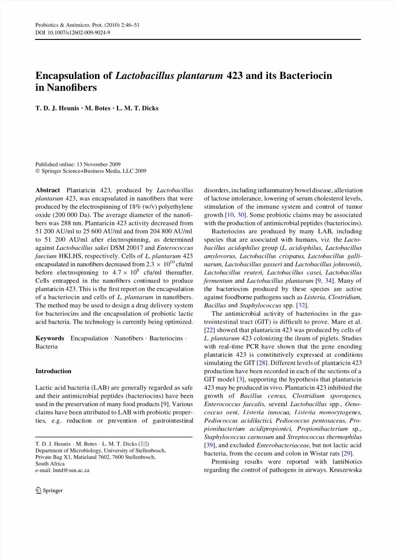
The polymer solutions were each placed in a tapered
sterile glass tube, fixed at a 15° angle (Fig. 1). The positive
electrode (cathode) was placed in the polymer solution, and
the anode on a collector plate was covered with sterile foil
and positioned 15 cm from the cathode. A constant electric
field of 10 kV was applied to the polymer solution and
-5 kV to the collector. The relative humidity was kept
constant at 50% and the temperature at 25°C. Nanofibers,
ejected from the tip of the tapered glass tube, were col-
lected at the anode (Fig. 1). Nanofibers were coated with
gold particles and visualized using a LeoÒ 1430VP
Scanning Electron Microscope. The diameter of the
nanofibers was determined by using the SEM Image Studio
software (version 10.1).
Evaluation of the Bacteriocin and Cell-Encapsulation in
Nanofibers
Nanofibers with plantaricin 423 were collected from the
metal surface, and 25.0 mg suspended in 500 ll sterile
distilled water by vigorous mixing on a vortex. The anti-
microbial activity of the suspension was determined by
using the agar-spot method as described before. L. sakei
DSM 20017 and E. faecium HKLHS served as target
strains. The number of viable cells encapsulated in the
nanofibers was determined by vortexing 25.0 mg fibers in
500 ll sterile saline (0.75%, w/v, NaCl). Serial dilutions
were made in sterile saline and plated onto MRS agar
(Biolab). Colonies were counted after 48 h of incubation at
37°C. Plantaricin 423 production by encapsulated cells was
evaluated by overlaying the colonies with E. faecium
HKLHS embedded in MRS agar. The plates were
Polymer jet
Anode collector with anon-woven nanofiber mat
High
Voltage
Polymer solution
Tapered glass tubewith catode
+
Fig. 1 Schematic representation of the electrospinning process with a
high voltage supply, tapered glass tube and collector
Probiotics & Antimicro. Prot. (2010) 2:46–51 47
123
8/7/2019 LOS LACTOCOMPUESTOS Y LAS NANOFIBRAS LACTEAS
http://slidepdf.com/reader/full/los-lactocompuestos-y-las-nanofibras-lacteas 3/6
incubated at 37°C for 48 h and then inspected for forma-
tion of inhibition zones.
Results
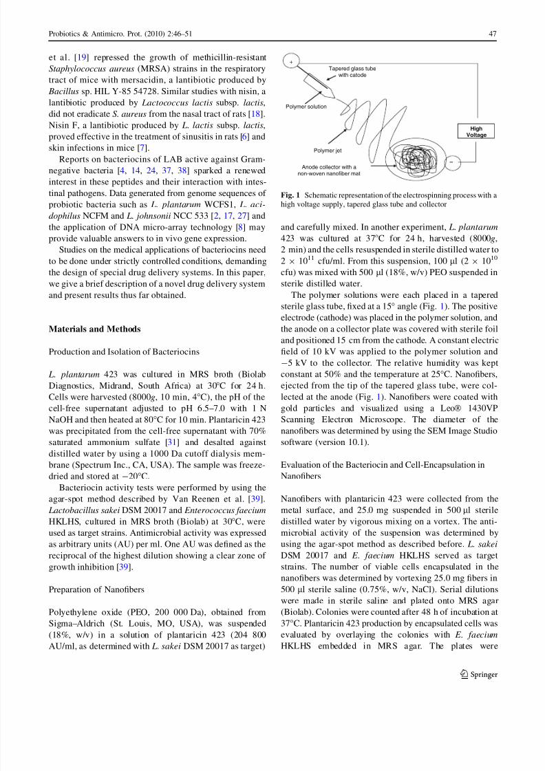
The activity of plantaricin 423 in the cell-free supernatant
was recorded as 51 200 AU/ml against L. sakei DSM20017 and 204 800 AU/ml against E. faecium HKLHS.
After electrospinning, the activity of plantaricin 423 in the
nanofibers was 25 600 AU/ml against L. sakei and 51 200
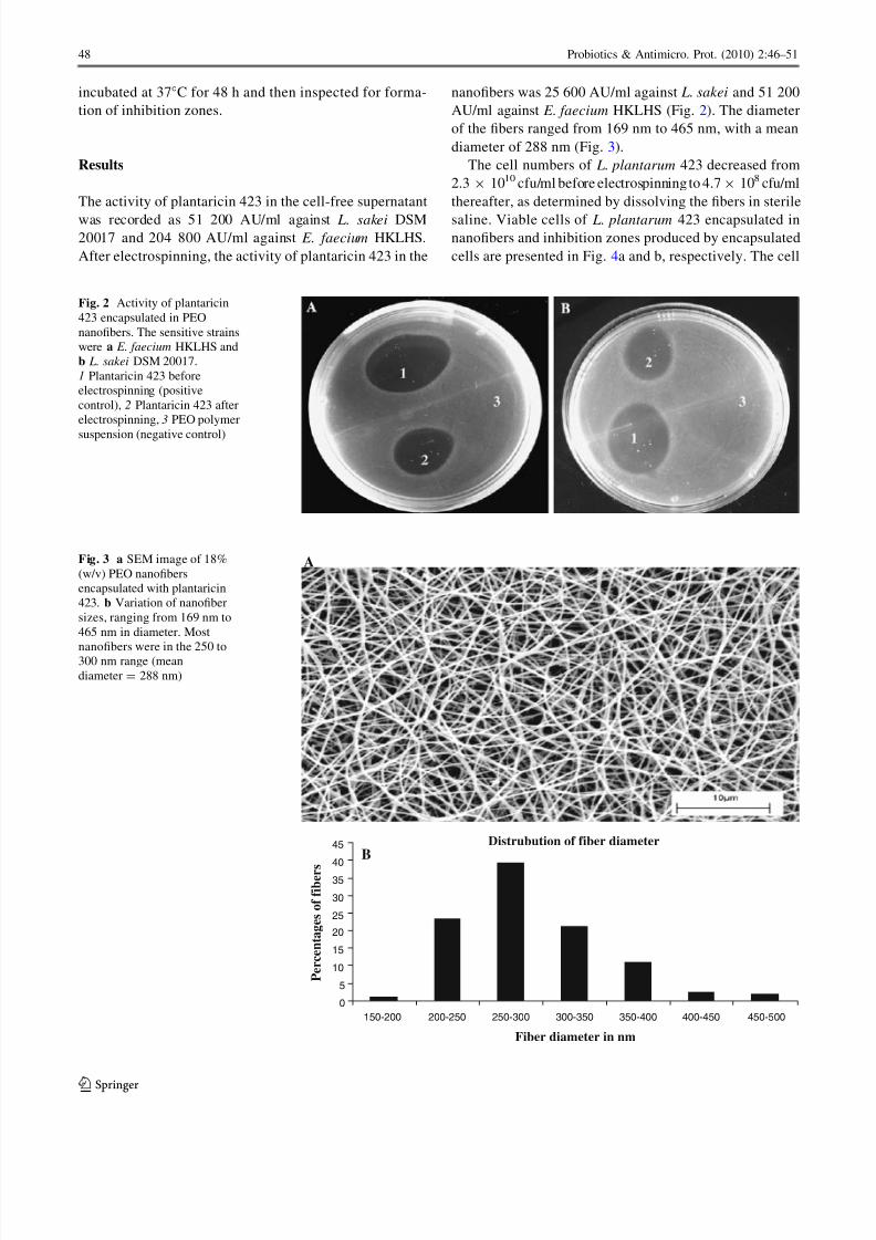
AU/ml against E. faecium HKLHS (Fig. 2). The diameter
of the fibers ranged from 169 nm to 465 nm, with a mean
diameter of 288 nm (Fig. 3).
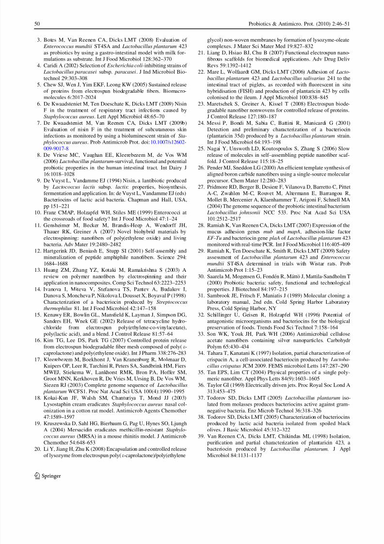
The cell numbers of L. plantarum 423 decreased from
2.3 9 1010 cfu/ml before electrospinning to 4.7 9 108 cfu/ml
thereafter, as determined by dissolving the fibers in sterile
saline. Viable cells of L. plantarum 423 encapsulated innanofibers and inhibition zones produced by encapsulated
cells are presented in Fig. 4a and b, respectively. The cell
Fig. 2 Activity of plantaricin
423 encapsulated in PEO
nanofibers. The sensitive strains
were a E. faecium HKLHS and
b L. sakei DSM 20017.
1 Plantaricin 423 before
electrospinning (positive
control), 2 Plantaricin 423 after
electrospinning, 3 PEO polymer
suspension (negative control)
Distrubution of fiber diameter
0
5
10
15
20
25
30
35
40
45
150-200 200-250 250-300 300-350 350-400 400-450 450-500
Fiber diameter in nm
Percentages of fibers
B
AFig. 3 a SEM image of 18%
(w/v) PEO nanofibers
encapsulated with plantaricin
423. b Variation of nanofiber
sizes, ranging from 169 nm to
465 nm in diameter. Most
nanofibers were in the 250 to300 nm range (mean
diameter = 288 nm)
48 Probiotics & Antimicro. Prot. (2010) 2:46–51
123

8/7/2019 LOS LACTOCOMPUESTOS Y LAS NANOFIBRAS LACTEAS
http://slidepdf.com/reader/full/los-lactocompuestos-y-las-nanofibras-lacteas 4/6
size of L. plantarum 423 decreased during encapsulation, as
shown in Fig. 4a. No bacterial cells were observed on the
surface of the nanofibers.
Discussion
Several methods are used to produce ultrafine fibers, e.g.
self assembly of polymers [12, 25], template synthesis [26],
phase separation [35] and electrospinning [5, 42]. Elec-
trospinning is a versatile and relatively easy technique to
produce large amounts of ultrathin fibers. During the
spinning process, an electric field is applied to a polymer
solution to form a Taylor cone [36, 40]. When the electric
forces overcome the surface tension of the solution, a
charged jet is ejected from the Taylor cone and accelerates
toward the collector. The solvent evaporates and nanofibers
are formed [1, 21]. The quality and characteristics of the
final product are determined by the elasticity, viscosity,
temperature, conductivity and surface tension of the poly-
mer, strength of the electric field, distance between the
Taylor cone and collector, and humidity [13, 21].
Electrospinning has been used to encapsulate antibiotics
[15, 41], growth factors, proteins [5, 20, 23] and silver
nanoparticles [33]. As far as we could determine, we are
the first to report on the encapsulation of a bacteriocin and
viable cells of L. plantarum. The slight decrease in anti-
microbial activity of plantaricin 423 recorded after elec-
trospinning could be due to the high voltage applied.
However, Kim et al. [16] have reported only a slight
decrease (10%) in the activity of lysozyme after electros-
pinning. Human b-nerve growth factor released from
electrospun fibers was still able to differentiate PC12 cells
into neurons [5].
Judged from the decrease in cell numbers (from
2.3 9 1010 cfu/ml before electrospinning to 4.7 9 108 cfu/ml
when encapsulated), only 2% of the L. plantarum cells
remained viable after encapsulation into nanofibers (Fig. 4).
A higher percentage of cells might have been encapsulated,
but could have been damaged by the high voltage and
whipping or shearing action of electrospinning. It is also
possible that cells could have lost their viability after
encapsulation. Only 0.1% of Escherichia coli cells encap-sulated in PEO nanofibers survived, whereas 74% of
Micrococcus luteus cells survived electrospinning [11]. It
would seem as if smaller cells and Gram-positive bacteria
are more resistant to electrospinning. Further research is
needed to determine the exact fate of the cells. The smallsize
of the fibers and the large surface to volume ratio provide for
encapsulation of high cell numbers, thus compensating for
the loss in viability. The fact that no cells were observed on
the surface of the nanofibers may be ascribed to the high
voltage applied during the spinning process. Cells and bac-
teriocins not enclosed in nanofibers are not protected by the
high voltage and are prone to destruction.In this paper, we demonstrate the feasibility of encap-
sulating bacteriocins and lactic acid bacteria into spun
nanofibers. Encapsulation of bacteriocin-producing cells
and cells with probiotic properties may have specific food
and medical applications. Through careful selection of
polymers, larger concentrations of bacteriocins and higher
cell numbers may be spun into nanofibers. We are currently
conducting further research to optimize the technology.
Acknowledgments Cipla Medpro (Pty) Ltd and the National
Research Foundation, South Africa, for funding the research.
References
1. Agarwal S, Wendorff JH, Greiner A (2008) Use of electrospin-
ning technique for biomedical applications. Polymer 49:5603–
5621
2. Alterman E, Russell WM, Azcarate-Peril MA, Barrangou R, Buck
BL, McAuliffe O, Souther N, Dobson A, Duong T, Callanan M,
Lick S, Hamrick A, Cano R, Klaenhammer TD (2005) Complete
genome sequence of the probiotic lactic acid bacterium Lacto-
bacillus acidophilus NCFM. Proc Nat Acad Sci (USA) 102:3906–
3912
Fig. 4 a Viable cells of L. plantarum 423 encapsulated in 18% (w/v) PEO nanofibers, clearly showing a ‘‘bead’’ structure where cells were
entrapped. b Growth inhibition of E. faecium HKLHS by plantaricin 423 produced from encapsulated cells
Probiotics & Antimicro. Prot. (2010) 2:46–51 49
123
8/7/2019 LOS LACTOCOMPUESTOS Y LAS NANOFIBRAS LACTEAS
http://slidepdf.com/reader/full/los-lactocompuestos-y-las-nanofibras-lacteas 5/6
3. Botes M, Van Reenen CA, Dicks LMT (2008) Evaluation of
Enterococcus mundtii ST4SA and Lactobacillus plantarum 423
as probiotics by using a gastro-intestinal model with milk for-
mulations as substrate. Int J Food Microbiol 128:362–370
4. Caridi A (2002) Selection of Escherichia coli-inhibiting strains of
Lactobacillus paracasei subsp. paracasei. J Ind Microbiol Bio-
technol 29:303–308
5. Chew SJ, Wen J, Yim EKF, Leong KW (2005) Sustained release
of proteins from electrospun biodegradable fibers. Biomacro-
molecules 6:2017–2024
6. De Kwaadsteniet M, Ten Doeschate K, Dicks LMT (2009) Nisin
F in the treatment of respiratory tract infections caused by
Staphylococcus aureus. Lett Appl Microbiol 48:65–70
7. De Kwaadsteniet M, Van Reenen CA, Dicks LMT (2009b)
Evaluation of nisin F in the treatment of subcutaneous skin
infections as monitored by using a bioluminescent strain of Sta-
phylococcus aureus. Prob Antimicrob Prot. doi:10.1007/s12602-
009-9017-8
8. De Vriese MC, Vaughan EE, Kleerebezem M, de Vos WM
(2006) Lactobacillus plantarum-survival, functional and potential
probiotic properties in the human intestinal tract. Int Dairy J
16:1018–1028
9. De Vuyst L, Vandamme EJ (1994) Nisin, a lantibiotic produced
by Lactococcus lactis subsp. lactis: properties, biosynthesis,
fermentation and application. In: de Vuyst L, Vandamme EJ (eds)
Bacteriocins of lactic acid bacteria. Chapman and Hall, USA,
pp 151–221
10. Franz CMAP, Holzapfel WH, Stiles ME (1999) Enterococci at
the crossroads of food safety? Int J Food Microbiol 47:1–24
11. Gensheimer M, Becker M, Brandis-Heep A, Wendorff JH,
Thauer RK, Greiner A (2007) Novel biohybrid materials by
electrospinning: nanofibers of poly(ethylene oxide) and living
bacteria. Adv Mater 19:2480–2482
12. Hartgerink JD, Beniash E, Stupp SI (2001) Self-assembly and
mineralization of peptide amphiphile nanofibers. Science 294:
1684–1688
13. Huang ZM, Zhang YZ, Kotaki M, Ramakrishna S (2003) A
review on polymer nanofibers by electrospinning and their
application in nanocomposites. Comp Sci Technol 63:2223–2253
14. Ivanova I, Miteva V, Stefanova TS, Pantev A, Budakov I,
Danova S, Moncheva P, Nikolova I, Dousset X, Boyaval P (1998)
Characterization of a bacteriocin produced by Streptococcus
thermophilus 81. Int J Food Microbiol 42:147–158
15. Kenawy ER, Bowlin GL, Mansfield K, Layman J, Simpson DG,
Sanders EH, Wnek GE (2002) Release of tetracycline hydro-
chloride from electrospun poly(ethylene-co-vinylacetate),
poly(lactic acid), and a blend. J Control Release 81:57–64
16. Kim TG, Lee DS, Park TG (2007) Controlled protein release
from electrospun biodegradable fiber mesh composed of poly(e-
caprolactone) and poly(ethylene oxide). Int J Pharm 338:276–283
17. Kleerebezem M, Boekhorst J, Van Kranenburg R, Molenaar D,
Kuipers OP, Leer R, Tarchini R, Peters SA, Sandbrink HM, Fiers
MWEJ, Stiekema W, Lankhorst RMK, Bron PA, Hoffer SM,
Groot MNN, Kerkhoven R, De Vries M, Ursing B, De Vos WM,Siezen RJ (2003) Complete genome sequence of Lactobacillus
plantarum WCFS1. Proc Nat Acad Sci USA 100(4):1990–1995
18. Kokai-Kun JF, Walsh SM, Chanturiya T, Mond JJ (2003)
Lysostaphin cream eradicates Staphylococcus aureus nasal col-
onization in a cotton rat model. Antimicrob Agents Chemother
47:1589–1597
19. Kruszewska D, Sahl HG, Bierbaum G, Pag U, Hynes SO, Ljungh
A (2004) Mersacidin eradicates methicillin-resistant Staphylo-
coccus aureus (MRSA) in a mouse rhinitis model. J Antimicrob
Chemother 54:648–653
20. Li Y, Jiang H, Zhu K (2008) Encapsulation and controlled release
of lysozyme from electrospun poly(e-caprolactone)/poly(ethylene
glycol) non-woven membranes by formation of lysozyme-oleate
complexes. J Mater Sci Mater Med 19:827–832
21. Liang D, Hsiao BJ, Chu B (2007) Functional electrospun nano-
fibrous scaffolds for biomedical applications. Adv Drug Deliv
Revs 59:1392–1412
22. Mare L, Wolfaardt GM, Dicks LMT (2006) Adhesion of Lacto-
bacillus plantarum 423 and Lactobacillus salivarius 241 to the
intestinal tract of piglets, as recorded with fluorescent in situ
hybridisation (FISH) and production of plantaricin 423 by cells
colonised to the ileum. J Appl Microbiol 100:838–845
23. Maretschek S, Greiner A, Kissel T (2008) Electrospun biode-
gradable nanofiber nonwovens for controlled release of proteins.
J Control Release 127:180–187
24. Messi P, Bondi M, Sabia C, Battini R, Manicardi G (2001)
Detection and preliminary characterization of a bacteriocin
(plantaricin 35d) produced by a Lactobacillus plantarum strain.
Int J Food Microbiol 64:193–198
25. Nagai Y, Unswoth LD, Koutospoulos S, Zhang S (2006) Slow
release of molecules in self–assembling peptide nanofiber scaf-
fold. J Control Release 115:18–25
26. Pender MJ, Sneddon LG (2000) An efficient template synthesis of
aligned boron carbide nanofibers using a single-source molecular
precursor. Chem Mater 12:280–283
27. Pridmore RD, Berger B, Desiere F, Vilanova D, Barretto C, Pittet
A-C, Zwahlen M-C, Rouvet M, Altermann E, Barrangou R,
Mollet B, Mercenier A, Klaenhammer T, Arigoni F, Schnell MA
(2004) The genome sequence of the probiotic intestinal bacterium
Lactobacillus johnsonii NCC 533. Proc Nat Acad Sci USA
101:2512–2517
28. Ramiah K, Van Reenen CA, Dicks LMT (2007) Expression of the
mucus adhesion genes mub and mapA, adhesion-like factor
EF-Tu and bacteriocin gene plaA of Lactobacillus plantarum 423
monitored with real-time PCR. Int J Food Microbiol 116:405–409
29. Ramiah K, Ten Doeschate K, Smith R, Dicks LMT (2009) Safety
assessment of Lactobacillus plantarum 423 and Enterococcus
mundtii ST4SA determined in trials with Wistar rats. Prob
Antimicrob Prot 1:15–23
30. Saarela M, Mogensen G, Fonden R, Matto J, Mattila-Sandholm T
(2000) Probiotic bacteria: safety, functional and technological
properties. J Biotechnol 84:197–215
31. Sambrook JE, Fritsch F, Maniatis J (1989) Molecular cloning: a
laboratory manual, 2nd edn. Cold Spring Harbor Laboratory
Press, Cold Spring Harbor, NY
32. Schillinger U, Geisen R, Holzapfel WH (1996) Potential of
antagonistic microorganisms and bacteriocins for the biological
preservation of foods. Trends Food Sci Technol 7:158–164
33. Son WK, Youk JH, Park WH (2006) Antimicrobial cellulose
acetate nanofibers containing silver nanoparticles. Carbohydr
Polym 65:430–434
34. Tahara T, Kanatani K (1997) Isolation, partial characterization of
crispacin A, a cell-associated bacteriocin produced by Lactoba-
cillus crispatus JCM 2009. FEMS microbiol Letts 147:287–290
35. Tan EPS, Lim CT (2004) Physical properties of a single poly-
meric nanofiber. Appl Phys Letts 84(9):1603–160536. Taylor GI (1969) Electrically driven jets. Proc Royal Soc Lond A
313:453–475
37. Todorov SD, Dicks LMT (2005) Lactobacillus plantarum iso-
lated from molasses produces bacteriocins active against gram-
negative bacteria. Enz Microb Technol 36:318–326
38. Todorov SD, Dicks LMT (2005) Characterization of bacteriocins
produced by lactic acid bacteria isolated from spoiled black
olives. J Basic Microbiol 45:312–322
39. Van Reenen CA, Dicks LMT, Chikindas ML (1998) Isolation,
purification and partial characterization of plantaricin 423, a
bacteriocin produced by Lactobacillus plantarum. J Appl
Microbiol 84:1131–1137
50 Probiotics & Antimicro. Prot. (2010) 2:46–51
123
8/7/2019 LOS LACTOCOMPUESTOS Y LAS NANOFIBRAS LACTEAS
http://slidepdf.com/reader/full/los-lactocompuestos-y-las-nanofibras-lacteas 6/6
40. Yarin AL, Koombhongse S, Reneker DH (2001) Taylor cone and
jetting from liquid droplets in electrospinning of nanofibers. J
Appl Phys 90(9):4836–4846
41. Zeng J, Xu X, Chen X, Liang Q, Bian X, Yang L, Jing X (2003)
Biodegradable electrospun fibers for drug delivery. J Control
Release 92:227–231
42. Zhou Y, Yang D, Chen X, Xu Q, Lu F, Nie J (2008) Electrospun
water-soluble carboxyethyl chitosan/poly(vinyl alcohol) nanofi-
brous membrane as potential wound dressing for skin regenera-
tion. Biomacromolecules 9:349–354
Probiotics & Antimicro. Prot. (2010) 2:46–51 51
123