Molecular Autoimmunity€¦ · novel immunointervention strategies. The first part of the book is...
30
Molecular Autoimmunity
Transcript of Molecular Autoimmunity€¦ · novel immunointervention strategies. The first part of the book is...

Molecular Autoimmunity
Prelims.qxd 07/06/05 6:07 PM Page i

Molecular Autoimmunity
Edited by
MONCEF ZOUALIInstitut National de Santé et de Recherche Médicale (INSERM), Paris, France
Prelims.qxd 07/06/05 6:07 PM Page iii

A C.I.P. Catalogue record for this book is available from the Library of Congress.
ISBN 0-387-24533-2
©2005 Springer Science+Business Media, Inc.All rights reserved. This work may not be translated or copied in whole or in part without the writtenpermission of the publisher (Springer Science+Business Media, Inc., 233 Spring Street, New York, NY10013, USA), except for brief excerpts in connection with reviews or scholarly analysis, Use in con-nection with any form of information storage and retrieval, electronic adaptation, computer software,or by similar or dissimilar methodology now known or hereafter developed is forbidden.The use in this publicaiton of trade names, trademarks, service marks and similar terms, even if theyare not identified as such, is not to be taken as an expression of opinion as to whether or not theyare subject to proprietary rights.
Printed in the United States of America.
9 8 7 6 5 4 3 2 1
springeronline.com
Prelims.qxd 07/06/05 6:07 PM Page iv

Preface
A unique feature of the normal immune system is that, while being able to mountresponses to virtually all antigens of the environment, it also exhibits tolerance toits own components, a property that prevents attack of the body’s own tissues. Attimes, however, self-tolerance breaks down, and the immune system fails to rec-ognize self-antigens and mounts a misguided immune attack against its own tis-sues, which culminates in autoimmune disease. Currently, a growing number ofdisorders affecting virtually all organs or tissues of the human body have a provenor a strongly suspected autoimmune etiology. Their prevalence is worldwide andtheir etiology remains under investigation. In the past few years, our understand-ing of autoimmunity has witnessed important advances. This volume commemo-rates the 100th anniversary of the discovery of the first human autoimmunedisease by Julius Donath and Karl Landsteiner in 1904 in Vienna. It comprises acollection of papers that show some of the ways in which insight into autoimmu-nity is opening new avenues for understanding their etiology and for designingnovel immunointervention strategies.
The first part of the book is concerned with innate immunity, a branch ofthe immune system mainly directed to recognition of invariant molecules ofinfectious agents. Most of them are essential for pathogen survival and are con-served and shared by groups of pathogens. The innate immune system is essen-tial for the activation of the adaptive immune response, capable of coping with ahigh mutation rate and antigen heterogeneity of infectious agents, and generatinga long-lasting immune memory. Addressing its role in induction, progression, andprotection of myocarditis, a disease linked to adenovirus or coxsackievirus thataccounts for approximately 25% of all heart failure in North America, Noel Roseet al. tackle brilliantly the daunting task of bringing together the many facets ofinnate immunity in autoimmunity. They discuss in detail four of the major com-ponents of the innate response found to contribute to disease susceptibility: thecomplement system, natural killer cells, macrophages/dendritic cells, and early-acting proinflammatory cytokines. Also important in the innate immunity branchare toll-like receptors (TLRs) present on a variety of cell types. Paul N. Moynaghreviews the crucial involvement of TLR9 in mediating the immunostimulatoryeffects of bacterial DNA, potentially leading to activation of B cells and produc-tion of autoantibodies independently of T cells. Another example of the interplaybetween innate immunity and autoimmunity is discussed in the paper from theTerry Du Clos laboratory. Here, members of the pentraxin family, a phylogeneti-cally ancient, highly conserved component of the innate immune system, areshown to bind microbial determinants and autoantigens, and to have the potential
v
Prelims.qxd 07/06/05 6:07 PM Page v

to interact with the adaptive immune system through the complement system andFcγ receptors. In studies of autoimmune type 1 diabetes, an autoimmune diseasecaused by T cell–mediated destruction of insulin-producing β cells in the pancre-atic islets of Langerhans, Terry L. Delovitch and coworkers show how a subset ofT cells act as regulators of both innate and adaptive immune responses. Since thiscell population seems to be important in maintaining immune homeostasis, a fur-ther understanding of its role offers promise for the development of novel thera-pies for the prevention of diabetes.
The second part of this book focuses on genetic susceptibility. While earlystudies revealed that human autoimmune diseases require an inherited contribu-tion, their genetics remains the focus of much investigation. Marta E. Alarcón-Riquelme discusses how the availability of the human genome sequence isplaying an essential role in unraveling complex disease genetics, and how humangenome scans are providing new discoveries. Most interesting is the observationthat some of the genes identified are shared among various autoimmune diseases.In a search for factors that promote autoimmunity, Bruce Richardson’s laboratoryis exploring DNA methylation, an important determinant of chromatin structurethat modifies gene expression through localized effects on the nucleosome poly-mers. Their article elegantly describes how the results can be used to predict func-tional, biochemical, and genetic alterations in T cells from patients withidiopathic lupus, and how failure to maintain T cell DNA methylation and chro-matin structure contributes to human lupus. In myasthenia gravis, an organ-specific antibody-mediated autoimmune disease characterized by an immuneresponse against the nicotinic acetylcholine receptor on the neuromuscular junc-tion, the data described by Ann Kari Lefvert support the notion that the diseaseis polygenic, with subgroups of patients having different genetic backgrounds.Also polygenic is human lupus. C. Yung Yu and coworkers describe the strongassociation of complete C4A and C4B deficiencies with human lupus, providingsupport for the interpretation that C4A deficiency is a genetic risk factor for thisdisease.
Discussed in the third part of this volume are some potential triggers ofautoimmunity that affect different organs. In rheumatic fever, a disease occurringas a delayed sequel of throat infection by Streptococcus pyogenes in 3–4 % ofuntreated children, Jorge Kalil and coworkers clearly show how molecular mim-icry between streptococcal antigens and human heart tissue leads to rheumaticheart lesions. In this process, CD4+ T lymphocytes are the major effectors of heartlesions, and several histocompatibility leucocyte antigen (HLA) class II mole-cules are associated with the disease worldwide, leading to multiple valvularlesions and/or mitral valve regurgitation. By contrast, in other disorders, no infec-tious agent has been identified. For example, celiac disease is an intestinal disor-der caused by an inflammatory T cell response to gluten peptides bound toHLA-DQ2 or -DQ8, molecules with a preference for peptides that contain nega-tively charged amino acids. As described in the article from Frits Koning’s labo-ratory, posttranslational modification of gluten is critical for the generation of arepertoire of T cell stimulatory peptides, an observation that may be relevant forother HLA-associated disease. The paper from Michael Hertl’s laboratory dis-
vi Preface
Prelims.qxd 07/06/05 6:07 PM Page vi
cusses the pathogenic role of autoantibodies and the potential role of autoreactiveT cells to desmogleins in pemphigus vulgaris. Aiming to develop antigen-specificimmunotherapies, the authors put the emphasis on autoaggressive T cell epitopesand on a subset of T cells that may be critical in the maintenance/restoration oftolerance against desmogleins. Also unclear is the trigger involved in myastheniagravis pathogenesis. Here, circumstantial evidence suggests a primary role of thethymus. Having established a model of intrathymic inflammation localized to thethymic medulla, Arnold I. Levinson et al. attempt to determine how intrathymicexpression of the neuromuscular muscle type of acetylcholine receptors isinvolved in immunopathogenesis.
The fourth part of this volume is devoted to targets of autoimmunity. PaolaMigliorini et al. focus on development of autoantibody-mediated nephritis. Theyreview data indicating that distinct damage mechanisms probably coexist andplay a role in the different phases of poststreptococcal nephritis, Goodpasture’ssyndrome, and systemic lupus nephritis. The contribution from Ansar Ahmed’slaboratory lucidly addresses the role of hormonal factors in autoimmunity. Theireffects have been demonstrated in many experimental settings. In humans, expo-sure to estrogens occurs through various sources, including physiological estro-gens that vary during the lifetimes of women, pharmacological estrogens givenfor medical reasons and environmental estrogens, or endocrine-disrupting chem-icals, (pesticides, plastic products, detergents, industrial by-products, municipalsewage–contaminated water that contains metabolites of estrogen-based contra-ceptive drugs). Nevertheless, their effects are complex and remain incompletelyunderstood. Also important in deciphering autoimmunity are studies of the roleof CD4+CD25+ regulatory T cells, discussed by Yi-chi M. Kong et al. in experi-mental murine autoimmune thyroiditis. Yet, our lack of understanding autoimmu-nity is perhaps best illustrated by the complexity of immune phenomenadescribed in multiple sclerosis. This chronic inflammatory disease of the centralnervous system represents one of the most common neurological diseases ofyoung adults in developed countries. Its hallmarks include focal plaques of whitematter demyelination, presence of autoreactive T cells in the blood of mostpatients, and autoreactive T cells and antibodies in the lesions. Arguing that theautoimmune responses in the affected patients are not invariably detrimental, butmay even be beneficial, the provocative article from Hans Lassmann’s laboratorychallenges the “autoimmune hypothesis” of multiple sclerosis. Future work isrequired to provide a better understanding of the pathogenesis of this disease.
What could underlie the loss of tolerance in autoimmunity? As discussed inthe fourth part of this volume, the reason may well relate to crippling of signal-ing pathways that govern the discriminatory potential of lymphocytes. Ourimmune system functions properly only because lymphocytes communicate withone another constantly. Recognizing molecules that are part of our body as self-antigens and distinguishing them from those that come from the external envi-ronment, lymphocytes instruct their relatives to attack invaders or produce growthfactors or antibodies. This high-fidelity recognition is achieved through a networkof intracellular communications wherein lymphocyte receptors are able to sensethe nature of encountered molecules and to generate signals that are appropriately
Preface vii
Prelims.qxd 07/06/05 6:07 PM Page vii
delivered to the internal machinery, allowing specific functional responses. As inother cells, the amount of signals generated is fine-tuned for optimal transmis-sion, and kinases and phosphatases control most activities. The chapter on B cellssheds light on the biochemical and molecular aberrations that are responsible forthe aberrant lupus B cell biology. Inactivation of genes encoding B cell signalingmolecules leads to autoimmune phenomena, and crippled signaling pathways aredetectable in the B lymphocytes from patients with systemic autoimmunity.Focusing on rheumatoid arthritis, Ana M. Blasini and Martín A. Rodríguez sum-marize how abnormalities in T cell responses seen in patients with systemicautoimmunity can be related to identifiable signaling abnormalities. In lupus too,T cells display diverse cellular and cytokine abnormalities. The paper formGeorge C. Tsokos’s laboratory elegantly describes biochemical abnormalities thatunderwrite the diverse T cell abnormalities in lupus. Here, the decreased T cellreceptor–associated ζ chain in effector T cells is due mostly to increased degra-dation, rather than to decreased transcription. Lupus T cells also expressincreased amounts of the transcriptional repressor CREM that binds IL-2 pro-moter, thereby limiting its expression. Of further importance is the increasedspontaneous aggregation of lipid rafts on the surface membrane of lupus T cells,an abnormality that may contribute to the well-established overexcitable T cellphenotype. Altered signaling in both B and T cells also might account for theaberrant rates of apoptosis in lupus. Koji Yasutomo argues that the resultingincreased levels of free-circulating chromatin represent a potential source of anti-gen trigger in systemic autoimmunity.
Finally, the recent advances in the field of autoimmunity have given clini-cians exciting new tools for diagnosing and treating autoimmune disorders. Thefinal section of the book discusses state-of-the-art therapeutic intervention strate-gies. The rationale for B lymphocyte depletion therapy in autoimmune disordersstems from the paramount role of B cells in autoimmunity. Jonathan Edwardset al. discuss in detail its potential for clinical applications, the logisticsemployed, and the clinical results obtained with anti-CD20 antibody. While theprecise mechanisms of action remain to be elucidated, an alternative to this ther-apy is based on study of B lymphocyte longevity. Following migration to theperiphery, the selection and survival of B cells are controlled by a variety of sig-nals. Longevity factors, such as B lymphocyte stimulator (BLyS), also calledBAFF, TALL-1, THANK, or zTNF4, that support differentiation of selected Bcells into mature long-lived B cells are critical in determining the capacity tomount protective immune responses and to generate deleterious autoimmuneresponses. Their vital role in survival and maturation of B cells is discussed byWilliam Stohl. In experimental animals, treatment with BLyS/BAFF antagonistsameliorates disease progression and enhances survival. Since patients with lupus,rheumatoid arthritis, or systemic sclerosis overexpress this longevity factor, andbecause a phase I clinical trial in lupus patients with a neutralizing anti-BLySmonoclonal antibody has documented the safety and biological activity of thisBLyS antagonist, additional phase I and phase II clinical trials with a variety of BLySantagonists are currently under way. Another unifying theme in autoimmune dis-eases is the involvement of cytokines that play key roles throughout the whole
viii Preface
Prelims.qxd 07/06/05 6:07 PM Page viii
course of the disease, from induction to effector functions. Hence, the controlof autoimmunity by cytokine and anti-cytokine treatments represents a potentialimmunointervention strategy. The simplest approach, already in practice, is thespecific inhibition of their action. As discussed in detail by Pierre Miossec, theuse of TNFα inhibitors has provided clear evidence of the direct role of cytokinesin complex inflammatory diseases. Another more physiological approach consistsin stimulating endogenous regulatory mechanisms to restore an adequate bal-ance. However, as Alan Tyndall and Paul Hasler point out, just as there is no con-sensual unifying mechanism in autoimmune diseases, there is no singlesuccessful treatment strategy. Most patients with severe autoimmune diseases aretreated with a combination of glucocorticosteroids and immunosuppressiveagents, but some either do not respond or require more toxic drugs to achieve ormaintain clinical remission, and this subgroup poses a serious treatment dilemma.Rather than total eradication of clonal autoimmunity, hematopoietic stem celltransplantation techniques aim at resetting an imbalance in the complex immunenetwork. The authors posit that this emerging alternative could be a viable optionfor selected autoimmune diseases patients. Currently, through an internationalcollaboration, around 700 patients have received such treatment. The experiencegained from the phase I and II clinical studies is sufficiently encouraging tobe exploited in designing phase III randomized comparative trials in the majordiseases.
Other potential immunointervention strategies have not reached the stage ofclinical trials. In autoimmune uveitis, a disease that affects the inner eye of about2% of the Western population, CD4+ T helper1 cells recruit inflammatory cellsthat can irreversibly destroy photoreceptors and neuronal tissue within the eye,leading to decreased vision or even blindness. Gerhild Wildner et al. describe sev-eral peptides mimicking a retinal autoantigen. Even though some of them arepathogenic in a rat model of experimental uveitis, they do not induce oral toler-ance, thus indicating that pathogenic antigens are not obligatory oral tolerogens.The paper by Marc Monestier and coworker reviews the pathogenesis of athero-sclerosis, with a particular emphasis on the role of the immune system. They alsodiscuss studies that have addressed the importance of autoantibodies in this dis-ease. Although their exact function is still not understood, manipulating humoralautoimmunity may represent a novel therapeutic or prophylactic approach in ath-erosclerosis. In their chapter, Silvia S. Pierangeli et al. review the molecular andintracellular pathways mediated by anti-phospholipid antibodies in platelets andendothelial cells that lead to thrombotic events. A better definition of the natureof the antiphospholipid antibody–target tissue interaction and the mechanism(s)by which these antibodies cause thrombosis may lead to devising new targetedtreatment modalities. Finally, antigen-specific therapy represents a promisingavenue for treating autoimmune diseases. It involves vaccination with autoanti-gens in a tolerogenic fashion, i.e., by nasal administration, oral feeding, and DNAvaccination, thought to induce regulatory T cells that produce anti-inflammatorycytokines. In the closing chapter, Matthias G. von Herrath and coworker focus onfactors that influence the induction of autoantigen-specific regulatory T cells. Inanimal models, vaccination with autoantigens was successful in the prevention of
Preface ix
Prelims.qxd 07/06/05 6:07 PM Page ix
autoimmune diseases, such as type 1 autoimmune diabetes and experimentalallergic encephalomyelitis. In contrast, it has been more difficult to see an imme-diate benefit in human clinical trials.
Thus, the study of autoimmunity has penetrated several fields of medicine,such as neurology, cardiology, nephrology, endocrinology, gastroenterology, der-matology, and rheumatology. Integrating autoimmunity concepts with a variety ofdisorders, this book aims to provide both researchers and clinicians with a basicunderstanding of discoveries tangential to their own areas. As these advancespush back the frontiers of our understanding of autoimmunity, it is likely that fur-ther studies of these and related pathways will provide means to tease apart someof the molecular strands involved in the complex interactions that culminate inautoaggressive immune reactions. Future insight into elucidating autoimmunitywill have an impact on the pursuit of new and better designs of improved diag-nosis and treatments.
October 2004 Moncef Zouali
x Preface
Prelims.qxd 07/06/05 6:07 PM Page x
Contents
Part I. Innate Immunity in Autoimmune Diseases
1. Innate Immunity in Experimental Autoimmune Myocarditis
Ziya Kaya and Noel R. Rose
1. Introduction . . . . . . . . . . . . . . . . . . . . . . . . . . . . . . . . . . . . . . . . . . . . . . . . . . . 12. Experimental Models of Myocarditis in Mice . . . . . . . . . . . . . . . . . . . . . . . . . 2
2.1. Coxsackievirus B3 (CB3)-Induced Autoimmune Myocarditis . . . . . . . . . . 22.2. Cardiac Myosin–Induced Autoimmune Myocarditis . . . . . . . . . . . . . . . . . 22.3. Peptide-Induced Myocarditis . . . . . . . . . . . . . . . . . . . . . . . . . . . . . . . . . . 3
3. Susceptibility to Myocarditis . . . . . . . . . . . . . . . . . . . . . . . . . . . . . . . . . . . . . . 34. Mouse Genotype . . . . . . . . . . . . . . . . . . . . . . . . . . . . . . . . . . . . . . . . . . . . . . . 45. Innate Immune System and Myocarditis . . . . . . . . . . . . . . . . . . . . . . . . . . . . . 5
5.1. Complement and Myocarditis . . . . . . . . . . . . . . . . . . . . . . . . . . . . . . . . . 65.2. NK Cells and Myocarditis . . . . . . . . . . . . . . . . . . . . . . . . . . . . . . . . . . . . 85.3. Cytokines and Myocarditis . . . . . . . . . . . . . . . . . . . . . . . . . . . . . . . . . . . . 85.4. Chemokines and Myocarditis . . . . . . . . . . . . . . . . . . . . . . . . . . . . . . . . . . 10
6. Conclusions . . . . . . . . . . . . . . . . . . . . . . . . . . . . . . . . . . . . . . . . . . . . . . . . . . . 11Acknowledgments . . . . . . . . . . . . . . . . . . . . . . . . . . . . . . . . . . . . . . . . . . . . . . 11References . . . . . . . . . . . . . . . . . . . . . . . . . . . . . . . . . . . . . . . . . . . . . . . . . . . 12
2. Toll-like Receptor 9 and Autoimmunity
Paul N. Moynagh
1. Introduction . . . . . . . . . . . . . . . . . . . . . . . . . . . . . . . . . . . . . . . . . . . . . . . . . . 172. TLRs as Receptors for Pathogen-Associated Molecules . . . . . . . . . . . . . . . . . . 173. TLR9 and the Immunostimulatory Effects of Bacterial DNA . . . . . . . . . . . . . 184. TLR9 and Intracellular signaling . . . . . . . . . . . . . . . . . . . . . . . . . . . . . . . . . . . 185. CpG Sequences in Self-DNA Trigger Autoantibody Production . . . . . . . . . . . . 206. TLR9 as a Target for Regulating RF Production . . . . . . . . . . . . . . . . . . . . . . . 217. Concluding Remarks . . . . . . . . . . . . . . . . . . . . . . . . . . . . . . . . . . . . . . . . . . . . 22
Acknowledgments . . . . . . . . . . . . . . . . . . . . . . . . . . . . . . . . . . . . . . . . . . . . . . 22References . . . . . . . . . . . . . . . . . . . . . . . . . . . . . . . . . . . . . . . . . . . . . . . . . . . 22
xi
Prelims.qxd 07/06/05 6:07 PM Page xi
3. C-Reactive Protein as a Regulator of Autoimmune Disease
Terry W. Du Clos and Carolyn Mold
1. Introduction . . . . . . . . . . . . . . . . . . . . . . . . . . . . . . . . . . . . . . . . . . . . . . . . . 272. Structural Features of CRP . . . . . . . . . . . . . . . . . . . . . . . . . . . . . . . . . . . . . . 273. CRP as an Acute-Phase Reactant . . . . . . . . . . . . . . . . . . . . . . . . . . . . . . . . . . 284. CRP Interaction with Nuclear Antigens . . . . . . . . . . . . . . . . . . . . . . . . . . . . . 295. CRP, SAP, and Nuclear Antigen Clearance . . . . . . . . . . . . . . . . . . . . . . . . . . . 306. CRP Genetics and Autoimmunity . . . . . . . . . . . . . . . . . . . . . . . . . . . . . . . . . 317. CRP Levels in Human SLE . . . . . . . . . . . . . . . . . . . . . . . . . . . . . . . . . . . . . . 318. CRP in Animal Models of Autoimmunity . . . . . . . . . . . . . . . . . . . . . . . . . . . 329. CRP in Immune Complex Nephritis . . . . . . . . . . . . . . . . . . . . . . . . . . . . . . . 33
10. CRP in Inflammation . . . . . . . . . . . . . . . . . . . . . . . . . . . . . . . . . . . . . . . . . . . 3411. Identification of FcγR as CRP Receptors . . . . . . . . . . . . . . . . . . . . . . . . . . . . 3412. Role of FcγR in CRP Effects on Inflammation . . . . . . . . . . . . . . . . . . . . . . . . 3513. Essential Role of IL-10 in Anti-inflammatory Activities of CRP . . . . . . . . . . 3714. Current Perspective on CRP in Autoimmune Disease . . . . . . . . . . . . . . . . . . . 37
References . . . . . . . . . . . . . . . . . . . . . . . . . . . . . . . . . . . . . . . . . . . . . . . . . . . 38
4. NKT Cells and Autoimmune Type 1 Diabetes
Shabbir Hussain, Dalam Ly, Melany Wagner, and Terry L. Delovitch
1. Introduction . . . . . . . . . . . . . . . . . . . . . . . . . . . . . . . . . . . . . . . . . . . . . . . . . . . 432. Type 1 Diabetes . . . . . . . . . . . . . . . . . . . . . . . . . . . . . . . . . . . . . . . . . . . . . . . . 443. NKT Cells . . . . . . . . . . . . . . . . . . . . . . . . . . . . . . . . . . . . . . . . . . . . . . . . . . . . 444. Role of iNKT Cells in the Pathogenesis of Type 1 Diabetes . . . . . . . . . . . . . . . 45
4.1. iNKT Cell Deficiency and T1D . . . . . . . . . . . . . . . . . . . . . . . . . . . . . . . . 454.2. iNKT Cell Activation Induces Protection against Type 1 Diabetes . . . . . . 47
5. Future Directions . . . . . . . . . . . . . . . . . . . . . . . . . . . . . . . . . . . . . . . . . . . . . . . 49Acknowledgments . . . . . . . . . . . . . . . . . . . . . . . . . . . . . . . . . . . . . . . . . . . . . . 50References . . . . . . . . . . . . . . . . . . . . . . . . . . . . . . . . . . . . . . . . . . . . . . . . . . . . 50
Part II. Genetics of Autoimmune Diseases
5. The Genetics of Human Autoimmune Diseases
Marta E. Alarcón-Riquelme
1. Introduction . . . . . . . . . . . . . . . . . . . . . . . . . . . . . . . . . . . . . . . . . . . . . . . . . . . 552. Analysis of the Genetics of Complex Diseases . . . . . . . . . . . . . . . . . . . . . . . . 56
2.1. Linkage Analysis . . . . . . . . . . . . . . . . . . . . . . . . . . . . . . . . . . . . . . . . . . . 562.2. Association Analysis . . . . . . . . . . . . . . . . . . . . . . . . . . . . . . . . . . . . . . . . 572.3. Combining Linkage and Association . . . . . . . . . . . . . . . . . . . . . . . . . . . . 58
3. Genetic Analysis in Autoimmunity . . . . . . . . . . . . . . . . . . . . . . . . . . . . . . . . . 583.1. Genome Scans and Linkage Analysis in Autoimmune Diseases . . . . . . . . 583.2. Autoimmune Diabetes (T1D) . . . . . . . . . . . . . . . . . . . . . . . . . . . . . . . . . . 583.3. Multiple Sclerosis (MS) . . . . . . . . . . . . . . . . . . . . . . . . . . . . . . . . . . . . . . 59
xii Contents
Prelims.qxd 07/06/05 6:07 PM Page xii

3.4. Rheumatoid Arthritis (RA) . . . . . . . . . . . . . . . . . . . . . . . . . . . . . . . . . . . . 603.5. Crohn’s Disease (CD) and Ulcerative Colitis (UC) . . . . . . . . . . . . . . . . . . 613.6. Systemic Lupus Erythematosus (SLE) . . . . . . . . . . . . . . . . . . . . . . . . . . . 623.7. Genes Shared between Autoimmune Diseases . . . . . . . . . . . . . . . . . . . . . 63References . . . . . . . . . . . . . . . . . . . . . . . . . . . . . . . . . . . . . . . . . . . . . . . . . . . . 64
6. Failure to Maintain T Cell DNA Methylation and ChromatinStructure Contributes to Human Lupus
Donna Ray and Bruce Richardson
1. Introduction . . . . . . . . . . . . . . . . . . . . . . . . . . . . . . . . . . . . . . . . . . . . . . . . . . . 692. DNA Methylation, Chromatin Structure, and Gene Expression . . . . . . . . . . . . 703. DNA Methylation and Drug-Induced Lupus . . . . . . . . . . . . . . . . . . . . . . . . . . 73
3.1. DNA Methylation and Autoimmunity . . . . . . . . . . . . . . . . . . . . . . . . . . . 733.2. DNA Methylation and Drug-Induced Lupus . . . . . . . . . . . . . . . . . . . . . . . 753.3. T Cell Genes Affected by DNA Methylation Inhibitors . . . . . . . . . . . . . . 76
4. Aberrant T cell DNA Methylation, Gene Expression, and Cellular Function in Idiopathic Lupus . . . . . . . . . . . . . . . . . . . . . . . . . . . . . . . . . . . . . . 774.1. DNA Methylation . . . . . . . . . . . . . . . . . . . . . . . . . . . . . . . . . . . . . . . . . . 774.2. Gene Expression and Cellular Function . . . . . . . . . . . . . . . . . . . . . . . . . . 78
5. Conclusions . . . . . . . . . . . . . . . . . . . . . . . . . . . . . . . . . . . . . . . . . . . . . . . . . . . 80References . . . . . . . . . . . . . . . . . . . . . . . . . . . . . . . . . . . . . . . . . . . . . . . . . . . 81
7. Complement Components C4A and C4B in Human Lupus
Yan Yang, Erwin K. Chung, Karl Lhotta, Yee Ling Wu, Gloria C. Higgins,Robert M. Rennebohm, Lee A. Hebert, Daniel J. Birmingham, Brad H. Rovin, and C. Yung Yu
1. Introduction . . . . . . . . . . . . . . . . . . . . . . . . . . . . . . . . . . . . . . . . . . . . . . . . . . . 852. Diversities of Complement Components C4A and C4B in Human
Populations . . . . . . . . . . . . . . . . . . . . . . . . . . . . . . . . . . . . . . . . . . . . . . . . . . . 862.1. Dichotomy in Gene Sizes, Polygenes, and RCCX Module Variants . . . . . 862.2. Diversity of Human C4A and C4B Proteins . . . . . . . . . . . . . . . . . . . . . . . 872.3. Genetic Determinants of C4 Plasma/Serum Protein Levels . . . . . . . . . . . . 89
3. Complete Deficiencies of C4A and C4B in SLE and Immune-Complex Diseases . . . . . . . . . . . . . . . . . . . . . . . . . . . . . . . . . . . . . . . . . . . . . . . . . . . . . 903.1. Molecular Basis of Complete C4 Deficiency . . . . . . . . . . . . . . . . . . . . . . 903.2. Impairment of Immune Response in C4-Deficient Patients . . . . . . . . . . . 91
4. Deficiencies of C4A or C4B in Human SLE . . . . . . . . . . . . . . . . . . . . . . . . . . 924.1. Low Complement Activity and C4 Protein Concentrations in SLE . . . . . . 924.2. Homozygous or “partial” Deficiency of C4A in SLE across multiple
ethnic groups . . . . . . . . . . . . . . . . . . . . . . . . . . . . . . . . . . . . . . . . . . . . . . 924.3. Deficiency of C4B in SLE Patients from Spanish, Mexican,
and Australian Aborigines . . . . . . . . . . . . . . . . . . . . . . . . . . . . . . . . . . . . 934.4. Partial Deficiencies versus Polygenic Variations of C4A and C4B . . . . . . 94
5. Concluding Remarks and Perspectives . . . . . . . . . . . . . . . . . . . . . . . . . . . . . . . 94Acknowledgments . . . . . . . . . . . . . . . . . . . . . . . . . . . . . . . . . . . . . . . . . . . . . . 95References . . . . . . . . . . . . . . . . . . . . . . . . . . . . . . . . . . . . . . . . . . . . . . . . . . . 96
Contents xiii
Prelims.qxd 07/06/05 6:07 PM Page xiii
8. Non-MHC Genetic Polymorphisms with Functional Importancefor Human Myasthenia Gravis
Ann Kari Lefvert
1. Introduction . . . . . . . . . . . . . . . . . . . . . . . . . . . . . . . . . . . . . . . . . . . . . . . . . . . 1012. Pro- and Anti-inflammatory Cytokines in MG . . . . . . . . . . . . . . . . . . . . . . . . . 102
2.1. Association of MG to the High Secretory Alleles of TNF-α . . . . . . . . . . 1022.2. Functional Implications of the Association with the TNF-α-308
A2 Allele . . . . . . . . . . . . . . . . . . . . . . . . . . . . . . . . . . . . . . . . . . . . . . . . . 1032.3. Association of MG to the high secretory Allele of IL-1β . . . . . . . . . . . . . 1032.4. Functional Implications of the Association with the IL-1β TaqI RFLP
A2 Allele . . . . . . . . . . . . . . . . . . . . . . . . . . . . . . . . . . . . . . . . . . . . . . . . . 1042.5. Lack of Associations of MG to Genetic Variants of IL-4 and IL-6 . . . . . . 1052.6. IL-10 Is Associated to MG with High Autoantibody Levels . . . . . . . . . . . 105
3. The β2-Adrenergic Receptor in MG . . . . . . . . . . . . . . . . . . . . . . . . . . . . . . . . 1054. The T Cell Receptor Cofactor CTLA-4 in MG . . . . . . . . . . . . . . . . . . . . . . . . 106
4.1. Association to MG with Thymoma and Increased Activation of the Immune System . . . . . . . . . . . . . . . . . . . . . . . . . . . . . . . . . . . . . . . 106
4.2. Functional Correlates to the Genetic Variants of Ctla-4 . . . . . . . . . . . . . . 1074.3. The C/T SNP at −318 . . . . . . . . . . . . . . . . . . . . . . . . . . . . . . . . . . . . . . . 1084.4. The A/G SNP in CDS1 . . . . . . . . . . . . . . . . . . . . . . . . . . . . . . . . . . . . . . 1084.5. Promoter SNPs −1772 (C/T) and −1661 (A/G) . . . . . . . . . . . . . . . . . . . . . 1084.6. CTLA-4 and Thymomas . . . . . . . . . . . . . . . . . . . . . . . . . . . . . . . . . . . . . 1094.7. Ctla-4 (AT)n Is Associated to ADCC . . . . . . . . . . . . . . . . . . . . . . . . . . . . 109
5. Conclusions . . . . . . . . . . . . . . . . . . . . . . . . . . . . . . . . . . . . . . . . . . . . . . . . . . . 109Acknowledgments . . . . . . . . . . . . . . . . . . . . . . . . . . . . . . . . . . . . . . . . . . . . . . 110References . . . . . . . . . . . . . . . . . . . . . . . . . . . . . . . . . . . . . . . . . . . . . . . . . . . . 110
Part III. Triggers of the Autoimmune Attack
9. Rheumatic Heart Disease: Molecular Basis of AutoimmuneReactions Leading to Valvular Lesions
Luiza Guilherme, Kellen Faé, and Jorge Kalil
1. Introduction . . . . . . . . . . . . . . . . . . . . . . . . . . . . . . . . . . . . . . . . . . . . . . . . . . . 1152. The Etiopathogenic Agent: Streptococcus Pyogenes . . . . . . . . . . . . . . . . . . . . . 1163. Genetic Susceptibility . . . . . . . . . . . . . . . . . . . . . . . . . . . . . . . . . . . . . . . . . . . 1164. Molecular Mimicry and RF/RHD . . . . . . . . . . . . . . . . . . . . . . . . . . . . . . . . . . 118
4.1. The Humoral Immune Response . . . . . . . . . . . . . . . . . . . . . . . . . . . . . . . 1194.2. The Cellular Immune Response . . . . . . . . . . . . . . . . . . . . . . . . . . . . . . . . 1194.3. Humoral and Cellular Immune Responses Interface in RF/RHD . . . . . . . 1214.4. T Cell Receptor (TCR) Usage . . . . . . . . . . . . . . . . . . . . . . . . . . . . . . . . . 121
5. Cytokines . . . . . . . . . . . . . . . . . . . . . . . . . . . . . . . . . . . . . . . . . . . . . . . . . . . . 1226. Animal Models . . . . . . . . . . . . . . . . . . . . . . . . . . . . . . . . . . . . . . . . . . . . . . . . 1227. Conclusions . . . . . . . . . . . . . . . . . . . . . . . . . . . . . . . . . . . . . . . . . . . . . . . . . . . 123
References . . . . . . . . . . . . . . . . . . . . . . . . . . . . . . . . . . . . . . . . . . . . . . . . . . . 123
xiv Contents
Prelims.qxd 07/06/05 6:07 PM Page xiv
10. Autoimmunity against Desmogleins in Pemphigus Vulgaris
Christian Veldman and Michael Hertl
1. Introduction . . . . . . . . . . . . . . . . . . . . . . . . . . . . . . . . . . . . . . . . . . . . . . . . . . 1272. Clinical Phenotype of Pemphigus Vulgaris . . . . . . . . . . . . . . . . . . . . . . . . . . . 1283. Epidemiology of Pemphigus and Association with HLA Class II
Alleles . . . . . . . . . . . . . . . . . . . . . . . . . . . . . . . . . . . . . . . . . . . . . . . . . . . . . . 1284. Pathogenesis of Pemphigus . . . . . . . . . . . . . . . . . . . . . . . . . . . . . . . . . . . . . . 1285. Autoantibody Reactivity against Desmogleins . . . . . . . . . . . . . . . . . . . . . . . . 1296. Autoreactive T Lymphocytes in Pemphigus . . . . . . . . . . . . . . . . . . . . . . . . . . 1327. Regulatory T Lymphocytes in Pemphigus . . . . . . . . . . . . . . . . . . . . . . . . . . . 1348. Passive Animal Models of Pemphigus Vulgaris . . . . . . . . . . . . . . . . . . . . . . . 1359. Active Animal Model of Pemphigus Vulgaris . . . . . . . . . . . . . . . . . . . . . . . . . 135
10. Conclusions . . . . . . . . . . . . . . . . . . . . . . . . . . . . . . . . . . . . . . . . . . . . . . . . . . 135References . . . . . . . . . . . . . . . . . . . . . . . . . . . . . . . . . . . . . . . . . . . . . . . . . . . 136
11. The Molecular Basis of Celiac Disease
Liesbeth Spaenij-Dekking and Frits Koning
1. Introduction . . . . . . . . . . . . . . . . . . . . . . . . . . . . . . . . . . . . . . . . . . . . . . . . . . . 1412. T Cell Recognition of Gluten Peptides . . . . . . . . . . . . . . . . . . . . . . . . . . . . . . . 1423. The Specificity of tTG Is Linked to Gluten Toxicity . . . . . . . . . . . . . . . . . . . . 1454. Additional T cell Stimulatory Peptides in Barley, Rye, and Oats . . . . . . . . . . . 1455. The HLA Gene Dose Effect Is Linked to the Level of Gluten
Presentation . . . . . . . . . . . . . . . . . . . . . . . . . . . . . . . . . . . . . . . . . . . . . . . . . . 1466. Generation of Safer Foods for Patients . . . . . . . . . . . . . . . . . . . . . . . . . . . . . . . 1477. A Hypothesis for Disease Development . . . . . . . . . . . . . . . . . . . . . . . . . . . . . . 1478. Future Research and Perspectives . . . . . . . . . . . . . . . . . . . . . . . . . . . . . . . . . . 148
Acknowledgments . . . . . . . . . . . . . . . . . . . . . . . . . . . . . . . . . . . . . . . . . . . . . . 149References . . . . . . . . . . . . . . . . . . . . . . . . . . . . . . . . . . . . . . . . . . . . . . . . . . . 149
12. Intrathymic Expression of Neuromuscular AcetylcholineReceptors and the Immunopathogenesis of Myasthenia Gravis
Arnold I. Levinson, Yi Zheng, Glen Gaulton, and Decheng Song
1. Introduction . . . . . . . . . . . . . . . . . . . . . . . . . . . . . . . . . . . . . . . . . . . . . . . . . . . 1512. Evidence Supporting the Role of the Thymus in MG Pathogenesis . . . . . . . . . 1523. Expression of Neuromuscular AChRs by Thymic Cells . . . . . . . . . . . . . . . . . . 1534. The Thymus and Central Immune Tolerance . . . . . . . . . . . . . . . . . . . . . . . . . . 1575. The Thymus and T Cell Trafficking . . . . . . . . . . . . . . . . . . . . . . . . . . . . . . . . . 1576. Development of an Experimental Model to Examine Peripheral T Cell
Entry and Activation in the Thymus . . . . . . . . . . . . . . . . . . . . . . . . . . . . . . . . 1587. Conclusions . . . . . . . . . . . . . . . . . . . . . . . . . . . . . . . . . . . . . . . . . . . . . . . . . . . 160
Acknowledgments . . . . . . . . . . . . . . . . . . . . . . . . . . . . . . . . . . . . . . . . . . . . . . 160References . . . . . . . . . . . . . . . . . . . . . . . . . . . . . . . . . . . . . . . . . . . . . . . . . . . 161
Contents xv
Prelims.qxd 07/06/05 6:07 PM Page xv
Part IV. Targets of the Autoimmune Attack
13. Autoantibodies and Nephritis: Different Roads May Lead to Rome
Paola Migliorini, Consuelo Anzilotti, Laura Caponi, and Federico Pratesi
1. Introduction . . . . . . . . . . . . . . . . . . . . . . . . . . . . . . . . . . . . . . . . . . . . . . . . . . . 1652. Acute Poststreptococcal Glomerulonephritis . . . . . . . . . . . . . . . . . . . . . . . . . . 1673. Goodpasture’s Syndrome . . . . . . . . . . . . . . . . . . . . . . . . . . . . . . . . . . . . . . . . . 1694. Lupus Nephritis . . . . . . . . . . . . . . . . . . . . . . . . . . . . . . . . . . . . . . . . . . . . . . . . 1705. Other Nephritogenic Autoantibodies . . . . . . . . . . . . . . . . . . . . . . . . . . . . . . . . 1746. Conclusions . . . . . . . . . . . . . . . . . . . . . . . . . . . . . . . . . . . . . . . . . . . . . . . . . . . 175
References . . . . . . . . . . . . . . . . . . . . . . . . . . . . . . . . . . . . . . . . . . . . . . . . . . . 176
14. Estrogen, Interferon-Gamma, and Lupus
S. Ansar Ahmed and Ebru Karpuzoglu-Sahin
1. Introduction . . . . . . . . . . . . . . . . . . . . . . . . . . . . . . . . . . . . . . . . . . . . . . . . . . . 1812. Estrogen and Lupus: Human and Animal Studies . . . . . . . . . . . . . . . . . . . . . . 1823. Mechanisms of Estrogen Effects on the Immune System . . . . . . . . . . . . . . . . . 185
3.1. Estrogen Exerts Its Biological Effects on Cells by Both EstrogenReceptor–Dependent and –Independent Mechanisms . . . . . . . . . . . . . . . . 185
3.2. Estrogen Alterations of B cells . . . . . . . . . . . . . . . . . . . . . . . . . . . . . . . . . 1893.3. Estrogen Effects on Cytokines . . . . . . . . . . . . . . . . . . . . . . . . . . . . . . . . . 189
4. IFNγ in SLE and Other Autoimmune Diseases . . . . . . . . . . . . . . . . . . . . . . . . 1905. Conclusions . . . . . . . . . . . . . . . . . . . . . . . . . . . . . . . . . . . . . . . . . . . . . . . . . . . 191
Acknowledgments . . . . . . . . . . . . . . . . . . . . . . . . . . . . . . . . . . . . . . . . . . . . . . 192References . . . . . . . . . . . . . . . . . . . . . . . . . . . . . . . . . . . . . . . . . . . . . . . . . . . 193
15. Extent of Regulatory T Cell Influence on MajorHistocompatibility Complex Class II Gene Control ofSusceptibility in Murine Autoimmune Thyroiditis
Yi-chi M. Kong, Gerald P. Morris, and Chella S. David
1. Introduction . . . . . . . . . . . . . . . . . . . . . . . . . . . . . . . . . . . . . . . . . . . . . . . . . . . 1972. Major Histocompatibility Complex (MHC) Class II Gene Control
of Susceptibility . . . . . . . . . . . . . . . . . . . . . . . . . . . . . . . . . . . . . . . . . . . . . . . 1983. Establishment of CD4+ T Cells as Mediators of Induced Resistance . . . . . . . . 199
3.1. Protection from EAT Induction by Elevating Circulatory Thyroglobulin Level . . . . . . . . . . . . . . . . . . . . . . . . . . . . . . . . . . . . . . . . . 199
3.2. CD4+ Regulatory T Cells as Mediators of Induced Resistance . . . . . . . . . 2003.3. Effect of Cytokines on CD4+ Regulatory T Cell Induction and
Function . . . . . . . . . . . . . . . . . . . . . . . . . . . . . . . . . . . . . . . . . . . . . . . . . . 2004. CD25 Expression on CD4+ Regulatory T Cells in Induced Resistance . . . . . . . 201
4.1. Abrogation of Established Tolerance by CD4+CD25+ T Cell Depletion . . . . . . . . . . . . . . . . . . . . . . . . . . . . . . . . . . . . . . . . . . . . . . . . . 201
xvi Contents
Prelims.qxd 07/06/05 6:07 PM Page xvi
4.2. Interference with CD4+CD25+ Regulatory T Cell Function by Cross-Linking TNFR Family Molecules . . . . . . . . . . . . . . . . . . . . . . . . . . 203
5. Naturally Existing CD4+CD25+ T Cells as Peripheral Barrier to Autoimmune Thyroiditis . . . . . . . . . . . . . . . . . . . . . . . . . . . . . . . . . . . . . . . 204
6. T Cell Regulation and MHC Restriction . . . . . . . . . . . . . . . . . . . . . . . . . . . . . 2047. Conclusion . . . . . . . . . . . . . . . . . . . . . . . . . . . . . . . . . . . . . . . . . . . . . . . . . . . 206
Acknowledgment . . . . . . . . . . . . . . . . . . . . . . . . . . . . . . . . . . . . . . . . . . . . . . 206References . . . . . . . . . . . . . . . . . . . . . . . . . . . . . . . . . . . . . . . . . . . . . . . . . . . 206
16. The Role of Autoimmunity in Multiple Sclerosis
Monika Bradl and Hans Lassmann
1. Introduction . . . . . . . . . . . . . . . . . . . . . . . . . . . . . . . . . . . . . . . . . . . . . . . . . . . 2092. The “Autoimmune Hypothesis” of MS . . . . . . . . . . . . . . . . . . . . . . . . . . . . . . 2103. The Multiple Facets of Multiple Sclerosis . . . . . . . . . . . . . . . . . . . . . . . . . . . . 210
3.1. The Clinical Spectrum of MS . . . . . . . . . . . . . . . . . . . . . . . . . . . . . . . . . 2103.2. The Pathological Spectrum of MS . . . . . . . . . . . . . . . . . . . . . . . . . . . . . . 2113.3. Evidence for T Cell–Mediated Autoimmunity . . . . . . . . . . . . . . . . . . . . . 2123.4. Evidence for B Cell– or Antibody-Mediated Autoimmunity . . . . . . . . . . . 2153.5. Evidence for Autoimmunity from Immunotherapies of MS . . . . . . . . . . . 216
4. The Triggers for Autoimmune Reactions in MS Patients . . . . . . . . . . . . . . . . . 2164.1. Autoimmune Reactions Caused by a Defect in Immune
Regulation . . . . . . . . . . . . . . . . . . . . . . . . . . . . . . . . . . . . . . . . . . . . . . . . 2174.2. Autoimmune Reactions Caused by Infections . . . . . . . . . . . . . . . . . . . . . 218
5. Protective Autoimmunity . . . . . . . . . . . . . . . . . . . . . . . . . . . . . . . . . . . . . . . . . 2206. What Remains of the “Autoimmune Hypothesis” of MS? . . . . . . . . . . . . . . . . 220
References . . . . . . . . . . . . . . . . . . . . . . . . . . . . . . . . . . . . . . . . . . . . . . . . . . . 221
Part V. Immune Receptor Signaling Pathways
17. Crippled B Lymphocyte Signaling Checkpoints in SystemicAutoimmunity
Moncef Zouali
1. Introduction . . . . . . . . . . . . . . . . . . . . . . . . . . . . . . . . . . . . . . . . . . . . . . . . . . . 2272. B Lymphocytes Participate in Both Innate and Adaptive Immunity . . . . . . . . . 2283. The Critical Role of B Cells in Autoimmunity . . . . . . . . . . . . . . . . . . . . . . . . . 2294. B Cell Receptor–Mediated Signaling Checkpoints . . . . . . . . . . . . . . . . . . . . . . 2305. Critical Regulators of B Cell Receptor Signaling . . . . . . . . . . . . . . . . . . . . . . . 2316. Negative Regulators of B Cell Receptor–Mediated Signal
Transduction . . . . . . . . . . . . . . . . . . . . . . . . . . . . . . . . . . . . . . . . . . . . . . . . . . 2347. Disrupted B Cell Signaling Pathways in Human Autoimmunity . . . . . . . . . . . . 2378. Conclusions . . . . . . . . . . . . . . . . . . . . . . . . . . . . . . . . . . . . . . . . . . . . . . . . . . . 239
References . . . . . . . . . . . . . . . . . . . . . . . . . . . . . . . . . . . . . . . . . . . . . . . . . . . 239
Contents xvii
Prelims.qxd 07/06/05 6:07 PM Page xvii
18. Disrupted T Cell Receptor Signaling Pathways in SystemicAutoimmunity
Ana M. Blasini and Martín A. Rodríguez
1. Introduction . . . . . . . . . . . . . . . . . . . . . . . . . . . . . . . . . . . . . . . . . . . . . . . . . . . 2452. Signaling Pathways in T Cells . . . . . . . . . . . . . . . . . . . . . . . . . . . . . . . . . . . . . 2463. T Cell Signaling Abnormalities in Systemic Autoimmune Disease . . . . . . . . . . 250
3.1. Signaling Abnormalities in Antigen-Presenting Cells and Autoimmune Disease . . . . . . . . . . . . . . . . . . . . . . . . . . . . . . . . . . . . . . . . . . . . . . . . . . 250
3.2. Signaling Abnormalities in T Cells and Autoimmune Disease . . . . . . . . . 2524. Conclusions . . . . . . . . . . . . . . . . . . . . . . . . . . . . . . . . . . . . . . . . . . . . . . . . . . . 255
Acknowledgments . . . . . . . . . . . . . . . . . . . . . . . . . . . . . . . . . . . . . . . . . . . . . . 257References . . . . . . . . . . . . . . . . . . . . . . . . . . . . . . . . . . . . . . . . . . . . . . . . . . . 257
19. Immune Cell Signaling and Gene Transcription in HumanSystemic Lupus Erythematosus
Christina G. Katsiari and George C. Tsokos
1. Introduction . . . . . . . . . . . . . . . . . . . . . . . . . . . . . . . . . . . . . . . . . . . . . . . . . . . 2632. Altered Pattern of Tyrosine Phosphorylation and Calcium Responses . . . . . . . 2643. TCR ζ Chain Deficiency . . . . . . . . . . . . . . . . . . . . . . . . . . . . . . . . . . . . . . . . . 265
3.1. Impaired TCR ζ Chain Gene Transcription . . . . . . . . . . . . . . . . . . . . . . . . 2663.2. Impaired Translation and Posttranscription Events . . . . . . . . . . . . . . . . . . 2663.3. Impaired Posttranslational Functions . . . . . . . . . . . . . . . . . . . . . . . . . . . . 2673.4. Oxidative Stress . . . . . . . . . . . . . . . . . . . . . . . . . . . . . . . . . . . . . . . . . . . . 2673.5. Role of IFNγ . . . . . . . . . . . . . . . . . . . . . . . . . . . . . . . . . . . . . . . . . . . . . . 268
4. Mechanisms of Increased TCR/CD3-Mediated [Ca2+]i Response in SLE T Cells . . . . . . . . . . . . . . . . . . . . . . . . . . . . . . . . . . . . . . . . . . . . . . . . . . 2684.1. FcRγ Chain Substitutes for Defective ζ Chain . . . . . . . . . . . . . . . . . . . . . 2684.2. Altered Composition and Dynamics of Lipid Rafts . . . . . . . . . . . . . . . . . 269
5. Protein Kinase A (PKA) Function . . . . . . . . . . . . . . . . . . . . . . . . . . . . . . . . . . 2716. Regulation of Transcription Determines Interleukin 2 Deficiency
in SLE T Cells . . . . . . . . . . . . . . . . . . . . . . . . . . . . . . . . . . . . . . . . . . . . . . . . 2727. Conclusions . . . . . . . . . . . . . . . . . . . . . . . . . . . . . . . . . . . . . . . . . . . . . . . . . . . 274
References . . . . . . . . . . . . . . . . . . . . . . . . . . . . . . . . . . . . . . . . . . . . . . . . . . . 275
20. Accumulation of Self-Antigens in Systemic LupusErythematosus
Koji Yasutomo
1. Introduction . . . . . . . . . . . . . . . . . . . . . . . . . . . . . . . . . . . . . . . . . . . . . . . . . . . 2792. T Cell in Human Lupus . . . . . . . . . . . . . . . . . . . . . . . . . . . . . . . . . . . . . . . . . . 2803. Antigen Clearance and Autoimmunity . . . . . . . . . . . . . . . . . . . . . . . . . . . . . . . 281
3.1. DNASE1-Deficient Patients: Gene Mutation and Clinical Features . . . . . 2813.2. DNASE1-Deficient Patients: Laboratory Findings . . . . . . . . . . . . . . . . . . 2823.3. DNASE1-Deficient Patients: Effect on Autoreactivity . . . . . . . . . . . . . . . 282
4. Defective Clearance of Self-Antigens in SLE . . . . . . . . . . . . . . . . . . . . . . . . . 2824.1. Evidence from Knockout Mice . . . . . . . . . . . . . . . . . . . . . . . . . . . . . . . . 282
xviii Contents
Prelims.qxd 07/06/05 6:07 PM Page xviii
4.2. Mechanisms of Accumulation of Self-Antigens in SLE . . . . . . . . . . . . . . 2844.3. Clearance of Self-Antigens as a Therapeutic Strategy . . . . . . . . . . . . . . . . 286References . . . . . . . . . . . . . . . . . . . . . . . . . . . . . . . . . . . . . . . . . . . . . . . . . . . 286
Part VI. Immunointervention Strategies
21. B Lymphocyte Depletion Therapy in Autoimmune Disorders:Chasing Trojan Horses
Jonathan C. W. Edwards, Geraldine Cambridge, and Maria J. Leandro
1. Introduction . . . . . . . . . . . . . . . . . . . . . . . . . . . . . . . . . . . . . . . . . . . . . . . . . . . 2912. Human Autoimmunity: An Abnormality of B Cell Function . . . . . . . . . . . . . . 291
2.1. A Brief History of Investigation of B and T Cell Autoreactivity in Human Autoantibody-Associated Diseases . . . . . . . . . . . . . . . . . . . . . . 292
2.2. Generation of Autoreactive T Cells . . . . . . . . . . . . . . . . . . . . . . . . . . . . . 2932.3. Autoantibodies as Effector Molecules . . . . . . . . . . . . . . . . . . . . . . . . . . . 2942.4. Autoantibodies as Trojan Horse Immunomodulators . . . . . . . . . . . . . . . . 294
3. Clinical Significance of the Trojan Horse Concept . . . . . . . . . . . . . . . . . . . . . 2973.1. Effector Mechanisms in RA . . . . . . . . . . . . . . . . . . . . . . . . . . . . . . . . . . . 2983.2. Logistics of B Cell Depletion . . . . . . . . . . . . . . . . . . . . . . . . . . . . . . . . . . 2993.3. Anti-CD20 Therapeutic Agents . . . . . . . . . . . . . . . . . . . . . . . . . . . . . . . . 2993.4. Rituximab . . . . . . . . . . . . . . . . . . . . . . . . . . . . . . . . . . . . . . . . . . . . . . . . 3003.5. Efficacy . . . . . . . . . . . . . . . . . . . . . . . . . . . . . . . . . . . . . . . . . . . . . . . . . . 3013.6. Failure of Seronegative Disease to Respond . . . . . . . . . . . . . . . . . . . . . . . 3023.7. Adverse Events Associated with BLyD . . . . . . . . . . . . . . . . . . . . . . . . . . 3023.8. Repeated Cycles of B Cell Depletion . . . . . . . . . . . . . . . . . . . . . . . . . . . . 303
4. Do Data from BLyD Support the Trojan Horse Concept? . . . . . . . . . . . . . . . . 3034.1. Autoantibody Levels Fall Selectively Compared with Antimicrobial
and Total Immunoglobulin Levels . . . . . . . . . . . . . . . . . . . . . . . . . . . . . . 3034.2. Total Immunoglobulin Levels May Fall after Repeat Cycles . . . . . . . . . . . 3044.3. Clinical Response Follows Serological Response, not B Cell Numbers . . 3054.4. The Kinetics of Relapse Follow Autoantibody Rises Rather than
B Cell Return . . . . . . . . . . . . . . . . . . . . . . . . . . . . . . . . . . . . . . . . . . . . . . 3054.5. Why Are There Two Patterns of Relapse? . . . . . . . . . . . . . . . . . . . . . . . . . 306
5. Conclusions . . . . . . . . . . . . . . . . . . . . . . . . . . . . . . . . . . . . . . . . . . . . . . . . . . . 306References . . . . . . . . . . . . . . . . . . . . . . . . . . . . . . . . . . . . . . . . . . . . . . . . . . . 309
22. B lymphocyte Stimulator (BLyS) and Autoimmune Rheumatic Diseases
William Stohl
1. Introduction . . . . . . . . . . . . . . . . . . . . . . . . . . . . . . . . . . . . . . . . . . . . . . . . . . . 3132. BLyS and Its Receptors . . . . . . . . . . . . . . . . . . . . . . . . . . . . . . . . . . . . . . . . . . 313
2.1. General Biology . . . . . . . . . . . . . . . . . . . . . . . . . . . . . . . . . . . . . . . . . . . . 313
Contents xix
Prelims.qxd 07/06/05 6:07 PM Page xix
2.2. In Vivo Deficiency of BLyS or Its Receptors . . . . . . . . . . . . . . . . . . . . . . 3152.3. Supranormal Levels of BLyS In Vivo . . . . . . . . . . . . . . . . . . . . . . . . . . . . 3162.4. APRIL and Its Relevance to BLyS . . . . . . . . . . . . . . . . . . . . . . . . . . . . . . 318
3. BLyS Antagonism as a Therapeutic Modality . . . . . . . . . . . . . . . . . . . . . . . . . 3193.1. Mouse Models . . . . . . . . . . . . . . . . . . . . . . . . . . . . . . . . . . . . . . . . . . . . . 3193.2. The Human Experience . . . . . . . . . . . . . . . . . . . . . . . . . . . . . . . . . . . . . . 3203.3. Which Patients Are Candidates for BLyS Antagonist Therapy? . . . . . . . . 3213.4. Concluding Comments . . . . . . . . . . . . . . . . . . . . . . . . . . . . . . . . . . . . . . . 322References . . . . . . . . . . . . . . . . . . . . . . . . . . . . . . . . . . . . . . . . . . . . . . . . . . . 322
23. Control and Induction of Autoimmunity by Cytokine and Anti-cytokine Treatments
Pierre Miossec
1. Introduction . . . . . . . . . . . . . . . . . . . . . . . . . . . . . . . . . . . . . . . . . . . . . . . . . . 3292. TNFα and Its Receptors . . . . . . . . . . . . . . . . . . . . . . . . . . . . . . . . . . . . . . . . 3303. Mode of Action of the Specific TNFα Inhibitors . . . . . . . . . . . . . . . . . . . . . . 3314. The Local and Systematic Effects of TNFα Inhibition . . . . . . . . . . . . . . . . . . 3325. Understanding the Side Effects of TNFα Inhibitors . . . . . . . . . . . . . . . . . . . . 3356. Other Cytokine Inhibitors . . . . . . . . . . . . . . . . . . . . . . . . . . . . . . . . . . . . . . . 3367. Other Cytokines as Treatment Targets . . . . . . . . . . . . . . . . . . . . . . . . . . . . . . 3378. Targeting One or More than One Cytokine . . . . . . . . . . . . . . . . . . . . . . . . . . 3379. Understanding the Heterogeneity of the Response to TNFα Inhibitors . . . . . . 338
10. Autoimmune Manifestations with Cytokine Administration . . . . . . . . . . . . . . 33811. Conclusions . . . . . . . . . . . . . . . . . . . . . . . . . . . . . . . . . . . . . . . . . . . . . . . . . . 341
References . . . . . . . . . . . . . . . . . . . . . . . . . . . . . . . . . . . . . . . . . . . . . . . . . . . 341
24. Hematopoietic Stem Cell Transplantation for the Treatment of Severe Autoimmune Diseases
Alan Tyndall and Paul Hasler
1. Introduction . . . . . . . . . . . . . . . . . . . . . . . . . . . . . . . . . . . . . . . . . . . . . . . . . . 3472. Autoimmune Disease Mechanisms . . . . . . . . . . . . . . . . . . . . . . . . . . . . . . . . 3483. Coincidental AD in Patients Receiving HSCT for Another Indication . . . . . . 3494. Animal Models . . . . . . . . . . . . . . . . . . . . . . . . . . . . . . . . . . . . . . . . . . . . . . . 3505. Treatment of Human Autoimmune Disease with Hematopoietic Stem
Cell Transplantation . . . . . . . . . . . . . . . . . . . . . . . . . . . . . . . . . . . . . . . . . . . . 3516. Systemic Sclerosis (SSc) . . . . . . . . . . . . . . . . . . . . . . . . . . . . . . . . . . . . . . . . 3537. Rheumatoid Arthritis . . . . . . . . . . . . . . . . . . . . . . . . . . . . . . . . . . . . . . . . . . . 3538. Juvenile Idiopathic Arthritis . . . . . . . . . . . . . . . . . . . . . . . . . . . . . . . . . . . . . . 3549. Systemic Lupus Erythematosus . . . . . . . . . . . . . . . . . . . . . . . . . . . . . . . . . . . 354
10. Prospective Randomized Controlled Clinical Trials . . . . . . . . . . . . . . . . . . . . 35511. Open Issues . . . . . . . . . . . . . . . . . . . . . . . . . . . . . . . . . . . . . . . . . . . . . . . . . . 356
11.1. Allogeneic HSCT . . . . . . . . . . . . . . . . . . . . . . . . . . . . . . . . . . . . . . . . . 35611.2. Immune Reconstitution . . . . . . . . . . . . . . . . . . . . . . . . . . . . . . . . . . . . 35711.3. Ablative Therapy without HSCT . . . . . . . . . . . . . . . . . . . . . . . . . . . . . 358
12. Conclusions . . . . . . . . . . . . . . . . . . . . . . . . . . . . . . . . . . . . . . . . . . . . . . . . . . 359References . . . . . . . . . . . . . . . . . . . . . . . . . . . . . . . . . . . . . . . . . . . . . . . . . . . 359
xx Contents
Prelims.qxd 07/06/05 6:07 PM Page xx
25. Molecular Mimicry in Autoimmune Uveitis: From Pathogenesis to Therapy
Gerhild Wildner, Maria Diedrichs-Moehring, and Stephan R. Thurau
1. Introduction . . . . . . . . . . . . . . . . . . . . . . . . . . . . . . . . . . . . . . . . . . . . . . . . . . . 3652. Retinal Autoantigens and Mimicry Peptides . . . . . . . . . . . . . . . . . . . . . . . . . . 3663. HLA Peptide B27PD in EAU . . . . . . . . . . . . . . . . . . . . . . . . . . . . . . . . . . . . . 3674. Pathogenic and Tolerogenic Epitopes of the Retinal Peptide PDSAg
and Its Mimotope B27PD . . . . . . . . . . . . . . . . . . . . . . . . . . . . . . . . . . . . . . . . 3685. Antigenic Mimicry of Retinal Autoantigen and Environmental Antigens . . . . . 3706. Treatment of Uveitis Patients with Oral Peptide B27PD . . . . . . . . . . . . . . . . . 372
References . . . . . . . . . . . . . . . . . . . . . . . . . . . . . . . . . . . . . . . . . . . . . . . . . . . 374
26. Molecular Pathogenesis of the Antiphospholipid Syndrome:Toward Novel Therapeutic Targets
Silvia S. Pierangeli, Mariano Vega-Ostertag, Azzudin E. Gharavi, and E. Nigel Harris
1. Introduction . . . . . . . . . . . . . . . . . . . . . . . . . . . . . . . . . . . . . . . . . . . . . . . . . . . 3772. Antiphospholipid Antibodies and Platelets . . . . . . . . . . . . . . . . . . . . . . . . . . . . 378
2.1. Effects of aPL on Platelets In Vitro and In Vivo . . . . . . . . . . . . . . . . . . . . 3782.2. Hydroxychloroquine in aPL-mediated thrombosis . . . . . . . . . . . . . . . . . . 3792.3. Intracellular Events in aPL-Mediated Platelet Activation . . . . . . . . . . . . . 380
3. Antiphospholipid Antibodies and Endothelial Cells . . . . . . . . . . . . . . . . . . . . . 3813.1. Effects of aPL on Endothelial Cells . . . . . . . . . . . . . . . . . . . . . . . . . . . . . 3813.2. The Statins and Antiphospholipid Antibodies . . . . . . . . . . . . . . . . . . . . . . 3853.3. Activation of the Complement Cascade and Antiphospholipid
Antibodies . . . . . . . . . . . . . . . . . . . . . . . . . . . . . . . . . . . . . . . . . . . . . . . . 3854. Conclusions . . . . . . . . . . . . . . . . . . . . . . . . . . . . . . . . . . . . . . . . . . . . . . . . . . . 386
Acknowledgement . . . . . . . . . . . . . . . . . . . . . . . . . . . . . . . . . . . . . . . . . . . . . . 387References . . . . . . . . . . . . . . . . . . . . . . . . . . . . . . . . . . . . . . . . . . . . . . . . . . . 388
27. A Novel Approach to the Prevention of Atherosclerosis
Sun-Ah Kang and Marc Monestier
1. Introduction . . . . . . . . . . . . . . . . . . . . . . . . . . . . . . . . . . . . . . . . . . . . . . . . . . . 3932. Atherosclerosis . . . . . . . . . . . . . . . . . . . . . . . . . . . . . . . . . . . . . . . . . . . . . . . . 393
2.1. Lesion Initiation . . . . . . . . . . . . . . . . . . . . . . . . . . . . . . . . . . . . . . . . . . . . 3942.2. Fatty Streak Formation . . . . . . . . . . . . . . . . . . . . . . . . . . . . . . . . . . . . . . . 3952.3. Fibrous Plaques . . . . . . . . . . . . . . . . . . . . . . . . . . . . . . . . . . . . . . . . . . . . 3952.4. Plaque Rupture and Thrombosis . . . . . . . . . . . . . . . . . . . . . . . . . . . . . . . . 396
3. Immune Cells in Atherosclerosis . . . . . . . . . . . . . . . . . . . . . . . . . . . . . . . . . . . 3964. Cellular Immunity in Atherosclerosis . . . . . . . . . . . . . . . . . . . . . . . . . . . . . . . . 3975. Humoral Immunity in Atherosclerosis . . . . . . . . . . . . . . . . . . . . . . . . . . . . . . . 4006. Vaccination or Immunoglobulin Administration in Atherosclerosis . . . . . . . . . 4017. Conclusions . . . . . . . . . . . . . . . . . . . . . . . . . . . . . . . . . . . . . . . . . . . . . . . . . . . 402
References . . . . . . . . . . . . . . . . . . . . . . . . . . . . . . . . . . . . . . . . . . . . . . . . . . . 403
Contents xxi
Prelims.qxd 07/06/05 6:07 PM Page xxi
28. Antigen-Specific Regulation of Autoimmunity
Amy E. Juedes and Matthias G. von Herrath
1. Introduction . . . . . . . . . . . . . . . . . . . . . . . . . . . . . . . . . . . . . . . . . . . . . . . . . . . 4072. Antigen-Specific Therapy . . . . . . . . . . . . . . . . . . . . . . . . . . . . . . . . . . . . . . . . 4083. Antigen-Induced Regulatory T cells . . . . . . . . . . . . . . . . . . . . . . . . . . . . . . . . 4084. Factors Involved in Treg Induction . . . . . . . . . . . . . . . . . . . . . . . . . . . . . . . . . . 410
4.1. Mechanisms of Protection . . . . . . . . . . . . . . . . . . . . . . . . . . . . . . . . . . . . 4114.2. Application to Human Disease . . . . . . . . . . . . . . . . . . . . . . . . . . . . . . . . . 412
5. Conclusions . . . . . . . . . . . . . . . . . . . . . . . . . . . . . . . . . . . . . . . . . . . . . . . . . . . 414References . . . . . . . . . . . . . . . . . . . . . . . . . . . . . . . . . . . . . . . . . . . . . . . . . . . 414
Index . . . . . . . . . . . . . . . . . . . . . . . . . . . . . . . . . . . . . . . . . . . . . . . . . . . . . . . . . . 419
xxii Contents
Prelims.qxd 07/06/05 6:07 PM Page xxii
Marta E. Alarcón-RiquelmeDepartment of Genetics and PathologyUppsala UniversityDag Hammarsjkölds väg 20, 751 85 UppsalaSweden
S. Ansar AhmedCenter for Molecular Medicine and Infectious
Diseases1410, Prices Fork RoadVirginia–Maryland Regional College
of Veterinary MedicineVirginia TechBlacksburg, Virginia 24061United States
Consuelo AnzilottiClinical Immunology UnitDepartment of Internal MedicineVia Roma 67, 56126 PisaItaly
Daniel J. BirminghamDepartment of Internal MedicineThe Ohio State University700 Children’s DriveColumbus, Ohio 43205United States
Ana M. BlasiniCentro Nacional de Enfermedades ReumáticasServicio de ReumatologíaHospital Universitario de Caracas, CaracasVenezuela
Monika BradlDivision of Neuroimmunology, Brain Research
InstituteMedical University of ViennaSpitalgasse 4, A-1090 WienAustria
Geraldine CambridgeUniversity College London Centre for
RheumatologyArthur Stanley House, 40-50 Tottenham StreetLondon W1T 4NJUnited Kingdom
Laura CaponiDepartment of Experimental PathologyUniversity of Pisa, PisaItaly
Erwin K. ChungCenter for Molecular and Human
GeneticsColumbus Children’s Research Institute700 Children’s DriveColumbus, Ohio 43205United States
Chella S. DavidDepartment of Immunology, Mayo ClinicRochester, Minnesota 5590United States
Terry L. DelovitchAutoimmunity/Diabetes GroupRobarts Research InstituteLondon, Ontario N6G 2V4Canada
Maria Diedrichs-MoehringSection of Immunobiology, Department
of OphthalmologyLudwig-Maximilians-UniversityMathildenstrasse 8, 80336 MunichGermany
Terry W. Du ClosThe Department of Veterans Affairs Medical
Center andThe University of New Mexico School
of MedicineDepartment of Internal MedicineAlbuquerque, New Mexico 87108United States
Jonathan C. W. EdwardsUniversity College London Centre
for RheumatologyArthur Stanley House40-50 Tottenham StreetLondon W1T 4NJUnited Kingdom
Contributors
xxiii
Prelims.qxd 07/06/05 6:07 PM Page xxiii
Kellen FaéInstitute for Immunology InvestigationMillenium Institute, São PauloBrazil
Glen GaultonDepartment of Laboratory Medicine and
PathologyUniversity of Pennsylvania School of Medicine421 Curie BoulevardPhiladelphia, Pennsylvania 19104United States
Azzudin E. GharaviDepartment of MedicineMorehouse School of Medicine720 Westview Drive SWAtlanta, Georgia 30310-1495United States
Luiza GuilhermeHeart Institute (InCor)School of MedicineUniversity of São Paulo, São PauloBrazil
E. Nigel HarrisOffice of the DeanMorehouse School of MedicineAtlanta, GeorgiaUnited States
Paul HaslerDepartment of RheumatologyKantonsspital, AarauSwitzerland
Lee A. HebertDepartment of Internal MedicineThe Ohio State University700 Children’s DriveColumbus, Ohio 43205United States
Michael HertlDepartment of DermatologyUniversity of ErlangenHartmannstrasse 14, D-91054 ErlangenGermany
Gloria C. HigginsDepartment of PediatricsThe Ohio State University700 Children’s DriveColumbus, Ohio 43205United States
Shabbir HussainAutoimmunity/Diabetes GroupRobarts Research InstituteLondon, Ontario N6G 2V4Canada
Amy E. JuedesDivision of Developmental ImmunologyLa Jolla Institute for Allergy and ImmunologySan Diego, California 92121United States
Jorge KalilClinical Immunology and AllergyDepartment of Clinical MedicineUniversity of São PauloSchool of Medicine, São PauloBrazil
Sun-Ah KangDepartment of Microbiology and ImmunologyTemple University School of Medicine3400 North Broad StreetPhiladelphia, Pennsylvania 19140United States
Ebru Karpuzoglu-SahinCenter For Molecular Medicine and Infectious
Diseases1410, Prices Fork RoadVirginia–Maryland Regional College of
Veterinary MedicineVirginia TechBlacksburg, Virginia 24061United States
Christina G. KatsiariDepartment of Cellular InjuryWalter Reed Army Institute of ResearchSilver Spring, Maryland 20190United States
Ziya KayaDepartment of Pathology and Feinstone
Department of Molecular Microbiology andImmunology
The Johns Hopkins Medical InstitutionsBaltimore, Maryland 21205United States
Yi-chi M. KongDepartment of Immunology and MicrobiologyWayne State University School of MedicineDetroit, Michigan 48201United States
Frits KoningDepartment of Immunohematology and Blood
TransfusionE3-Q, Leiden University Medical CenterPO Box 9600, 2300 RC LeidenThe Netherlands
Hans LassmannDivision of NeuroimmunologyBrain Research InstituteMedical University of ViennaSpitalgasse 4, A-1090 WienAustria
xxiv Contributors
Prelims.qxd 07/06/05 6:07 PM Page xxiv

Maria J. LeandroUniversity College London Centre for
RheumatologyArthur Stanley House40-50 Tottenham Street, London W1T 4NJUnited Kingdom
Ann Kari LefvertImmunological Research LaboratoryCenter for Molecular Medicine and Department
of MedicineKarolinska Institutet, Karolinska HospitalS-171 76 StockholmSweden
Arnold I. LevinsonSection of Allergy and ImmunologyUniversity of Pennsylvania School
of MedicineSuite 1014, 421 Curie BoulevardPhiladelphia, Pennsylvania 19104United States
Karl LhottaDivision of Clinical NephrologyInnsbruck University Hospital, InnsbruckAustria
Dalam LyDepartment of Microbiology and
ImmunologyUniversity of Western OntarioLondon, Ontario N6A 5C1Canada
Paola MiglioriniClinical Immunology UnitDepartment of Internal MedicineVia Roma 67, 56126 PisaItaly
Pierre MiossecDepartment of Immunology and RheumatologyHôpital Edouard Herriot69437 Lyon Cedex 03France
Carolyn MoldMolecular Genetics and MicrobiologyThe University of New Mexico School of
MedicineAlbuquerque, New Mexico 87108United States
Marc MonestierDepartment of Microbiology and
ImmunologyTemple University School of Medicine3400 North Broad StreetPhiladelphia, Pennsylvania 19140United States
Gerald P. MorrisDepartment of Immunology and
MicrobiologyWayne State University School
of MedicineDetroit, Michigan 48201United States
Paul N. MoynaghDepartment of PharmacologyConway Institute of Biomolecular and
Biomedical ResearchUniversity College Dublin, Belfield,
Dublin 4Ireland
Silvia S. PierangeliDepartments of Microbiology, Biochemistry
and ImmunologyMorehouse School of Medicine720 Westview Drive SWAtlanta, Georgia 30310-1495United States
Federico PratesiClinical Immunology UnitDepartment of Internal MedicineVia Roma 67, 56126, PisaItaly
Donna Ray5310 Cancer Center and Geriatrics Center
Building1500 E. Medical Center DriveAnn Arbor, Michigan 48109-0940United States
Robert M. RennebohmDepartment of PediatricsThe Ohio State University700 Children’s DriveColumbus, Ohio 43205United States
Bruce RichardsonDepartment of MedicineUniversity of Michigan5310 Cancer Center and Geriatrics Center
Building1500 E. Medical Center DriveAnn Arbor, Michigan 48109-0940United States
Martín A. RodríguezCentro Nacional de Enfermedades
ReumáticasServicio de ReumatologíaHospital Universitario de Caracas, CaracasVenezuela
Contributors xxv
Prelims.qxd 07/06/05 6:07 PM Page xxv
Noel R. RoseDepartment of Pathology and Feinstone
Department of Molecular Microbiology and Immunology
The Johns Hopkins Medical InstitutionsBaltimore, Maryland 21205United States
Brad H. RovinDepartment of Internal MedicineThe Ohio State University700 Children’s DriveColumbus, Ohio 43205United States
Decheng SongSection of Allergy and ImmunologyUniversity of Pennsylvania School
of MedicineSuite 1014, 421 Curie BoulevardPhiladelphia, Pennsylvania 19104United States
Liesbeth Spaenij-DekkingDepartment of Immunohematology and Blood
TransfusionE3-Q, Leiden University Medical CenterPO Box 9600, 2300 RC LeidenThe Netherlands
William StohlDivision of RheumatologyUniversity of Southern California Keck
School of Medicine2011 Zonal Avenue HMR 711Los Angeles, California 90033United States
Stephan R. ThurauSection of ImmunobiologyDepartment of OphthalmologyLudwig-Maximilians-University
Mathildenstrasse 880336 MunichGermany
George C. TsokosDepartment of Cellular InjuryWalter Reed Army Institute of ResearchSilver Spring, Maryland 20190United States
Alan TyndallDepartment of RheumatologyFelix-Platter Spital,CH-4012 BaselSwitzerland
Mariano Vega-OstertagDepartments of Microbiology, Biochemistry,
and ImmunologyMorehouse School of Medicine720 Westview Drive SWAtlanta, Georgia 30310-1495United States
Christian VeldmanDepartment of DermatologyUniversity of Erlangen, Hartmannstrasse 14D-91054 ErlangenGermany
Matthias G. von HerrathDivision of Developmental ImmunologyLa Jolla Institute for Allergy and ImmunologySan Diego, California 92121United States
Melany WagnerDepartment of Microbiology and ImmunologyUniversity of Western OntarioLondon, Ontario N6A 5C1Canada
Gerhild WildnerSection of ImmunobiologyDepartment of OphthalmologyLudwig-Maximilians-UniversityMathildenstrasse 8, 80336 MunichGermany
Yee Ling WuCenter for Molecular and Human GeneticsColumbus Children’s Research Institute700 Children’s DriveColumbus, Ohio 43205United States
Yan YangCenter for Molecular and Human GeneticsColumbus Children’s Research Institute700 Children’s DriveColumbus, Ohio 43205United States
Koji YasutomoDepartment of Immunology and ParasitologyInstitute of Health BiosciencesThe University of Tokushima Graduate School3-18-15 KuramotoTokushima 770-8503Japan
C. Yung YuCenter for Molecular and Human GeneticsColumbus Children’s Research Institute700 Children’s DriveColumbus, Ohio 43205United States
xxvi Contributors
Prelims.qxd 07/06/05 6:07 PM Page xxvi
Yi ZhengSection of Allergy and ImmunologyUniversity of Pennsylvania School of MedicineSuite 1014, 421 Curie BoulevardPhiladelphia, Pennsylvania 19104United States
Moncef ZoualiUnité d’Immunopathologie HumaineINSERM U 43015 rue de l’Ecole de MédecineF-75006, ParisFrance
Contributors xxvii
Prelims.qxd 07/06/05 6:07 PM Page xxvii
1Innate Immunity in ExperimentalAutoimmune Myocarditis
Ziya Kaya and Noel R. Rose
1. Introduction
A century has passed since the epic publication by Donath and Landsteiner(1904) on the pathogenesis of paroxysmal cold hemoglobunaria (PCH). The workprovided the first hint that autoimmunity could be the cause of human disease.The concept remained fallow for half a century until improved immunologicmethods and a broader view of the basis of the immune response validated theidea. Landsteiner associated PCH with his concurrent studies of syphilis, leadingto the suggestion that infection may serve as the initiating factor for an autoim-mune reaction. The idea became embedded in immunologic thought. Yet there arefew firmly established examples of a human autoimmune disease caused byinfection and little information about mechanisms by which infection might insti-gate such a pathologic autoimmune response. With the goal of elucidating thelikely mechanisms, our group undertook a detailed study of one clear experi-mental model in mice of an autoimmune disease triggered by a viral infection,myocarditis (Rose et al., 1988a).
Myocarditis accounts for approximately 25% of all heart failure in NorthAmerica and is especially prevalent among young adults. Although most viralmyocarditis patients recover, a few progress to chronic myocarditis and dilatedcardiomyopathy (DCM), an often-fatal condition and a frequent reason for car-diac transplantation. The most common cause of myocarditis in the USA is infec-tion with adenovirus or coxsackievirus. Progressive forms of myocarditis arecharacterized by the presence of cardiac myosin–specific autoantibodies (Caforioet al., 2001). In this chapter, we review recent studies on the role of the innateimmune system in induction, progression, and protection of the disease.
Ziya Kaya and Noel R. Rose • Department of Pathology and Feinstone Department of MolecularMicrobiology and Immunology, The Johns Hopkins Medical Institutions, Baltimore, Maryland 21205.
Molecular Autoimmunity: In commemoration of the 100th anniversary of the first description ofhuman autoimmune disease, edited by Moncef Zouali. Springer Science+Business Media, Inc., New York, 2005.
1
Ch01.qxd 30/05/05 4:00 PM Page 1

2. Experimental Models of Myocarditis in Mice
There is strong evidence that cardiac myosin is a dominant autoantigen invirus–induced myocarditis in mice (Neu et al., 1987a). The disease can be repro-duced by immunization of susceptible strains of mice with cardiac myosin (Neuet al., 1987b). Myosin-induced myocarditis can be adoptively transferred by CD4+
T lymphocytes (Smith and Allen, 1991). In addition to T cells, passive administra-tion of antimyosin monoclonal antibody was found to induce myocarditis inDBA/2 but not in BALB/c mice because of the presence of myosin or a myosin-likeprotein in the extracellular matrix of DBA/2 mice (Liao et al., 1995). Therefore,both antibody and T cells may contribute to the pathogenesis of inflammatorymyocardial lesions. Gauntt et al. (1995) and Cunningham (2004) investigated therelationship between coxsackievirus and myosin and suggested that molecularmimicry between myosin and coxsackieviruses may play a role in myocarditis.Anti-coxsackievirus-neutralizing antibody produced myocardial inflammation inmice (Gauntt et al., 1995). On the other hand, Horwitz et al. (2000) presented evi-dence that virus-mediated damage to the heart is necessary for the induction of theautoimmune response, a finding that challenges the idea of molecular mimicry.
Studies to explore the inductive and the effector mechanisms involved inthe development of experimental autoimmune myocarditis (EAM) implicate bothinnate and adaptive immune responses. Thus, important roles have been shownfor autoreactive T cells (Smith and Allen, 1991), cardiac-specific autoantibodies(Neumann et al., 1991; Liao et al., 1995), various cytokines and chemokines(Afanasyeva and Rose, 2002a; Eriksson et al., 2003a, 2003b; Fairweather et al.,2003, 2004), natural killer (NK) cells (Fairweather et al., 2001, 2003), and thecomplement system (Kaya et al., 2001; Afanasyeva and Rose, 2002b) in thedevelopment of myocarditis.
2.1. Coxsackievirus B3 (CB3)–Induced Autoimmune Myocarditis
Our murine model of autoimmune myocarditis is based on genetic differ-ences among inbred mouse strains in the immune response to CB3. In certainmouse strains, CB3-mediated myocarditis resolves into an early phase character-ized by myocyte damage due to viral cytotoxicity and a late phase that is associ-ated with the production of heart muscle–specific autoantibodies (Rose et al.,1988a). The later phase of CB3-induced heart disease can be mimicked by immu-nization of mice with purified murine cardiac myosin in the absence of viralinfection, and experimental cardiac myosin–induced myocarditis has immuno-logic and histopathologic features that resemble postviral heart disease in miceand myocarditis in humans (Neu et al., 1987b). Thus, the myocarditis modeloffers a unique opportunity to study the factors contributing to the transition froma viral infection to an autoimmune disease.
2.2. Cardiac Myosin–Induced Autoimmune Myocarditis
Immunization of susceptible mice with cardiac myosin emulsified in com-plete Freund adjuvant induces myocarditis in mice with a peak of inflammation
2 Ziya Kaya and Noel R. Rose
Ch01.qxd 30/05/05 4:00 PM Page 2
in the heart around day 21 (Afanasyeva et al., 2001b). This inflammation is sim-ilar to that seen in the CB3-induced autoimmune myocarditis during the chronicphase. The immunization with cardiac myosin is linked with production of IgG1autoantibodies to cardiac myosin and autoreactive CD4 T cells (Afanasyeva et al.,2004).
2.3. Peptide-Induced Myocarditis
Unique epitopes within cardiac myosin have been described to producemyocarditis. Myocarditis can be induced in BALB/c mice by amino acid residues736–1032 in cardiac myosin (Liao et al., 1993), by amino acid residues 334–352,located in the S1 region of mouse cardiac myosin (Donermeyer et al., 1995), orby acetylated amino acid residues 614–643 of rat cardiac myosin (Pummereret al., 1996). Myocarditis can also be induced in Lewis rats by amino acidresidues 1070–1165 of porcine cardiac myosin (Inomata et al., 1995), by aminoacid residues 1107–1186 of the rat myosin heavy chain (Kohno et al., 2002), andby acetylated amino acid residues 1539–1555 of rat cardiac myosin alpha-chain(Wegmann et al., 1994).
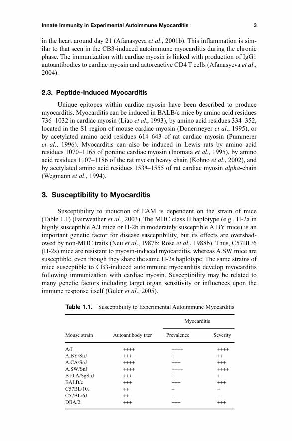
3. Susceptibility to Myocarditis
Susceptibility to induction of EAM is dependent on the strain of mice(Table 1.1) (Fairweather et al., 2003). The MHC class II haplotype (e.g., H-2a inhighly susceptible A/J mice or H-2b in moderately susceptible A.BY mice) is animportant genetic factor for disease susceptibility, but its effects are overshad-owed by non-MHC traits (Neu et al., 1987b; Rose et al., 1988b). Thus, C57BL/6(H-2s) mice are resistant to myosin-induced myocarditis, whereas A.SW mice aresusceptible, even though they share the same H-2s haplotype. The same strains ofmice susceptible to CB3-induced autoimmune myocarditis develop myocarditisfollowing immunization with cardiac myosin. Susceptibility may be related tomany genetic factors including target organ sensitivity or influences upon theimmune response itself (Guler et al., 2005).
Innate Immunity in Experimental Autoimmune Myocarditis 3
Table 1.1. Susceptibility to Experimental Autoimmune Myocarditis
Myocarditis
Mouse strain Autoantibody titer Prevalence Severity
A/J ++++ ++++ ++++A.BY/SnJ +++ + ++A.CA/SnJ ++++ +++ +++A.SW/SnJ ++++ ++++ ++++B10.A/SgSnJ +++ + +BALB/c +++ +++ +++C57BL/10J ++ – −C57BL/6J ++ − −DBA/2 +++ +++ +++
Ch01.qxd 30/05/05 4:00 PM Page 3
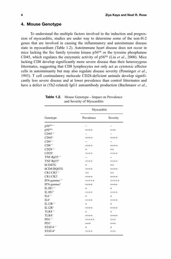
4. Mouse Genotype
To understand the multiple factors involved in the induction and progres-sion of myocarditis, studies are under way to determine some of the non-H-2genes that are involved in causing the inflammatory and autoimmune diseasestate in myocardium (Table 1.2). Autoimmune heart disease does not occur inmice lacking the Src family tyrosine kinase p56lck or the tyrosine phosphataseCD45, which regulates the enzymatic activity of p56lck (Liu et al., 2000). Micelacking CD8 develop significantly more severe disease than their heterozygouslittermates, suggesting that CD8 lymphocytes not only act as cytotoxic effectorcells in autoimmunity but may also regulate disease severity (Penninger et al.,1993). T cell costimulatory molecule CD28-deficient animals develop signifi-cantly less severe disease and at lower prevalence than control littermates andhave a defect in (Th2-related) IgG1 autoantibody production (Bachmaier et al.,
4 Ziya Kaya and Noel R. Rose
Table 1.2. Mouse Genotype—Impact on Prevalenceand Severity of Myocarditis
Myocarditis
Genotype Prevalence Severity
p56lck−/− − −p56lck+ ++++ +++CD45−/− − −CD45+ ++++ ++++CD4−/− − −CD8−/− ++++ ++++CD28−/− + ++CD28+ ++++ ++++TNF-Rp55−/− − −TNF-Rp55+ ++++ ++++hCD4TG + ++hCD4/DQ6TG ++++ ++++CR1/CR2−/− ++ ++CR1/CR2+ ++++ ++++IFN-gamma−/− +++++ +++++IFN-gamma+ ++++ ++++IL1R1−/− + +IL1R1+ ++++ ++++IL6−/− + +IL6+ ++++ ++++IL12R−/− + +IL12R+ ++++ ++++TLR4−/− + +TLR4+ ++++ ++++PD1−/− +++++ +++PD1+ +++ +++STAT-4−/− + +STAT-4+ ++++ +++
Ch01.qxd 30/05/05 4:00 PM Page 4