Molecular phylogeny of Paradermamoeba valamo (Amoebozoa ...
8
© 2020 The Author(s) Protistology © 2020 Protozoological Society Affiliated with RAS Protistology Protistology 14 (4), 219–226 (2020) Molecular phylogeny of Paradermamoeba valamo (Amoebozoa, Discosea, Dermamoebida) Alexey V. Smirnov 1 , Nikita S. Kulishkin 1 , Alina A. Surkova 1 , Yelisei S. Mesentsev 1 , Natalia I. Bondarenko 1 , Elena S. Nassonova 1,2 and Yuri A. Mazei 3 1 Department of Invertebrate Zoology, Faculty of Biology, St. Petersburg State University, 199034 St. Petersburg, Russia 2 Laboratory of Cytology of Unicellular Organisms, Institute of Cytology RAS, 194064 St. Petersburg, Russia 3 Department of General Ecology and Hydrobiology, Lomonosov Moscow State University, 119991 Moscow, Russia | Submitted November 12, 2020 | Accepted December 2, 2020 | Summary A remarkable amoeba species – Paradermamoeba valamo Smirnov et Goodkov,1993 was re-isolated from the bottom sediment in Izmailovo Park pond (Moscow, Russia). For the first time, we obtained modern DIC images of this species. High-quality light-microscopy allowed us to resolve some subcellular structures, including trichocyst-like bodies, and show congruence of the present optical images with the results of earlier electron-microscopic studies. We applied single-cell PCR to obtain full sequence of the 18S rRNA gene of this species. The analysis of genetic distances, as well as the result of phylogenetic analysis, shows that P. valamo is a valid species, related to P. levis but showing clear morphological and molecular differences. Key words: Amoebozoa, Discosea, morphology, phylogeny, SSU gene, light microscopy doi:10.21685/1680-0826-2020-14-4-1 Introduction An amoebae genus Paradermamoeba was estab- lished by Smirnov and Goodkov (1993) to accom- modate P. valamo – a remarkable species of flat- tened, lanceolate amoebae possessing thick cell coat consisting of spiral glycostyles. One year later, another species of this genus, P. levis was described (Smirnov and Goodkov, 1994). This organism, being similar in appearance to P. valamo, was smaller in size and had almost twice thinner cell coat, hence of the same structure. At the time of description, both these species were found to be difficult objects for TEM studies, so only the images of the cell coat were published in both above-cited paper, also this was sufficient for morphological species distinction. The comprehensive ultrastructure of P. valamo and P. levis was published ten years later
Transcript of Molecular phylogeny of Paradermamoeba valamo (Amoebozoa ...
© 2020 The Author(s)
Protistology © 2020 Protozoological Society Affiliated with RAS
ProtistologyProtistology 14 (4), 219–226 (2020)
Molecular phylogeny of Paradermamoeba valamo (Amoebozoa, Discosea, Dermamoebida)
Alexey V. Smirnov1, Nikita S. Kulishkin1, Alina A. Surkova1,Yelisei S. Mesentsev1, Natalia I. Bondarenko1,Elena S. Nassonova1,2 and Yuri A. Mazei3
1 Department of Invertebrate Zoology, Faculty of Biology, St. Petersburg State
University, 199034 St. Petersburg, Russia2 Laboratory of Cytology of Unicellular Organisms, Institute of Cytology RAS,
194064 St. Petersburg, Russia3 Department of General Ecology and Hydrobiology, Lomonosov Moscow State
University, 119991 Moscow, Russia
| Submitted November 12, 2020 | Accepted December 2, 2020 |
Summary
A remarkable amoeba species – Paradermamoeba valamo Smirnov et Goodkov,1993
was re-isolated from the bottom sediment in Izmailovo Park pond (Moscow, Russia).
For the first time, we obtained modern DIC images of this species. High-quality
light-microscopy allowed us to resolve some subcellular structures, including
trichocyst-like bodies, and show congruence of the present optical images with the
results of earlier electron-microscopic studies. We applied single-cell PCR to obtain
full sequence of the 18S rRNA gene of this species. The analysis of genetic distances,
as well as the result of phylogenetic analysis, shows that P. valamo is a valid species,
related to P. levis but showing clear morphological and molecular differences.
Key words: Amoebozoa, Discosea, morphology, phylogeny, SSU gene, light
microscopy
doi:10.21685/1680-0826-2020-14-4-1
Introduction
An amoebae genus Paradermamoeba was estab-
lished by Smirnov and Goodkov (1993) to accom-
modate P. valamo – a remarkable species of flat-
tened, lanceolate amoebae possessing thick cell
coat consisting of spiral glycostyles. One year later,
another species of this genus, P. levis was described
(Smirnov and Goodkov, 1994). This organism, being
similar in appearance to P. valamo, was smaller in
size and had almost twice thinner cell coat, hence
of the same structure. At the time of description,
both these species were found to be difficult objects
for TEM studies, so only the images of the cell
coat were published in both above-cited paper,
also this was sufficient for morphological species
distinction. The comprehensive ultrastructure of
P. valamo and P. levis was published ten years later
· 220
(Smirnov and Goodkov, 2004). This publication
highlighted differences in the ultrastructure of the
nucleus between these two species and confirmed
their status of individual species. The same paper
showed that while P. levis was found to be widely
distributed in Europe and Asia, the only confirmed
finding of P. valamo remained the original finding
at Valamo Island (North-West Russia). Mrva (2005;
2006) listed P. valamo (as well as P. levis) among
species recovered in Slovakia (from mosses of the
Malé Karpaty and rainwater pool in Bratislava,
respectively), but the taxonomic identification was
based only on the light-microscopic morphology.
The SSU sequence of P. levis was obtained by
Smirnov et al. (2011) and the phylogenetic analysis
performed in this study resulted in the establishment
of the order Dermamoebida, unifying the genera
Mayorella, Paradermamoeba and Dermamoeba.
High-quality light microscopic images of P. levis and
further data on its ultrastructure became available
from Kamyshatskaya et al. (2016), while the re-iso-
lation of P. valamo was found to be a problem. This
species appeared in the area of our attention several
times, but we were never successful in culturing it. In
the year 2020, we isolated P. valamo from a sample,
collected in Izmailovo Park pond (Moscow), and
managed to obtain SSU sequence of this species.
The results of phylogenetic analysis and the first
high-quality DIC images of this species are reported
in the present paper.
Material and methods
The strain of Paradermamoeba valamo designa-
ted as strain MSK1, which is the subject of the pre-
sent paper was isolated from the sample, containing
material from the top 5 cm layer of the sandy bottom
sediments of a freshwater pond in Izmailovo park,
Moscow, Russia (55°46’46.8”N, 37°46’09.2”E).
Amoebae were found in the dish, inoculated with
the sampled sediments diluted 1:1000 with 0.25%
WG infusion (see Geisen et al., 2014 for protocol),
made on PJ medium (Prescott and James, 1955).
After two weeks of incubation, the initial culture
contained about a hundred of P. valamo cells.
Species identification was performed basing on the
unique locomotive morphology, nuclear structure
and size data on this species.
All attempts to maintain or to clone culture
failed, so observations and study were done in ini-
tial enrichment culture. All cells in this culture clear-
Alexey V. Smirnov, Nikita S. Kulishkin, Alina A. Surkova, et al.
ly belong to the same species (further molecular stu-
dies confirmed this). Live cells in culture were
observed using Leica M205C dissection microscope
equipped with Rottermann contrast and Leica
DMI3000 inverted microscope equipped with
phase-contrast and IMC (Integrated Modulation
Contrast) optics. Cells were studied, measured and
photographed on the glass object slides using
Leica DM2500 microscope equipped with DIC
optics on PL-fluotar and plan-apochromatic
lenses. Amoebae were photographed and video-
recorded using DS-Fi3 Nikon camera powered by
NisElements – AR software (Nikon).
For DNA isolation, individual cells were trans-
ferred in sterile 40 mm Petri dishes containing a
layer of NN agar (see Page, 1988) covered with
Millipore-filtered (0.22 µm) PJ medium. Cells were
left to starve for three days, every day they were
transferred to the fresh dish with sterile medium.
To isolate DNA, individual amoeba cells were
collected manually with the tapered-end Pasteur
pipette, washed in two subsequent changes of the
same medium and placed with 1–2 µl of the medium
in a 200 µl PCR tube. DNA was extracted using the
Arcturus PicoPure DNA Extraction Kit (Thermo
Fischer Scientific, USA). The extraction mixture
was prepared according to the manufacturer’s inst-
ructions; 12 µl of the mixture was added to the tube
containing the single cell.
The SSU sequence was obtained in two ways.
First, PCR amplification was performed using
RibA (forward, 5’> ac ctg gtt gat cct dcc agt <3’)
and reverse S12.2R ( 5’> gac tac gac ggt atc tra tc
<3’) primers (Pawlowski, 2000). PCR products were
sequenced directly, without purification, using the
Big Dye Terminator Cycle sequencing kit and an
ABI PRISM automatic sequencer using the same
primers. In total, 12 different cells were treated and
sequenced, one sequence was found to belong to a
contaminant, while all other were almost identical.
The consensus sequence was assembled using
CodoneCode software (https://www.codoncode.
com) on the base of six sequences showing the best
trace quality.
The second way included the whole genome
amplification followed by NGS sequencing. We
performed the Multiple Displacement Amplifica-
tion (MDA) of the DNA from the sample showing
good trace quality in Sanger sequencing (cell #2),
using the REPLI-g Single Cell DNA Amplification
Kit (Qiagen, Hilden, Germany) according to the
manufacturer’s instructions. The resulting MDA
· 221 Protistology
products were sequenced, using Illumina HiSeq
2500 system at the core facility center “Development
of Molecular and Cell Technologies” of the SPSU
Research Park according to the manufacturer’s
protocols; as a result, 20M paired reads with the
length about 150 bp were obtained. Quality control
check of raw sequence data was performed, using
FastQC (http://www.bioinformatics.babraham.
ac.uk/projects/fastqc). SPAdes assembler was used
for de novo genome assembly (Bankevich et al.
2012). The contig containing the SSU rRNA gene
fragment was identified, using BLAST (Altschul et
al. 1990) and the sequence fragment of this isolate
obtained by Sanger sequencing as a query.
The alignment constructed for this study was
based on one used by Glotova et al. (2018) for Mayo-rella analysis. It contained all named culture-derived
18S rRNA gene sequences of Dermamoebida, two
full-length sequences belonging to unnamed strains
of Mayorella and a number of discosean sequences
used to form a proper set of outgroups. Other
available Mayorella sequences were not used because
of their short length. Sequences were aligned using
the Muscle algorithm as implemented in SeaView
4.0 (Gouy et al., 2010); the alignment was further
polished manually. The phylogenetic analysis was
performed using maximum likelihood method as
implemented in PhyML program (Guindon and
Gascuel, 2003) with GTR (eight rate categories)
+ γ + I model; 1651 sites were selected for the
analysis. The initial selection was made using the
g-blocks algorithm as implemented in SeaView
4.0, further the set was manually polished. The
number of invariant sites, alpha parameter, and
tree topology was optimized by PhyML. The
program generated 25 random starting trees; the
best tree was further optimized. To test the stability
of branching, 1000 bootstrap pseudoreplicates
were used. Bayesian analysis of the same dataset
was performed using MrBayes 3.2.7, GTR model
with gamma correction for intersite rate variation
(8 categories) and the covarion model (Ronquist
and Huelsenbeck, 2003). Trees were run as two
separate chains (default heating parameters) for 10
million generations, by which time they had ceased
converging (final average standard deviation of the
split frequencies was less than 0.01). The quality of
chains was estimated using built-in MrBayes tools
and additionally – using the software Tracer 1.6
(Rambaut et al., 2014); based on the estimates by
Tracer, the first 25 % of generations were discarded
as burn-in. MrBayes was run at Cipres V.3.3 website
(Miller et al., 2010). Sequence identity level was
calculated using an online tool “Ident and Sim”
provided at http://www.bioinformatics.org/sms2/
ident_sim.html website.
The obtained sequence was deposited with the
GenBank under the number MW293873 (Para-dermamoeba valamo strain MSK1, length 2096 bp).
Results and discussion
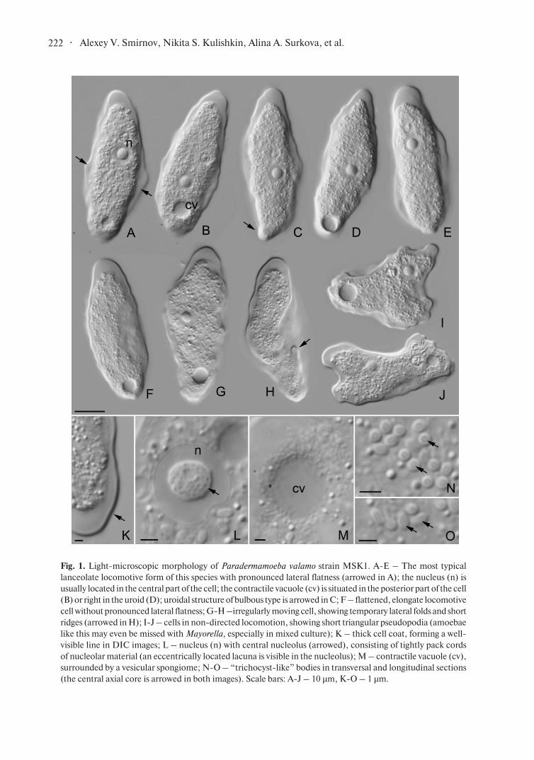
Light-microscopic observations made in culture,
as well as those made on the surface of the object
slide show almost identical cell morphology and
locomotive behaviour. Amoebae were flattened
and oblong in locomotion. They demonstrated
lanceolate morphotype (Smirnov and Goodkov,
1999; Smirnov and Brown, 2004) and show clear
lateral flatness in moving cells, characteristic to
this species (Smirnov and Goodkov, 1993, 2004)
(Fig. 1, A-E). Rapidly moving cells could be more
flattened and often adopted oblong, lingulate shape
without prominent lateral flatness (Fig. 1, F). When
the cell moved more slowly and appeared to be
“uncertain” with the direction of locomotion, it
could temporarily form short prominent folds and
wrinkles (Fig. 1, G-H). These structures were never
stable and could be observed for a short time, often
counted by seconds. When the cell moved slower,
without the single, clear direction of movement it
could form short triangular pseudopodia (Fig. 1, I-J).
Amoebae like this resembled small representatives
of the genus Mayorella and could be even missed
with them, especially in mixed culture. However,
much thicker cell coat of Paradermamoeba helped
to distinguish these two genera.
We have not observed a differentiated floating
form in our strain; few amoebae cells detached from
the substrate only for a short time, and soon settled
down and continue the movement. This might be a
physiological thing because to get proper locomotive
forms we worked with actively growing culture while
floating forms are usually observed in older cultures,
which are on the stationary phase.
The cell coat of P. valamo was well visible with
DIC microscope as a thick line bordering the cell
(Fig. 1, K). The nucleus was of the vesicular type and
had a central nucleolus showing non-homogenous
structure (Fig. 1, L). In the best DIC images, it was
possible to see the “cords” of nucleolar material and
eccentrically located homogeneous area looking
as a lacuna inside the nucleolus. The observed
· 222 Alexey V. Smirnov, Nikita S. Kulishkin, Alina A. Surkova, et al.
Fig. 1. Light-microscopic morphology of Paradermamoeba valamo strain MSK1. A-E – The most typical
lanceolate locomotive form of this species with pronounced lateral flatness (arrowed in A); the nucleus (n) is
usually located in the central part of the cell; the contractile vacuole (cv) is situated in the posterior part of the cell
(B) or right in the uroid (D); uroidal structure of bulbous type is arrowed in C; F – flattened, elongate locomotive
cell without pronounced lateral flatness; G-H –irregularly moving cell, showing temporary lateral folds and short
ridges (arrowed in H); I-J – cells in non-directed locomotion, showing short triangular pseudopodia (amoebae
like this may even be missed with Mayorella, especially in mixed culture); K – thick cell coat, forming a well-
visible line in DIC images; L – nucleus (n) with central nucleolus (arrowed), consisting of tightly pack cords
of nucleolar material (an eccentrically located lacuna is visible in the nucleolus); M – contractile vacuole (cv),
surrounded by a vesicular spongiome; N-O – “trichocyst-like” bodies in transversal and longitudinal sections
(the central axial core is arrowed in both images). Scale bars: A-J – 10 µm, K-O – 1 µm.
· 223 Protistology
structure is congruent with the TEM data obtained
by Smirnov and Goodkov (2004); it is possible to
suggest that the “lacuna” observed in DIC images
(which could look as elevation depending on the
side of the interference spectrum selected with the
upper DIC prism) correspond to the central body
of the nucleolar material visible with TEM in this
species (op. cit.). No trace of the nuclear lamina
was seen in DIC optics, this is congruent with TEM
data as well.
The contractile vacuole was surrounded with
numerous small bodies (0.3-0.75 µm across),
rounded or oblong, which probably represented
mitochondria, located around the vacuole.
DIC microscopy allowed us to observe myste-
rious trichocyst-like bodies mentioned in all studied
strains of P. valamo and P. levis (Smirnov and
Goodkov, 1993, 1994, 2004; Kamyshatskaya et al.,
2016). These bodies were oblong in longitudinal
section (0.75-1 µm in length) and circular in cross-
-section, ca 0.5 µm across. The central core pene-
trating the body was distinguishable in the best
DIC photographs (Fig. 1, N-O). This observation
confirms the structure of these bodies, earlier reve-
aled by TEM and further show that the presence of
these structures is characteristic for the entire genus
Paradermamoeba. They are still not known in any
other organism outside this genus.
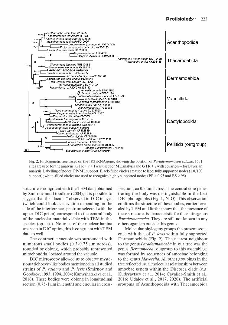
Molecular phylogeny groups the present sequ-
ence with that of P. levis within fully supported
Dermamoebida (Fig. 2). The nearest neighbour
to the genusParadermamoeba in our tree was the
genus Dermamoeba, outgroup to this assemblage
was formed by sequences of amoebae belonging
to the genus Mayorella. All other groupings in the
tree reflected usual molecular relationships between
amoebae genera within the Discosea clade (e.g.
Kudryavtsev et al., 2014; Cavalier-Smith et al.,
2016; Udalov et al., 2017, 2020). The artificial
grouping of Acanthopodida with Thecamoebida
Fig. 2. Phylogenetic tree based on the 18S rRNA gene, showing the position of Paradermamoeba valamo. 1651
sites are used for the analysis; GTR + γ + I was used for ML analysis and GTR + γ with covarion – for Bayesian
analysis. Labelling of nodes: PP/ML support. Black-filled circles are used to label fully supported nodes (1.0/100
support); white-filled circles are used to recognize highly supported nodes (PP > 0.95 and BS > 95).
· 224 Alexey V. Smirnov, Nikita S. Kulishkin, Alina A. Surkova, et al.
was caused with the usage of Pellitida as an outgroup
clade.
Before the present sequence was obtained, a little
doubt on the validity of P. valamo as an individual
species anyway remained. The major difference
between these two species is the size, the thickness
of the cell coat and the structure of the nucleus. Each
of these characters is believed to be reliable, and in
combination, they form a rather clear pattern of
differences. However, there are amoebae species,
showing a wide size range (Page, 1983, 1988; see
for example size data in Page, 1985). For example,
the smallest known specimens of Flabellula baltica
are about three times smaller than the largest ones
(Smirnov, 1999; Smirnov et al., 2017). The cell coat
in P. valamo is twice thicker than in P. levis, but the
overall structure of the cell coat is almost identical.
Differences in the organization of the nucleolus are
significant, but there are cases when the details of
the nuclear structure in amoebae vary depending on
the stage of the cell cycle or the fixation procedure
used (e.g. Demin et al., 2016; Mesentsev et al.,
2020). However, the present study dispels the last
doubts. The level of identity between sequences of
P. valamo and P. levis on the compared fragment
shared by both sequences (1785 bp) was only 78.61%
which is a rather low value. For comparison, the
SSU sequence identity level among species of the
genus Stygamoeba varies in the range from 83 to
85% (Lotonin and Smirnov, 2020). The identity
between sequences of Thecamoeba quadrilineata
and T. cosmophorea is lower and reaches only 70%
(Mesentsev and Smirnov, 2019). However, the
level of sequence identity between morphologically
different Vannella species may be as high as 0.97
or even 0.99, and anyway, these species have clear
morphological differences (Smirnov et al., 2016).
So, the observed value is in the range, common for
a reliable, well-differing amoebae species.
The present study indicates that Paradermamo-eba valamo is a valid species, showing a clear diffe-
rence from P. levis at the morphological level and well
distant from it at the molecular level. The sequence
of its SSU gene will facilitate reliable identification
of this species at the organismal level as well as
its recovery among the results of environmental
DNA sequencing. This, in turn, should improve
our knowledge on the geographic distribution of
this amoeba species. DIC observation made with
the top-quality optical system with the resolution
close to the theoretical limit for conventional light
microscopy shows perfect congruence with earlier
LM and TEM data on this species.
Acknowledgements
Supported with RFBR project 20-54-53017 to
YM (sampling, treatment and LM study of cultures)
and RSF project 20-14-00195 to AS (molecular
studies).
References
Altschul S.F., Gish W., Miller W., Myers E.W.
and Lipman D.J. 1990. Basic local alignment search
tool. Journal of Molecular Biology. 215, 403–410.
Bankevich A., Nurk S., Antipov D., Gurevich
A.A., Dvorkin M., Kulikov A.S., Lesin V.M.,
Nikolenko S.I., Pham S., Prjibelski A.D., Pyshkin
A.V., Sirotkin A.V., Vyahhi N., Tesler G., Alekseyev
M.A. and Pevzner P.A. 2012. SPAdes: A new
genome assembly algorithm and its applications
to single-cell sequencing. J. Comput. Biol. 19,
455– 477.
Cavalier-Smith T., Chao E.E. and Lewis R.
2016. 187-gene phylogeny of protozoan phylum
Amoebozoa reveals a new class (Cutosea) of deep-
branching, ultrastructurally unique, enveloped
marine Lobosa and clarifies amoeba evolution.
Mol. Phylogenet. Evol. 99, 275–296. https://doi.
org/10.1016/j.ympev.2016.03.023
Demin S.Yu., Berdieva M.A., Podlipaeva Yu.I.,
Yudin A.L. and Goodkov A.V. 2016. Optical tomo-
graphy analysis of Amoeba proteus chromatin orga-
nization at various cell cycle stages. Cell Tissue Biol.
10, 84–94. https://doi.org/10.1134/S1990519X
16010041.
Geisen S., Weinert J., Kudryavtsev A., Glotova
A., Bonkowski M. and Smirnov A. 2014. Two
new species of the genus Stenamoeba (Discosea,
Longamoebia): Cytoplasmic MTOC is present in
one more amoebae lineage. Europ. J. Protistol. 50,
153–165. https:// 10.1016/j.ejop.2014.01.007.
Glotova A., Bondarenko N. and Smirnov A.
2018. High genetic diversity of amoebae belonging
to the genus Mayorella (Amoebozoa, Discosea,
Dermamoebida) in natural habitats. Acta Protozool.
57, 29–42. https://doi.org/10.4467/16890027AP.18.
002.8396
Gouy M., Guindon S. and Gascuel O. 2010.
Sea view version 4: A multiplatform graphical user
interface for sequence alignment and phylogenetic
tree building. Mol. Biol. Evol. 27, 221–224. https://
doi.org/10.1093/molbev/msp259.
Guindon S. and Gascuel O. 2003. A simple, fast,
and accurate algorithm to estimate large phylogenies
· 225 Protistology
by maximum likelihood. Syst. Biol. 52, 696–704.
https://doi.org/10.1080/10635150390235520
Kamyshatskaya O. and Smirnov A. 2016. New
data on the ultrastructure of Paradermamoeba levis (Amoebozoa, Discosea, Dermamoebida):
Cytoplasmic MTOCs are found among Derm-
amoebida. Eur. J. Protistol. 54, 74–82. https://doi.
org/10.1016/j.ejop.2016.03.004.
Kudryavtsev A., Brown M.W., Tice A., Spiegel
F.W., Pawlowski J. and Anderson O.R. 2014. A
revision of the order Pellitida Smirnov et al. 2011
(Amoebozoa, Discosea) based on ultrastructural and
molecular evidence, with description of Endostelium crystalliferum n. sp. Protist. 165, 208–229. https://
doi.org/10.1016/j.protis.2014.02.003.
Lotonin K. and Smirnov A. 2020. Stygamoeba cauta n. sp. (Amoebozoa, Discosea) – a new bra-
ckish-water species from Nivå Bay (Baltic Sea, The
Søund). Eur. J. Protistol. 72, 125660. https://doi.
org/10.1016/j.ejop.2019.125660.
Mesentsev Y., Kamyshatskaya O. and Smir-
nov A. 2020. Thecamoeba foliovenanda n. sp.
(Amoebozoa, Discosea, Thecamoebida) – one more
case of sibling species among amoebae of the genus
Thecamoeba. Eur. J. Protistol. 76, 125716. https://
doi.org/10.1016/j.ejop.2020.125716.
Mesentsev Y.S. and Smirnov A.V. 2019. Thec-amoeba cosmophorea n. sp. (Amoebozoa, Discosea,
Thecamoebida) — an example of sibling species
within the genus Thecamoeba. Eur. J. Protistol.
67, 132–141. https://doi.org/10.1016/j.ejop.2018.
12.003.
Miller M.A., Pfeiffer W. and Schwartz T. 2010.
Creating the CIPRES Science Gateway for inference
of large phylogenetic trees. Gateway Computing
Environments Workshop (GCE). IEEE. pp. 1–8.
https://doi.org/10.1109/GCE.2010.5676129.
Mrva M. 2005. Diversity of active gymnamoebae
(Rhizopoda, Gymnamoebia) in mosses of the Malé Karpaty Mts (Slovakia). Ekológia (Bratislava). Vol.
24, Suppl. 2, pp. 51–58.
Mrva M. 2006. Diversity of gymnamoebae (Rhi-
zopoda, Gymnamoebia)in a rain-water pool. Bio-
logia (Bratislava). 61, 627–629. https://doi.org/10.
2478/s11756-006-0100-2.
Page F.C. 1983. Marine Gymnamoebae. Insti-
tute of Terrestrial Ecology, Cambrige, England.
Page F.C. 1985. The limax amoebae: compara-
tive fine structure of the Hartmannellidae (Lobosea)
and further comparisons with the Vahlkampfiidae
(Heterolobosea). Protistologica. 21, 361–383.
Page F.C. 1988. A new key to freshwater and soil
Gymnamoebae. Freshwater Biological Association,
The Ferry House, Ambleside, Cumbria.
Pawlowski J. 2000. Introduction to the molecu-
lar systematics of foraminifera. Micropaleontology.
46, 1–12. https://doi.org/10.2307/1486176.
Prescott D.M. and James T.W. 1955. Culturing
of Amoeba proteus on Tetrahymena. Exp. Cell Res.
8, 256–258. https://doi.org/10.1016/0014-4827
(55)90067-7.
Rambaut A., Suchard M., Xie D. and Drum-
mond A. 2014. Tracer v1.6.2014.
Ronquist F. and Huelsenbeck J.P. 2003. MrBa-
yes 3: Bayesian phylogenetic inference under mixed
models. Bioinformatics. 19, 1572–1574. https://doi.
org/10.1093/bioinformatics/btg180.
Smirnov A.V. 1999. An illustraited survey of
gymnamoebae isolated from anaerobic sediments
of the Nivo Bay (the Sound). Ophelia. 50, 113–148.
Smirnov A.V., Bondarenko N., Glotova A. and
Nassonova E. 2016. Morphology and phylogeny
of Vannella croatica n. sp. (Amoebozoa, Discosea,
Vannellida). Eur. J. Protistol. 52, 65–72. https://
doi.org/10.1016/j.ejop.2015.11.002.
Smirnov A.V. and Brown S. 2004. Guide to the
methods of study and identification of soil gymn-
amoebae. Protistology. 3, 148–190.
Smirnov A.V., Chao E., Nassonova E.S. and
Cavalier-Smith T. 2011. A revised classification
of naked lobose amoebae (Amoebozoa: Lobosa).
Protist. 162, 545–570. https://doi.org/10.1016/j.
protis.2011.04.004.
Smirnov A.V. and Goodkov A.V. 1993. Para-dermamoeba valamo gen. n., sp. n. (Gymnamoebia,
Thecamoebidae) – a freshwater amoeba from bottom
sediments. Zool. Zhurn. 72, 5–11. (in Russian with
English summary).
Smirnov A.V and Goodkov A.V 1994. Fresh-
water gymnamoebae with a new type of surface
structure Paradermamoeba valamo and P. levis sp.
n. (Thecamoebidae), and notes on the diagnosis of
the family. Acta Protozool. 33, 109–115.
Smirnov A.V. and Goodkov A.V. 1999. An illu-
strated list of basic morphotypes of Gymnamoebia
(Rhizopoda, Lobosea). Protistology. 1, 20–29.
Smirnov A. and Goodkov A. 2004. Ultra-
structure and geographic distribution of the genus
Paradermamoeba (Gymnamoebia, Thecamoebi-
dae). Eur. J. Protistol. 40, 113–118. https://doi.org/
10.1016/j.ejop.2003.12.001.
Smirnov A., Nassonova E., Geisen S., Bonko-
wski M., Kudryavtsev A., Berney C., Glotova A.,
Bondarenko N., Dyková I., Mrva M., Fahrni J.
and Pawlowski J. 2017. Phylogeny and systematics
of leptomyxid amoebae (Amoebozoa, Tubulinea,
· 226 Alexey V. Smirnov, Nikita S. Kulishkin, Alina A. Surkova, et al.
Address for correspondence: Alexey Smirnov. Department of Invertebrate Zoology, Faculty of
Biology, St. Petersburg State University, Universitetskaya Emb. 7/9, 199034 St. Petersburg, Russia;
e-mail: [email protected]
Leptomyxida). Protist. 168, 220–252. https://doi.
org/10.1016/j.protis.2016.10.006.
Udalov I.A., Lee W.J., Lotonin K. and Smirnov
A. 2020. Pseudoparamoeba garorimi n. sp., with notes
on species distinctions within the genus. J. Eukaryot.
Microbiol. 67, 132–139. https://doi.org/10.1111/
jeu.12763.
Udalov I.A., Völcker E. and Smirnov A. 2017.
Korotnevella novazelandica n. sp. (Amoebozoa, Dis-
cosea, Dactylopodida) — a new freshwater amoeba
with unusual combination of scales. Protistology.
11, 238–247. https://doi.org/10.21685/1680-0826-
2017-11-4-5.