Optical coherence tomography for the diagnosis of ...
11
Optical coherence tomography for the diagnosis of malignant skin tumors: a meta-analysis Yi-Quan Xiong Yun Mo Yu-Qi Wen Ming-Ji Cheng Shu-Ting Huo Xue-Jiao Chen Qing Chen Yi-Quan Xiong, Yun Mo, Yu-Qi Wen, Ming-Ji Cheng, Shu-Ting Huo, Xue-Jiao Chen, Qing Chen, “Optical coherence tomography for the diagnosis of malignant skin tumors: a meta-analysis, ” J. Biomed. Opt. 23(2), 020902 (2018), doi: 10.1117/1.JBO.23.2.020902. Downloaded From: https://www.spiedigitallibrary.org/journals/Journal-of-Biomedical-Optics on 23 Jan 2022 Terms of Use: https://www.spiedigitallibrary.org/terms-of-use
Transcript of Optical coherence tomography for the diagnosis of ...
Optical coherence tomography for thediagnosis of malignant skin tumors: ameta-analysis
Yi-Quan XiongYun MoYu-Qi WenMing-Ji ChengShu-Ting HuoXue-Jiao ChenQing Chen
Yi-Quan Xiong, Yun Mo, Yu-Qi Wen, Ming-Ji Cheng, Shu-Ting Huo, Xue-Jiao Chen, Qing Chen, “Opticalcoherence tomography for the diagnosis of malignant skin tumors: a meta-analysis,” J. Biomed.Opt. 23(2), 020902 (2018), doi: 10.1117/1.JBO.23.2.020902.
Downloaded From: https://www.spiedigitallibrary.org/journals/Journal-of-Biomedical-Optics on 23 Jan 2022Terms of Use: https://www.spiedigitallibrary.org/terms-of-use
Optical coherence tomography for the diagnosis ofmalignant skin tumors: a meta-analysis
Yi-Quan Xiong,† Yun Mo,† Yu-Qi Wen, Ming-Ji Cheng, Shu-Ting Huo, Xue-Jiao Chen, and Qing Chen*Southern Medical University, School of Public Health, Guangdong Provincial Key Laboratory of Tropical Disease Research,Department of Epidemiology, Guangzhou, China
Abstract. Optical coherence tomography (OCT) is an emergent imaging tool used for noninvasive diagnosis ofskin diseases. The present meta-analysis was carried out to assess the accuracy of OCT for the diagnosis ofskin cancer. We conducted a systematic literature search though EMBASE, Medline, PubMed, the CochraneLibrary, and Web of Science database for relevant articles published up to June 6, 2017. The quality of theincluded studies was assessed using the QUADAS-2 tool and the Oxford Levels of Evidence Scale.Statistical analyses were conducted using the software Meta-Disc version 1.4 and STATA version 12.0. Atotal of 14 studies involving more than 813 patients with a total of 1958 lesions were included in our analyses.The pooled sensitivity and specificity of OCT for skin cancer diagnoses were 91.8% and 86.7%, respectively.Subgroup analysis showed that the pooled sensitivities of OCT for detecting basal cell carcinoma (BCC), squ-amous cell carcinoma (SCC), actinic keratosis, and malignant melanoma were 92.4%, 92.3%, 73.8%, and81.0%, respectively. The pooled specificities were 86.9%, 99.5%, 91.5%, and 93.8%, respectively. OCT appearsto be useful for the detection of BCC and SCC. It is a valuable diagnostic method when screening for early skincancers. © 2018 Society of Photo-Optical Instrumentation Engineers (SPIE) [DOI: 10.1117/1.JBO.23.2.020902]
Keywords: optical coherence tomography; skin cancer; basal cell carcinoma; squamous cell carcinoma; actinic keratosis; malignantmelanoma.
Paper 170502VR received Aug. 6, 2017; accepted for publication Jan. 29, 2018; published online Feb. 22, 2018.
1 IntroductionAlarming increases in the incidence and prevalence of malignantskin tumors have occurred in recent decades, especially formalignant melanoma (MM), basal cell carcinoma (BCC), andsquamous cell carcinoma (SCC).1–5 Nonmelanoma skin cancers(NMSC), including Bowen’s disease, actinic keratosis (AK),BCC, and SCC, are the most prevalent malignancies affectinglight-skinned individuals worldwide and represent almost95% of all cutaneous cancer diagnoses.6,7 The World HealthOrganization reported an increase of more than 2 to 3 millionnew cases of NMSC per year,8 of which about 80% are BCCs.9
Meanwhile, MM is also increasing in frequency both in the UKand Europe.3 MM accounts for 2% of skin cancers and isresponsible for nearly 1% of deaths caused by oncologicaletiologies.10 Both NMSC and MM have excellent prognosiswhen they are diagnosed and treated early.11 If melanoma istreated while in situ or stage 1, the overall 5-year survivalrate is over 90%, but for metastatic disease at stage 4, therate drops rapidly to 10% to 15%.11 Thus, diagnosis at anearly stage is crucial for improving survival rates and overallprognosis,12–14 and it is important that dermatologists notonly provide preventative counseling but also be astute in theearly diagnosis and treatment of any suspected malignancies.15
Several new methods offer improved diagnoses of skinlesions.16 In particular, the development of optospectral technol-ogies for imaging of living tissues is reshaping the dermatologicalpractice. This technique offers in vivo/ex vivo, noninvasive,painless, real-time visualization of skin structures and physiologi-cal parameters, at high resolution and without interfering with themorphology and the functions of the examined integument.17–19
Optical coherence tomography (OCT) was first used medically in1988 in ophthalmology by Fercher et al.20 This method is basedon the physical principle of low-coherence interferometry anduses infrared light21 to measure the scattering properties of tissues(echo time delay and the intensity of backscattered or backre-flected light).22–24 It provides a real-time, cross-sectional aswell as en-face sectional images of the subsurface microstructureof tissues with micrometer resolution, enabling visualization ofskin morphology (epidermis, dermis, dermoepidermal junction,nodular and fibrous structures, cellular elements, hair follicleunits, blood vessels, and sweat glands) and investigation of thealtered skin architecture present in superficial skinlesions.13,19,25,26 High-definition OCT (HD-OCT) is a techniquebased on the principal of conventional OCT using a combinationof parallel time-domain interferometry and adaptive optics.27
Conventional OCT techniques, such as VivoSight OCT, polarizedsensitivity OCT (PS-OCT), Skintell HD-OCT, or full-field OCT(FF-OCT), offer two-dimensional (brightness-scan mode) andthree-dimensional (3-D) (c-scan mode) reconstruction of theimages with acquisition speeds reported to be up to severalvolumes per second, as well as noise removal.28
In recent years, it has been demonstrated that melanocyticlesions such as AK, SCC, and BCC have distinctive featuresrevealed using OCT that can differentiate them from noncancer-ous skin lesions.29–32 However, the reported diagnostic accuracyof OCT is inconsistent. To formulate a comprehensive basis foruse of OCT, we performed a meta-analysis of the literature toassess OCT for differentiating skin tumors from normal skin.
2 MethodsThis meta-analysis was conducted according to the publishedstandards for reviews of diagnostic accuracy and performed
*Address all correspondence to: Qing Chen, E-mail: [email protected]
†Authors contributed equally to this work. 1083-3668/2018/ $25.00 © 2018 SPIE
Journal of Biomedical Optics 020902-1 February 2018 • Vol. 23(2)
Journal of Biomedical Optics 23(2), 020902 (February 2018) REVIEW
Downloaded From: https://www.spiedigitallibrary.org/journals/Journal-of-Biomedical-Optics on 23 Jan 2022Terms of Use: https://www.spiedigitallibrary.org/terms-of-use
in accordance with the Preferred Reporting Items for SystematicReviews and Meta-Analyses guidelines.33
2.1 Literature Search
Electronic searches were conducted of EMBASE, Medline,PubMed, the Cochrane Library, and the Web of Science data-bases from the earliest date available until June 6, 2017. Weused the following key words, separately and in combination:“skin,” “tumor,” “cancer,” “neoplasm,” “carcinoma,” “mela-noma,” “basal cell carcinoma,” “BCC,” “squamous cell carci-noma,” “SCC,” “optical coherence tomography,” and “OCT.”No language restriction was applied to the searches, but thesearch was restricted to studies on humans. Forward citationsearching of the reference lists of the original studies and reviewarticles was also conducted.
2.2 Eligibility Criteria
Studies were screened for eligibility using the following criteria:(i) evaluation by diagnostic clinical trials of the accuracy of OCTfor the diagnosis of skin cancer in patients, (ii) retrospective orprospective study design, (iii) results reported with sufficientdata to construct a diagnostic 2 × 2 contingency table, includingtrue-positive, false-positive, true-negative, and false-negativedata, and (iv) all diagnoses were confirmed by the histologyseen in a biopsy sample. Comment papers, small case series,case reports, reviews, or guideline articles were excluded.When more than one article reported the same study, the pub-lication with more information was selected.
2.3 Data Selection and Extraction
Citations were merged in EndNote (version X7) to facilitatemanagement. Two authors independently evaluated all retrievedarticles by title and abstract according to the previous inclusioncriteria in a nonblinded, standardized manner. After full-textscreening, articles that fulfilled the inclusion criteria wereselected for the final analysis. Data were extracted from eacheligible study by two authors independently and a consensuswas reached on all items. Relevant data included: (i) first author,year of publication, country of origin, study design (prospectiveor retrospective), number of centers; (ii) type of OCT system,(iii) in vivo or ex vivo examination; (iv) type of skin cancer;(v) number of investigators and their experience, OCT analysis(blinded or others); (vi) number of enrolled patients and spec-imens, baseline patient characteristics (age distribution and sexratio); and (vii) accuracy of OCT (sensitivity and specificity)with reference to histopathology.
2.4 Quality Assessment
The quality of the included studies and the risk of bias wereassessed according to the Oxford Centre for Evidence-basedMedicine-Levels of Evidence (March 2009)34 and the QualityAssessment of Diagnostic Accuracy Studies-2 (QUADAS-2)35
tools by two authors independently. QUADAS-2 consists of fourkey domains regarding patient selection, index test, referencestandard, and flow of patients (through the study and timingof the index tests). Risk of bias was judged as “low,” “high,”and “unclear.”
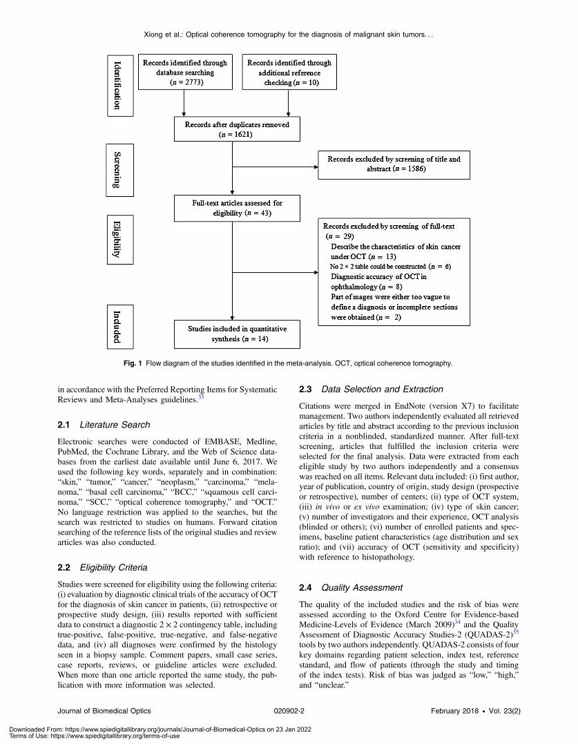
Fig. 1 Flow diagram of the studies identified in the meta-analysis. OCT, optical coherence tomography.
Journal of Biomedical Optics 020902-2 February 2018 • Vol. 23(2)
Xiong et al.: Optical coherence tomography for the diagnosis of malignant skin tumors. . .
Downloaded From: https://www.spiedigitallibrary.org/journals/Journal-of-Biomedical-Optics on 23 Jan 2022Terms of Use: https://www.spiedigitallibrary.org/terms-of-use

Tab
le1
Bas
icch
arac
teris
ticsof
selected
stud
ies.
Autho
rYea
rCou
ntry
Des
ign
Cen
ter
(n)
Typ
eof
OCT
Typ
eof
canc
erInve
stigators
Exp
erienc
edPatients
(n)
Age
(mea
nor
med
ian)
Male
(%)
Lesion
sLe
velsof
eviden
ce
Gam
bich
lere
tal.1
220
15German
yPro
Three
SkintellH
D-O
CT
MM
One
Exp
64NA
NA
931a
Hus
sain
etal.1
2016
Den
mark
Pro
One
VivoS
ight
OCT
BCC
NA
NA
5865
.9(41to
88)
37.5
581b
Mog
ense
net
al.13
2009
Den
mark
Retro
One
OCTan
dPS-O
CT
NMSC
Two
Exp
104
69.3
(37to
90)
43.0
220
1b
Marko
witz
etal.39
2015
USA
Pro
Three
VivoS
ight
OCT
BCC
NA
NA
NA
NA
NA
115
1a
Maier
etal.40
2014
German
yPro
One
SkintellH
D-O
CT
BCC
One
Exp
20NA
(53to
90)
55.0
801b
Durkinet
al.41
2014
USA
Retro
One
FF-O
CT
SCC,
BCC
One
NA
11NA
NA
181b
Ulrich
etal.42
2015
German
yPro
Six
VivoS
ight
OCT
BCC
NA
Exp
155
70(33to
90)
NA
235
1a
Wah
rlich
etal.43
2015
German
yPro
One
VivoS
ight
OCT
BCC
One
Exp
5062
.846
.050
1b
Boo
neet
al.45
2016
Den
mark
Retro
One
SkintellH
D-O
CT
AK,SCC
NA
NA
108
66.3
(39to
92)
47.8
108
1b
Marvd
ashtie
tal.47
2016
USA
Retro
One
PS-O
CT
BCC
One
Exp
42NA
NA
520
1b
Olsen
etal.46
2016
Den
mark
Pro
One
VivoS
ight
OCT
AK,BCC
Five
Exp
NA
69.4
54.9
142
1b
Boo
neet
al.48
2016
Den
mark
Retro
One
SkintellH
D-O
CT
MM
NA
NA
4551
(20to
70)
44.4
451b
Marne
ffeet
al.44
2016
Belgium
Retro
One
SkintellH
D-O
CT
AK,SCC
Three
Exp
7166
.9(35to
91)
50.7
106
1b
Che
nget
al.49
2016
Aus
tralia
Pro
One
OCT
BCC
Three
Exp
103
66(31to
88)
61.2
168
1b
Note:
OCT,o
ptical
cohe
renc
etomog
raph
y;HD-O
CT,h
igh-de
finition
OCT;P
S-O
CT,p
olarized
sens
itivity
OCT;M
M,m
aligna
ntmelan
oma;
BCC,b
asal
cellca
rcinom
a;SCC,s
quam
ousce
llca
rcinom
a;AK,ac
tinic
keratosis;
NMSC,no
nmelan
omask
inca
ncer;AHM,am
elan
otic/hyp
omelan
otic
melan
oma;
NA,no
data
available;
Pro,pros
pective;
Retro,retros
pective;
andExp
,ex
perie
nced
.
Journal of Biomedical Optics 020902-3 February 2018 • Vol. 23(2)
Xiong et al.: Optical coherence tomography for the diagnosis of malignant skin tumors. . .
Downloaded From: https://www.spiedigitallibrary.org/journals/Journal-of-Biomedical-Optics on 23 Jan 2022Terms of Use: https://www.spiedigitallibrary.org/terms-of-use

2.5 Statistical Analysis
Sensitivity, specificity, positive likelihood ratio (PLR), and neg-ative likelihood ratio (NLR) were meta-analyzed. Between-stud-ies heterogeneity was estimated by I2 statistics; significantheterogeneity was defined as I2 > 50%. Pooled results andtheir corresponding 95% confidence intervals (CIs) were calcu-lated with a fixed-effects model (Mantel and Haenszel method)when heterogeneity was not significant (I2 < 50%); otherwise, arandom-effects model (DerSimonian and Laird method) wasapplied. Forest plots were constructed for visual display ofpooled results if necessary. A weighted symmetric summaryreceiver operating characteristic (sROC) curve was drawn, andthe area under the curve (AUC) was calculated.36 Thresholdanalysis was performed using the Spearman’s coefficient (>0.5with p < 0.05).37 Meta-regression was also performed toexplore the potential heterogeneity among studies. Statisticalanalyses were conducted using Meta-Disc software (version1.4; Unit of Clinical Biostatistics, Ramon y Cajal Hospital,Madrid, Spain).38 Publication bias was assessed using STATA(version 12.0; Stata Corp; College Station, Texas), and theMidas command was used for all statistical analyses.
3 Results
3.1 Description of the Included Studies
The initial search yielded 2773 references, of which 1621 poten-tially relevant studies were unique. Fourteen studies,1,12,13,39–49
including eight prospective and six retrospective studies, thatinvolved more than 813 patients and 1958 lesions were includedin the final analysis. The selection process for the studies is
shown in Fig. 1. The main characteristics of the 14studies1,12,13,39–49 are summarized in Table 1. All of the studieswere conducted in the USA, Germany, Belgium, Australia, andDenmark, and three were multicenter studies.12,39,42 Five studiesused VivoSight OCT,1,39,42,43,46 five used Skintell HD-OCT,12,40,44,45,48 two used PS-OCT,13,47 and the remaining oneused FF-OCT.41 All included studies covered most types ofskin cancer, including MM, AK, BCC, and SCC. Twelvestudies1,12,13,39–42,44–48 reported the diagnostic performance ofOCT in differentiating cancerous from noncancerous skin, andsome studies validated the distinction of BCC,1,39–43,46,47,49,50
SCC,41,44,48 AK,44,46,48 or MM12,45 from other cancerous or non-cancerous lesions, respectively. Three studies 43,47,48 used anewly developed scoring systems [the “Berlin score” (BS)]or algorithms as assisting tools in detection. Ninestudies12,13,40,42–44,46,47,49 noted the experience of the observerswho examined the outcomes of OCT images.
3.2 Quality Assessment
Two scales were used for assessing the study quality. The resultsassessed using the Oxford Levels of Evidence Scale are shown inTable 1, and the results assessed using QUADAS-2 are shown inTable 2. Most of the studies included in our analysis were of highquality. One study13 had a high risk of bias in patient selectionbecause normal adjacent skin areas were selected to use as thecontrol sites. Four studies, in which some patients were excludedfrom the final analysis because of lack of information1,42 orabsence of optimal quality OCT images,13,47 were classified ashaving a high risk of bias in flow and timing. And in one
Table 2 QUADAS-2 risk of bias assessment.
Study
Risk of bias Applicability concerns
Patient selection Index test Reference standard Flow and timing Patient selection Index test Reference standard
Gambichler et al.12 Low Low Low Low Low Low Low
Hussain et al.1 Low Low Low High Low Low Low
Mogensen et al.13 High Low Low High Low Low Low
Markowitz et al.39 Low Low Unclear Low Low Low Low
Maier et al.40 Low Low Low Low Unclear Low Low
Durkin et al.41 Low Low Low Low Low Low Low
Ulrich et al.42 Low Low Low High Low Low Low
Wahrlich et al.43 Low Low Low Low Low Low Low
Boone et al.45 Low Unclear Low Low Low Low Low
Marvdashti et al.47 Low Unclear Low High Low Low Low
Olsen et al.46 Low Low Low Low Low Low Low
Boone et al.48 Low Unclear Low Low Low Low Low
Marneffe et al.44 Low Low Low High Low Low Low
Cheng et al.49 Low Low Low Low Low Low Low
Note: Low, low risk; High, high risk; and Unclear, unclear risk.
Journal of Biomedical Optics 020902-4 February 2018 • Vol. 23(2)
Xiong et al.: Optical coherence tomography for the diagnosis of malignant skin tumors. . .
Downloaded From: https://www.spiedigitallibrary.org/journals/Journal-of-Biomedical-Optics on 23 Jan 2022Terms of Use: https://www.spiedigitallibrary.org/terms-of-use

study,44 the control patients did not have the same reference stan-dards as the cases, suggesting a high risk of selection bias.
3.3 Diagnostic Accuracy of OCT for Skin Cancer
Twelve studies,1,12,13,39–42,44–48 including a total of 1740 lesions,reported the diagnostic accuracy of OCT for distinguishing can-cerous from noncancerous features at the per-lesion level. Thesensitivity ranged from 66.7% to 100.0% and specificity rangedfrom 64.4% to 100.0%. The corresponding pooled sensitivityand specificity were 91.8% (95% CI: 0.90 to 0.94, I2 ¼72.9%) and 86.7% (95% CI: 0.84 to 0.89 I2 ¼ 85.5%), respec-tively (Fig. 2). The pooled PLR and NLR were 6.94 (95% CI:4.21 to 11.47, I2 ¼ 86.3%) and 0.12 (95% CI: 0.07 to 0.20,I2 ¼ 79.1%), respectively. The AUC for sROC was 0.95 (Fig. 3).The distribution of scatter points in the sROC was not ina shoulder-like form and the Spearman’s coefficient was 0.14(p ¼ 0.67), suggesting that there was no threshold effect.
3.4 Diagnostic Accuracy of OCT for BCC
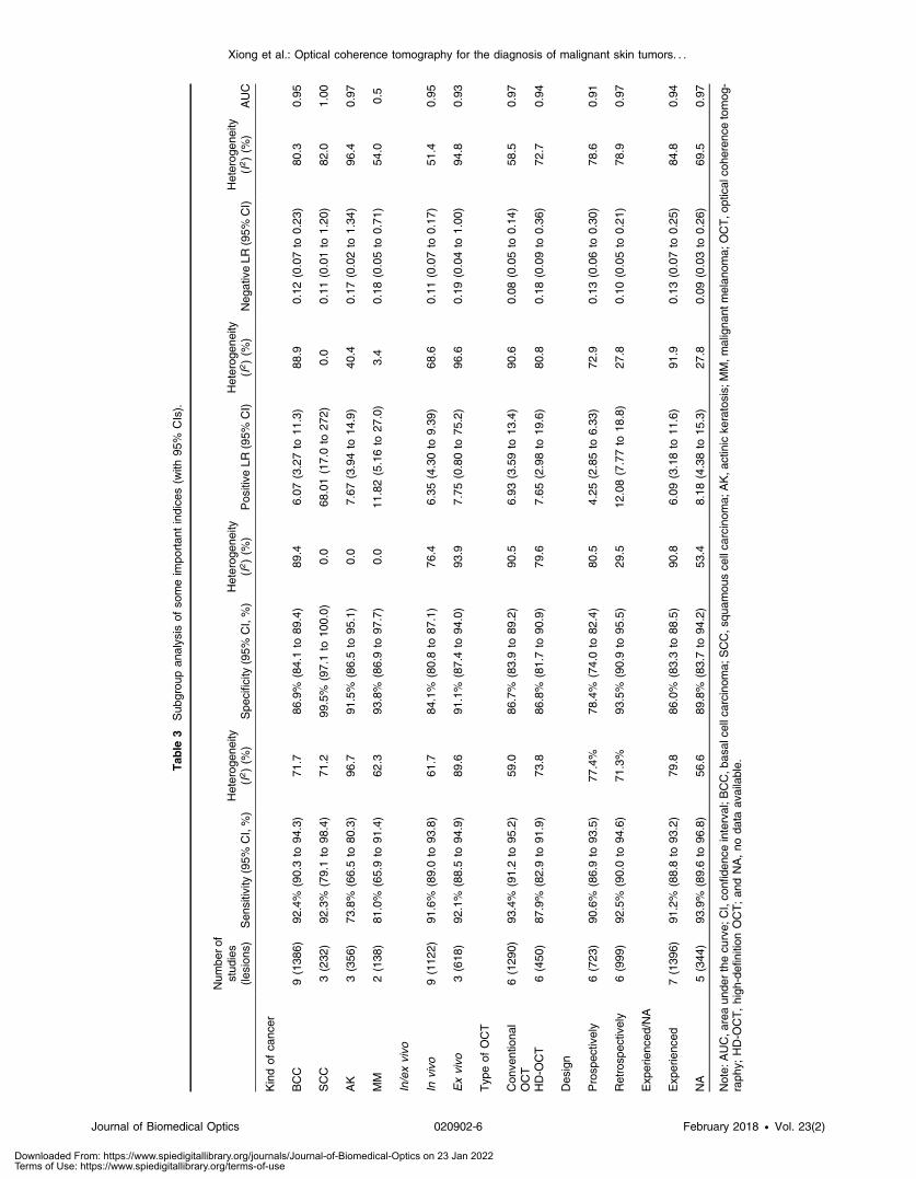
Nine studies,1,39–43,46,47,49 covering a total of 1386 lesions,reported the diagnoses of BCC by OCT. The pooled sensitivitywas 92.4% (95% CI: 0.90 to 0.94, I2 ¼ 71.7%) and specificitywas 86.9% (95% CI: 0.84 to 0.89, I2 ¼ 89.4%); whereas thepooled PLR and NLR were 6.07 (95% CI: 3.27 to 11.26,I2 ¼ 88.9%) and 0.12 (95% CI: 0.07 to 0.23, I2 ¼ 80.3%),respectively. The AUC for sROC was 0.95 (Table 3).
3.5 Diagnostic Accuracy of OCT for SCC
Three studies,41,44,48 including the diagnoses of SCC, achieveda pooled sensitivity of 92.3% (95% CI: 0.79 to 0.98,I2 ¼ 71.2%) and a high specificity of 99.5% (95% CI: 0.97to 1.00, I2 ¼ 0.0%); then the pooled PLR and NLR were
Fig. 2 Forest plot of the (a) pooled sensitivity and (b) specificity of OCT for detection of malignant skintumors.
Fig. 3 The SROC with 95% CI of OCT for detection of malignant skintumors. AUC, area under the curve.
Journal of Biomedical Optics 020902-5 February 2018 • Vol. 23(2)
Xiong et al.: Optical coherence tomography for the diagnosis of malignant skin tumors. . .
Downloaded From: https://www.spiedigitallibrary.org/journals/Journal-of-Biomedical-Optics on 23 Jan 2022Terms of Use: https://www.spiedigitallibrary.org/terms-of-use
Tab
le3
Sub
grou
pan
alys
isof
someim
portan
tindice
s(w
ith95
%CIs).
Num
bero
fstud
ies
(lesion
s)Sen
sitivity
(95%
CI,%)
Heterog
eneity
(I2)(%
)Spe
cificity
(95%
CI,%)
Heterog
eneity
(I2)(%
)Pos
itive
LR(95%
CI)
Heterog
eneity
(I2)(%
)Neg
ativeLR
(95%
CI)
Heterog
eneity
(I2)(%
)AUC
Kindof
canc
er
BCC
9(138
6)92
.4%
(90.3to
94.3)
71.7
86.9%
(84.1to
89.4)
89.4
6.07
(3.27to
11.3)
88.9
0.12
(0.07to
0.23
)80
.30.95
SCC
3(232
)92
.3%
(79.1to
98.4)
71.2
99.5%
(97.1to
100.0)
0.0
68.01(17.0to
272)
0.0
0.11
(0.01to
1.20
)82
.01.00
AK
3(356
)73
.8%
(66.5to
80.3)
96.7
91.5%
(86.5to
95.1)
0.0
7.67
(3.94to
14.9)
40.4
0.17
(0.02to
1.34
)96
.40.97
MM
2(138
)81
.0%
(65.9to
91.4)
62.3
93.8%
(86.9to
97.7)
0.0
11.82(5.16to
27.0)
3.4
0.18
(0.05to
0.71
)54
.00.5
In/exvivo
Invivo
9(112
2)91
.6%
(89.0to
93.8)
61.7
84.1%
(80.8to
87.1)
76.4
6.35
(4.30to
9.39
)68
.60.11
(0.07to
0.17
)51
.40.95
Exvivo
3(618
)92
.1%
(88.5to
94.9)
89.6
91.1%
(87.4to
94.0)
93.9
7.75
(0.80to
75.2)
96.6
0.19
(0.04to
1.00
)94
.80.93
Typ
eof
OCT
Con
ventiona
lOCT
6(129
0)93
.4%
(91.2to
95.2)
59.0
86.7%
(83.9to
89.2)
90.5
6.93
(3.59to
13.4)
90.6
0.08
(0.05to
0.14
)58
.50.97
HD-O
CT
6(450
)87
.9%
(82.9to
91.9)
73.8
86.8%
(81.7to
90.9)
79.6
7.65
(2.98to
19.6)
80.8
0.18
(0.09to
0.36
)72
.70.94
Des
ign
Prosp
ectively
6(723
)90
.6%
(86.9to
93.5)
77.4%
78.4%
(74.0to
82.4)
80.5
4.25
(2.85to
6.33
)72
.90.13
(0.06to
0.30
)78
.60.91
Retrosp
ectively
6(999
)92
.5%
(90.0to
94.6)
71.3%
93.5%
(90.9to
95.5)
29.5
12.08(7.77to
18.8)
27.8
0.10
(0.05to
0.21
)78
.90.97
Exp
erienc
ed/NA
Exp
erienc
ed7(139
6)91
.2%
(88.8to
93.2)
79.8
86.0%
(83.3to
88.5)
90.8
6.09
(3.18to
11.6)
91.9
0.13
(0.07to
0.25
)84
.80.94
NA
5(344
)93
.9%
(89.6to
96.8)
56.6
89.8%
(83.7to
94.2)
53.4
8.18
(4.38to
15.3)
27.8
0.09
(0.03to
0.26
)69
.50.97
Note:
AUC,a
reaun
derthecu
rve;
CI,co
nfiden
ceinterval;B
CC,b
asal
cellca
rcinom
a;SCC,s
quam
ousce
llca
rcinom
a;AK,a
ctinicke
ratosis;
MM,m
aligna
ntmelan
oma;
OCT,o
ptical
cohe
renc
etomog
-raph
y;HD-O
CT,high
-definition
OCT;an
dNA,no
data
available.
Journal of Biomedical Optics 020902-6 February 2018 • Vol. 23(2)
Xiong et al.: Optical coherence tomography for the diagnosis of malignant skin tumors. . .
Downloaded From: https://www.spiedigitallibrary.org/journals/Journal-of-Biomedical-Optics on 23 Jan 2022Terms of Use: https://www.spiedigitallibrary.org/terms-of-use
68.01 (95% CI: 17.01 to 272.4, I2 ¼ 0.0%) and 0.11 (95% CI:0.01 to 1.20, I2 ¼ 82.0%), respectively. The AUC for sROCwas1.00 (Table 3).
3.6 Diagnostic Accuracy of OCT for AK
Three studies44,46,48 addressed the diagnosis of AK with pooledsensitivity and specificity of 73.8% (95% CI: 0.67 to 0.80,I2 ¼ 96.7%) and 91.5% (95% CI: 0.87 to 0.95, I2 ¼ 0.0%),respectively. The pooled PLR and NLR were 7.67 (95% CI:3.94 to 14.94, I2 ¼ 40.4%) and 0.17 (95% CI: 0.02 to 1.34,I2 ¼ 96.4%), respectively. The AUC for sROC was 0.97(Table 3).
3.7 Diagnostic Accuracy of OCT for MM
Only two publications12,45 reported diagnoses of MM andachieved 81.0% (95% CI: 0.66 to 0.91, I2 ¼ 62.3%), 93.8%(95% CI: 0.87 to 0.98, I2 ¼ 0.0%), 11.82 (95% CI: 5.16 to27.09, I2 ¼ 3.4%), and 0.18 (95% CI: 0.05 to 0.71, I2 ¼54.0%) for the pooled sensitivity, specificity, PLR, and NLR,respectively (Table 3).
3.8 Heterogeneity Analysis
Between-study heterogeneity of the 12 studies that reporteddiagnostic accuracy of OCT for skin cancers was exploredusing subgroup analyses classified by types of OCT (HD-OCT or conventional OCT), study design (prospective versusretrospective), and diagnosis mode (in vivo versus ex vivo).
Six studies used HD-OCT, such as Skintell HD-OCT12,40,44,45,48 and FF-OCT;41 the other six studies used con-ventional OCT, including VivoSight OCT1,39,42,46 and PS-OCT.13,47 The pooled sensitivity of HD-OCT was 87.9% (95%CI: 0.83 to 0.92, I2 ¼ 73.8%), which was slightly lower thanthat of conventional OCT at 93.4% (95% CI: 0.92 to 0.95,I2 ¼ 59.0%). However, the pooled specificity of HD-OCTwas similar to that of conventional OCT [86.8% (95% CI:0.82 to 0.91, I2 ¼ 79.6%) versus 86.7% (95% CI: 0.84 to0.89, I2 ¼ 90.5%)] (Table 3).
Moreover, the pooled sensitivity of nine studies with invivo1,12,13,39,42,44–46,48 and three with ex vivo40,41,47 imagingwas 91.6% (95% CI: 0.89 to 0.94, I2 ¼ 62.3%) and 92.1%(95% CI: 0.89 to 0.95, I2 ¼ 89.6%); and their pooled specificitywas 84.1% (95% CI: 0.81 to 0.87, I2 ¼ 76.4%) and 91.1% (95%CI: 0.87 to 0.94, I2 ¼ 93.9%), respectively (Table 3).
We also conducted a meta-regression to explore potentialheterogeneity. In the meta-regression, we included six variables:(i) diagnosis mode (in vivo versus ex vivo), (ii) number of lesions(≥50 versus <50), (iii) study center (single versus multiple), (iv)type of cancer, (v) observer’s experience (experienced versusnonexperienced), and (vi) type of OCT (HD-OCT versus con-ventional OCT). The meta-regression analysis failed to revealany factor that contributed to the heterogeneity.
3.9 Publication Bias
The funnel plots for publication bias were symmetrical, andDeek’s test indicated no statistically significant publication bias(p ¼ 0.19).
4 DiscussionDermoscopy is a widely used method for the clinical diagnosisof skin diseases.12 A meta-analysis reported the diagnostic accu-racy of dermoscopy has a sensitivity of 83.2% and specificity of85.8%,51 which were both superior to the diagnostic accuracyachieved with the unaided eye. Among high-resolution opticalimaging technologies, reflectance confocal microscopy (RCM)is reported to be one of the best techniques for noninvasivediagnosis.12 Previous reports have demonstrated that RCMhas a sensitivity of 94% and specificity of 83% for skin cancerdiagnoses.12,52 However, as one of the most time-consuming andcostly diagnostic methods, RCM can be difficult to apply indaily practice.12,52 OCT is an alternative method with the advan-tage of avoiding pre- and posttreatment biopsies. Moreover,functional quantitative information can be extracted (that is,flow information, layer thickness, and attenuation coefficientof the OCT signal).53,54 To the best of our knowledge, this isthe first meta-analysis on the accuracy of OCT technique indiagnosing skin tumors. We assessed the accuracy of OCT indetecting dermatologic lesions as reported in 14 studies involv-ing more than 813 patients and 1958 lesions. Our results indicatethat OCT can accurately differentiate cancerous from noncan-cerous lesions with a pooled sensitivity of 91.8% (95% CI:0.90 to 0.94) and specificity of 86.7% (95% CI: 0.84 to0.89), values that are higher than those of dermoscopy,51 andapproximately equal to those of RCM.12,52
BCC is one of the most common skin cancers, with a preva-lence exceeding all other skin cancers combined. Most clinicalstudies of OCTare focused on diagnostic accuracy for BCC, andthey report a sensitivity of 79% to 94% and specificity of 85% to96%.13 From our analysis, the pooled sensitivity and specificityof OCT for BCC were 92.4% (95% CI: 0.90 to 0.94) and 86.9%(95% CI: 0.87 to 0.89), respectively. Additionally, we assessedthe diagnostic accuracy of OCT for SCC, AK, and MM, with apooled sensitivity of 92.3% (95% CI: 0.79 to 0.99), 73.8% (95%CI: 0.67 to 0.80), and 81.0% (95% CI: 0.66 to 0.92), respec-tively. Similarly, the specificities for the diagnoses were99.5% (95% CI: 0.97 to 1.00) for SCC, 91.5% (95% CI:0.87 to 0.96) for AK, and 93.8% (95% CI: 0.87 to 0.98) for MM.
Several studies reported that the main limitation of OCT fordiagnosing BCC is an inability to identify its subtypes.13,16 Thismay suggest that the diagnostic features of BCC are too subtle torecognize in the OCT images.46 Cheng et al.49 reported that thesensitivity and specificity of OCT for superficial BCC were87.0% and 80.0%, respectively, which indicated that theimprovements of the OCT technique now allow detailed imag-ing of structures within the uppermost layers of the skin, andhave made it possible to identify the morphology of the differentsubtypes of BCC in the clinic.55,56 Newer studies are focusing ondetermining diagnostic accuracy based on scoring systems43 oralgorithms47,48 as adjunctive tools in the clinical setting.Wahrlich et al.43 validated diagnostic BCC using OCT with anewly developed scoring system (BS). Their study found thatapplication of the OCT by students revealed a sensitivity andspecificity of 92.8% and 24.1%, respectively. However, the sen-sitivity and specificity amounted to 96.6% and 75.2% when theOCT used by dermatological specialists and experts, showingthat experience markedly increased the sensitivity and specific-ity of the diagnosis of BCC.
The thickness of BCC is an important prognostic factor. Astudy of 127 BCC patients treated with imiquimod showed arecurrence rate of 58% in lesion thickness >0.40 mm, whereas
Journal of Biomedical Optics 020902-7 February 2018 • Vol. 23(2)
Xiong et al.: Optical coherence tomography for the diagnosis of malignant skin tumors. . .
Downloaded From: https://www.spiedigitallibrary.org/journals/Journal-of-Biomedical-Optics on 23 Jan 2022Terms of Use: https://www.spiedigitallibrary.org/terms-of-use
there was no recurrence in lesion thickness ≤0.4 mm (mean fol-low-up period 34 months).57 However, it is difficult to accuratelydetermine the thickness of BCC tumors using clinical examina-tion alone.58 OCT provides a useful tool for depth measurementof BCC thickness, particularly for thin tumors <0.4 mm. Chenget al.49 reported that there was excellent correlation betweenOCT and biopsy for tumor depth among ≤0.4 mm (Pearson’scorrelation r ¼ 0.86, p < 0.001), but the correlation was becom-ing smaller with increase of the depth of thickness. Therefore,the use of OCT can guide an appropriate treatment for BCCbased on tumor thickness.
The conventional OCT system has a penetration depth of upto 2 mm, with an optical lateral resolution of at least 7.5 μm andaxial resolution of at least 5 μm.42 The HD-OCT devices pos-sess a scan area of 1.8 × 0.5 mm and a penetration depth of 450to 750 μm in optimal conditions.40 Although HD-OCT has alower penetration depth than conventional OCT, it provides im-aging with a higher lateral and axial resolution of about 3 μm,making it possible to visualize specific 3-D image details ineither mode (i.e., en-face visualization in slice mode and viceversa) with real-time scanning.40,59 Nonetheless, it seems thatthe diagnostic limitations of HD-OCT are such that it cannotreliably rule out melanoma based on a morphologyanalysis.12 Our subgroup analysis found that HD-OCT has asensitivity of 87.9%, which was lower than that of conventionalOCT (93.4%), whereas the specificity of HD-OCT was similarto conventional OCT (86.8 versus 86.7%).
There is a limited penetration depth for in vivo examination(<1 mm),60,61 which does not allow a complete evaluation of thetumor depth, whereas in ex vivo imaging, the scanning process isnot limited to a certain depth.40 In a recent study, OCT wasapplied ex vivo in the detection of BCC on frozen sections pre-pared for Mohs micrographic surgery and resulted in a lowspecificity (56%) and low sensitivity (19%).62 This resultderived mainly from the reduced contrast in OCT images thathave previously been noted in ex vivo samples and possiblyas a result of increased optical scatter in nonperfused tissue.62
Our study shows that the sensitivity of ex vivo and in vivo detec-tion was 92.1% and 91.6%, and the specificity was 91.1% and84.1%, respectively. The field of ex vivo imaging is rapidlyevolving, and perhaps the next generation of devices will cap-ture sufficient nuclear detail.41
Use of noninvasive techniques for determining the depth ofBCC thickness would obviate the need for invasive tissuebiopsy. Cheng et al.49 reported that there was a potential 76%reduction of biopsy with a 5% error rate using OCT. Ulrichet al.42 found that the diagnostic accuracy of BCC increasedfrom 65.8% (clinical) to 76.2% (clinical + dermoscopy) to87.4% (clinical + dermoscopy + OCT). Markowitz et al.39
also found that the overall diagnostic accuracy was 57% withclinical examination alone; however, the rate rose to 70%with dermoscopy and 88% with OCT, and the results showedthat more than 1 in 3 patients would avoid diagnostic biopsyin combination of these three methods by the improvedspecificity.
As previous studies have reported, OCT has several primarybenefits for clinical practice. First, OCT can increase the diag-noses of cancerous lesions and reduce the incidence of false-negative examinations.39 Second, by identifying lesions earlier,treatment measures can be initiated in a timely manner, withimproved clinical and cosmetic outcomes and reducedmorbidity.39,42 Third, OCT could provide information on
tumor margins and depth, so that surgical interventions canbe planned better.63,64 Fourth, OCT may complement other im-aging methods, such as RCM or dermoscopy. Fifth, OCT couldmonitor the progress of noninvasive treatment. Sixth, OCTcould reduce biopsy. OCT with sophisticated lasers has notyet achieved cellular resolution images,65 although its imagingcharacteristics indicate that this should be possible. In one studyto assess OCT imaging in the diagnosis of NMSC, 59% of thelesions were discarded,13 mainly as a result of artifacts caused bytoo little gel, hyperkeratosis, motion, and mismatch of the probeand skin.15,62 These findings suggest that refining OCT diagnos-tic criteria and improving image quality and resolution canincrease diagnostic accuracy.13,66
We reported results of diagnostic accuracy of OCT for skintumors in the meta-analysis, including the limitations and poten-tial biases. The sources of bias include variation in (i) studydesign, (ii) inclusion and exclusion criteria, (iii) type of OCTdevice, and (iv) the clinical experience of the observer. Onestudy12 met our inclusion criteria but was excluded becausethe images obtained in more than half of the cases were inad-equate. All of the aforementioned contribute to heterogeneity.Although we used a random-effects model, between-studyvariation was taken into account, and there was still some influ-ence on the results. In addition, not all the studies were of highquality, which might lead to some bias in the final statisticalresults.
5 ConclusionsOCT appears to have significant potential for improving diag-nosis of BCC and SCC, and for monitoring and therapy of skincancers. Combinations of OCT with other imaging techniques,such as RCM, refining the OCT morphological diagnostic cri-teria, as well as improving image quality and resolution of OCTimages could improve our capability to easily and accuratelydiagnose many skin cancers.
DisclosuresNo conflicts of interest, financial or otherwise, are declared bythe authors.
Compliance with Ethical StandardsThis article does not contain any studies with human participantsperformed by any of the authors.
References1. A. A. Hussain et al., “Adjunct use of optical coherence tomography
increases the detection of recurrent basal cell carcinoma over clinicaland dermoscopic examination alone,” Photodiagn. Photodyn. Ther.14, 178–184 (2016).
2. F. Birch-Johansen et al., “Trends in the incidence of nonmelanoma skincancer in Denmark 1978–2007: rapid incidence increase among youngDanish women,” Int. J. Cancer 127, 2190–2198 (2010).
3. E. de Vries and J. W. Coebergh, “Cutaneous malignant melanoma inEurope,” Eur. J. Cancer 40, 2355–2366 (2004).
4. M. Trakatelli et al., “Epidemiology of nonmelanoma skin cancer(NMSC) in Europe: accurate and comparable data are needed for effec-tive public health monitoring and interventions,” Br. J. Dermatol.156(Suppl. 3), 1–7 (2007).
5. D. Wielowieyska-Szybinska et al., “The use of reflectance confocalmicroscopy for examination of benign and malignant skin tumors,”Adv. Dermatol. Allergol. 31, 380–387 (2014).
6. C. A. Banzhaf et al., “Optical coherence tomography imaging of non-melanoma skin cancer undergoing imiquimod therapy,” Skin Res.Technol. 20, 170–176 (2014).
Journal of Biomedical Optics 020902-8 February 2018 • Vol. 23(2)
Xiong et al.: Optical coherence tomography for the diagnosis of malignant skin tumors. . .
Downloaded From: https://www.spiedigitallibrary.org/journals/Journal-of-Biomedical-Optics on 23 Jan 2022Terms of Use: https://www.spiedigitallibrary.org/terms-of-use

7. M. Mogensen and G. B. Jemec, “Diagnosis of nonmelanoma skincancer/keratinocyte carcinoma: a review of diagnostic accuracy of non-melanoma skin cancer diagnostic tests and technologies,” Dermatol.Surg. 33, 1158–1174 (2007).
8. World Health Organization, “Skin cancers. How common is skincancer,” http://www.who.int/uv/faq/skincancer/en/index1.html (2016).
9. A. Lomas, J. Leonardi-Bee, and F. Bath-Hextall, “A systematic reviewof worldwide incidence of nonmelanoma skin cancer,” Br. J. Dermatol.166, 1069–1080 (2012).
10. A. M. Forsea et al., “Melanoma incidence and mortality in Europe: newestimates, persistent disparities,” Br. J. Dermatol. 167, 1124–1130(2012).
11. C. M. Balch et al., “Final version of 2009 AJCC melanoma staging andclassification,” J. Clin. Oncol. 27, 6199–6206 (2009).
12. T. Gambichler et al., “A multicentre pilot study investigating high-def-inition optical coherence tomography in the differentiation of cutaneousmelanoma and melanocytic naevi,” J. Eur. Acad. Dermatol. Venereol.29, 537–541 (2015).
13. M. Mogensen et al., “Assessment of optical coherence tomography im-aging in the diagnosis of non-melanoma skin cancer and benign lesionsversus normal skin: observer-blinded evaluation by dermatologists andpathologists,” Dermatol. Surg. 35, 965–972 (2009).
14. C. Reggiani et al., “Update on non-invasive imaging techniques in earlydiagnosis of non-melanoma skin cancer,” G. Ital. Dermatol. Venereol.150, 393–405 (2015).
15. C. Wassef and B. K. Rao, “Uses of non-invasive imaging in the diag-nosis of skin cancer: an overview of the currently available modalities,”Int. J. Dermatol. 52, 1481–1489 (2013).
16. M. A. Calin et al., “Optical techniques for the noninvasive diagnosis ofskin cancer,” J. Cancer Res. Clin. Oncol. 139, 1083–1104 (2013).
17. T. Gambichler et al., “Applications of optical coherence tomography indermatology,” J. Dermatol. Sci. 40, 85–94 (2005).
18. S. Q. Wang et al., “Current technologies for the in vivo diagnosis ofcutaneous melanomas,” Clin. Dermatol. 22, 217–222 (2004).
19. N. Kollias and G. N. Stamatas, “Optical non-invasive approaches todiagnosis of skin diseases,” J. Invest. Dermatol. Symp. Proc. 7, 64–75 (2002).
20. A. F. Fercher, K. Mengedoht, and W. Werner, “Eye-length measurementby interferometry with partially coherent light,” Opt. Lett. 13, 186–188(1988).
21. H. M. Cheng and P. Guitera, “Systematic review of optical coherencetomography usage in the diagnosis and management of basal cell car-cinoma,” Br. J. Dermatol. 173, 1371–1380 (2015).
22. J. Welzel, “Optical coherence tomography in dermatology: a review,”Skin Res. Technol. 7, 1–9 (2001).
23. A. Knuttel and M. Boehlau-Godau, “Spatially confined and temporallyresolved refractive index and scattering evaluation in human skin per-formed with optical coherence tomography,” J. Biomed. Opt. 5, 83–92(2000).
24. T. Hinz et al., “Assessment of tumor thickness in melanocytic skinlesions: comparison of optical coherence tomography, 20-MHz ultra-sound and histopathology,” Dermatology 223, 161–168 (2011).
25. T. Gambichler et al., “In vivo optical coherence tomography of basal cellcarcinoma,” J. Dermatol. Sci. 45, 167–173 (2007).
26. C. Salvini et al., “Application of optical coherence tomography in non-invasive characterization of skin vascular lesions,” Skin Res. Technol.14, 89–92 (2008).
27. M. Boone et al., “High-definition optical coherence tomography enablesvisualization of individual cells in healthy skin: comparison to reflec-tance confocal microscopy,” Exp. Dermatol. 21, 740–744 (2012).
28. T. M. Jorgensen et al., “Machine-learning classification of non-mela-noma skin cancers from image features obtained by optical coherencetomography,” Skin Res. Technol. 14, 364–369 (2008).
29. T. Gambichler et al., “Characterization of benign and malignant mela-nocytic skin lesions using optical coherence tomography in vivo,”J. Am. Acad. Dermatol. 57, 629–637 (2007).
30. M. Mogensen et al., “How histological features of basal cell carcinomasinfluence image quality in optical coherence tomography,”J. Biophotonics 4, 544–551 (2011).
31. A. M. Forsea et al., “Clinical application of optical coherence tomog-raphy for the imaging of non-melanocytic cutaneous tumors: a pilotmulti-modal study,” J. Med. Life 3, 381–389 (2010).
32. M. A. Boone et al., “Imaging actinic keratosis by high-definition opticalcoherence tomography. Histomorphologic correlation: a pilot study,”Exp. Dermatol. 22, 93–97 (2013).
33. D. Moher et al., “Preferred reporting items for systematic reviews andmeta-analyses: the PRISMA statement,” PLoS Med. 6, e1000097(2009).
34. J. Howick et al., “Oxford centre for evidence-based medicine-levels ofevidence,” http://www.cebm.net/oxford-centre-evidence-based-medicine-levels-evidence-march-2009/ (2009).
35. P. F. Whiting et al., “QUADAS-2: a revised tool for the quality assess-ment of diagnostic accuracy studies,” Ann. Intern. Med. 155, 529–536(2011).
36. L. E. Moses, D. Shapiro, and B. Littenberg, “Combining independentstudies of a diagnostic test into a summary ROC curve: data-analyticapproaches and some additional considerations,” Stat. Med. 12,1293–1316 (1993).
37. R. DerSimonian and N. Laird, “Meta-analysis in clinical trials,”Controlled Clin. Trials 7, 177–188 (1986).
38. J. Zamora et al., “Meta-DiSc: a software for meta-analysis of test accu-racy data,” BMC Med. Res. Method. 6, 31 (2006).
39. O. Markowitz et al., “Evaluation of optical coherence tomography as ameans of identifying earlier stage basal cell carcinomas while reducingthe use of diagnostic biopsy,” J. Clin. Aesthetic Dermatol. 8, 14–20(2015).
40. T. Maier et al., “Ex vivo high-definition optical coherence tomographyof basal cell carcinoma compared to frozen-section histology in micro-graphic surgery: a pilot study,” J. Eur. Acad. Dermatol. Venereol. 28,80–85 (2014).
41. J. R. Durkin et al., “Imaging of Mohs micrographic surgery sectionsusing full-field optical coherence tomography: a pilot study,”Dermatol. Surg. 40, 266–274 (2014).
42. M. Ulrich et al., “The sensitivity and specificity of optical coherencetomography for the assisted diagnosis of nonpigmented basal cell car-cinoma: an observational study,” Br. J. Dermatol. 173, 428–435 (2015).
43. C. Wahrlich et al., “Assessment of a scoring system for basal cell car-cinoma with multi-beam optical coherence tomography,” J. Eur. Acad.Dermatol. Venereol. 29, 1562–1569 (2015).
44. A. Marneffe et al., “Validation of a diagnostic algorithm for the discrimi-nation of actinic keratosis from normal skin and squamous cell carci-noma by means of high-definition optical coherence tomography,” Exp.Dermatol. 25(9), 684–687 (2016).
45. M. A. L. M. Boone et al., “In vivo assessment of optical properties ofmelanocytic skin lesions and differentiation of melanoma from non-malignant lesions by high-definition optical coherence tomography,”Arch. Dermatol. Res. 308(1), 7–20 (2016).
46. J. Olsen et al., “Diagnostic accuracy of optical coherence tomography inactinic keratosis and basal cell carcinoma,” Photodiagn. Photodyn.Ther. 16, 44–49 (2016).
47. T. Marvdashti et al., “Classification of basal cell carcinoma in humanskin using machine learning and quantitative features captured bypolarization sensitive optical coherence tomography,” Biomed. Opt.Express 7(9), 3721–3735 (2016).
48. M. A. L. M. Boone et al., “A new algorithm for the discrimination ofactinic keratosis from normal skin and squamous cell carcinoma basedon in vivo analysis of optical properties by high-definition optical coher-ence tomography,” Acad. Dermatol. Venereol. 30(10), 1714–1725(2016).
49. H. M. Cheng et al., “Accuracy of optical coherence tomography for thediagnosis of superficial basal cell carcinoma: a prospective, consecutive,cohort study of 168 cases,” Br. J. Dermatol. 175, 1290–1300 (2016).
50. A. A. Hussain, L. Themstrup, and G. B. Jemec, “Optical coherencetomography in the diagnosis of basal cell carcinoma,” Arch.Dermatol. Res. 307, 1–10 (2015).
51. H. Kittler et al., “Diagnostic accuracy of dermoscopy,” Lancet Oncol. 3,159–165 (2002).
52. Y. D. Xiong et al., “A meta-analysis of reflectance confocal microscopyfor the diagnosis of malignant skin tumours,” J. Eur. Acad. Dermatol.Venereol. 30, 1295–1302 (2016).
53. E. J. van Zeeburg et al., “Early perfusion of a free RPE-choroid graft inpatients with exudative macular degeneration can be imaged with spec-tral domain-OCT,” Invest. Ophthalmol. Visual Sci. 52, 5881–5886(2011).
Journal of Biomedical Optics 020902-9 February 2018 • Vol. 23(2)
Xiong et al.: Optical coherence tomography for the diagnosis of malignant skin tumors. . .
Downloaded From: https://www.spiedigitallibrary.org/journals/Journal-of-Biomedical-Optics on 23 Jan 2022Terms of Use: https://www.spiedigitallibrary.org/terms-of-use
54. V. J. Srinivasan et al., “OCT methods for capillary velocimetry,”Biomed. Opt. Express 3, 612–629 (2012).
55. T. Maier et al., “Morphology of basal cell carcinoma in high definitionoptical coherence tomography: en-face and slice imaging mode, andcomparison with histology,” J. Eur. Acad. Dermatol. Venereol. 27,e97–e104 (2013).
56. M. A. Boone et al., “Imaging of basal cell carcinoma by high-definitionoptical coherence tomography: histomorphological correlation. A pilotstudy,” Br. J. Dermatol. 167, 856–864 (2012).
57. K. M. McKay et al., “Thickness of superficial basal cell carcinoma(sBCC) predicts imiquimod efficacy: a proposal for a thickness-based definition of sBCC,” Br. J. Dermatol. 169, 549–554 (2013).
58. E. Christensen et al., “Comparison of clinical and histopathologicalevaluations of basal cell carcinoma thickness,” Br. J. Dermatol. 173,578–580 (2015).
59. M. A. Boone et al., “High-definition optical coherence tomography im-aging of melanocytic lesions: a pilot study,” Arch. Dermatol. Res. 306,11–26 (2014).
60. A. Kardynal and M. Olszewska, “Modern non-invasive diagnostic tech-niques in the detection of early cutaneous melanoma,” J. Dermatol.Case Rep. 8, 1–8 (2014).
61. T. Hinz et al., “Preoperative characterization of basal cell carcinomacomparing tumour thickness measurement by optical coherence tomog-raphy, 20-MHz ultrasound and histopathology,” Acta Derm.–Venerol.92, 132–137 (2012).
62. D. Cunha et al., “Comparison of ex vivo optical coherence tomographywith conventional frozen-section histology for visualizing basal cellcarcinoma during Mohs micrographic surgery,” Br. J. Dermatol. 165,576–580 (2011).
63. S. A. Alawi et al., “Optical coherence tomography for presurgical mar-gin assessment of non-melanoma skin cancer-a practical approach,”Exp. Dermatol. 22, 547–551 (2013).
64. K. X. Wang et al., “Optical coherence tomography-based optimizationof Mohs micrographic surgery of Basal cell carcinoma: a pilot study,”Dermatol. Surg. 39, 627–633 (2013).
65. F. Spoler et al., “High-resolution optical coherence tomography as anon-destructive monitoring tool for the engineering of skin equivalents,”Skin Res. Technol. 12, 261–267 (2006).
66. G. Isenberg et al., “Accuracy of endoscopic optical coherence tomog-raphy in the detection of dysplasia in Barrett’s esophagus: a prospective,double-blinded study,” Gastrointest. Endoscopy 62, 825–831 (2005).
Yi-Quan Xiong is a doctoral candidate at the Southern MedicalUniversity, who majored in clinical epidemiology and evidence-based medicine. He has published several system reviews focusingon the application of optical imaging technology in tumor diagnosis.
Biographies for the other authors are not available.
Journal of Biomedical Optics 020902-10 February 2018 • Vol. 23(2)
Xiong et al.: Optical coherence tomography for the diagnosis of malignant skin tumors. . .
Downloaded From: https://www.spiedigitallibrary.org/journals/Journal-of-Biomedical-Optics on 23 Jan 2022Terms of Use: https://www.spiedigitallibrary.org/terms-of-use