Retina invertida- Diseño superior de Jehová Dios (en inglés)
12
Introduction One of the most common examples of putative poor design in both the popular and scientific literature is the mammalian retina. The retina is the thin, light-sensitive organ located at the back of the eyeball. The claim is made that the vertebrate eye is functionally suboptimal because the retina photore- ceptors are oriented way from incom- ng light (Ayoub, 1996, p. 19). Oxford professor Richard Dawkins considers this an example of poor design because he concludes that an engineer would naturally assume hat the photocells would point Abstract is often claimed that the human retina is poorly designed because photoreceptor cells, which are located behind the eye’s wiring. Many specific reasons exist for this so -called backward placement of the pho- torecepto rs. A major one is that it allows close association bet ween the rods and cones and t he pigment epitheli um required to maintain the photoreceptors. It is also essential in both the development and the normal function of the retina. Both the rods and cones must physically interact with retinal pigment epithelial cells, which provide nutrien ts to the retina, recycle photopigments, and provide an opaque layer to absorb excess light. * Jerry Bergman, Ph.D., Biology Department, Northwest State College, 2-600 State Rt 34 Archbold, OH 43543, [email protected]. ** Joseph Calkins, MD, ophthalmologist at Eye Group of Lancaster County, Lancaster, PA., [email protected] Accepted for publication June 11, 2008 owar s t e ig t, wit t eir wires eading backwards towards the brain. He would laugh at any suggestion hat the photocells might point away rom the light, with their wires de- arting on the side nearest the light. Ye t this is e xactly what happens in all vertebrate retinas. Each photocell is, n effect, wired in backwards, with its wire sticking out on the side nearest he light. The wire has to travel over he surface of the retina, to a point where it dives through a hole in the etina (the so-called ‘blind spot’) to join the optic nerve. This means t hat he light, instead of being granted n u nr es tr icte pa ss ag e to t e p o- ocells, has to pass through a forest of connecting wires, presumably uffering at least some attenuation nd distortion (actually probably ot much but, still, it is the principle of the thing that would offend any idy-minded engineer!) (Dawkins, 1986, p. 93). Tuffs University Professor Daniel Dennett argued that, although the eye design is brilliant, t betrays its origin with a tell-tale aw: the retina is inside out. The erve fibers that carry the signals rom the eye’s rods and cones (which ense light and color) lie on top of hem, and have to plunge through a arge hole in the retina to get to the rain, cr eati ng t e in spot . No ntelligent designer woul d put such a clumsy arrangement in a camcorder, nd this is just one of hundreds of ccidents frozen in evolutionary istory that confirm the mindlessness
-
Upload
jorgeduardo9219 -
Category
Documents
-
view
222 -
download
0
Transcript of Retina invertida- Diseño superior de Jehová Dios (en inglés)
8/2/2019 Retina invertida- Diseño superior de Jehová Dios (en inglés)
http://slidepdf.com/reader/full/retina-invertida-diseno-superior-de-jehova-dios-en-ingles 1/12
IntroductionOne of the most common examples
of putative poor design in both the
popular and scientific literature is the
mammalian retina. The retina is the
thin, light-sensitive organ located at the
back of the eyeball. The claim is made
that the vertebrate eye is functionally
suboptimal because the retina photore-
ceptors are oriented way from incom-
ng light (Ayoub, 1996, p. 19). Oxford
professor Richard Dawkins considers
this an example of poor design because
he concludes that an
engineer would naturally assumehat the photocells would point
Abstract
is often claimed that the human retina is poorly designed because
photoreceptor cells, which are located behind the eye’s wiring. Many
specific reasons exist for this so-called backward placement of the pho-
toreceptors. A major one is that it allows close association between the
rods and cones and the pigment epithelium required to maintain the
photoreceptors. It is also essential in both the development and the
normal function of the retina. Both the rods and cones must physically
interact with retinal pigment epithelial cells, which provide nutrients
to the retina, recycle photopigments, and provide an opaque layer to
absorb excess light.
* Jerry Bergman, Ph.D., Biology Department, Northwest State College,
2-600 State Rt 34 Archbold, OH 43543, [email protected].
** Joseph Calkins, MD, ophthalmologist at Eye Group of Lancaster County,
Lancaster, PA., [email protected]
Accepted for publication June 11, 2008
owar s t e ig t, wit t eir wires
eading backwards towards the brain.
He would laugh at any suggestion
hat the photocells might point away
rom the light, with their wires de-
arting on the side nearest the light.
Yet this is exactly what happens in all
vertebrate retinas. Each photocell is,
n effect, wired in backwards, with its
wire sticking out on the side nearest
he light. The wire has to travel over
he surface of the retina, to a point
where it dives through a hole in the
etina (the so-called ‘blind spot’) to
join the optic nerve. This means thathe light, instead of being granted
n unrestricte passage to t e p o-
ocells, has to pass through a forest
of connecting wires, presumably
uffering at least some attenuation
nd distortion (actually probably
ot much but, still, it is the principle
of the thing that would offend any
idy-minded engineer!) (Dawkins,
1986, p. 93).
Tuffs University Professor Daniel
Dennett argued that, although the eye
design is brilliant,
t betrays its origin with a tell-tale
aw: the retina is inside out. The
erve fibers that carry the signalsrom the eye’s rods and cones (which
ense light and color) lie on top of
hem, and have to plunge through a
arge hole in the retina to get to the
rain, creating t e in spot. No
ntelligent designer would put such a
clumsy arrangement in a camcorder,
nd this is just one of hundreds of
ccidents frozen in evolutionary
istory that confirm the mindlessness

8/2/2019 Retina invertida- Diseño superior de Jehová Dios (en inglés)
http://slidepdf.com/reader/full/retina-invertida-diseno-superior-de-jehova-dios-en-ingles 2/12
214 Creation Research Society Quarterly
of the historical process (Dennett,
005, p. 4).
Williams claimed the retina is not
just an example but one of the est ex-
amples of “poor design” in vertebrates
that proves a “blind watchmaker” cre-
ated life.
Every organism shows features that
re functionally arbitrary or even
aladaptive…. y chosen classic
is the vertebrate eye. It was used by
Paley as a particularly forceful partof his theological argument from
esign. As he claimed, the eye is
urely a superbly fashioned optical
nstrument. It is also something else,
super examp e o ma a aptive
istorical legacy.…Unfortunately
or Paley’s argument, the retina is
pside down. The rods and cones are
he bottom layer, and light reaches
hem only after passing through the
erves and blood vessels (Williams,
1 2, p. 72, italics added).
Williams (1992) admitted that the
vertebrate eye still functions extremely
well in spite of the backward retina and
argued that this does not negate the “fact
of maladaptive design, however minimal
n effect,” which disproves “Paley’s argu-
ment that the eye shows intelligent prior
planning” (p. 73). Barash and Barash
(2000) even claimed that the human
eye, for all its effectiveness, has aajor design flaw. The optic nerve,
fter accumulating information
rom our rods and cones, does not
ravel directly inward from the retina
owar t e rain as any minima y
competent engineer would demand.
Rather, for a variety of reasons related
o the accidents of evolutionary his-
ory plus the vagaries of embryonic
evelopment, optic-nerve fibers first
ead away from the brain, into the
eye cavity, e ore coa escing an
nally turning 180 degrees, exiting
t last through a hole in the retina
nd going to the brain’s optic regions
(p. 296). After noting that the backward retina
s a “classic” example of the “stupid fea-
tures which support the idea that they
are the result of evolution by natural
selection” Frymire (2000) concluded
that the inverted retina “results in an
absurd situation in which the light has to
travel through blood vessels and nerves
before it reaches the rods and cones” (p.
36). Diamond (1985) added that, of all
of our features,
one is more often cited by creation-sts in their attempts to refute natural
election than the human eye. In
eir opinion, so comp ex an per-
ect an organ could only have been
created by design. Yet while it’s true
hat our eyes serve us well, we would
ee even better if they weren’t flawed
y some bad design. Like other cells
n our o ies, t e retina’s p otore-
ceptor cells are linked to a network of
lood vessels and nerves. However,
he vessels and nerves aren’t located
ehind the photoreceptors, where
ny sensible engineer would have
laced them, but out in front of
hem, where they screen some of
he incoming light.…By contrast,
he eyes of the lowly squid, with the
erves artfully hidden behind the
hotoreceptors, are an example of
esign perfection. If the Creator had
ndeed lavished his best design on
he creature he shaped in his ownmage, creationists would surely
ave to conclude that God is really
squid (p. 91).
Kenneth Miller claimed that a
prime example of “poor design” is the
fact that in the human eye light has to
travel through the neuron layers before
t reaches the retina photoreceptors. He
argued that this design provides clear
evidence that the eye evolved by muta-
Figure 1. The basic design of the verted (left) and inverted (right) retinas, show-
ing the light-sensitive cells. The arrow shows the direction the light travels into
the retina. Note in the verted retina (left) the light-sensitive cells face toward the
light, and in the inverted retina (right) design used in humans the light-sensitive
cells face away from the light source. Drawing by B. L. Lindley-Anderson after
Land and Nilsson (2005).
8/2/2019 Retina invertida- Diseño superior de Jehová Dios (en inglés)
http://slidepdf.com/reader/full/retina-invertida-diseno-superior-de-jehova-dios-en-ingles 3/12
Volume 45, Winter 2009 215
tions and natural selection and was not
designed. An intelligent designer, he
maintained, would not have placed “the
neural wiring of the retina on the side
facing incoming light. This arrangement
scatters the light, making our vision lessdetailed than it might be” (Miller, 1999,
p. 10). Thwaites (1982) argued that the
nverted retina problem hits at the ore of
the design argument, historically a major
basis of theism, because the “vertebrate
eye shows poor design when compared
to the eye evolved by the cephalopods”
because vertebrates see everything
hrough the nerves and blood
vessels of the retina since the pho-
osensitive elements of the retina
re on the far side of the retinaway from the light source. Clearly
he cephalopod solution to retinal
tructure is more ogica , or t ey
ave the photosensitive elements of
he retina facing the light. Certainly
he creationists need to explain why
we got the inferior design. I had
hought that people were supposed
o e t e reator’s c osen organism
(p. 210).
Shermer claimed that anatomy of
the human eye shows it is not intelli-
gently designed because it is
uilt upside down and backward,
with photons of light having to travel
hrough the cornea, lens, aqueous
uid, blood vessels, ganglion cells,
macrine cells, horizontal cells, and
ipolar cells, before reaching the
ight-sensitive rods and cones that
will transduce the light signal into
eural impulses (Shermer, 2005,
. 186). Williams (1997) added that “our
eyes, and those of all other vertebrates,
have the functionally stupid upside-
down orientation of the retina” and that
the “functionally sensible arrangement
s in fact what is found in the eye of a
squid and other mollusks” (pp. 9-10).
The so-called inversion of the retina
s considered a suboptimal design pri-
marily because of its simplistic compari-
son with a camera. Diamond argued that
placing the rods and cones at the bottom
layer and requiring light to pass through
the nerves and blood vessels is the oppo-site of how an engineer would have de-
signed the eye, and “a camera designer
who committed such a blunder would
be fired immediately” (Diamond, 1985,
p. 91). And Edinger (1997) concluded
that the “vertebrate eye is like a camera
with the film loaded backward…if an
engineer at Nikon designed a camera
like that, he would be fired” (p. 761).
This conclusion is based not only on
the assumption that placing nerves
and blood vessels in front of the retina
reduces the retina’s overall effectiveness,
but also that another design would, as awhole, be superior. An evaluation of this
argument reveals it is not only naive but
also grossly erroneous.
Verted and Inverted EyesResearch has clearly shown why the
human retina must have an “inverted”
design, forcing the incoming light to
travel through the front of the retina to
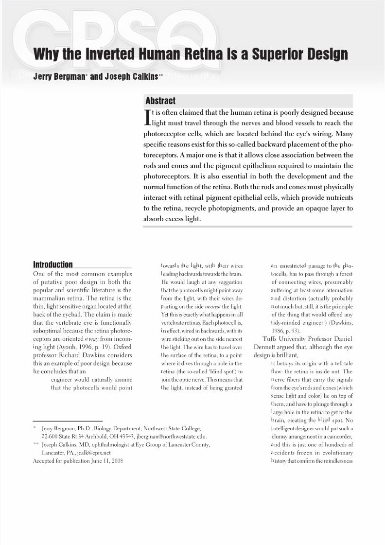
Figure 2. A cross section of the cepha-
lopod eye receptors, called rhabdo-
meric receptors, which contain light-
sensitive cells directly exposed to the
incoming light. Note that the function
of the pigment retina epithelium is
served by the supporting cells located
between the light-sensitive cells, re-
ducing their number. Drawing by B.
L. Lindley-Anderson after Land and
Nilsson (2005).
Figure 3. A cross section of the ver-
tebrate retina, showing the retina
pigment epithelium and other retina
structures. Note the ganglion cells, the
amacrine cells, the bipolar cells, and
the horizontal cells. These are some
of the structures that light must pass
through before striking the photocells,
the rods and cones. The retina pig-
ment epithelium absorbs the light and
supplies the photocells with nutrients
among other functions. Drawing by B.
L. Lindley-Anderson after Land and
Nilsson (2005).

8/2/2019 Retina invertida- Diseño superior de Jehová Dios (en inglés)
http://slidepdf.com/reader/full/retina-invertida-diseno-superior-de-jehova-dios-en-ingles 4/12
216 Creation Research Society Quarterly
reach the photoreceptors. The opposite
placement (where the photoreceptors
face the front of the eye) is a “verted”
design. Verted eyes are wired so that
the photoreceptors face toward the
light and the nerves are placed behind
the photoreceptor layer (Miller, 1994,
p. 30).
ost invertebrates and the pineal or
dorsal eyes of lower vertebrates use theverted eye design, and most vertebrates
(including mammals, birds, amphib-
ans, and fish) use the inverted design.
Most verted eye designs are very simple,
although a few, such as the cephalopod
eye (squids and octopi), are almost as
complex as the vertebrate eye (Abbott
et al., 1995). Even the better verted
eyes are still “overall quite inferior to
the vertebrate eye,” a conclusion usually
determined by measuring performance
n response to visual stimuli (Hamilton,
1985, p. 60).
The Cephalopod Visual SystemThere are several reasons to conclude
that evolutionists incorrectly understand
the design of the inverted eye. The mostadvanced invertebrate eye known today
s that used by certain cephalopods,
but the most advanced eye may actu-
ally be the extinct trilobite (Bergman,
2007). The cephalopod visual system
s poorly understood, both because it is
so complex and because understanding
ts design is not a funding priority—as
s research related to cancer or heart
disease. It is known that the major ana-
tomical difference between the human
eye and the advanced cephalopod eye,
such as the octopus, is the retina. The
cephalopod retina is not only verted
but also lacks the most sensitive part of
the retina, the fovea centralis (Land andNilsson, 2005, p. 64).
In contrast to the claims of Dawkins
and others, no evidence exists that even
the most advanced verted cephalopod
eye is superior to the inverted eye. The
sensitivity of the existing human inverted
design is so great that a single photon
s able to elicit an electrical response
(Baylor et al., 1979). Consequently,
functional sensitivity of the verted retina
could not be significantly improved:
eurobiologists have yet to deter-ine how such a negative system
of operation might be adaptive, but
ey marve over t e acute sensitiv-
ty possible in rod cells. Apparently
od cells are excellent amplifiers. A
ingle photon (unit of light) can pro-
uce a detectable electrical signal in
he retina, and the human brain can
ctua y “see” a c uster o ve p o-
ons—a small point of light, indeed
(Ferl and Wallace, 1996, p. 611).
Greater sensitivity than this single
photon threshold, if this were possible,
might actually result in poorer vision due
to sensory overload. In a similar fashion,
Williams syndrome patients have su-
perior hearing compared to those with
average hearing, allowing them to hear
a faint whisper; however, this sensitivity
causes them serious sensory overload
problems such as in dealing with loud
noises like thunder, which is actually
physically painful.Physiologically, the verted cephalo-
pod retina is simpler compared to the
nverted vertebrate retina. An example
s there are “no equivalents of the ama-
crine, bipolar or ganglion cells in the
cephalopod retina” (Wells, 1978, p.
150). The optic lobes, located behind
the eyeball in cephalopods, must assume
many of the image processing functions
that occur in the inverted retina in ver-
Figure 4. A cross section of the vertebrate eye illustrating some of its major
structures. Drawing by B. L. Lindley-Anderson after Land and Nilsson (2005).
8/2/2019 Retina invertida- Diseño superior de Jehová Dios (en inglés)
http://slidepdf.com/reader/full/retina-invertida-diseno-superior-de-jehova-dios-en-ingles 5/12
Volume 45, Winter 2009 217
tebrates. As an underwater animal that
usually lives on the ocean bottom, its eye
s designed to detect motion, not detail,
as is true of human eyes. It must also
maximize its utilization of light, since
the ocean usually has little or no light atlower depths. The cephalopod eye
ndoubtedly forms an image, but
he animal’s visual perception is
certainly quite different from that
of man, which is greatly dependent
pon interpretation by the brain.
The cephalopod optic connections
ppear to be especially adapted for
nalyzing vertical and horizontal
rojections of objects in the visual
eld (Barnes, 1980, p. 454).
Pechenik (1991) indicated that al-though cephalopods can perceive shape,
light intensity, and texture, they lack
many of the advantages of an inverted
retina, such as the ability to perceive
small details. The cephalopod visual
system is designed very differently from
the inverted eye in other ways to enable
them to function in their water world.
Most cephalopods, including octopi,
have only one visual pigment and are
thus color-blind (Land and Nilsson,
2005).
Furthermore, the maximum resolv-
able spatial frequency in cycles per
radian is 4,175 for humans and only
2,632 for octopi (Land and Nilsson,
2005). Their photoreceptor cell popula-
tion is composed entirely of rods, which
contain a “mere” 20 million retina
receptor cells, compared to 126 million
n humans (Young, 1971). Their rod
outer segments contain rhodopsin pig-
ment that has a maximum absorptionn the blue-green part of the spectrum
(475 nanometers [nm]), which is the
predominant color in their environment.
Photons change the rhodopsin to metar-
hodopsin, and no further breakdown
or bleaching occurs (Wells, 1978). A
second octopus retina pigment, retino-
chrome, has an absorption maximum of
490 nm, which is more sensitive to dim
light (Wells, 1978). Humans have one
rod type and three cone types. One cone
type has a broad peak light frequency
of 440 nm (blue), another type 540 nm
(green), and the third type 570 nm (red)
(Stoltzmann, 2006).
The squid’s visual system must func-
tion in an aqueous medium. Water acts
as a filter, and, as a result, the light is of
a much lower intensity. Consequently,
a squid’s vision sensitivity is for shorterwavelengths (below around 400 nm)
than a human’s, which is from 400 to
00 nm (Peet, 1999). In bright light the
cephalopod’s pupils become thin and
slit-shaped and are held in a horizontal
position by a statocyst, an organ that
uses gravity to determine the horizontal
(Young, 1971). Their visual process is
“quite similar to that of the batrachians,
reptiles and insects. A ‘photograph’ of
the recorded image is not traced on the
retina as in man; instead cephalopods
respond only to ‘light and color varia-
tions of a moving object’” (Grzimek,
1972, p. 191).
Significantly, the octopus responds
to certain motions of nonfood objects
as if they were prey, but will not react
to their normal food-objects if motion-
less (Spigel, 1965). The importance of motion supports the observation that
the octopus eye is actually a simple
“compound eye with a single lens” be-
cause each receptor cell is surrounded
by photopigment containing microvilli,
which form a rhabdomeric structure like
a compound lens (Budelmann, 1994,
p. 15). Each facet in a compound eye is
either on or off, and object movement
produces a change in the on-and-off pat-
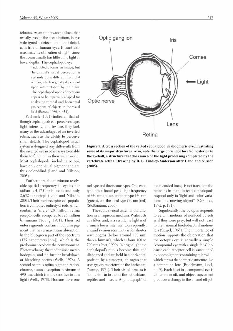
Figure 5. A cross section of the verted cephalopod rhabdomeric eye, illustrating
some of its major structures. Also, note the large optic lobe located posterior to
the eyeball, a structure that does much of the light processing completed by the
vertebrate retina. Drawing by B. L. Lindley-Anderson after Land and Nilsson
(2005).

8/2/2019 Retina invertida- Diseño superior de Jehová Dios (en inglés)
http://slidepdf.com/reader/full/retina-invertida-diseno-superior-de-jehova-dios-en-ingles 6/12
218 Creation Research Society Quarterly
tern—similar to the manner in which a
series of light bulbs produces the illusion
of movement by changing on-and-off
patterns.
Our ignorance about the function
of major parts of the cephalopod visualsystem, such as the optic lobe, prevents
researchers from completing a more
detailed analysis of cephalopod vision.
How the basic eye types could have
evolved from the putative primitive type
s also unknown, in part because no
transitional forms exist, nor do plausible
hypothetical intermediate forms exist.
An essential difference between verte-
brate and invertebrate eyes is that the
vertebrate eye photoreceptors face
outwards towards the choroid,whereas in invertebrates they mostly
oint inwards towards the lens. But
or t at o stac e we s ou ave
een deluged with theories on the
original evolution of the vertebrate
eye from the invertebrate. As it is,
vertebrate visual origins have to
e approached with great caution,
n … t ere is not ing in isputa e
which can be used to explain the
origins of the vertebrate eye from
n invertebrate organ (Prince, 1956,
p. 334, 348).
All known animals have either verted
or inverted retina eyes, and no evidence
exists of transitional forms. Invertebrate
eyes use either some type of a lens-
based eye, such as cephalopods, or a
compound eye, as used in trilobites and
nsects today. All known vertebrates have
nverted eyes, and there are no known
ntermediates between the two.
As Ayoub asked, would “hundredsof thousands of vertebrate species—in
a great variety of terrestrial, marine and
aerial environments—really see better
with a visual system used by a handful
of exclusive marine vertebrates? In the
absence of any rigorous comparative
evidence, all claims that the cephalopod
retina is functionally superior to the
vertebrate retina remain entirely con-
jectural” (Ayoub, 1996, p. 20).
Rod and Cone Functionsin Vertebrates
The rods and cones are photorecep-
tor cells located in the retina used to
transduce light into electrical signals.
Black-and-white transduction occursn the rod shaped receptors, and color
transduction occurs largely in the cone-
shaped receptors (Ryan, 1994). The
nverted retina vision system requires
light to first pass through the cornea,
then through the anterior chamber filled
with aqueous fluid, and last, the lens,
and the vitreous humor . Before reaching
the retina, the light passes through the
nner retina’s cell layers (which contain
a dense array of neural processing cells)
and on past the rods and cones until itreaches the posterior (distal) end of these
cells—wherein lie the so-called outer
cell segments. The outer cell segments
contain the photoreceptors, light-sensi-
tive structures including the photopig-
ment, where the transduction of light
nto receptor potentials occurs.
The photopigment family of pro-
teins undergoes physical changes when
they absorb light energy. The principal
photopigment, opsin glycoprotein, is a
derivative of retinal (a modified vitamin
A molecule). Rods contain a single phot-
opigment type called rhodopsin (rhodo
meaning rose and opsis meaning vision).
The cones contain one of three different
kinds of photopigments called iodopsins,
namely erythrolabe (most sensitive to
red), chlorolabe (most sensitive to green)
and yanolabe (most sensitive to blue)
(Shier et al., 1999, p. 482).
Vision functions by changes in the
retina photopigments molecule causedby light. The molecule has a bent shape
(cis-retinal ) in darkness, and when it
absorbs light, isomerization occurs, caus-
ng the molecule to form the “straight”
orm (trans-retinal). This causes sev-
eral unstable intermediate chemicals
to form, and, after about a minute, the
trans-retinal form completely separates
from opsin, causing the photopigment
to appear colorless (for this reason the
process is called bleaching). In order
for the rods and cones to again function
for vision, retinal must be converted
from the trans back to the cis form. This
resynthesis process, called regeneration,
requires that the retina pigment epithe-lium (RPE) cells be located next to the
rod and cone outer segments.
An average of five minutes is required
for rhodopsin regeneration in rods,
compared to 1.5 minutes for iodopsin
regeneration in cones (Tortora and
Grabowski, 1996). Excessive light causes
blindness in the affected rods and cones
until this regeneration process occurs, as
shown by the temporary blindness that
occurs after watching a very bright light
flash from a camera strobe light (Snelland Lemp, 1989).
When rods and cones are stimulated
by light, they release neurotransmitters
that induce graded, local potentials
n both bipolar and horizontal cells.
By this means the rod and cone outer
segments transduce light into electrical
signals. The signals are then carried by
the central nervous system neurons to
bipolar cells that, in turn, synapse onto
the ganglion cells, then to the lateral ge-
niculate body of the thalamus, and, last,
to the occipital region of the brain stem,
where the information is organized into
a useful image (Stoltzmann, 2006).
The RetinalPigment EpitheliumOne of the many reasons for the inverted
design is that behind the photoreceptors
lies a multifunctional and indispensable
structure, the retinal pigment epithelium(RPE) (Martínez-Morales et al., 2004).
RPE is a single-cell-thick tissue layer
consisting of relatively uniform polygo-
nal-shaped cells whose apical end is
covered with dense microvilli and basal
membrane infoldings. Posterior to the
RPE is the vascular choroid layer, and
posterior to it is the connective tissue
known as the sclera. The RPE touches
the extremities of both the rod and the
8/2/2019 Retina invertida- Diseño superior de Jehová Dios (en inglés)
http://slidepdf.com/reader/full/retina-invertida-diseno-superior-de-jehova-dios-en-ingles 7/12
Volume 45, Winter 2009 219
cone photoreceptors, and the microvilli
nterdigitate with their sides (Steinberg
and Wood, 1994).
The photoreceptors (rods and cones)
must face away from the front of the
eye in order to be in close contact withthe vascular choroid, which supplies
the photoreceptors with nutrients and
oxygen. This arrangement also allows a
steady stream of the vital molecule reti-
nal to flow to the rods and cones, without
which vision would be impossible (Kolb,
2003). The verted design, on the other
hand, would place the photoreceptors
away from their source of nutrition,
oxygen, and retinal. This design would
fail because the rods and cones require
an enormous amount of energy for theirhigh metabolism required to function.
In addition, due to phototoxicity dam-
age from light, the rods and cones must
completely replace themselves approxi-
mately every seven days or so. Seemingly
simple in appearance, the RPE has “a
complex structural and functional polar-
ty that allows them to perform highly
specialized roles” (Hewitt and Adler,
1994, p. 58). One of their major func-
tions is to recycle the used retinal from
the photoreceptors.
Vision depends on the isomeriza-
tion of 11-cis-retinal to 11-trans-retinal
n the rods and cones outer segments.
Each light photon striking a photorecep-
tor isomerizes retinal, and billions of
photons can strike the retina at any one
second. The RPE constantly restores the
chromophore to cis-retinal from its trans
configuration caused by photostimula-
tion, permitting visual pigment synthesis
and regeneration (Dowling, 1987). The11- is-retinal must also be regularly re-
placed to maintain the cycle, a task for
which the RPE is critical (Hewitt and
Adler, 1994). The RPE manufactures
retinal isomerase and other enzymes
and stores large quantities of vitamin A
to regenerate retinal.
Since RPE cells use enormous
amounts of energy and nutrients, they
must be in intimate contact with both
the photoreceptors and the blood supply
(in this case the choroid) to carry out
this critical function (Marshall, 1996).
Research on the eyes of different spe-
cies has found that, although major
differences among them exist, the RPEshows “little variation” (Kuwabara, 1994,
p. 58). The small RPE variations are due
to differences in the retina structure,
ndicating its critical role in the vision of
all vertebrates. One study found retinol
somerase in all the major vertebrates
tested and was lacking in all three
cephalopods tested (Bridges, 1989).
Bridges concluded that reciprocal flow
of retinoids between the retina and the
site of isomerase action in the RPE is a
feature common to the visual cycle inall vertebrates (Bridges, 1989).
Phagocytic Role of the RPE A major role of the RPE is to recycle
the used rod and cone outer segment
membranes, the cone portion closest to
the RPE. The photoreceptors and RPE
absorb an enormous amount of light on a
continuous basis when the eyes are open.
This light is converted largely into heat,
requiring a very effective cooling system.
The choroidal blood supply directly be-
hind the RPE carries away, not only this
heat, but also the relatively large amount
of waste products produced by the high
level of rod and cone metabolism. Which
compounds are allowed to pass though
this area is determined by basal mem-
brane receptors. Cones usually contain
from 1,000 to 1,200 disks, and rods from
00 to 1,000. The enormous amount of
outer segment activity requires continual
replacement of these disks (Bok, 1994). As the outer segment lengthens from
ts base, the oldest membrane, which is
at the distal end, is shed in segments of
one to three disks at a time. Those that
are sloughed off are phagocytized by
enzymes stored in RPE lysosomes and
ts components recycled (Tortora and
Grabowski, 1996, p. 467).
The RPE phagocytosizes about ten
percent of the outer segment disks of nor-
mal rod photoreceptors at its apex and
renews the same amount daily (Benson,
1996). To replace those segments that
are lost, new outer segment membranes
are continually being produced at the
outer photoreceptor segment base. Pho-toreceptor outer segments are renewed
at “an astonishingly rapid pace” (Tortora
and Grabowski, 1996, p. 467).
After RPE breaks down the ingested
material, the free radicals and superox-
des produced must be neutralized by
superoxide dismutase, peroxidase, and
other enzymes (Hewitt and Adler, 1994).
This process is continuous, effectively
maintaining the photoreceptor’s high
sensitivity (Benson, 1996). Bok and
Young (1994) summarized this cycle,noting that the
etinal pigment epithelium carries
out severa unctions t at are crucia
or the normal operation of the visual
ystem. One of these important roles,
ppreciated for about a decade, is the
hagocytosis of rod outer segment
ebris. This scavenging activity goes
on ai y at an impressive rate in t e
ormal retina. It can be accelerated
o extraordinary levels when outer
egments are damaged. Disruption
of this phagocytic function may un-
erlie a variety of clinical disorders,
ome of which result in blindness
(p. 148).
RPE microvilli interdigitate and
surround the photoreceptor outer seg-
ments so as to effectively carry out their
phagocytic and recycling role (Bok and
Young, 1994).
Nutrient Role of the RPEThe RPE selectively transports nutrients
from choroidal circulation to both the
photoreceptors and retinal cells. The
RPE also helps maintain water and
on flow between the neural retina
and the choroid, protects against free
radical damage, and regulates retinoid
metabolism (Martínez-Morales et al.,
2004). The RPE functions similarly to a
placenta to ensure that the outer retina
8/2/2019 Retina invertida- Diseño superior de Jehová Dios (en inglés)
http://slidepdf.com/reader/full/retina-invertida-diseno-superior-de-jehova-dios-en-ingles 8/12
220 Creation Research Society Quarterly
s protected from injurious compounds
and yet allows the necessary nutrients
to pass into the rod and cone area. RPE
cell tight junctions are also part of the
outer blood-retinal barrier, prevent-
ng diffusion of even small moleculesnto the vitreous humor and ensuring
that the metabolites required by the
outer retina can move to where they are
needed when they are needed (Hewitt
and Adler, 1994).
To ensure that enough of the needed
nutrients pass the RPE barrier, the basal
membrane is highly infolded to produce
more surface area. This role is critical
because the rods and cones require a
greater blood supply than any other
bodily tissue (Hewitt and Adler, 1994).This is important because of the high
level of metabolism due to the complex
chemistry required for vision, which
necessitates a higher level of oxygen
and nutrients. The RPE also synthesizes
and secretes various extracellular matrix
molecules that must be produced near
the location where they are to be used.
If the photoreceptors were anterior to
the neurons, as in the verted design, the
blood supply would have to be either di-
rectly in the light path of the receptors or
on their side, which would reduce enor-
mously the number of photoreceptors
used for sight. If the pigment epithelium
tissue were placed in front of the retina,
sight would be seriously compromised.
The verted design would make vision
mpossible because the photoreceptors
must be embedded in the retinal pig-
ment epithelium to obtain the nutrients
required to function.
Müller Cells Functionas Optical FibersPlacing the retina neural components
n front of the photoreceptors does
not produce an optical handicap for
yet other reasons (Land and Nilsson,
2005). One is that the neural elements
are separated by less than a wavelength
of light. Consequently, very little or no
scattering or diffraction occurs, and the
light travels through this area as if it were
at near-perfect transparency.
The Müller cells (which are radial
glial cells) in front of the retina have
both shape and optical properties that
contribute to optimizing light transferraland reducing light scatter (Franze et al.,
2007). Müller cells “have an extended
funnel shape, a higher refractive index
than their surrounding tissue and are
oriented along the direction of light
propagation” (Franze et al., 2007, p.
8287). The effect provides a “low-scat-
tering passage for light from the retinal
surface to the photoreceptor cells,” func-
tioning as fiber optic plates that are ef-
fective for low-distortion transfer of light
mages. Franze et. al. (2007) concludedthat cells thought to interfere with light
transmission are actually highly effective
n reducing light scatter and distortion,
helping to produce a sharp image.
The MaculaThe importance of the RPE is indicated
by the fact that one of the most common
causes of blindness in the developed
world, macular degeneration, is the
result of RPE deterioration (Zhang,
et al., 1995). In this disease the eye’s
macula loses its ability to function, caus-
ng major central vision loss. Without
the nourishment and waste removal role
of the pigment epithelium, retina cells
will also die. Among the other diseases
affecting the macula is central serous
retinopathy, an ion pump malfunction
and/or a result of choroidal vascular
hyperpermeability.
Detached Retina and theRole of Pigment Epithelial CellsThe retina is connected to the RPE
largely by the interphotoreceptor matrix.
When the retina pulls away from the
RPE at the interphotoreceptor matrix
area, a detached retina results (Zamir,
1997). The RPE can then no longer ef-
fectively function to regenerate the rods
and cones, causing vision to become
distorted, and eventually the death of
significant levels of retina tissue. Progres-
sive detachment can often be halted by
laser therapy, a procedure that is only
minimally invasive because laser light
s able to pass through the cornea and
the lens without damaging them. Lasertherapy stimulates the migration of the
RPE cells, inducing the pigmentation
line to form.
Functions of the PigmentThe many diverse functions of the
retinal pigment epithelium cells that are
“essential for the normal functioning of
the outer retina” include producing a
black pigment called melanin (Hewitt
and Adler, 1994, p. 67). The melanin
functions to absorb most of the light notcaptured by the retina, preventing the
reflection and scattering of light within
the eyeball. This inhibits light from be-
ng reflected off the back of the eye onto
the retina, preventing degradation of the
visual image and ensuring that the image
cast on the retina by the cornea and lens
remains sharp and clear.
et another function of the pigment
s to form an opaque screen behind the
optical path of the photoreceptors. This
light absorptive property of the pigment
s critical to maintaining high visual
acuity. For this reason, normal retinal
function requires that the RPE and
photoreceptors be in close proximity.
Lack of the pigment, as in albinism,
can cause a variety of problems such as
fovea hypoplasia, an abnormal routing
of the optic nerve (Oetting and King,
1999; Lyle et al., 1997; Jeffery and Wil-
liams, 1994). As a result of this and other
factors, albinism victims lack detailedcentral vision (Snell and Lemp, 1989;
Williamson, 2005).
The Retina Pigment Epithelium’sRole in DevelopmentRPE is also critical for normal vertebrate
eye development. A series of reciprocal
cellular interactions during vertebrate
eye development determine the fate of
the eye components, and the
8/2/2019 Retina invertida- Diseño superior de Jehová Dios (en inglés)
http://slidepdf.com/reader/full/retina-invertida-diseno-superior-de-jehova-dios-en-ingles 9/12
Volume 45, Winter 2009 221
resence of the RPE is required for
e norma eve opment o t e eye
in vivo. Its presence early in devel-
opment is necessary for the correct
orphogenesis of the neural retina
(Raymond and Jackson, 1995, p.12 ).
The RPE actually plays a succession
of roles during embryonic development,
ncluding trophic influence, transport
functions, retinomotor response, and
phagocytic and inductive interaction
(Coulombre, 1994).
Does the Backward DesignBlock Light?
Nerve cell fibers and the small branchesof the central retina artery and vein
produce minimal hindrance to light
reaching the photoreceptors because
most cells are 60 to 70% water and,
consequently, are largely transparent.
When viewed under the microscope,
most cells are largely transparent. It is
for this reason stains, such as Eosin-Y
and Hematoxylin 2, are required to
better visualize the various cell parts.
Myelin, an opaque whitish lipid that
coats nerve axons, would block much
light, but, in contrast to most periph-
eral nerves, nerve fibers in front of the
retina are not mylinated. Furthermore,
the larger blood vessels and nerve fibers
skirt around the area centralis, where
visual acuity is most important (Gregory,
1976). The vertebrate eye is highly effec-
tive in spite of the retina reversal because
t is a precise visual instrument designed
to function with the rods and cones fac-
ng away from the light.The tissues intervening between the
ransparent humors of the eye cavity
nd the optically sensitive layer are
icroscopically thin. The absorp-
ion an scatter o ig t is or inari y
inor, and functional impairment
eldom serious.…Red blood cells are
oor transmitters of light, but when
oving single file through capillaries
can cause only a negligible shading
of the light sensors (Williams, 1992,
. 73).
These facts have forced Dawkins to
note that many
hotocells point backwards, away
rom the light. This is not as silly as itounds. Since they are very tiny and
ransparent, it doesn’t much matter
which way they point: most photons
will go straight through and then run
he gauntlet of pigment-laden baffles
waiting to catch them (Dawkins,
1 , p. 17 ).
oving shadows produced by the
venules and arterioles are also highly
functional because they produce mo-
mentary darkness to aid in the rod and
cone regeneration. Constant bright lightwould excessively bleach the photopig-
ment, and the lower light achieved by
the existing design allows their regen-
eration.
Other Possible Designs A major concern when critiquing the
existing vertebrate retina design involves
speculation on the quality of vision that
would result from another design. No
evidence exists that a verted human
retina design, as in octopi, would result
n better vision, and it would likely be
worse. Comparisons of different eyes are
difficult to make because, although the
quality of the image projected on the
retina can be evaluated by a study of the
lens system’s optical traits, direct knowl-
edge about the actual image produced
n the brain is lacking.
If the retina were reversed, the retinal
pigment epithelium or its analog and itscellular support system would have to
be placed either in front of the photo-
receptors or on their side. Both of these
approaches are clearly inferior to the ex-
sting vertebrate system, which produces
superior sight for terrestrial animals. If
located in front of the retina, depending
on the transparency of these cells, this
design could prevent most light from
reaching the photoreceptors.
If the RPE functioning cells were
located on each side of the rods and
cones, as in the cephalopods, primarily
only the sensory cell face would be able
to respond to light. Octopi use support
cells located next to the light-sensitivecells called rhabdomeric receptors that
use photopigments containing microvilli
(Land and Nilsson, 2005). The support
cells also require increasing the space
between the photoreceptors, further
decreasing light able to strike the pho-
toreceptors, and consequently lowering
vision resolution. Prince (1956) even
claims the cephalopod’s side design “is
protective and shields the receptors from
excess light” (p. 343). Opaque wastes
would accumulate in the light path,and the presence of required nutrients
would further diminish the amount
of light reaching the photoreceptors.
Recycling the outer segments to allow
rapid regeneration of the photorecep-
tors would also be a major problem if
the photoreceptors faced the vision light
path line. Verted designs produce the
following concerns:
Should the disk end of the rods and
cones be reversed in direction so as
o face the light…we would probably
ave a visual disaster. What would
erform the essential function of
bsorbing the some 10,000 million
isks produced each day in each
of our eyes? They would probably
ccumulate in the vitreous humor
egion and soon interfere with light
en route to the retina. If the pigment
epithelium layer were placed on the
nside of the retina so as to absorb
he disks, it would also interferewith light trying to reach the rods
nd cones. Furthermore, the pig-
ent epithelium, which is closely
ssociated with the disk ends of the
o s an cones, a so provi es t em
with nutrients for making new disks.
The epithelium gets its nutrients
rom the rich blood supply in the
choroid layer next to it. In order for
he pigment epithelium to function

8/2/2019 Retina invertida- Diseño superior de Jehová Dios (en inglés)
http://slidepdf.com/reader/full/retina-invertida-diseno-superior-de-jehova-dios-en-ingles 10/12
222 Creation Research Society Quarterly
roperly, it needs this blood supply.
To put ot t e pigment epit e ium
nd its choroid blood supply on the
nside of the eye, between the light
ource and the light-sensitive rods
nd cones, would severely disrupt thevisual process (Roth, 1998, p. 109).
Although higher visual acuity may
mprove night vision, in humans it
would result in difficultly seeing during
daylight hours, which would not be
functional for persons that must work
n normal human-light environments
(Sjostrand, 1989). Actually, a case can
be made that more light blockage of the
retina would be functional. Many per-
sons must wear sunglasses because nor-
mal outdoor light is often too bright. Ina review of the literature, Young (1992)
found that excess solar radiation can be
a serious health problem, and may
explain the distinctive global pat-
ern of age-related cataract among
uman populations—the risk of
cataract depends on where one lives
on the surface of the earth.…Cur-
ent evi ence provi es t e asis or
he design of protective lenses that
inimize the hazards of sunlight
exposure without significantly inter-
ering with vision. The prescription
as two components—one to protect
he lens, the other to protect the
etina.…Use of sunglasses…should
egin early in childhood and be
continued throughout the life span
whenever exposure to bright sunlight
s desirable or necessary. Radiation
amage to delicate ocular structures
can occur at any age and tends to be
cumulative (pp. 335-357). Albinos lack iris pigment, requiring
them to wear sunglasses in daylight be-
cause even moderately bright light may
severely adversely affect their vision (Tor-
tora and Grabowski, 1996, p. 461). Even
blue-eyed persons are at a disadvantage
because the blue pigment allows in more
light than the darker iris pigments. Con-
sequently, they suffer from more vision
problems (Young, 1992). Being able to
effectively read by very dim light may
be an improvement in some situations,
but since most human activities occur
during daylight hours and darkness is
functional to induce sleep due to pineal
gland activity, the existing system ap-pears to be the most effective.
Furthermore, although the light yel-
low tint of the eye lens filters out some
ultraviolet light, the inverted eye design
serves to filter out much of the remain-
ng ultraviolet light. The incoming light
must pass through the overlying neural
components and blood vessels, and the
penetrating power of ultraviolet light is
markedly inferior to white light (Lums-
den, 1994). The verted eye is used in
animals such as the octopus, which liveunderwater, where most of the ultravio-
let light is filtered out. Consequently,
they have less need for this protection.
Given the role of the pigmented epithe-
lium, it is clear that the existing design
s ideal.
Conclusions A review of research on the vertebrate
retina consistently concludes that each
design is perfectly suited for the envi-
ronment the organism normally lives
n—even the system used by the most
advanced cephalopods (Bergman, 2000;
Bergman and Calkins, 2005; Wieland,
1996; Marshall, 1996). The design maxi-
mized for life in our environment would
no doubt function poorly in another
environment, such as that experienced
by undersea bottom dwellers. The RPE
metabolic machinery is “essential for the
normal functioning of the outer retina[and] because of the nature of these
nteractions, it is essential that the RPE
and photoreceptors be in close proxim-
ty” for normal retina function (Hewitt
and Adler, 1994, p. 67). This review sup-
ports Hamilton’s conclusion that
nstead of being a great disadvantage,
or a “curse” or being incorrectly
constructed, the inverted retina is
tremendous advance in function
nd design compared with the
imp e an ess comp icate verte
rrangement. One problem amongst
any, for evolutionists, is to explain
ow this abrupt major retinal trans-
ormation from the verted type innvertebrates to the inverted verte-
rate model came about as nothing
n paleontology offers any support
(Hamilton, 1985, p. 63).
Rather than being fired, our camera
designer would no doubt be promoted
for utilizing a less obvious but far more
functional design. It is clear that “eye-
sight is a compelling testimony to cre-
ative design” (DeYoung, 2002, p. 190).
This short review covers only a few of
the many reasons for the superiority of the existing mammalian retina design.
Gratitude rather than impertinence
seems the more appropriate response to
ts ingenious design.
AcknowledgmentsI wish to thank Dr. Tara Richmond,
O.D.; Clifford Lillo, MS; John Up-
Church; David Stoltzmann, optical
engineer; Don DeYoung, Ph.D., physi-
cist; Jody Allen, RN; B. L. Lindley-An-
derson for producing the illustrations;
and George F. Howe for editorial
assistance.
Jos eph Calkin s, MD, currently an
ophthalmologist in private practice, was
formally Assistant Professor of Ophthal-
mology at Johns Hopkins University in
Baltimore, Maryland. His MD is from
the University of Michigan Medical
School in Ann Arbor, MI.
ReferencesCRSQ: Creation Research Society Quar-
terly.
Abbott, J., R. Williamson, and L. Maddock.
1995. Cephalopod Neurobiology. Oxford
University Press, Oxford, NY.
Ayoub, G. 1996. On the design of the
vertebrate retina. Origins and Design
17:1 –22.
8/2/2019 Retina invertida- Diseño superior de Jehová Dios (en inglés)
http://slidepdf.com/reader/full/retina-invertida-diseno-superior-de-jehova-dios-en-ingles 11/12
Volume 45, Winter 2009 223
Barash, D.P., and I.A. Barash. 2000. The
Mammal in the Mirror: Understanding
Our Place in the Natural World. W.H.
Freeman, New York, NY.
Barnes, R.D. 1980. Invertebrate Zoology.
Saunders, Philadelphia, PA.Baylor, D. A., T.D. Lamb, and K.W.
Yau. 1979. Response of retinal rods to
single photons. Journal of Physiology
88:613–634.
Benson, E. 1996. Retinitis pigmentosa:
nfolding its mystery. Proceedings of
the National Academy of Science USA
3: 52 – 52 .
Bergman, J. 2000. Is the inverted human eye
a poor design? ournal of the American
Scientific Affiliation 52:18–30.
Bergman, J. 2007. Lack of fossil evidencefor arthropod evolution is a major
ifficulty for neo-Darwinism. R
3:222–23 .
Bergman, J., and J. Calkins. 2005. Is the
backwards human retina evidence of
poor design? Impact 388:1–4.
Bok, D. 1994. Retinal photoreceptor disc
shedding and pigment epithelium
p agocytosis. In Zinn, K.M., an M.F.
armor (editors), The Retinal Pigment
Epithelium, pp. 81–94. Harvard Univer-
sity Press, Cambridge, MA.
Bok, D., and R. Young. 1994. Phagocytic
properties of the retinal pigment. In
Zinn, K.M., and M.F. Marmor (edi-
tors), The Retinal Pigment Epithelium,
pp. 148–174. Harvard University Press,
Cambridge, MA.
Bridges, C.D.B. 1989. Distribution of retinol
somerase in vertebrate eyes and its emer-
gence during reti nal deve lopment .
Vision Research 29:1711–1717.
Budelmann, B.V. 1994. Cephalopod senseorgans, nerves and the brain: adapta-
tions for high performance and life
style. In Portner, Hans, et al. (editors),
Physiology of Cephalopod Mollusks,
pp. 13–33. or on an Breac , Base ,
Switzerland.
Coulombre, A. 1994. Roles of the retinal
pigment epithelium in the development
of ocular tissue. In Zinn, K.M., and M.F.
armor (editors), The Retinal Pigment
Epithelium, pp. 53–57. Harvard Univer-
sity Press, am ri ge, M .
Dalton, R. 2004. True colours. Nature
428:596–597.
Dawkins, R. 1986. he Blind Watchmaker . W.
W. Norton, New York, NY.Dawkins, R.. 1996. ClimbingMount Improb-
able. W.W. Norton, New York, NY.
Dennett, D. 2005. Show me the science.
New York Times, August 28.
DeYoung, D.B. 2002. Vision. CRSQ 38:190–
192.
Diamond J. 1985. Voyage of the overloaded
ark. Discover : 2– 2.
Dowling, J.E. 1987. The Retina: An Ap-
proachable Part of the Brain . The
Belknap Press of Harvard University
Press, Cambridge, MA.Edinger, S. 1997. Is there a scientific basis
for creationism? The Congressional
uarterly Researcher 7:7 1.
Ferl, R., and R.A. Wallace. 1996. Biology,
the Realm of Life. Harper Collins, New
York, NY.
Franze, K., J. Grosche, S.N. Skatchkov, S.
Schinkinger, C. Foja, D. Schild, O.
Uc ermann, K. Travis, . Reic en ac ,
and J. Guck. 2007. Müller cells are liv-
ng optical fibers in the vertebrate retina.
Proceedings of the National Academy of
cience 104:8287–8292.
Frymire, P. 2 . Impeaching Mere Creation-
ism. Writers Club Press, San Jose, CA.
Gregory, R. 1976. Eye and Brain. World
Universal Library, New York, NY.
Grzimek, B. 1972. Grzimek’s Animal Life
Encyclopedia. Van Nostrand Reinhold,
ew York, NY.
Hamilton, H.S. 1985. The retina of the
eye—an evolutionary road block. CRSQ
2:59–64.Hewitt, A.T., and R. Adler. 1994. The retinal
pigment epithelium and interphotore-
ceptor matrix: structure and specialized
functions. In Ryan, S.J. (editor), The
Retina, econ E i tion, pp. 5 –71 .
osby, St. Louis, MO.
Jeffery, G., and A. Williams. 1994. Is abnor-
al retinal development in albinism
only a mammalian problem? Normality
of a hypopigmented avian retina. Experi-
mental Brain Research 100:47–57.
Ko , H. 2 3. How t e retina wor s. meri-
can Scientist 91:28–35.
Kuwabara, T. 1994. Species differences in
the retinal pigment epithelium. In Zinn,
K.M., and M.F. Marmor (editors), heRetinal Pigment Epithelium, pp. 58–82.
Harvard University Press, Cambridge,
A.
Land, M.F., and D.E. Nilsson. 2005. nimal
Eyes. Oxford University Press, Oxford,
Y.
Lumsden, R. 1994. Not so blind a watch-
aker. R 31:13–21.
Lyle, W.M., J.O.S. Sangster, and T.D.
Wil liams.1997. Albinism: an update
and review of the literature. Journal of
the American Optometric Association68:623–645.
Marshall, G. 1996. An eye for creation: an
nterview wit eye- isease researc er Dr.
George Marshall, University of Glasgow,
Scotland. Creation 18:19–21.
Martínez-Morales, J.R., I. Rodrigo, and P. Bo-
volenta. 2004. Eye development: a view
from the retina pigmented epithelium.
BioEssays 2 :7 –777.
Meglitsch, P. 1972. Invertebrate Zoology.
Oxford University Press, New York, NY.
Miller, K.R. 1994. Life’s grand design. ech-
nology Review 97:25–32.
Miller, K.R.. 1999. Finding Darwin’s God: A
cientist’s earch or ommon round
between God and Evolution. Cliff Street
Books, New York, NY.
Oetting, W.S. and R.A. King. 1999. Mo-
lecular basis of albinism: mutations and
polymorphisms of pigmentation genes
associated with albinism. Human Mu-
tatagens 13:99–115.
Pechenik, J. 1991. Biology of the Invertebrates. William C. Brown, Dubuque, IA.
Peet, J.H.J. 1999. Creation in the news:
Dawkins’ blind spot. rigins 26:2–4.
Prince, J. 1956. Comparative Anatomy of the
Eye. ar es T omas, pring e , IL.
Raymond, S.M., and I.J. Jackson. 1995. The
etinal pigment epithelium is required
for development and maintenance of
the mouse neural retina. Current Biology
5:1286–1295.
8/2/2019 Retina invertida- Diseño superior de Jehová Dios (en inglés)
http://slidepdf.com/reader/full/retina-invertida-diseno-superior-de-jehova-dios-en-ingles 12/12
224 Creation Research Society Quarterly
Roth, A. 1998. rigins. Review and Herald,
Hagerstown, MD.
Ryan, S.J. (Editor). 1994. The etina, nd
Edition. Mosby, St. Louis, MO.
Sarfati, J. 1998. A review of Climbing Mount
Improbable by Richard Dawkins.Techni-cal Journal. 12:2 –3 .
Shermer, M. 2005. cience Friction: Where
the Known Meets the Unknown. Holt/
Times Books, New York, NY.
Shier, D., J. Butler, and R. Lewis. 1999.
Hole’s Human Anatomy and Physiology.
William C. Brown, Dubuque, IA.
Sjostrand, F. 1989. An elementary infor-
ation processing component in the
circuitry of the ret ina generat ing the
on-response. Journal of Ultrastruc-
ture and Molecular Structure Research102:24–38.
Snell, R., and M. Lemp. 1989. Clinical
natomy o the Eye. B ac we cienti c
Publications, Boston, MA.
Spalton, D.J., R.A. Hitchings, and P.A.
Hunter (editors). 2005. Atlas of Clinical
Ophthalmology, nd Edition. Mosby, St.
Louis, M .
pige , I.M. (E itor). 1 5. Readings in the
Study of Visually Perceived Movement.
Harper and Row, New York, NY.
Steinberg, R.H., and I. Wood. 1994. The re-
ations ip o t e retina pigment epit e-
lium to the photoreceptor outer segment
n the human retina. In Zinn, K.M.,
and M.F. Marmor (editors), he Retinal
Pigment Epithelium, pp. 32–44. HarvardUniversity Press, Cambridge, MA.
Stoltzmann, D.E. 2006. The specified
complexity of retinal imagery. CRSQ
43:4–12.
Thwaites, W. 1982. Design, can we see the
hand of evolution in the things it has
wrought? Proceedings f the 3rd Annual
Meeting o the aci c Division; merican
Association of the dvancement f ci-
ence 1:206–213.
Tortora, G., and S. Grabowski. 1996. Prin-
ciples of Anatomy and Physiology. Harperand Collins, New York, NY.
Wells, M.J. 1978. Octopus; Physiology and
Behavior o an dvanced Invertebrate.
Chapman and Hall, London, UK.
Wieland, C. 1996. Seeing back to front: are
evolutionists right when they say our
eyes are wired the wrong way? reation
1 :3 – .
Wi iams, . . 1 2. Natural election:
Domains, Levels, and Challenges. Oxford
University Press, New York, NY.
Williams, G.C. 1997. The Pony ish Glow
and ther lues o Plan and urpose in
Nature. BasicBooks, New York, NY.
Williamson, T. 2005. Vitreous and vitreo-
Retinal disorders. In Spalton, D.J., R.A.
Hitchings, and P.A. Hunter (editors), Atlas of Clinical Ophthalmology, 2nd
Edition, pp. 263–297. Mosby, St. Louis,
O.
Young, J.Z. 1971. he Anatomy of the Ner-
vous System: Octopus Vulgaris. Oxford
University Press, New York, NY.
Young, R. 1992. Sunlight and age-related eye
disease. Journal o the National Medical
Association 84:353–358.
Zamir, E. 1997. Central serous retinopathy
associated with adrenocorticotrophic
hormone therapy. Graefes Archives for Clinical Ophthalmology 235:339–344.
Zhang, K., E. Nguyen, A. Crandall, and L.
Donoso. 1 5. enetic an mo ecu ar
studies of macular dystrophies: recent
developments. Survey of Ophthalmology
40:51–61.
Zinn, K.M., and M.F. Marmor (editors).
1 . The Retinal Pigment Epithelium.
Harvar University Press, am ri ge,
A.