SEMINARIO DE CASOS DE PATOLOGÍA PEDIÁTRICA CON …Figure 3 FNA smear of a case of HL exhibiting a...
47
SEMINARIO DE CASOS DE PATOLOGÍA PEDIÁTRICA CON CORRELACIÓN CITO-HISTOLÓGICA Dra. Cristina Jou Marta Marginet Anatomía Patológica Hospital Sant Joan de Deu
Transcript of SEMINARIO DE CASOS DE PATOLOGÍA PEDIÁTRICA CON …Figure 3 FNA smear of a case of HL exhibiting a...

SEMINARIO DE CASOS DE PATOLOGÍA PEDIÁTRICA CON
CORRELACIÓN CITO-HISTOLÓGICA
Dra. Cristina Jou
Marta Marginet
Anatomía Patológica
Hospital Sant Joan de Deu

CASO CLÍNICO:
Adolescente varón de 16 años con dolor lumbar de tres meses de evolución.
Fiebre en los últimos días.
Astenia.
AF: Tía materna Ca tiroides
EF: Neurológica: Fuerza 4/5 en MID con sensibilidad conservada.
No adenopatías palpables
PC: Analítica sangre: sin alteraciones.

Pruebas de imagen:
• Lesión ósea lítica con importante componente de partes blandas centrado en cuerpo vertebral D12.
• Invasión de los agujeros de conjunción D11-D12 y D12-L1 con componente intradural epidural de e importante compresión del saco tecal.
• Lesiones líticas óseas en cuerpos vertebrales D2, D7, D8, D9, D11, D12 y sacro derecho.
• Lesión en hueso iliaco izquierdo y rama iliopubiana izquierda.

Pruebas de imagen:
• Lesión ósea lítica con importante componente de partes blandas centrado en D12.
• Invasión de agujeros de conjunción D11-D12 y D12-L1 bilateral con componente intrarraquídeo epidural y con importante compresión del saco tecal.
• Alteración de señal en todos los cuerpos vertebrales, excepto L2 y pelvis.
Orientación diagnóstica radiológica: Sarcoma de Ewing
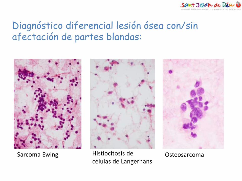
Diagnóstico diferencial lesión ósea con/sin afectación de partes blandas:
Osteosarcoma
Histiocitosis de células de Langerhans
Sarcoma Ewing

Biopsia intraoperatoria: Fragmentos tisulares elásticos y firmes de coloración blanco rosada 3x2’5 cm.


DIAGNOSTICO DIFERENCIAL CITOLÓGICO:
1. Tumor miofibroblástico inflamatorio
2. Linfoma anaplásico de células grandes
3. Linfoma de Hodgkin
Células ganglion-like Fondo inflamatorio benigno

1. Tumor miofibroblástico inflamatorio
• Neoplasia rara miofibroblástica de bajo potencial maligno. • Localización:
– Hígado 45% – Pulmón 40% – Partes blandas 15% – Hueso (¿?%)
• Histológicamente tres patrones: 1. Patrón vascular mixoide 2. Patrón fusocelular 3. Patrón colagenizado • Clínica: Síndrome constitucional con fiebre, sudoración nocturna, perdida peso y astenia.


Citología de TMI pulmonar Citología de TMI extra-pulmonar


2. Linfoma anaplásico células grandes ALK+
• 10-20% de los linfomas no Hodgkin en edad pediátrica: ALK + mas frec en las tres primeras décadas de la vida
• Translocación que afectando al gen ALK con expresión de CD30.
• Localización:
– Nodal
– Extranodal: piel, hueso, tejidos blandos, pulmón y hígado
• Distintos patrones histológicos.


From the Department of Pathology, King Faisal Specialist Hospital and Research Center, Riyadh, Saudi Arabia.
Drs. Mourad and Tulbah are Consultant Pathologists.
Dr. Nazer is Fellow.
Address correspondence to: Walid A. Mourad, M.D., FCAP, FRCPC, Department of Pathology (BMC 10), King Faisal Specialist Hospitaland Research Center, P.O. Box 3354, Riyadh, Saudi Arabia 11211 ([email protected]).
Financial Disclosure: The authors have no connection to any companies or products mentioned in this article.
Received for publication July 19, 2002.
Accepted for publication December 19, 2002.
0001-5547/ 03/ 4705-0744/ $19.00/ 0 © The International Academy of CytologyActa Cytologica
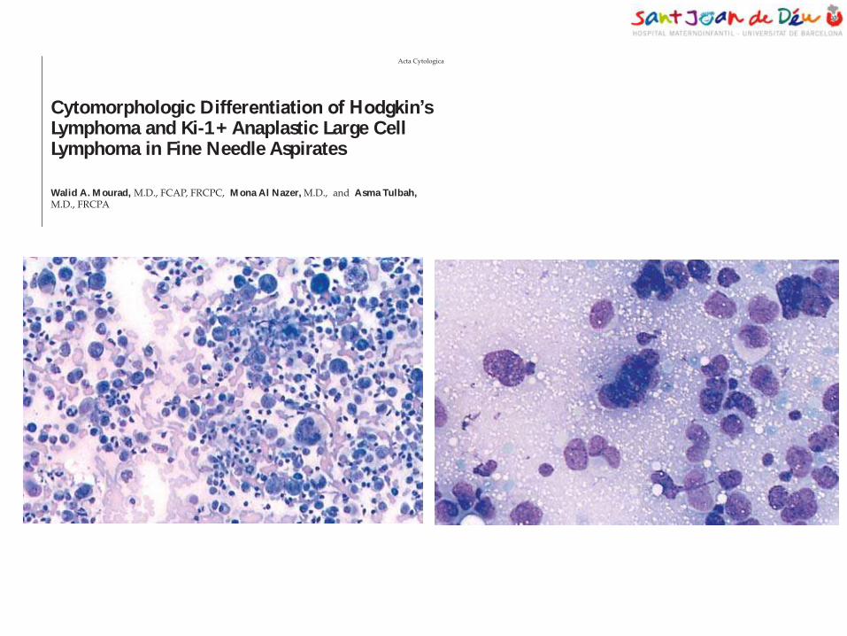
Cytomorphologic Differentiation of Hodgkin’sLymphoma and Ki-1+ Anaplastic Large CellLymphoma in Fine Needle Aspirates
Walid A. M ourad, M.D., FCAP, FRCPC, M ona Al Nazer, M.D., and Asma Tulbah, M.D., FRCPA
744
Acta Cytologica
OBJECTIVE: To cytomorphologically differentiate
Hodgkin’s lymphoma (HL) from Ki-1+ anaplastic large
cell lymphoma (ALCL) in
fine needle aspirates.
STUDY DESIGN: We
blindly reviewed 63 fine nee-
dle aspiration (FNA) smears
from histologically and im-
munophenotypically proven
cases of ALCL (n = 15) and
HL (n = 48). The smears
were reviewed for the follow-
ing criteria: (1) estimated percentages of abnormal cells,
(2) pattern of the smears (polymorphous vs. dimor-
phous), and (3) presence or absence of multilobated cells.
RESULTS: All cases were phenotyped by immunohisto-
chemistry for CD3, CD15, CD20, CD30 and CD45,
with flow cytometric immunophenotpyping in 41 cases.
Flow cytometric phenotyping was not successful in any
of the cases. The smears were polymorphous in all 15
cases of ALCL and in 1 case of HL (2%). The percentage
of abnormal cells ranged from 10% to 90% in cases of
ALCL (median, 30%) whereas it ranged from 1% to 25%
in HL (median 3%; P = .0003). Three cases of HL showed
abnormal cells constituting ³ 20% of the smears. They
were all grade 2 disease. Multilobated cells were identi-
fied in 14 of the 15 cases of ALCL (93%) and in 3 of the
48 cases of HL (6.25%;
P = .0008).
CONCLUSION: Our find-
ings indicate that the differ-
entiation of ALCL from HL
can be achieved in FNA
smears through identifica-
tion of abnormal cells repre-
senting > 30% of the popula-
tion, a spectrum of abnormal
cells and the presence of multilobated nuclei. Rare cases
of grade 2 HL may be difficult to differentiate from
ALCL. (Acta Cytol 2003;47:744–748)
Keywords: Hodgkin’s disease; lymphoma, large-cell, Ki-1; aspiration biopsy.
The diagnosis of malignant lymphoproliferativedisorders by fine needle aspiration (FNA) biopsy(FNAB) is becoming more acceptable as im-munophenotypic capabilities and cytomorpologicunderstanding of different disease entities im-prove.1-17 Many studies have shown that adopting
A detailed description ofcytomorphologic features of differententities of lymphoma can help reach
the diagnosis....
Dow
nlo
aded
by:
Univ
ers
itat d
e B
arc
elo
na
161.1
16.1
00.3
1 -
2/9
/201
5 1
0:1
4:0
9 A
M
746 Acta CytologicaMourad, Nazer and Tulbah
cells. The correct diagnosis was rendered on allcases except 1, providing diagnostic accuracy of98%.
Discussion
FNAB has become an acceptable tool in the primarydiagnosis of malignant lymphoma,25 mainly be-cause of better understanding of the cytomorpholo-gy of the different types of lymphomas and a sig-nificant improvement in their phenotyiccharacterization.9,25 More institutions are acceptingthe diagnosis and classification of a large percent-age of lymphomas by FNAB. Therapeutic decisionsare made solely on the diagnosis and classificationmade by FNAB in many instances.25 The proce-
dure, however, is not universally accepted, andmany traditional pathologists and oncologists ac-cept only the histologic diagnosis in treating pa-tients with lymphoma. Many clinical situationsarise where it is not feasible to perform an openbiopsy for the diagnosis.
One of the most difficult situations in diagnosticcytopathology is the differentiation between HLand ALCL,19,23 mainly because of the overlappingmorphologic features of the 2 diseases: both arecharacterized by abnormal cells in a background ofreactive lymphocytes and granulocytes. The num-ber of abnormal cells and their spectrum differ sig-nificantly in each disease. HL for the most part, ischaracterized by a very small number of abnormal
Figure 1 FNA smear of ALCL showing a spectrum of abnormal,
large cells in a background of benign inflammatory cells (Diff-
Quik, ´ 40).
Figure 2 FNA smear of a case of HL showing a Reed-Sternberg
cell standing out in a background of benign inflammatory cells
(Papanicolaou stain, ´ 100).
Figure 4 FNA smear of a case of ALCL showing abnormal cells
representing > 90% of the infiltrate.
Figure 3 FNA smear of a case of HL exhibiting a dichotomous
pattern, with abnormal cells representing 5% of the infiltrate.
Dow
nlo
aded
by:
Univ
ers
itat d
e B
arc
elo
na
161.1
16.1
00.3
1 -
2/9
/201
5 1
0:1
4:0
9 A
M
3. Linfoma de Hodgkin clásico
• Neoplasia monoclonal linfoide derivada de células B.
• Compuesta por: – Células de Hodgkin
– Células de Reed Sternberg
– Infiltrado inflamatorio: eosinófilos, linfocitos, células plasmáticas, neutrófilos y histiocitos.
• Subtipos histológicos: – Esclerosis nodular
– Rico en linfocitos
– Celularidad mixta
– Depleción linfocitaria


Objective
To assess the efficacy of fine needle aspiration cytology
(FNAC) in the diagnosis of nodular sclerosis variant of
Hodgkin’s lymphoma (NSHL) and to analyze cytologic fea-
tures that could help in subtyping a case of Hodgkin’s lym-
phoma into this variant.
Study Design
FNAC smears of 18 histo-
pathologically proven cases of
NSHL were analyzed for a
variety of features.
Results
On initial cytologic assess-
ment, 14 of 18 cases were diagnosed as Hodgkin’s lympho-
ma. No further subtyping was performed. In this retrospec-
tive analysis it was possible to revise the diagnosis in the
remaining 4 cases. Of the various cytologic features ana-
lyzed, presence of numerous lacunar-type cells along with fi-
broblasts and collagenous material were useful pointers to-
ward a diagnosis of nodular sclerosis variant. Fibroblasts
were seen in 83.33%, collagenous material in 27.77% and
numerous lacunar cells in 77.77%.
Conclusion
Subtyping of NSHL based on cytologic features alone has
been a matter of debate for a long time. Of the various sub-
types, nodular sclerosis poses the greatest diagnostic difficul-
ty. Though certain cytologic features may help in suggesting
a diagnosis of nodular sclerosis variant, the primary role of
fine needle aspiration is to diagnose a case of Hodgkin’s lym-
phoma as such and advise histopathologic examination for
further categorization. (Acta Cytol 2006;50:507–512)
Keywords: H odgkin’s disease; aspiration cytology,
fine-needle; nodular sclerosis variant of H odgkin’s
disease.
T he role of fine needle as-
piration (FN A) cytology in
the evaluation of lymph-
adenopathy is well estab-
lished. I t is the chosen
modality of investigation
for the assessment of lymph
node enlargement. I t is a useful diagnostic tool for
both non-H odgkin’s and H odgkin’s lymphoma
(H L)1-5 for both the initial diagnosis and recurrent
disease.2,6,7 T here are numerous reports describing
the FN A findings in H L and the cytologic criteria for
the diagnosis are well described.8-10 H owever, the
subtyping of H L based on cytologic features alone has
been a matter of debate for a long time. W hile some
authors have found that subtyping can be achieved on
FN A in a fair number of cases,11,12 others think that
this should be performed on histologic sections.7,13,14
N odular sclerosis H odgkin’s lymphoma (N SH L) is
difficult to diagnose and subtype on FN A because of
usually low cellularity due to sclerosis, lack of typical
Reed-Sternberg cells and difficult in identifying the
counterpart of lacunar cells on FN A smears. T his
A diagnosis of HL may be offered in a given case and
histopathologic examinationadvised for subtyping.
Role of Fine Needle Aspiration Cytology inNodular Sclerosis Variant of Hodgkin’sLymphoma
Sanjay Jogai, M.D., Pranab Dey, M.B.B.S., M .D., M .I.A.C., F.R.C.Path., Aisha Al Jassar,M.D., Amanguno H . G., F.M.C.P., and Aaron O. Adesina, F.M.C.P.
From the Departments of Cytology and H istopathology, Kuwait Cancer Control Center, Shuwaikh, Kuwait.
Dr. Jogai is Registrar, Department of Cytology.
Drs. Dey and Al Jassar are Specialists, Department of Cytology.
Drs. Amanguno and Adesina are Registrars, Department of H istopathology.
Address correspondence to: Pranab Dey, M.B.B.S., M .D., M .I.A.C., F.R.C.Path., Department of Cytology, Post Graduate Institute of Med-
ical Education and Research, Chandigarh, India ([email protected]).
Financial Disclosure: T he authors have no connection to any companies or products mentioned in this article.
Received for publication September 21, 2004.
Accepted for publication October 13, 2005.
0001-5547/06/5005-0507/$19.00/0 The International Academy of Cytology ACTA CYTOLOGICA 507
FINE NEEDLE ASPIRATION
Dow
nlo
aded
by:
Univ
ers
itat d
e B
arc
elo
na
161.1
16.1
00.3
1 -
2/9
/201
5 1
0:1
6:5
2 A
M
suggestive diagnosis of H L was offered in 14 cases.
Case 6 was classified as atypical lymphoid cells, case 7
as nondiagnostic aspirate, case 3 as suppurative in-
flammation and case 15 as malignant neoplasm with 3
differential diagnosis—H L, Langerhans cell histiocy-
tosis and metastatic carcinoma (Figure 1). All 18 cases
were reviewed for a variety of cytologic features
(T able II).
T he cellularity was moderate to high in 14 of 18 cases.
In the remaining 4 it was scant. T he background was
composed of lymphocytes, plasma cells, eosinophils
and neutrophils in varying proportions. Eosinophils
were conspicuous in 6 cases only. In 3 cases numerous
polymorphs were seen. N onneoplastic histiocytic cells
were seen in all the cases. W e also searched for fibro-
blastic cells and collagenous material. W e observed
spindle-shaped fibroblastic cells in 15 cases. T hese
were seen predominantly as scattered singly; however,
in some cases clusters of such cells were also seen. T he
nuclei were spindled to ovoid, with frail, wispy cyto-
plasm (Figure 2). Amorphous, eosinophilic material
was noted in 5 cases only. T his corresponded to the
collagenous bands seen on histologic sections.
D iscrete epithelioid cells were seen in 6 cases. and
in another 6, well-formed epithelioid cell granulomas
were noted (Figure 3).
T ypical Reed-Sternberg cells were found in 13
cases, and the cytologic counterpart of lacunar cells
was seen in all cases (Figure 4). In most cases these
were numerous and found easily. In the 4 cases (cases
3, 6, 7 and 15) in which the initial diagnosis was not
H L , we could find occasional H odgkin’s cells, albeit
with difficulty. In all cases histopathologic examina-
tion was advised in order to avoid a false negative di-
509
FNAC in Lymphoma
Volume 50 Number 5 2006 ACTA CYTOLOGICA
Figure 1 Lacunar cells with lobulated pleomorphic nuclei in a
background of polymorphs. Initial differential diagnoses included
metastatic carcinoma and Langerhans cell histiocytosis
(hematoxylin-eosin, ´ 480).
Table II Detailed Cytologic Features of NSHL
CaseInflammatory cells
no Cellularity L PC E P H Fibroblasts Collagen DEC EG RS cells Lacunar cells
1 + + + + + + + - - + + + - - - + + +
2 + + + + + + + - - + + - - - + + + +
3 + + + + + + + + + + - - - - - occ
4 + + + + + + - - + + + + + + + + + +
5 + + + + + + + + + + + - + + +
6 + + + + - - - - - + - - +
7 + + - - - + + - - - - +
8 + + + + + + - + + - - - + + + + +
9 + + + + + + + + + + - - - - + + +
10 + + + + + + + + + - - + + + +
11 + + + ++ + - + + + - - + + + + +
12 + + + + - + + + - + + +
13 + + + ++ + - - + + - - - + + + +
14 + + + ++ + - - + + - + - + + +
15 + + + - - + + + + + + + - + + +
16 + + + + - - + + - - + + + +
17 + + + - - + + - + - + + +
18 + + + + - - + + + - - - + +
L = lymphocytes, PC = plasma cells, E = eosinophils, P = polymorphs, H = histiocytes, DEC = discrete epithelioid cells, EG = e pithelioid granulomas, RS = Reed-Sternberg, + = mild, + + = moderate, + + + = high, occ = occasional.
Dow
nlo
aded
by:
Univ
ers
itat d
e B
arc
elo
na
161.1
16.1
00.3
1 -
2/9
/201
5 1
0:1
6:5
2 A
M
agnosis and to subtype the H L .
T o summarize, in this retrospective analysis we
were able to establish a diagnosis of H L in all 18 cases.
Discussion
FN A cytology is a well-established diagnostic tool in
the investigation of lymphadenopathies.15 I t is rou-
tinely used for the diagnosis of H L .4-7A conclusive di-
agnosis can be offered with a fair degree of accuracy in
a vast majority of cases.8,9T here are numerous reports
describing the FN A findings of H L .4-12 T he accuracy
varies between 88% and 98%.4
A few reports deal with the issue of subtyping of H L
based on cytologic features.11-13 H owever, there are
few detailed descriptions regarding the nodular scle-
rosis variant alone. W e undertook this study to find
the difficulties encountered in the diagnosis of this
variant and to determine whether it is possible to as-
certain any particular cytologic features favoring a di-
agnosis of this subtype.
Cellularity was moderate to high in a majority of
cases. N odular sclerosis variant has relatively low cel-
lularity because of the fibrosis in this subtype. I t is a
known cause of false negative diagnosis.6 In the pres-
ent series, this was a limiting factor in only 1 case, in
which the aspirated material was nondiagnostic. T he
probable reason may be the sampling of cellular nod-
ules during FN A.
T ypical Reed-Sternberg cells in our study were
noted as large cells (20–30 mm in diameter) with dou-
ble, multiple or multilobed nuclei. T he nuclear mem-
brane appeared thick, and there was a gigantic, inclu-
sionlike, eosinophilic nucleolus. Cytologic counterpart
of lacunar cells was characteristic of N SH L. T hey
possessed round or lobated nuclei, vesicular chro-
matin and multiple small to medium sized nucleoli.
T he cytoplasm was voluminous. T he characteristic la-
cunar cells were seen in all the cases, though the fre-
quency of their presence was variable. In most cases
these were numerous and seen as aggregates in a few
510
Jogai et al
ACTA CYTOLOGICA Volume 50 Number 5 2006
Figure 2 Fibroblasts with spindled nuclei and taillike, wispy
cytoplasm (hematoxylin-eosin, ´ 480).
Figure 3
(hematoxylin-eosin, ´ 480).
Figure 4 Group of lacunar cells with folded nuclear membrane
and single to multiple prominent nucleoli (hematoxylin-eosin,
´ 1,200).
Dow
nlo
aded
by:
Univ
ers
itat d
e B
arc
elo
na
161.1
16.1
00.3
1 -
2/9
/201
5 1
0:1
6:5
2 A
M

Tumor miofibroblástico inflamatorio
Linfoma anaplásico Linfoma de Hodgkin
DIAGNÓSTICO DIFERENCIAL:

DIAGNÓSTICO INTRAOPERATORIO:
SUGESTIVO DE LINFOMA DE HODGKIN


CD30

CD15

CD20 CD3
EMA
Detección de RNA del virus Epstein Barr mediante hibridación in situ es negativo.

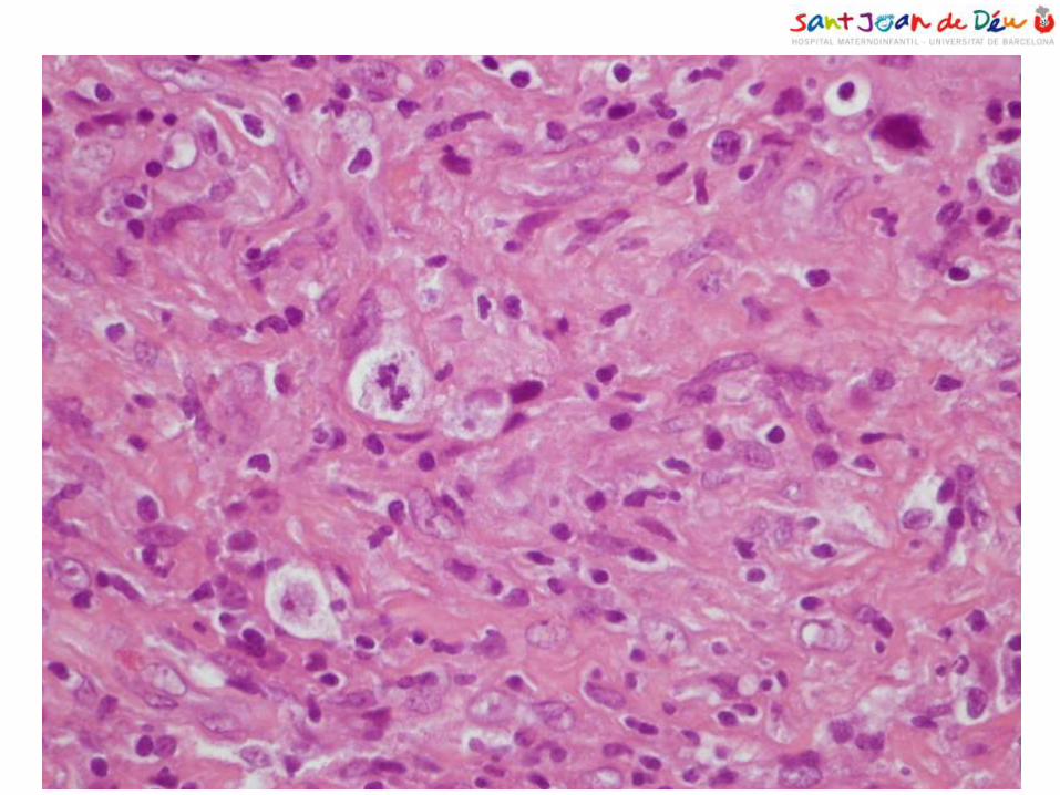
LINFOMA DE HODKGIN CLÁSICO,
TIPO ESCLEROSIS NODULAR
DIAGNÓSTICO DEFINITIVO:
CD30 CD15

• Dificultad del diagnóstico del linfoma de Hodgkin por PAAF es debido: – Grado de esclerosis – Distinta densidad de células de Reed-Sternberg – Dificultad en la identificación las células lacunares en los extendidos
citológicos. • Células de Reed-Sternberg-like en:
– Linfoma anaplásico – Melanoma metastásico – Carcinoma metastásico – Hiperplasia reactiva de ganglio linfático
En la edad pediátrica en el TUMOR MIOFIBROBLÁSTICO INFLAMATORIO

Linfoma de Hodgkin clásico, tipo esclerosis nodular
– Ganglios mediastino 80% – Pulmonar y/o esplénica 10%
– Ósea 5%
– Médula ósea 3%
– Hígado 2%
• Afectación primaria extranodal en un 1%.
• Afectación ósea:
– Diagnóstico <2%.
– Estadios avanzados 9–35% de los casos.
• Lesiones vertebrales 5’8% pacientes con LH

ONCOLOGY LETTERS 9: 677-680, 2015
Abstract. Hodgkin's lymphoma (HL) is one of the few adult
malignancies that most frequently presents with a progressive,
painless enlargement of the peripheral lymph nodes. A primary
osseous presentation of HL, without lymph node involvement,
is extremely rare. The present study describes a case of primary
multifocal osseous HL in a 22-year-old female. The patient
presented with pain in the lumbar-sacral-pelvic area and a
prolonged fever. Pathological examination led to a diagnosis
of primary multifocal osseous lymphoma, and the patient was
subsequently prescribed a course of Adriamycin, bleomycin,
vinblastine and dacarbazine (ABVD) chemotherapy. Following
this, the patient recovered with no pain or fever, and computed
tomography identified no further progression. The clinical,
radiological and histological features of HL are similar to
those of other medical conditions, such as tuberculosis and
eosinophilic granuloma. Furthermore, in rare cases, HL may
even occur in combination with multiple myeloma. This makes
it difficult to diagnose the condition, which often leads to a
delay in treatment. Clinicians should not ignore HL when it
manifests in the unusual primary osseous form.
Introduction
Hodgkin's lymphoma (HL) is one of the few adult malignan-
cies that is usually curable (1). HL frequently presents as a
progressive, painless enlargement of the peripheral lymph
nodes, particularly around the cervical region (2). Classical
HL is defined as a well‑established, proliferative neoplasm of
the lymph nodes that is composed of mononuclear Hodgkin
cells and multi-nucleated Reed-Sternberg (RS) cells in
variable proportions, along with neutrophils, eosinophils,
histiocytes, fibroblasts, collagen fibers, non-neoplastic
lymphocytes and plasma cells (3,4). Extra-nodal forms of HL
are rare, accounting for <1% of all HL cases (5,6). HL is a
systemic disease, with 10-20% of HL patients demonstrating
bone involvement throughout disease progression (7-9).
However, patients presenting with primary HL of the bone
are unusual (2,10). The histological, radiological and clinical
features of HL are similar to those of other medical conditions,
including tuberculosis and eosinophilic granuloma, and in
unusual cases, may even present in combination with multiple
myeloma (1,2,7,9,10). This makes HL difficult to diagnose and
often leads to delays in treatment.
The present study describes the case of a 22-year-old female
diagnosed with primary multifocal osseous HL. Informed
consent was provided by the patient and the patient's family.
Case report
A 22-year-old female patient presented to the Xiangya
Hospital (Central South University, Changsha, Hunan, China)
with a five‑month history of pain in the lumbar‑sacral‑pelvic
area, which gradually involved the left hip and subsequently
involved the left shoulder. The patient's symptoms were
accompanied by a prolonged fever (a recurrent fever type,
with a temperature of >39˚C for 1-2 days prior to returning
to normal and recurring repeatedly over the 5-month period),
night sweats and weight loss. The physical examination was
mostly normal, with normal appearance and range of motion.
However, pain and tenderness were evident upon percussion
of the lumbosacral area, left hip joint and left shoulder. The
patient was previously healthy, with no exposure to contami-
nated water or poisons, and no communicable diseases. The
patient's parents were also healthy, with no history of a heredi-
tary or similar disease in their families.
Upon presentation, the blood cell count revealed a persis-
tent elevation of white blood cells (>14.0x10⁹ g/l; normal
range, 4.0-10.0x10⁹ g/l), and a lowered hemoglobin level
(<90 g/l; normal range, 120-150 g/l). The platelet count was
also elevated (469.0x10⁹ g/l; normal range, 100-300x109 g/l)
and the mean corpuscular volume was <80 fl (normal range,
80‑100 fl). The mean corpuscular hemoglobin (MCH) level
was <27 pg (normal range, 27‑34 pg) and the MCH concen-
tration was <320 g/l (normal range, 320-360 g/l), which
suggested microcytic hypochromic anemia. The liver and
renal function tests were almost normal; the biochemical
evaluation demonstrated signs of inf lammation, with
levels of 130 mg/l C-reactive protein (CRP) (normal range,
0-8 mg/l) and 0.21 ng/ml procalcitonin (normal range,
<0.05 ng/ml), and a 75-mm/h erythrocyte sedimentation
rate (ESR) (normal range, 0-20 mm/h). Further serology
tests, including the human leukocyte antigen haplotype B27,
tuberculosis antibody, light chain protein and extractable
Unusual primary osseous Hodgkin's lymphoma: A case report
WEI LUO, FANGJIE ZHANG, JINPENG SUN and HONGBO HE
Department of Orthopedics, Xiangya Hospital, Central South University , Changsha, Hunan 410008, P.R. China
Received April 17, 2014; Accepted October 15, 2014
DOI: 10.3892/ol.2014.2724
Correspondence to: Dr Hongbo He, Department of Orthopaedics,
Xiangya Hospital, Central South University, 87 Xiangya Road,
Changsha, Hunan 410008, P.R. China
E-mail: [email protected]
Key words: Hodgkin's lymphoma, bone, diagnosis
50 casos 7 edad pediátrica

• VERTEBRA • Puede imitar procesos
inflamatorios

CONCLUSIONES:
• La extensión citológica en el acto intraoperatorio es de gran ayuda en la tipificación de las lesiones.
• Presencia de células “ganglion-like” y fondo inflamatorio en una lesión de partes blandas +/- afectación ósea en la edad pediátrica plantea el diagnostico diferencial entre:
– Tumor miofibroblástico inflamatorio
– Linfoma anaplásico
– Linfoma de Hodkgin

MUCHAS GRACIAS
AGRADECIMIENTOS: Dr. Emili Hinarejos. Servei de Diagnòstic per la Imatge. HSJD

• Un elevado porcentaje de linfomas no-Hodgkin se diagnostican y clasifican por PAAF.
• El uso de la PAAF es menos relevante y con un papel mas controvertido en el diagnostico del linfoma de Hodgkin.
• La sensibilidad de la PAAF es extremadamente variable, con rangos variables entre 30 y el 90% según las series publicadas en la literatura.
• La clasificación histológica en base a criterios citológicos únicamente es ampliamente debatido.

Pruebas de imagen:
Adenopatías en región: • Paraaorticas • Cadena iliaca común • Iliaca externa e interna • Inguinales
Tamaños entre 1 y 3’3 cm
![Convemar Power Point w[1].w](https://static.fdocuments.es/doc/165x107/5571fd784979599169992daa/convemar-power-point-w1w.jpg)