
Structure, dynamics and biochemical aspects of ... · structure, dynamics and biochemical aspects...
95
Structure, dynamics and biochemical aspects of phosphoranyl radicals : an ESR study Citation for published version (APA): Hamerlinck, J. H. H. (1982). Structure, dynamics and biochemical aspects of phosphoranyl radicals : an ESR study. Eindhoven: Technische Hogeschool Eindhoven. https://doi.org/10.6100/IR146278 DOI: 10.6100/IR146278 Document status and date: Published: 01/01/1982 Document Version: Publisher’s PDF, also known as Version of Record (includes final page, issue and volume numbers) Please check the document version of this publication: • A submitted manuscript is the version of the article upon submission and before peer-review. There can be important differences between the submitted version and the official published version of record. People interested in the research are advised to contact the author for the final version of the publication, or visit the DOI to the publisher's website. • The final author version and the galley proof are versions of the publication after peer review. • The final published version features the final layout of the paper including the volume, issue and page numbers. Link to publication General rights Copyright and moral rights for the publications made accessible in the public portal are retained by the authors and/or other copyright owners and it is a condition of accessing publications that users recognise and abide by the legal requirements associated with these rights. • Users may download and print one copy of any publication from the public portal for the purpose of private study or research. • You may not further distribute the material or use it for any profit-making activity or commercial gain • You may freely distribute the URL identifying the publication in the public portal. If the publication is distributed under the terms of Article 25fa of the Dutch Copyright Act, indicated by the “Taverne” license above, please follow below link for the End User Agreement: www.tue.nl/taverne Take down policy If you believe that this document breaches copyright please contact us at: [email protected] providing details and we will investigate your claim. Download date: 21. Jun. 2020
Transcript of Structure, dynamics and biochemical aspects of ... · structure, dynamics and biochemical aspects...
Structure, dynamics and biochemical aspects of phosphoranylradicals : an ESR studyCitation for published version (APA):Hamerlinck, J. H. H. (1982). Structure, dynamics and biochemical aspects of phosphoranyl radicals : an ESRstudy. Eindhoven: Technische Hogeschool Eindhoven. https://doi.org/10.6100/IR146278
DOI:10.6100/IR146278
Document status and date:Published: 01/01/1982
Document Version:Publisher’s PDF, also known as Version of Record (includes final page, issue and volume numbers)
Please check the document version of this publication:
• A submitted manuscript is the version of the article upon submission and before peer-review. There can beimportant differences between the submitted version and the official published version of record. Peopleinterested in the research are advised to contact the author for the final version of the publication, or visit theDOI to the publisher's website.• The final author version and the galley proof are versions of the publication after peer review.• The final published version features the final layout of the paper including the volume, issue and pagenumbers.Link to publication
General rightsCopyright and moral rights for the publications made accessible in the public portal are retained by the authors and/or other copyright ownersand it is a condition of accessing publications that users recognise and abide by the legal requirements associated with these rights.
• Users may download and print one copy of any publication from the public portal for the purpose of private study or research. • You may not further distribute the material or use it for any profit-making activity or commercial gain • You may freely distribute the URL identifying the publication in the public portal.
If the publication is distributed under the terms of Article 25fa of the Dutch Copyright Act, indicated by the “Taverne” license above, pleasefollow below link for the End User Agreement:www.tue.nl/taverne
Take down policyIf you believe that this document breaches copyright please contact us at:[email protected] details and we will investigate your claim.
Download date: 21. Jun. 2020
STRUCTURE, DYNAMICS AND BIOCHEMICALASPECTS OF PHOSPHORANYL RADICALS. AN ESR STUDY
STRUCTURE, DYNAMICS AND BIOCHEMICAL ASPECTS OF PHOSPHORANYL RADICALS. AN ESR STUDY
PROEFSCHRIFT
TER VERKRIJGING VAN DE GRAAD VAN DOCTOR IN DE TECHNISCHE WETENSCHAPPEN AAN DE TECHNISCHE HOGESCHOOL EINDHOVEN, OP GEZAG VAN DE RECTOR MAGNIFICUS, PROF. IR. J. ERKELENS, VOOR EEN COMMISSIE AANGEWEZEN DOOR HET COLLEGE VAN DEKANEN IN HET OPENBAAR TE VERDEDIGEN OP
VRIJDAG 14 MEI 1982 TE 16.00 UUR
DOOR
JOSEPHUS HELENA HUBERTUS HAMERLINCK
GEBOREN TE BANDUNG (INDONESIË)
DIT PROEFSCHRIFT IS GOEDGEKEURD DOOR
DE PROMOTOREN
PROF. DR. H.M. BUCK
EN
PROF. DR. TH.J. DE BOER
Chapter I
Chapter II
Chapter III
Chapter IV
Contents
INTRODUCTION
I.1.
I. 2.
I. 3.
I.4.
Scope of this thesis
The structure of penta-coor
dinated phosphorus compounds
Phosphoranyl radicals
ESR of trapped radicals in
solids
PHOSPHORANYL RADICALS IN A TRIGONAL
BIPYRAMIDAL CONFIGURATION (TBP)
7
7 8
10
12
21
II.1. Phosphorus in a TBP structure 21
with the unpaired electron
located in an equatorial
position (TBP-e)
II.2. Phosphorus in a TBP structure 30 with the unpaired electron
located in an apical position
(TBP-a)
II.3. TBP-e and TBP-a isomers
II.4. Discussion
PHOSPHORANYL RADICALS IN AN OCTA-
HEDRAL CONFIGURATION
III. 1. Phosphorus in an octahedral VI P geometry with the unpaired
electron in an axial position
III. 2. Discussion
INTRAMOLECULAR LIGAND REORGANISATION
IN PHOSPHORANYL RADICALS
IV.1. Pseudo rotating TBP-e
35 44
49
49
53
56
56
IV.2. X-ray structure determination 61 IV.3. Discussion 64
Chapter V INTRAMOLECULAR ELECTRON TRANSFER IN
PHOSPHORANYL RADICALS
V.1. Introduetion
V. 2. Results and diseussion
Chapter VI BIOCHEMICAL ASPECTS
VI.1. Introduetion
VI.2. Results and diseussion
Appendix
Summary
Samenvatting
Curriculum vitae
Dank-woord
68
68 68
75 75 76
80
85
87
89
90
CHAPTER I
Introduetion
I. 1 of this thesis
Much recent research concerns the effects of ionizing
radiation in living cells. It is found that strand breaks
in the DNA chains are induced 1
• The radical products in
this process have been studied by Electron Spin Resonance
(ESR). On comparison with model ESR studies on irradiated
nucleosides and nucleotides a number of mechanisms for
strand break in DNA has been proposed. However, the in
volvement of phosphorus centered radicals in reduction
processes upon irradiation has not been recognized,
although there is an increasing body of evidence that the
conformational properties of DNA are directly related to
the presence of phosphorus in the framework 2• Therefore
the study of radicals in which the unpaired electron is
centered on phosphorus, i.e. PV phosphoranyl radicals is
appropriate and is the subject of this thesis.
Moreover the determination of the electronic structure of
these radicals and their dynamic behaviour enables to test
the different predictions of rivaling theories adequately
by experiment. In spite of the large number of investi
gations concerning phosphoranyl radicals, a satisfactory
description of their structure could not be given hitherto,
since the majority of these studies concerns isotropie
hyperfine splittings which are not a reliable guide to the
overall electron spin distribution for these radicals. The
aim of this thesis is to establish the possible structures
of phosphoranyl radicals in order to understand the
behaviour of these intermediates in radiation damage
7
processes, in particular in DNA.
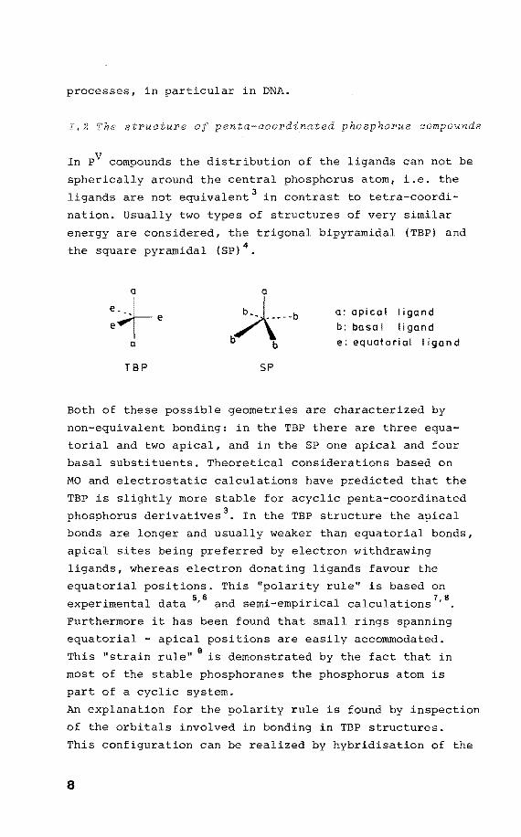
I.2 The structure of penta-coordinated p hor•us compounds
In PV compounds the distribution of the ligands can not be
spherically around the central phosphorus atom, i.e. the
ligands are not equivalent 3 in contrast to tetra-coordi
nation. Usually two types of structures of very similar
energy are considered, the bipyramidal (TBP) and
the square pyramidal (SP) 4•
a
b~:--b TBP SP
a: apical ligand
b:basal ligand
e: equotoriol ligond
Bath of these possible geometries are characterized by
non-equivalent bonding: in the TBP there are three equa
torial and two apical, and in the SP one apical and four
basal substituents. Theoretical considerations based on
MO and electrastatic calculations have predicted that the
TBP is slightly more stable for ie penta-coordinated
phosphorus derivatives 3• In the TBP structure the apical
bands are longer and usually weaker than equatorial bands,
apical sites being by electron withdrawing
ligands, whereas electron donating ligands favour the
equatorial positions. This "polarity rule" is based on . l d 56 d . . . 1 1 1 . 7 ' 8
exper~menta ata ' an sem~-emp~r~ca ca cu at~ons .
Purthermare it has been found that small rings spanning
equatorial - positions are easily accommodated.
This "strain rule" 9
is demonstrated by the fact that in
most of the stable phosphoranes the phosphorus atom is
part of a cyclic system.
An explanation for the polarity rule is found by inspeetion
of the orbitals involved in bonding in TBP structures.
This contiguration can be realized by hybridisation of the
8
P and d 2 orhitals for the apical honds, comhined with z z -three sp2 orhitals in the equatorial plane, resulting in
an sp3d hyhridisation 10
• The diffuse d 2 orhital accounts z for the elangation of the apical honds, favouring the
accomodation of electron withdrawing ligands, whereas the
shorter equatorial orhitals favour donation of electrans
from the ligands.
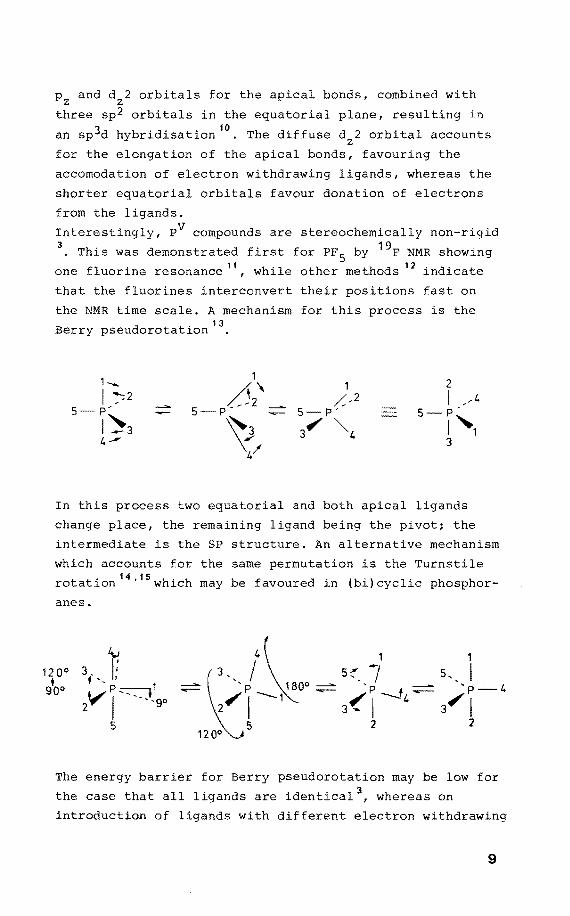
Interestingly, PV compounds are stereochemically non-rigid 3
• This was demonstrated first for PF 5 hy 19F NMR showing
one fluorine resonance 11, while other methods 12 indicate
that the fluorines interconvert their positions fast on
the NMR time scale. A mechanism for this process is the
Berry pseudorotatien 13
•
1-... 1 2 /52\
1 I /2 ,/-2 I ,4
5 p' 5 p- 5 P' 5-P'
1~3 ~ 3 ". ""'4 ,,1 4-' 3
4/
In this process two equatorial and hoth apical ligands
change place, the remaining ligand heing the pivot; the
intermediate is the SP structure. An alternative mechanism
which accounts for the same permutation is the Turnstile
rotation 14
'15 which may he favoured in (hi)cyclic phosphor
anes.
12 0° • goo
t 3 f '' 1
2 V ~ -- -. J. go
5
The energy harrier for Berry pseudorotation may he low for
the case that all ligands are identical 3 , whereas on
introduetion of ligands with different electron withdrawing
9
character the permutation is hindered. The same applies
for the presence of small rings7
•
It has been established by X-ray diffraction analysis that V in the solid state the structure of P compounds is
distorted more or less from an ideal TBP towards an SP
geometry4
• This depends on particular arrangment of ligands
differing in electronegativity or inclusion of phosphorus
in small four- or five-membered rings leading to stahili
zation of the SP.
No te
Since it will be demonstrated in chapter II.3 that a re
markable degree of s-character in the apical bands is
present, it is noted that the description À(sp 2 )+p(pd) is
not completely satisfying. However, this s-character is
higher for equatorial than for apical bands, accounting
for the observed differences in apical and equatorial
sites in the TBP structure.
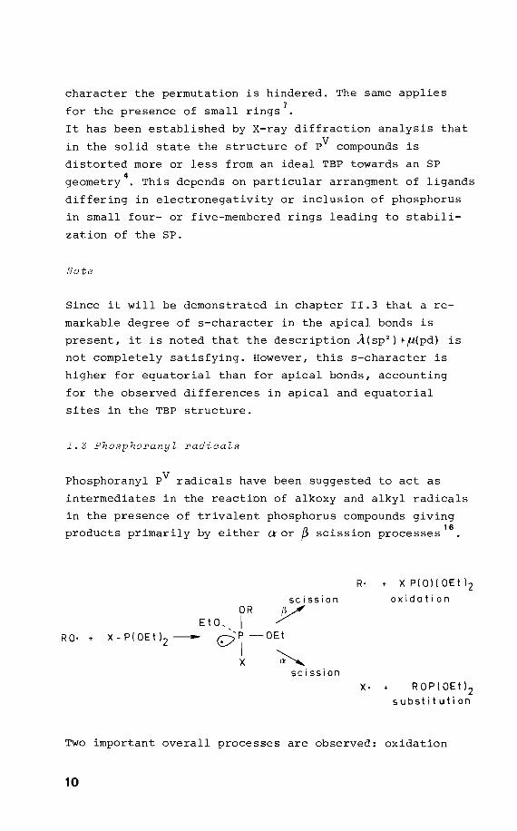
I.3 Phosphoranyl radicals
Phosphoranyl PV radicals have been suggested to act as
intermediates in the reaction of alkoxy and alkyl radicals
in the presence of trivalent phosphorus compounds giving
products primarily by either aor ~ scission processes16
•
RO· •
OR Et 0- I
scission y X-P(0Etl 2 -- Q~-OEt
x ~ scission
R· + XP(O)(OEtl2
oxidation
X· ROP(0Etl 2 substitution
Two important overall processes are observed: oxidation
10
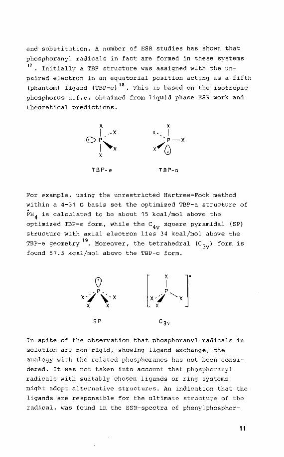
and substitution. A number of ESR studies has shown that
phosphoranyl radicals in fact are formed in these systems 17
• Initially a TBP structure was assigned with the un
paired electron in an equatorial position acting as a fifth
(phantom) ligand (TBP-e) 18• This is based on the isotropie
phosphorus h.f.c. obtained from liquid phase ESR work and
theoretical predictions.
TB P- e TB P-a
For example, using the unrestricted Hartree-Feek method
within a 4-31 G basis set the optimized TBP-a structure of
PH 4 is calculated to be about 15 kcal/mol above the
optimized TBP-e form, while the c4v square pyramidal {SP)
structure with axial electron lies 34 kcal/mol above the 19
TBP-e geometry • Moreover, the tetrahedral (c 3v) form is
found 57.5 kcal/mol above the TBP-e form.
SP
In spite of the observation that phosphoranyl radicals in
solution are non-rigid, showing ligand exchange, the
analogy with the related phosphoranes has not been consi
dered. It was not taken into account that phosphoranyl
radicals with suitably chosen ligands or ring systems
might adopt alternative structures. An indication that the
ligands are responsible for the ultimate structure of the
radical, was found in the ESR-spectra of phenylphosphor-
11
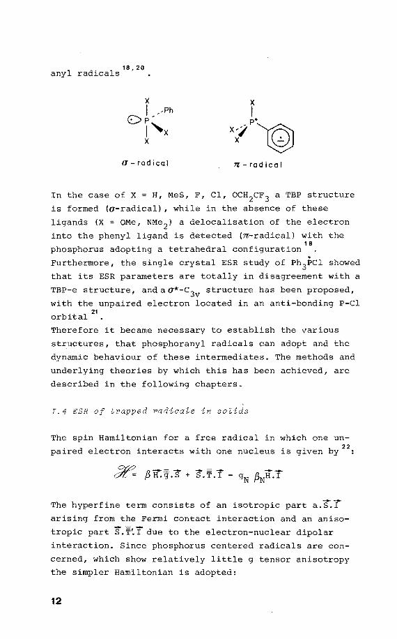
18' 20 anyl radicals •
a-radical 7l'- rad i cal
In the case of X = H, MeS, F, Cl, OCH2cF3 a TBP structure
is formed (a-radical) 1 while in the absence of these
ligands (X OMe, NMe 2 ) a delocalisation of the electron
into the ligand is detected (n-radical) with the 18
phosphorus adopting a tetrahedral configuration
Furthermore, the single crystal ESR study of Ph3PC1 showed
that its ESR parameters are totally in di with a
TBP-e structure, and a a*-c3v structure has been proposed,
with the unpaired electron located in an anti-bonding P-Cl
orbital 21
•
Therefore it became necessary to establish the various
structures, that phosphoranyl radicals can adopt and the
dynamic behaviour of these intermediates. The methods and
underlying theories by which this has been achieved, are
described in the following chapters.
I.4 ESR of radica~s in so~ids
The spin Hamiltonian for a free radical in which one un
paired electron interacts with one nucleus is given by22
:
The hyperfine term consists of an isotropie part a.if.j[
arising from the Fermi contact interaction and an anisa
tropie part S .. j[due to the electron-nuclear dipolar
interaction. Since phosphorus centered radicals are con
cerned, which show relatively little g tensor anisotropy
the simpler Hamiltonian is adopted:
12
in which g is a scalar.
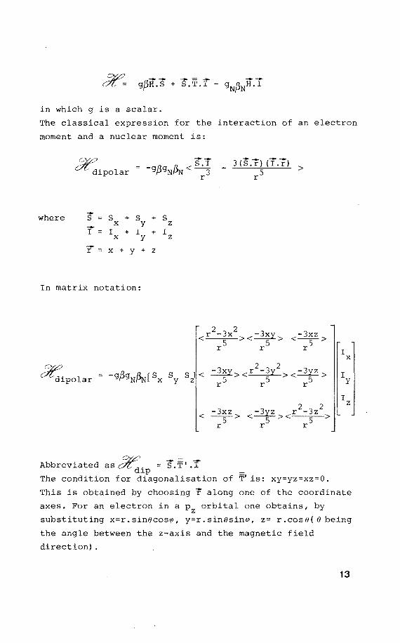
The classica! expression for the interaction of an electron
moment and a nuclear moment is:
where
CZ2? --Ol , = -g/Zg fl.. < S I dipolar fJ Nt-'N
s = s x I I
x
+ s y
+ I y
+ s z
+ I z
x + y + z
In matrix notation:
< >
dE: di po lar s y
2 2 S l < -3xy r -3y -3yz
z 5 >< 5 >< 5 > r r r
< >
Abbreviated as ~d. = .T' :r lp The condition for diagonalisation of T' is: xy=yz=xz=O.
I x
I y
I z
This is obtained by choosing r along one of the coordinate
axes. For an electron in a Pz orbital one obtains, by
substituting x=r.sinOcos~, y=r.sinOsin~, z= r.cosO( 0 being
the angle between the z-axis and the magnetic field
direction) .
13
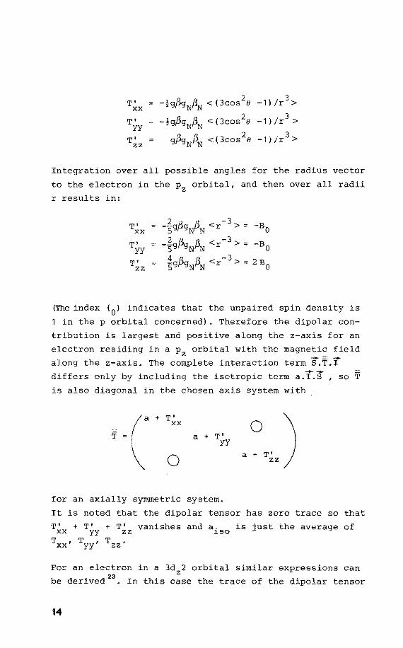
-!gpgN~ <(3cos2o 3
T' -1) /r > XX
-!gPgNpN <(3cos2o 3
T' -1) /r > yy
T' zz gpgNpN <(3cos2o -1)/r3 >
Integration over all possible angles for the radius vector
to the electron in the Pz orbital, and then over all radii
r results in:
(The index (0
) indicates that the unpaired spin density is
1 in the p orbital concerned). Therefore the dipolar con-
tribution is and positive along the z-axis for an
electron residing in a Pz orbital with the magne~i~ field
along the z-axis. The complete interaction term S.T.
differs only by including the isotropie term a.I.S , so T
is also diagonal in the chosen axis system with
T' XX
0
a + T' yy
for an axially symmetrie system.
a ~T') zz
It is noted that the dipolar tensor has zero trace so that
T~x + T~y + T~z vanishes and aiso is just the average of
Txx' Tyy' Tzz
For an electron in a 3dz2 orbital similar expressions can
be derived23
• In this case the trace of the dipolar tensor
14
T consists of:
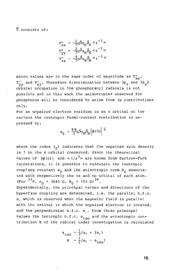
which values are in the same order of magnitude as T' , XX
T' and T' . Therefore discrimination between 3p and 3d 2 yy zz z z orbital accupation in the phosphoranyl radicals is not
possible and in this work the anisotropies observed for
phosphorus will be considered to arise from 3p contributions
only.
For an unpaired electron residing in an s orbital on the
nucleus the isotropie Fermi-contact contribution is ex
pressed by:
where the index (0 ) indicates that the unpaired spin density
is 1 in the s orbital concerned. Since the theoretical
values of ll/J(O) I and < 1/r3 > are known from Hartree-Fock
calculations, it is possible to calculate the isotropie
coupling constant a0
and the anisotropic term a0
associa
ted with respectively the ns and np orbital of each atom. 31 24
(For P, a 0 = 3640 G, B0 = 103 G) .
Experimentally, the principal values and directions of the
hyperfine coupl are determined, i.e. the parallel h.f.c.
a11 which is observed when the magnetic field is parallel
with the orbital in which the unpaired electron is located,
and the perpendicular h.f.c. From these principal
values the isotropie h.f.c. a. and the anisatrapie con-~so
tribution B of the radical under investigation is calculated
a. ~so
B
15
These values make it possible to give (after comparison
with the atomie constants) an estimate of the s character:
ps = a. !a0 ~so
and the p character:
of the atomie orbital which contains the unpaired electron.
Moreover, the direction of this orbital is obtained.
From an oriënted radical in a solid matrix (i.e. a single
crystal) all the ESR parameters, a11
, a.L, g11 and g.L can be
derived as well as their directions on rotatien around
suitably chosen axes. However, extracting the principal
directions and values of a and g from measurements on a
range of arbitrary orientations may be quite laborieus.
Fortunately, the ESR spectrum of a powdered sample con
tains also the anisatrapie information, because this
spectrum does not concern a motionally averaged system,
but results from randomly oriented crystallites 23• Since
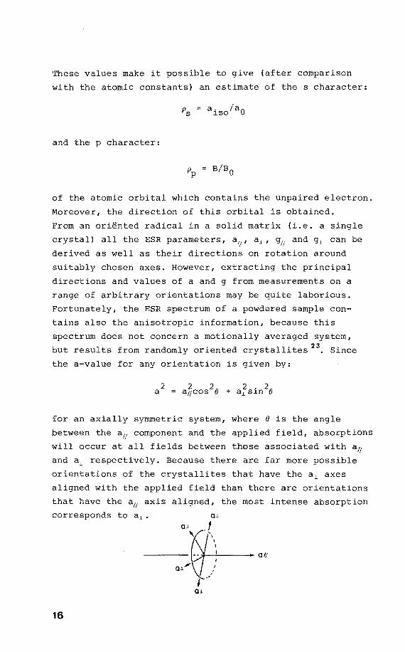
the a-value for any orientation is given by:
for an axially symmetrie system, where e is the angle
between the aH component and the applied field, absorptions
will occur at all fields between those associated with aH
and a.L respectively. Because there are far more possible
orientations of the crystallites that have the a.L axes
aligned with the applied field than there are orientations
that have the aH axis aligned, the most intense absarptien
corresponds to a.L. 0.1
16
_·"r;I: -.,, 0.1~~
# ,.' O.L
0
d
oj_
~ g.J.
0 I( g;,
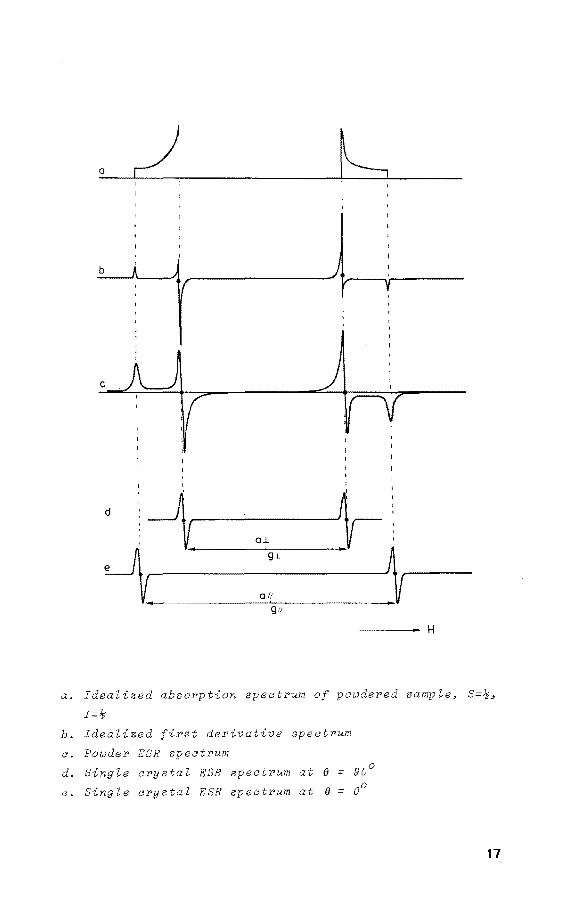
a. Idealized absorption spectrum
I=~
~ H
powdered sample, S=~,
b. Idealized t derivative spectrum
a. Powder ESR spectrum
d. Single crystaZ ESR spectrum at (} = 90°
e. Sing Ze crystaZ ESR spectrum at (} = 00
17
This results in the following absorption spectrum which is
converted into the first derivate spectrum. Therefore the
powder ESR spectrum serves as a useful check on the
validity of the parameters obtained from the ESR single
crystal experiments. It is emphasized that the single
crystal experiments are necessary to obtain the directional
information. Moreover, rather complex ESR spectra may
arise from powdered samples because of overlapping features
generated by aH and a~ and because of superimposed h.f.c.
due to atoms linked to the central atom. In contrast, in
single crystals the parallel and perpendicular features
are separated on rotation.
On this basis a methad of generating phosphorus centered
radicals is chosen which consists in rupture of a labile
P-H bond or electron addition at phosphorus initiated by
X-irradiation or UV laser irradiation in a single crystal
of which the molecular structure is known from X-ray dif
fraction analysis. Combination of the ESR directional
information and the orientation of the molecules in the
unit cell then provides a reliable determination of the
structure of the radical.
18
References and notes
1. M. Dizdaroglu, c. von Sonntag, and D. Schulte-Frohlinde,
J. Am. Chem. Soc . , 1 9 7 5 , 9 7 , 2 2 7 7 .
2. A.M.C.F. Castelijns, D. van Aken, P. Schipper, J.J.C.
van Lier, and H.M. Buck, Reel. Trav. Chim. Pays-Bas,
1980, 99, 380.
3. R. Luckenbach, "Dynamic Stereochemistry of Pentaco
ordinated Phosphorus and Related Elements", G. Thieme,
Stuttgart, 1973.
4. R.R. Holmes, Acc. Chem. Res., 1979, 12, 257.
5. E.L. Muetterties, W. Mahler and R. Schmutzler, Inorg.
Chem., 1963,2, 613.
6. E.L. Muetterties, K.J. Packer and R. Schmutzler, Inorg.
Chem., 1964, 3, 1298.
7. D. Marquarding, F. Ramirez, !. Ugi, and P. Gillespie,
Angew. Chem., 1973, 85, 99.
8. F. Keiland W. Kutzelnigg, J. Am. Chem. Soc., 1975,
97, 3623.
9. F.H. Westheimer, Acc. Chem. Res., 1968, 1, 70.
10. R.F. Hudson and M. Green, Angew. Chem., 1963, 75, 47.
11. H.S. Gutowski, D.H. McCall, and C.P. Slichter, J. Chem.
Phys., 1953, 21, 279.
12. H.S. Gutowski and A.D. Liehr, J. Chem. Phys., 1953,
20, 1652.
13. R.S. Berry, J. Chem. Phys., 1960, 32, 933.
14. F. Ramirez, S. Pfohl, E.A. Tsolis, J.F. Pilot, C.P.
Smith, I. Ugi, D. Jl.1arquarding, P. Gillespie, and P.
Hoffmann, Phosphorus, 1971, 1, 1.
15. I. Ugi, D. Marquarding, H. Klusacek, P. Gillespie, and
F. Ramirez, Acc. Chem. Res., 1971, 4, 288.
16. W.G. Bentrude, W. Del , N.A. Johnson, f-1. Murakami,
K. Nishikida, and H.W. Tan, J. Am. Chem. Soc., 1977,
99, 4383.
17. P.J. Krusic, W. Mahler, and J.K. Kochi, J. Am. Chem.
Soc., 1972, 94, 6.033.
18. A.G. Davies, M.J. Parrott, and B.P. Roberts, J. Chem.
Soc., Chem. Commun., 1974, 973.
19
19. J.M. Howell and J.F. Olsen, J. Am. Chem. Soc., 1976,
98, 7119.
20. G. Boekestein, E.H.J.M. Jansen, and H.M. Buck, J. Chem.
Soc. , Chem. Commun. , 1 9 7 4 , 11 8 .
21. T. Berclaz, M. Geoffroy, and E.A.C. Lucken, Chem.
Phys. Lett., 1975, 36, 677.
22. A. Carrington and A.D. McLachlan, "Introduction to
Magnetic Resonance", Harper and Row, New York, 1969.
23. R.S. Drago, "Physical Methods in Chemistry", W.B.
Saunders, London, 1977.
24. P.W. Atkins and M.C.R. Symons, "The Structure of
Inorganic Radicals", Elsevier, Amsterdam, 1967.
20
CHAPTER 11
Phosphoranyl radicals in a trigonal
bipyramidal contiguration (TBP)
II.l Phosphorus in a TBP structure with the unpaired
electron Zoaated in an equatoriaZ position (TBP-e)
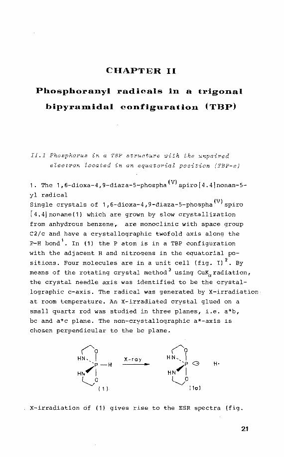
1. The 1,6-dioxa-4,9-diaza-5-phospha(V)spiro[4.4]nonan-5-
yl radical
Single crystals of 1,6-dioxa-4,9-diaza-5-phospha(V)spiro
[4.4]nonane(1) which are grown by slow crystallization
from anhydrous benzene, are mo:noclinic with space group
C2/c and have a crystallographic twofold axis along the
P-H bond1
• In (1) the P atom is in a TBP configuration
with the adjacent H and nitrogens in the equatorial po
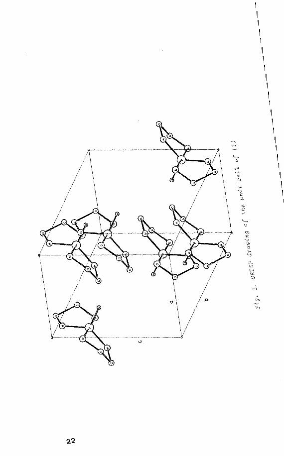
sitions. Four molecules are in a unit cell (fig. I) 2• By
means of the rotating crystal method 3 using CuKaradiation,
the crystal needle axis was identified to be the crystal
lographic c-axis. The radical was generated by X-irradiation
at room temperature. An X-irradiated crystal glued on a
small quartz rod was studied in three planes, i.e. a*b,
bc and a*c plane. The non-crystallographic a*-axis is
chosen perpendicular to the bc plane.
X ray H·
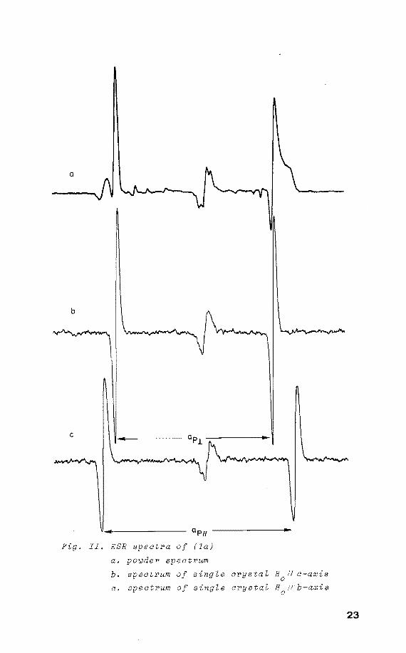
X-irradiation öf (1) gives rise to the ESR spectra (fig.
21
1 1 1 1 1 1 1 1 1 1 1 1 1 1 1 1 1 1 1 1 1
0 /~ . I
----~". . ----~-- I z û -- ___ I
..,
0
1 1 1 1 1 1 1 1 1 1 1 1 1 1 1 1 1 1 1 1 1 1 1 1 1 1 1 1 1
a
a P 11 ______ ____,_
. II. ESR spectra of (la)
a. powder spectrum
b. spectrum of single crystal H0
11 c-axis
c. spectrum of single crystal H Vb-axis 0
23
900
t Op (G)
850
800
750
800
750
750
24
. III.a.
oo
'* . 0-0XIS
Fig. III.b.
~0 I . C -QXJS
. III.c.
b-axis
180°
I* . 0-0XIS
180° I . C -QXI S
• 1 I I I I 1 a a • a 1 1 I I I I • I
oo I * ' 0-0XIS c-axis
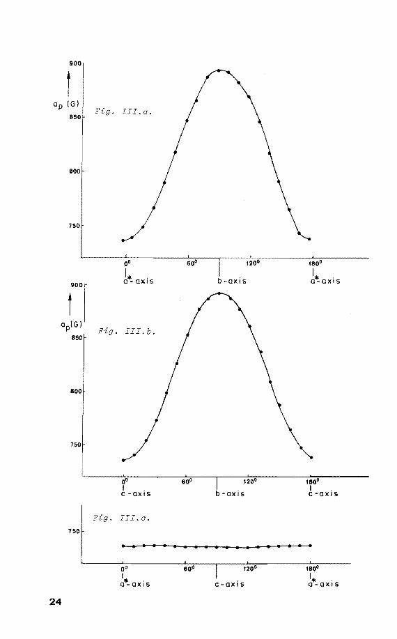
II) ascribed to (1a): only one 31 P doublet is observed, no
additional couplings have been detected 4• The angular
variations are shown in figure III a,b,c.
Rotating the single crystal of (1) around the crystallo
graphic c-axis reveals the maximum anisotropy ap to be I!
perpendicular to this axis. Similarly for a rotatien about
the a*-axis it was found that ap is perpendicular to this 1!
a*-axis. This corresponds with the direction of the P-H
linkage in its precursor as determined by x-ray diffraction
analysis (fig. I). From figure II it is seen that the
h.f.c. is axially symmetrie. The same applies to the g-ten
sor: g11 = 1.988 and cri = 2.005. It was found that x
irradiation and UV laser irradiation ( À= 248 nm) at 77 K
of a powdered sample of (1) gives rise to the same radical.
The parameters derived from the powder spectra agreed
nicely with those obtained from the single crystal measure
ments, only the directional information is lacking.
The orbital in which the unpaired electron resides, is
directed along the initial P-H linkage. Obviously the
radical (1a) is generated by P-H bond cleavage. Therefore
the structure of (la) has to be described as a nearly
perfect TBP with the unpaired electron and the nitrogen
ligands in the equatorial positions and the oxygen
ligands in the apical positions. From the anisotropic
values of the phosphorus h.f.c. ap~ = 893 G, ap-L 735 G,
the ap . is calculated 5 to be 788 G. These values in--lso dicate a P3s spin density (P~s) of 0.22, and a 3p spin
density (P~p) of 0.49, resulting in a total spin density
of 0.71 on phosphorus in an sp2 hybrid orbital. From the
line width the 14N coupling was estimated to be less than
5 G. The remaining spin density is distributed over the
apical oxygen ligands.
2. The 1,6-dioxa-4,9-diaza-2,3,7,8-dibenzo-5-phospha(V)
spiro [ 4. 4]nona-2, 7-dien-5-yl radical
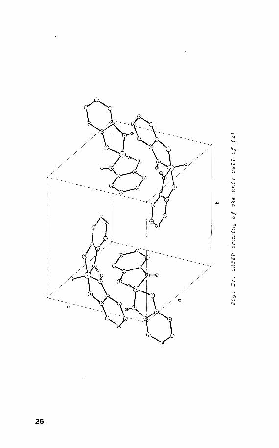
Single crystals of 1,6-dioxa-4,9-diaza-2,3,7,8-dibenzo-
5-phospha(V)spiro[4.4]nona-2,7-dien (2) were prepared by 1
slow crystallization from anhydrous benzene . Compound (2)
25
26
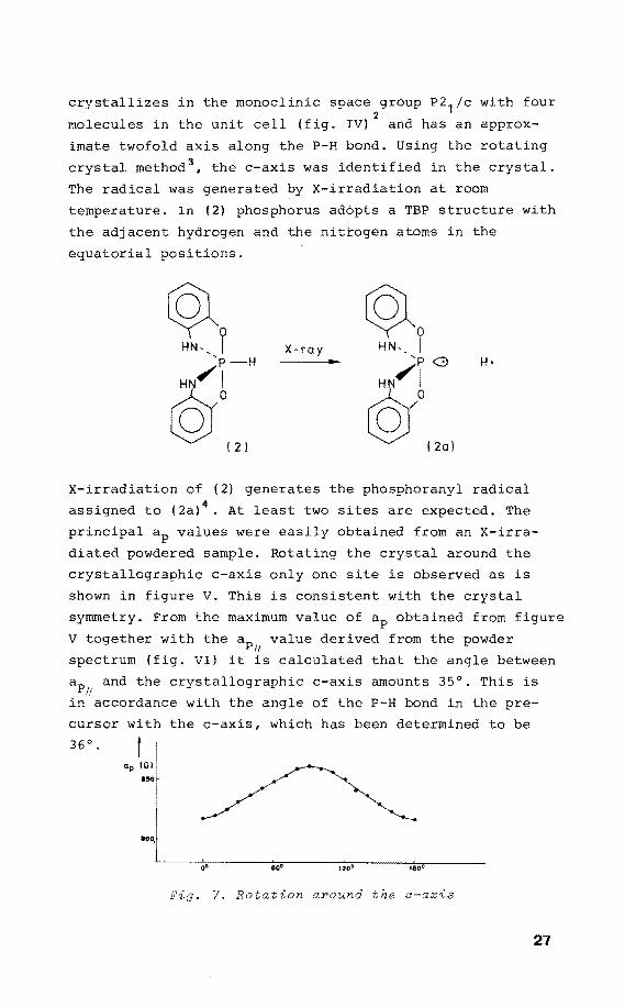
crystallizes in the monoclinic space group P2 1/c with four 2
molecules in the unit cell {fig. IV) and has an approx-
imate twofold axis along the P-H bond. Using the rotating
crystal method 3, the c-axis was identified in the crystal.
The radical was generated by X-irradiation at room
temperature. In {2) phosphorus adópts a TBP structure with
the adjacent hydrogen and the nittogen atoms in the
equatorial positions.
X ray ~0
H N-' I 'p G
HN~I
rQ{ l2a)
X-irradiation of (2) generates the phosphoranyl radical
assigned to (2a) 4• At least two sites are expected. The
principal ap values were easily obtained from an X-irra
diated powdered sample. Rotating the crystal around the
crystallographic c-axis only one site is observed as is
shown in figure V. This is consistent with the crystal
symmetry. From the maximum value of ap obtained from figure
V together with the ap value derived from the powder 11
spectrum (fig. VI) it is calculated that the angle between
ap and the crystallographic c-axis amounts 35°. This is I!
in accordance with the angle of the P-H bond in the pre-
cursor with the c-axis, which has been determined to be
36 0. t I ap IGI: ... ,
i
~i L_ ____ o~·.--------~eo~.------~,~.o7•------~,~ao~·--------
Fig. V. Rotation around the o-axis
27
a
b
c
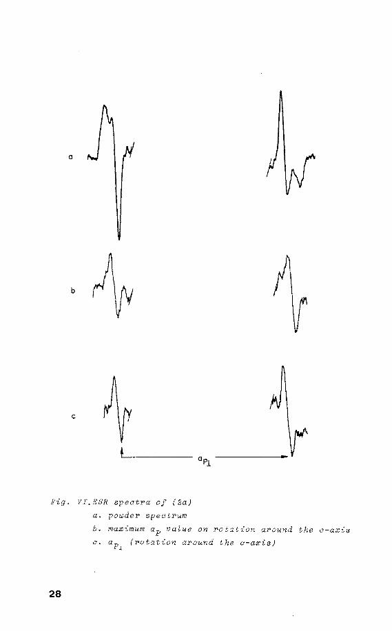
Fig. VI.ESR spectra of (2a)
a. powder spectrum
b. maximum ap value on rotation around the c-axis
c. (rotation around the a-axis)
28
In this radical the unpaired electron is directed along
the initial P-H bond. Therefore (2a) possesses a TBP struc
ture with the unpaired electron and the nitrogen atoms in
equatorial positions, and the oxygens in apical positions.
The axially symmetrie 31 P h.f.c., ap = 960 G, ap = 808 G, u 1
give ap . = 859 G and the anisotropy is 50 G. This in--lso P P
dicates a p3
= 0.24 and p3
= 0.47, resulting in 0.71 s p 2 5
spin density on phosphorus in an sp orbital . From the
line width it was estimated that aN<S G (not resolved).
The remaining spin density is distributed over the apical
ligands. The g values are gu = 1.983 and g1
= 2.008.
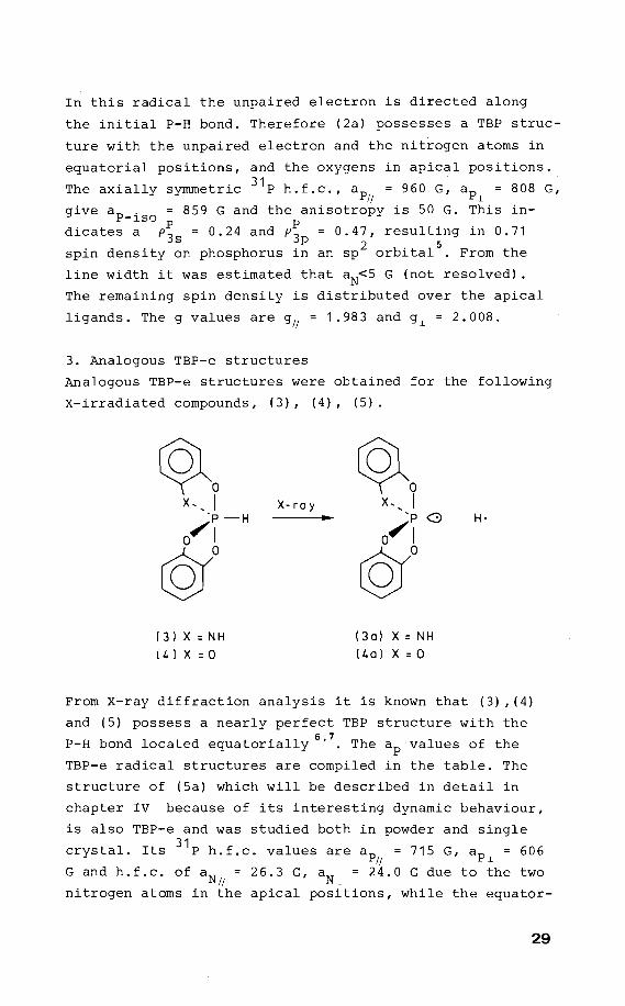
3. Analogous TBP-e structures
Analogous TBP-e structures were obtained for the following
X-irradiated compounds, (3), (4), (5).
~0 ~0 x,_ I X- ra y x,, I
·p-H 'p G H·
a~ I o~l ©a ©0 ( 3) X = NH ( 3a) X = NH
[ 4) x = 0 {4a) x = 0
From X-ray diffraction analysis it is known that (3) ,(4)
and (5) possess a nearly perfect TBP structure with the
P-H bond located equatorially6
'7
• The ap values of the
TBP-e radical structures are compiled in the table. The
structure of (Sa) which will be described in detail in
chapter IV because of its interesting dynamic behaviour,
is also TBP-e and was studied both in powder and single
crystal. Its
G and h.f.c.
31 P h.f.c. values are ap
/j 715 G, ap
1 = 606
of aN 26.3 G, aN = 24.0 G due to the two u 1
nitrogen atoms in the apical positions, while the equator-
29
ial nitrogens did nothave resolved h.f.c. (< 5 G from
linewidth).
('N) ~N) N-' I X- ray N'' I
<crJ ~-1J H·
( 5 J (Sa)
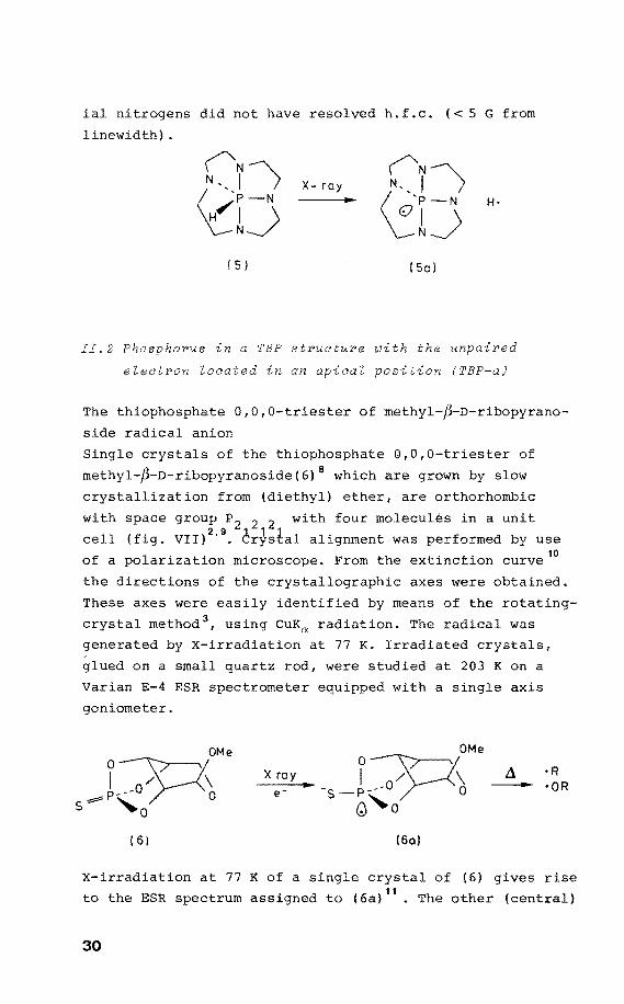
II.2 Phosphorus in a TBP strueture ~ith the unpaired
e~eetron ~oeated in an apiea~ tion (TBP-a)
The thiophosphate 0,0,0-triester of methyl-~-D-ribopyrano
side radical anion
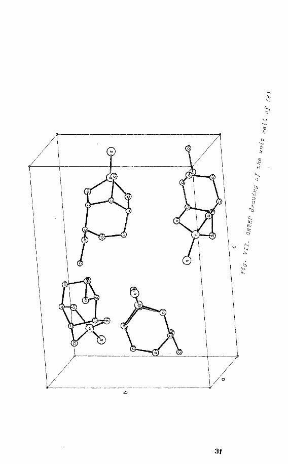
Single crystals of the thiophosphate 0,0,0-triester of
methyl-~-D-ribopyranoside(6) 8 which are grown by slow
crystallization from (diethyl) ether, are orthorhombic
with space group P2 2 2 with four molecules in a unit
cell (fig. VII)2
'9
• èr~stal alignment was performed by use
of a polarization microscope. From the extinction curve 10
the directions of the crystallographic axes were obtained.
These axes were easily identified by means of the rotating
crystal method 3, using CuKa radiation. The radical was
generated by X-irradiation at 77 K. Irradiated crystals,
glued on a small guartz rod, were studied at 203 K on a
Varian E-4 ESR spectrometer equipped with a single axis
goniometer.
O~OMe I .o/ \
~p~ 0 s ,..0
( 6)
X ray
e-
(6a}
---X-irradiation at 77 K of a single crystal of (6) gives rise
to the ESR spectrum assigned to ( 6a) 11
• The other (centra!)
30
31
"" ~ .,., ::s ':j
{; ~
~ u ::;
Ä
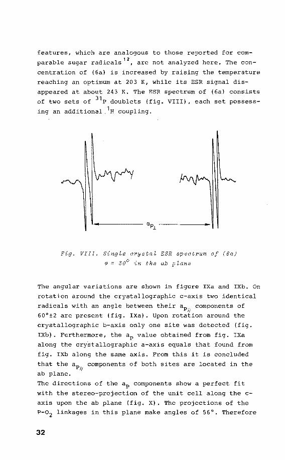
features, which are analogous to those reported for com
parable sugar radicals 12
, are not analyzed here. The con
centration of (6a) is increased by raising the temperature
reaching an optimum at 203 K, while its ESR dis-
appeared at about 243 K. The ESR spectrum of (6a) consists
of two sets of 31 P doublets (fig. VIII), each set possess
ing an additional, 1H coupling.
Fig. VIII. Single orystal ESR spectrum of (6a)
~ 30° in the ab plane
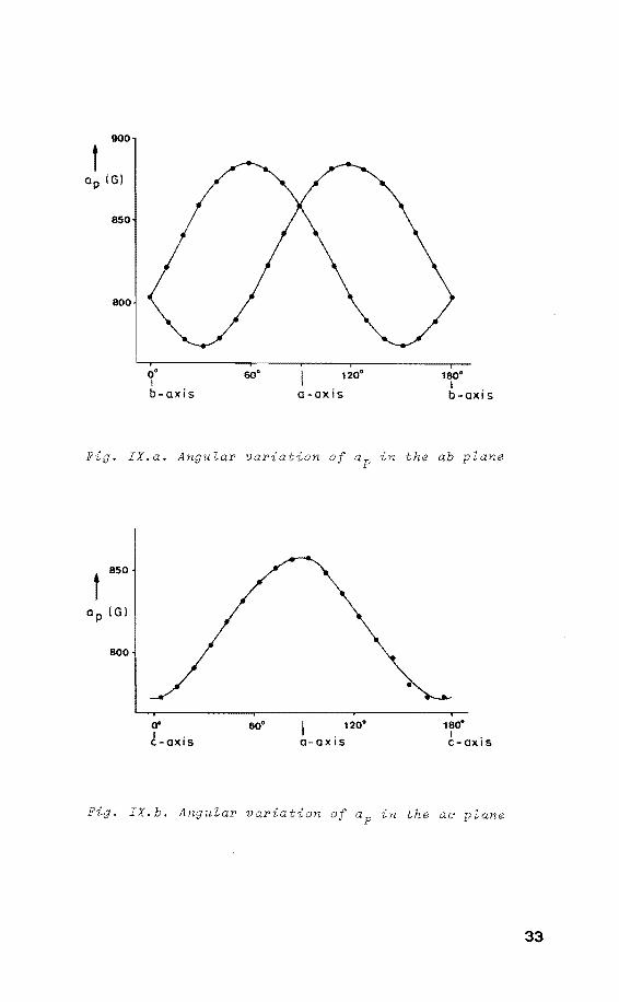
The angular variations are shown in figure IXa and IXb. On
rotation around the crystallographic c-axis two identical
radicals with an between their ap components of ,, 60°±2 are present IXa). Upon rotation around the
crystallographic b-axis only one site was detected
IXb). Furthermore, the ap value obtained from fig. IXa
along the crystallographic a-axis equals that found from
fig. IXb along the same axis. From this it is concluded
that the ap components of both sites are located in the I/
ab plane.
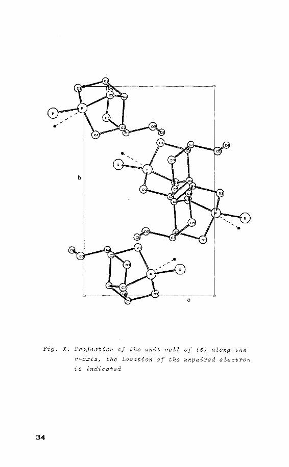
The directions of the ap components show a perfect fit
with the stereo-projection of the unit cell along the c
axis upon the ab plane (fig. X). The projections of the
P-02 linkages in this plane make angles of 56°. Therefore
32
900
t ap IGI
850
o• I b-axis
60. 120°
a -ax is
180° I b-axis
. IX.a. Angular variation of ap in the ab plane
t 850
Op IGl
800
00 I . C-QXIS
120"
o- a x is
•
180° I . c-axt s
. IX.b. Angular variation of ap in the ae plane
33
8
Fig. X. Projection of the unit cell of (6) along the
c-axis~ the location of the unpaired electron
is indicated
34
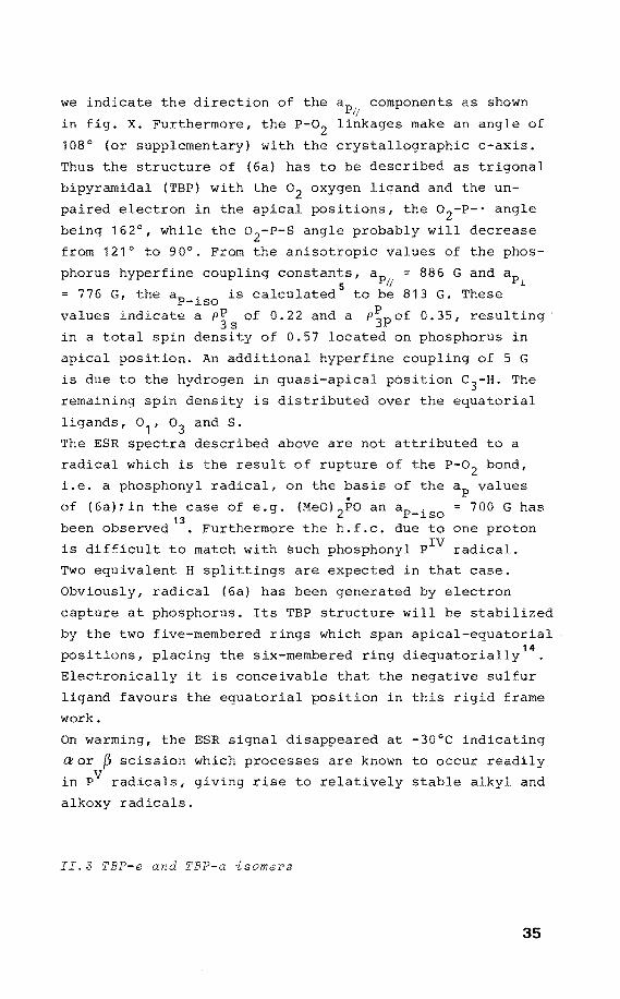
we indicate the direction of the components as shown
in fig. x. Furthermore, the P-02 make an angle of
108° (or supplementary) with the crystallographic c-axis.
Thus the structure of (6a) has to be described as trigonal
bipyramidal (TBP) with the o 2 oxygen ligand and the un
paired electron in the apical , the o 2-P-· angle
being 162°, while the o 2-P-S angle probably will decrease
from 121° to 90°. From the values of the phos-
phorus hyperfine coupling constants, 886 G and ap 5 l
= 776 G, the aP-iso is calculated to be 813 G. These
va lues indicate a p~ s of 0. 22 and a P~p of 0. 35, resulting
in a total spin density of 0.57 located on phosphorus in
apical position. An additional coupling of 5 G
is due to the hydrogen in pósition c3-H. The
remaining spin density is distributed over the equatorial
ligands, o 1 , o 3 and S.
The ESR spectra described above are not attributed to a
radical which is the result of of the P-02 bond,
i.e. a phosphonyl radical, on the basis of the values
of (6a)ïin the case of e.g. (Meo) 2Po an ap. = 700 G has \3 -l.SO
been observed . Furthermore the h.f.c. due to one proton
is difficult to match with such phosphonyl radical.
Two equivalent H splittings are
Obviously, radical (6a) has been
in that case.
by electron
capture at phosphorus. lts TBP structure will be stabilized
by the two five-membered rings which span apical-eguatorial
positions, placing the six-membered ring diequatorially14
•
Electronically it is conceivable that the negative sulfur
ligand faveurs the equatorial position in this rigid frame
work.
On warming, the ESR signal di at -30°C indicating
aor ~ scission which processes are known to occur readily
in PV radicals, giving rise to relatively stable alkyl and
alkoxy radicals.
II.3 TBP-e and TBP-a isomers
35
(,.) 0)
b
0
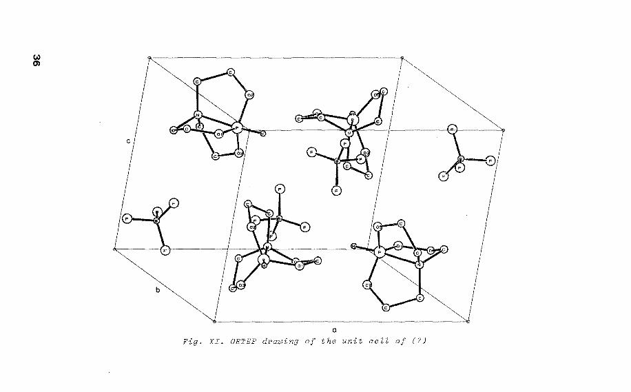
Fig. XI. ORTEP drawing of the unit cell of (7)
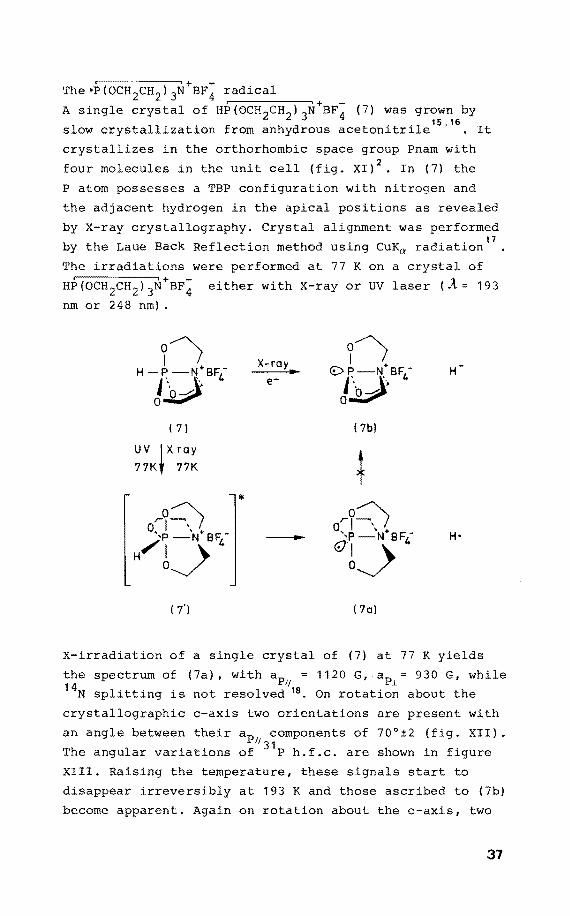
,--------,+ -The•P(OCH 2CH2 ) 3N BF4 radical
+ -A single crystal of HP(OCH2CH2 )
3N BF4 (7) was grown by
1 11 . . f h d . '1 15,16 s ow crysta ~zat~on rom an y rous aceton~tr~ e . It
crystallizes in the orthorhombic space group Pnam with
four molecules in the unit cell (fig. XI) 2• In (7) the
P atom possesses a TBP configuration with nitrogen and
the adjacent hydrogen in the apical positions as revealed
by X-ray crystallography. Crystal alignment was performed
by the Laue Back Reflection method using CuKa radiation17
The irradiations were performed at 77 K on a crystal of
ei ther wi th X-ray or UV laser (À= 19 3
nm or 248 nm) .
o) o) I • X-ray I +
H-P -N BF- €> P-N BF4 Jb) 4 e-
J~ H
( 7l 17b)
UV ~Xray 17K 77K
+ *
Or~~ r?~ 0, \ + ',p -N•BF- --- 'P -N BF4 -'ij 4 <Dbj H 0
( 7'l ( 7al
X-irradiation of a single crystal of (7) at 77 K yields
the spectrum of (7a), withap = 1120 G, ap 930 G, while 1! i 14N splitting is not resolved 18 • On rotatien about the
crystallographic c-axis two orientations are present with
an angle betweentheir ap componentsof 70°±2 (fig. XII). 11 31 The angular variations of P h.f.c. are shown in
XIII. Raising the temperature, these signals start to
disappear irreversibly at 193 K and those ascribed to (7b)
become apparent. Again on rotatien about the c-axis, two
37
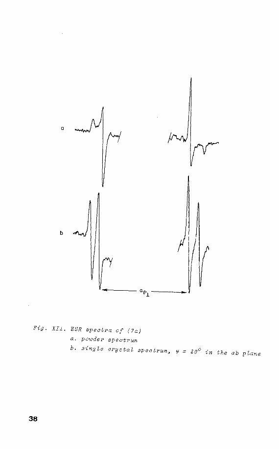
a
b
Fig. XII. ESR spectra of (?a)
a. powder spectrum
b. singLe crystaL spectrum~ ~ = 30° ~n the ab pLane
38
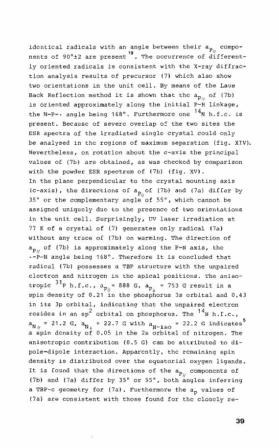
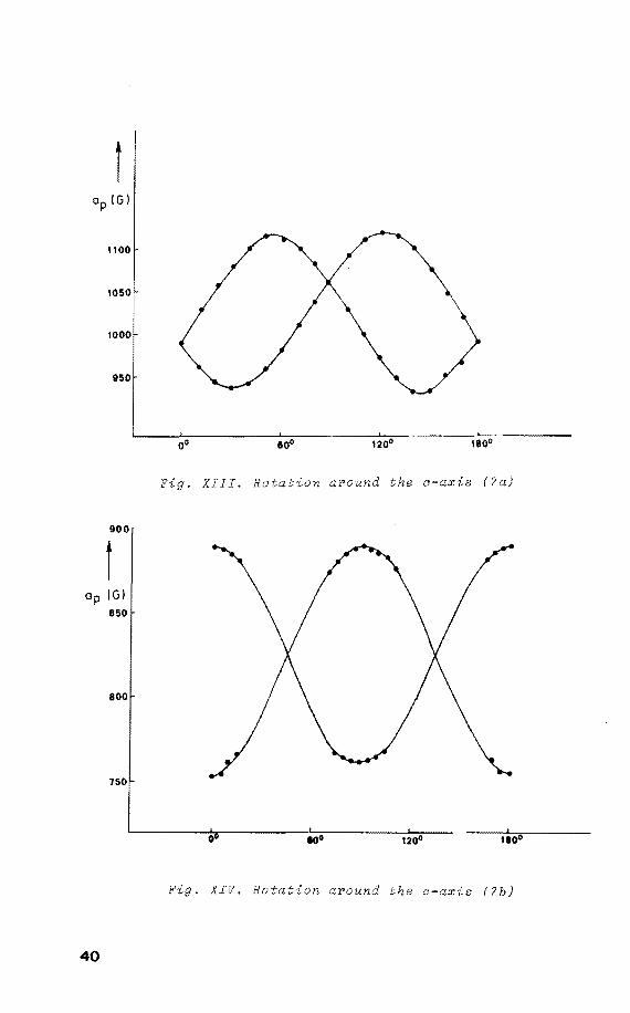
identical radicals with an angle between their ap compo-19 ~
nents of 90°±2 are present . The occurrence of different-
ly oriented radicals is consistent with the X-ray diffrac
tion analysis results of precursor (7) which also show
two orientations in the unit cell. By means of the Laue
Back Reflection methad it is shown that the ap~ of (7b)
is óriented approximately along the initial P-H linkage,
the N-P-· angle being 168°. Furthermore one 14N h.f.c. is
present. Because of severe overlap of the two sites the
ESR spectra of the irradiated single crystal could only
be analysed in the regions of maximum separation (fig. XIV).
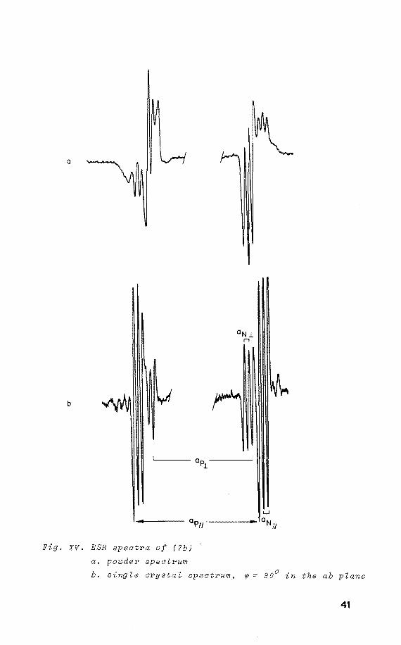
Nevertheless, on rotation about the c-axis the principal
values of (7b) are obtained, as was checked by comparison
with the powder ESR spectrum of (7b) (fig. XV).
In the plane perpendicular to the crystal mounting axis
(c-axis), the directions of ap of (7b) and (7a) differ by ~
35° or the complementary angle of 55°, which cannot be
assigned uniquely due to the presence of two orientations
in the unit cell. Surprisingly, UV laser irradiation at
77 K of a crystal of (7) generates only radical (7a)
without any trace of (7b) on warming. The direction of
ap of (7b) is approximately along the P-N axis, the ~
·-P-N angle being 168°. Therefore it is concluded that
radical (7b) possesses a TBP structure with the unpaired
electron and nitrogen in the apical positions. The anisa
tropie 31
P h.f.c., ap~ 888 G, ap~ = 753 G result in a
spin density of 0.21 in the phosphorus 3s orbital and 0.43
in its 3p orbital, indicating that the unpaired electron
resides in an sp2 orbital on phosphorus. The 14N h.f.c.,
aN = 21.2 G, a_ ~ N~
a spin density of
= 22.7 G with a . = 22.2 G indicates5
N-lSO 0.05 in the 2s orbital of nitrogen. The
anisatrapie contribution (0.5 G) can be attributed to di
pole-dipole interaction. Apparently, the remaining spin
density is distributed over the equatorial oxygen ligands.
It is found that the directions of the ap components of ~
(7b) and (7a) differ by 35° or 55°, bathangles inferring
a TBP-e geometry for (7a). Furthermore the ap values of
(7a) are consistent with those found for the closely re-
39
t
1100
1050
1000
950
900
1 Op (G)
850
800
750
40
Fig. XIII. Rotation around the e-axia (?a)
0 1200
Fig. XIV. Rotation around the c-axia (?b)
a
b
Fig. XV. ESR spectra of (?b)
a. powder spectrum
b. single crystal spectrum~ <p = 90° in the ab plane
41
lated phosphoranyl radical (3a) generated by X-irradiation
of (3) which is known to possess a nearby perfect TBP
structure as revealed by X-ray diffraction analysis 7• From
the anisatrapie 31 P h.f.c. values obtained for (7a) an
ap . = 993 G is calculated 5
, indicating the P P3
is -lSO S
0.27, while the anisatrapie contribution places 0.61 of
the spin density in the P3p orbital, which gives a total
spin density of 0.88 on phosphorus.
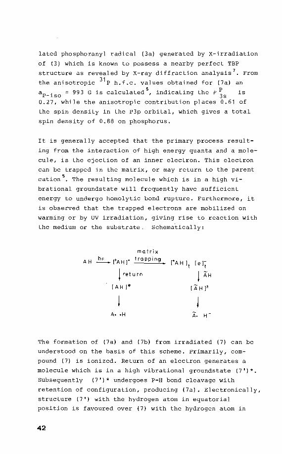
It is generally accepted that the primary process result
ing from the interaction of high energy quanta and a mole
cule, is the ejection of an inner electron. This electron
can be trapped in the matrix, or may return to the parent
cation5
• The resulting molecule which is in a high vi
brational groundstate will frequently have sufficient
energy to undergo homolytic bond rupture. Furthermore, it
is observed that the trapped electrans are mobilized on
warming or by UV irradiation, giving rise to reaction with
the medium or the substrate. Schematically:
matrix
AH ~[.AH]" trapping
t re t u r n
[AH]*
[+AH ]t [e]t
! AH
[AH f
The formation of (7a) and (7b) from irradiated (7) can be
understood on the basis of this scheme. Primarily, com
pound (7) is ionized. Return of an electron generates a
molecule which is in a high vibrational groundstate (7')*.
Subsequently (7')* undergoes P-H bond cleavage with
retention of configuration, producing (7a). Electronically,
structure (7') with the hydragen atom in equatorial
position is favoured over (7) with the hydragen atom in
42
apical position. In contrast, the strain energy in struc
ture (7') is enhanced with respect to that of (7). However,
the latter factor may be les& important in (7')* since in
this high vibrational groundstate the bond lengths are
increased. Therefore, in (7')* the electronic factor has
become dominant. It appears that the concentration of (7a)
increases on warming. Therefore, the return of an electron
to the parent cation is a thermodynamically controlled
process.
It is conceivable that trapped electrans attack (7) at
higher temperature, generating a radical anion which
produces (7b) by subsequent loss of H from the apical
axis of the TBP. It has been confirmed that trapped elec
trans are involved in the formation of (7b) by an experi
ment in which compound(7) is X-irradiated at 77 K, showing
(7a) . Subsequently this sample is UV irradiated at 77 K
which leads to loss of (7a). On warming the concentration
of (7a) is increased, whereas radical (7b) does nat show
up at all. Obviously, UV irradiation delocalizes the trapped
electrans which attack bath (7a) leading to a diamagnetic
product, and (7), resulting in (7b). However, in this
experiment (7b) is nat detected, simply because this ra
dical is lost upon UV irradiation as was proven independent
ly.
It is concluded that homolytic P-H bond rupture is favoured
in an equatorial position of the TBP, whereas this process
is inhibited in an apical position. In contrast, loss of
H takes place in an apical position preferentially.
43
II.4 Discussion
The single crystal ESR study of (1a) and (2a) in combination
with the X-ray diffraction results of the precursor
establishes unequivocally that the orbital the unpaired
electron resides in, is directed along the initial P-H
linkage. As a consequence the unpaired electron is
characterized as a real ligand in the TBP-e structure. The
TBP-e structure obtained here is in sharp contrast with
the structure assumed for Cl3Po- which was obtained by
Y-irradiation of a single crystal of cl 3Po 21
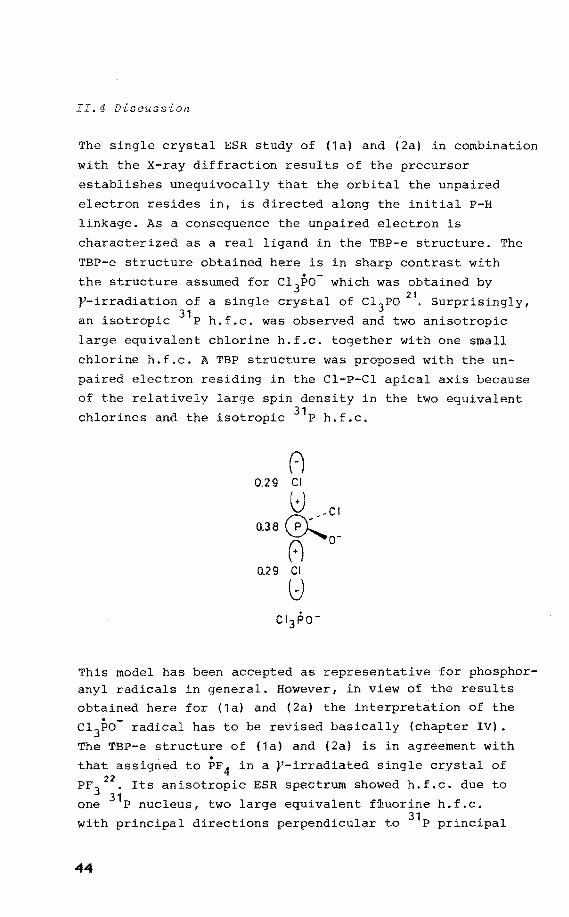
• Surprisingly,
an isotropie 31 P h.f.c. was observed and two anisatrapie
large equivalent chlorine h.f.c. tagether with one small
chlorine h.f.c. A TBP structure was proposed with the un
paired electron residing in the Cl-P-Cl apical axis because
of the relatively large spin density in the two equivalent
chlorines and the isotropie 31 P h.f.c.
(-) 0.29 Cl
8 -Cl o.3a rp-y--~o-8
0.29 Cl
u
This model has been accepted as representative for phosphoranyl radicals in general. However, in view of the results
obtained here for (1a) and (2a) the interpretation of the
cl3Po- radical has to be revised basically (chapter IV) •
The TBP-e structure of (1a) and (2a) is in agreement with
that assigned to PF4 in a y-irradiated single crystal of
PF3 22
• lts anisatrapie ESR spectrum showed h.f.c. due to
one 31 P nucleus, two large equivalent fiuorine h.f.c.
with principal directions perpendicular to 31 P principal
44
values, and two small fluorine h.f.c. From this a TBP-e
structure is deduced tentatively with the unpaired elec
tron and the two fluorines with small h.f.c. located
equatorially, and the other two fluorines in the apical
positions. In this case the assignment has been based
totally on the directions and magnitudes of the h.f.c. . However, since the radical was produced by addition of F
to PF3 no correlation could be made between the ESR para
meters and x-ray analysis. From the single crystal ESR
studies of (6a) and (7b) it is evident that the TBP-a and
TBP-e structures exhibit very similar ESR parameters. In
fact, the structures could only be assigned unambiguously
because of the availability of the X-ray diffraction re
sults. Consequently reliable structural information re
garding phosphoranyl radicals can only be obtained by the
methods outlined here, unless phosphorus bears only ligands
which have a magnetic moment. Therefore, much of the
earlier work on phosphoranyl radicals needs reinvestigation,
because invariably in these cases a TBP-e structure is
assigned in which the unpaired electron accupies an
equatorial position 23• This structure seems to be supported
by the p/s ratio of approximately 2 which is frequently
observed. However, in spite of similar speetral data for
{6a) and {7b) these compounds show a quite different {TBP-a)
geometry, probably as a result of the molecular and crystal
constraints.
The c 3 structure observed for (6a, 7b) has also been V • 24
found for Ph 3Pcl • In contrast, in this case the electron
spin density is assumed to reside in the P-Cl anti-bonding
{U*) orbital as infered from the fact that the principal
directions of and aCl are nearly parallel and from the
high density on chlorine. However, similar spin
densities onthe apical ligands adjacent to phosphorus
have been observed for TBP-e structures (e.g. PF 4 ) also,
and as a consequence this argument does not support the
u* structure at all. It is pointed out that this u* model
which concerns the excited state of the radical, should be
less stable than the TBP-a structure which deals with a
45
non-excited state of the phosphoranyl radical. Moreover,
since we demonstrated the ligand character of the unpaired
electron in these radicals, the TBP-a structure applies
very well to the Ph 3PC1 radical reconciling the directional
data excellently. As a matter of fact bath isoroers TBP-a
and TBP-e are detected in one precursor (7} affording a
direct comparison of bath types (7a}, (7b}. It appears
that the aP-iso of the TBP-a species is smaller than that
obtained for the TBP-e type. This indicates that the 3s
contribution in bonding in the apical axis is reduced
compared with the equatorial sites. A similar trend is
found for the analogous phosphoranes. However, the s
contribution is still so large, that structure assignment
purely on the basis of ap value without knowledge of the
direction of the orbital the unpaired electron resides in,
remains questionable.
46
Referenaes and notes
1. P.F. Meunier, R.O. Day, J.R. Devillers, and R.R.
Holmes, Inorg. Chem. 1978, 17, 3270.
2. The ORTEP drawing was kindly delivered by dr. G.J.
Visser, Computing Centre of the Eindhoven University
of Technology, The Netherlands.
3. G.H.W. Milburn, "X-ray Crystallography", Butterworth,
London, 1973. See also the appendix.
4. J.H.H. Hamerlinck, P. Schipper and H.M. Buck, J. Chem.
Soc., Chem. Commun., 1981, 104.
5. P.W. Atkins and M.C.R. Symons, "The Structure of In
organic Radicals", Elsevier, Amsterdam, 1967.
6. H. Wunderlich and H.G. Wussow, Acta Crystallogr., Sect.
B, 1978, 34, 2663.
7. T.E. Clark, R.O. Day and R.R. Holmes, Inorg. Chem.,
1979, 18, 1653.
8. Thiophosphate 0,0,0-triester of methyl~ -D-ribopyra
noside was a gift frorn dr. A.C. Bellaart, Department
of Organic Chemistry, Eindhoven University of Techno
logy, The Netherlands.
9. A.C. Bellaart, D. van Aken, H.M. Buck, C.H. Stam, and
A. van Herk, Reel. Trav. Chim. Pays-Bas, 1979, 98, 523.
10. I. Garaycochea and 0. Wittke, Acta Crystallogr., 1964,
17, 183. See also the appendix.
11. J.H.H. Hamerlinck, P. Schipper and H.M. Buck, J. Chem.
Phys., 1982 in press.
12. S.E. Locher and H.C. Box, J. Chem. Phys., 1980, 72,
828.
13. P. Schipper, E.H.J.M. Jansen, and H.M. Buck, "Topics
in Phosphorus Chernistry", Wiley-Interscience, New York,
1977, 9, 485.
14. R.F. Hudson and C. Brown, Acc. Chem. Res. 1972, 5, 204.
15. Compound (7) was a gift from dr. D. van Aken, Depart
ment of Organic Chemistry, Eindhoven University of
Technology, The Netherlands.
16. J.C. Clardy, D.S. Milbrath, J.P. Springer, and J.G.
Verkade, J. Am. Chern. Soc., 1976, 98, 623.
47
17. B.D. Cullity, "Elernents of X-ray diffraction",Addison
Wesley, London, 1959.
18. J.H.H. Harnerlinck, P. Schipper and H.M. Buck, J. Chern.
Soc., Chern. Cornrnun., 1981, 1149.
19. J.H.H. Harnerlinck, P. Schipper and H.M. Buck, J. Am.
Chern. Chern. Soc., 1980, 102, 5679.
20. E.E. Budzinski, W.R. Potter, G. Potienko, and H.C.
Box, J. Chern. Phys., 1979, 70, 5040.
21. T. Gillbro and F. Williarns, J. Am. Chern. Soc., 1974,
96, 5032.
22. A. Hasegawa, K. Ohnishi, K. Sogabe, and M. Miura, Mol.
Phys., 1975, 30, 1367.
23. R.W. Dennis and B.P. Roberts, J. Organornet. Chern.,
1 9 7 3 I 4 7 I C8.
24. T. Berclaz, M. Geoffroy, and E.A.C. Lucken, Chern. Phys.
Lett., 1975,36,677.
48
CHAPTER 111
Phosphoranyl radicals in an
octahedral configuration
III.l Phosphorus in an octahedraZ PVI geometry Mith the
unpaired electron in axiaZ position
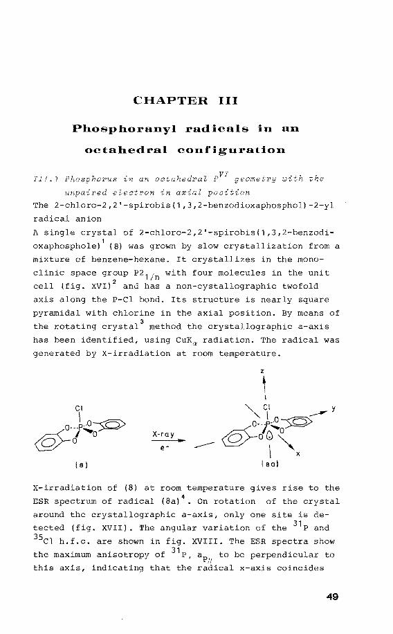
The 2-chloro-2,2'-spirobis(1,3,2-benzodioxaphosphol)-2-yl
radical anion
A single crystal of 2-chloro-2,2'-spirobis(1,3,2-benzodi
oxaphosphole)1 (8) was grown by slow crystallization from a
mixture of benzene-hexane. It crystallizes in the mono
clinic space group P2 1 . with four molecules in the unit 2 Jn
cell (fig. XVI) and has a non-cystallographic twofold
axis along the P-Cl bond. Its structure is nearly square
pyramidal with chlorine in the axial position. By means of
the rotating crystal3
methad the crystallographic a-axis
has been identified, using CuKa radiation. The radical was
generated by X-irradiation at room temperature.
Cl
I 0---P~~
~-, 0 ~0
( 8)
X-ra y
z
\
--- I sa)
X-irradiation of (8) at room temperature gives rise to the
ESR spectrum of radical (8a)4
• On rotatien of the crystal
around the crystallographic a-axis, only one site is de
tected (fig. XVII). The angular variatien of the 31 P and 35c1 h.f.c. are shown in fig. XVIII. The ESR spectra show
the maximum anisotropy of 31 P, ap to be perpendicular to I!
this axis, indicating that the radical x-axis coincides
49
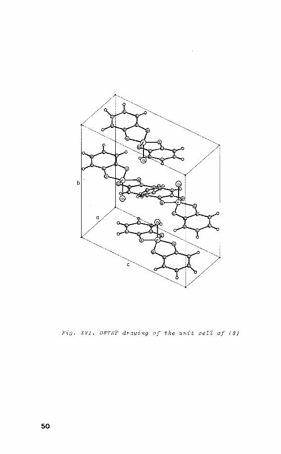
Fig. XVI. ORTEP drawing of the unit cell of (8)
50
40
30
1400
1350
1300
oo
'* . C -Q)CIS I b -ax is
180°
'* . c-a x 1 s
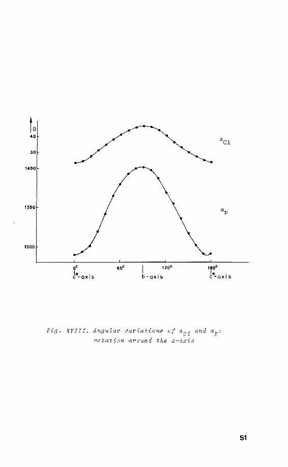
Fig. XVIII. Angular variations of aCZ and ap:
rotation around the a-axis
51
0
b
c
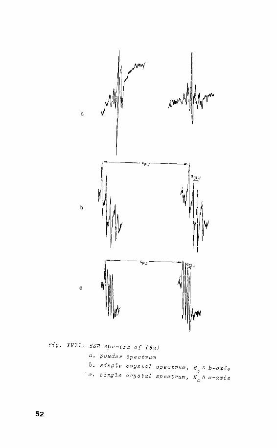
Pig. XVII. ESR spectra of (Ba) a. poüJder spectrum b. single crystaZ spectrum, 1/ b-axis a. single crystaZ spectrum, 11 a-axis
52
with the crystallographic a-axis. Purthermare the maximum
in the anisotropy of 35c1 and 37c1 appears to be parallel
with ap , indicating that the direction of ap corresponds ~ V
with the orientation of the P-Cl linkage, which is the
crystallographic b-axis in its precursor. From the ESR
spectrum of a powdered sample the principal values of ap
and aCl were obtained.
From this it is concluded that radical (8) has been gene
rated by electron capture rather than by rupture of the
P-0 linkage. In the latter case the aCl and ap directlans I! I!
should have been expected to be perpendicular. From the
ESR data it is derived that aP-iso = 1317 G, indicating a
phosphorus 3s spin density of 0.36. A spin density of 0.40
is located in the phosphorus 3pz orbital as inferred from
the anisotropy of ap. This results in a total spin density
of 0.76 on phosphorus. From the 35c1 h.f.c. one calculates
aCl-iso 31 G, indicating 5 a 3s spin density of 0.02, while
the anisatrapie contribution (B) accounts for a den-
sity of 0.16 in a chlorine 3pz orbital which is directed
along the P-Cl linkage. Similar spin densities are obtained
from the 37c1 tensor. Therefore, the structure of (8a) is
described as c4v with 76% of the unpaired electron located
in an axial spz hybrid orbital at the phosphorus nucleus
and 16% in the 3pz of chlorine.
III.2 Discussion
The PVI structure derived for (Ba) has been assigned pre
viously to PF~, obtained by Y-irradiation of hexafluoro
phosphate 6
'7• Its history is rather curieus since for many
years this radical centre was thought to be PF4 , which was
assumed to rotate rapidly in the solid matrix, accounting
for the existence of four equivalent anisatrapie fluorines
(aF-iso = 196 G) and the isotropie 31
p h.f.c. (
1346 G) 8• These values are close to those obtained for the
single
aF-iso and an
crystal of irradiated PF3 with anisatrapie fluorines,
306 G (2F apical), . = 61 G (2F equatorial) 31 ~sa
anisatrapie P h.f.c. (aP-iso = 1310 G), assigned
53
• 9 to PF4 in a TBP-e structure . However, on the basis of a
. 6 near zero h.f.c. ascribed to a fifth fluorine l1gand , it
was suggested that the radical centre in irradiated hexa-•
fluorophosphate should be PF5
insteadof PF 4 .
F I
F,/P.\-F F F
Furthermore it is believed that the fifth ligand in such
c 4v geometries, e.g. PF5 , SF5 , ÁsF5 and PCl~, possesses an 7 • 10 11
almast zero h.f.c. as a general rule ' • However, the
ClP(02c6H4); radical anion (8a) has been proven to possess
a similar c4v syrnrnetry in which the odd electron and
chlorine are located in axial positions, the chlorine ha-
ving a rather h.f.c. which is comparable to those
found for apical chlorine in PV phosphoranyl radicals 12
•
Therefore it is suggested that the small coupling detected
for the "PF~" radical 6 is due toa neighbouring fluorine
in the solid matrix, as was also the case in TBP-e PF49
•
Also the fact that the phosphorus.h.f.c. of the TBP-e PF4 radical and that for the assumed structure are almast
identical, in of the higher coordination of the
latter is indicative for a PV phosphoranyl radical. Com
pared to P(o2c
6H
4)
2 (4a) which has a TBP-e PV structure
with aP-iso~ 1005 G, the introduetion of one chlorine
ligand raises the ap . to 1317 G. On this basis the -lSO
radical becomes unbelievable, and here it is suggested
that there are two possibilities, i.e. the centre is pseu-•
do-rotati~g TBP-e , or it is PF 4 with a square pyrarnidal 13
structure (chapter IV)
54
References and notes
1. R.K. Brown and R.R. Holmes, Inorg. Chem., 1977, 16,
2294.
2. The ORTEP drawing of the unit cell of (8) was kindly
delivered by dr. G.J. Visser, Computing Centre of the
Eindhoven University of Technology, The Netherlands.
3. G.H.W . .!>Ulburn, "X-ray Crystallography", Butterworth,
London, 1973.
4. J.H.H. Hamerlinck, P. Schipper and H.M. Buck, Chem.
Phys. Lett., 1981, 80, 358.
5. P.W. Atkins and M.C.R. Symons, "The Structure of In
organic Radicals", Elsevier, Amsterdam, 1967.
6. S.P. Misbra and M.C.R. Symons, J. Chem. Soc., Chem.
Commun., 1974, 279.
7. J.R. Morton, K.F. Preston, and S.J. Strach, J. Magn.
Reson., 1980, 37, 321.
8. P.W. Atkins and M.C.R. Symons, J. Chem. Soc., 1964,
4 363.
9. A. Hasegawa, K. Ohnishi, K. Sogabe, and M. Miura, .t.lol.
Phys., 1975, 30, 1367.
10. A. Hasegawa and F. Williams, Chem. Phys. Lett., 1977,
45, 275.
11. S.P. Misbra and M.C.R. Symons, J. Chem. Soc., Dalton
Trans., 1976, 139.
12. P. Schipper. E.H.J.M. Jansen, and H.M. Buck, "Topics
in Phosphorus Chemistry", Wiley-Interscience, 1977, 9,
49 4.
13. J.H.H. Hamerlinck, P.H.H. Hermkens, P. Schipper, and
H.M. Buck, J. Chem. Soc., Chem. Commun., 1981, 358.
55
CHAPTER IV
Intramolecular ligand reorganisation
in phosphoranyl radicals
IV.I Pseudo-rotating TBP-e
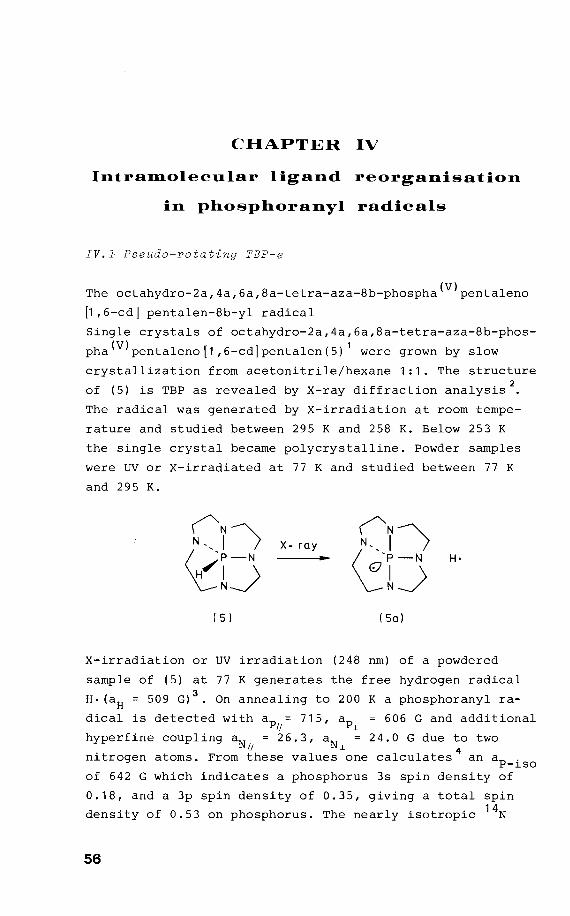
The octahydro-2a,4a,6a,8a-tetra-aza-8b-phospha(V)pentaleno
~,6-cd] pentalen-8b-yl radical
Single crystals of octahydro-2a,4a,6a,8a-tetra-aza-8b-phos
pha(V)pentaleno[1,6-cd]pentalen(5) 1 were grown by slow
crystallization from acetonitrile/hexane 1:1. The structure
of (5) is TBP as revealed by X-ray diffraction analysis2
The radical was generated by X-irradiation at room tempe
rature and studied between 295 K and 258 K. Below 253 K
the single crystal became polycrystalline. Powder samples
were UV or X-irradiated at 77 K and studied between 77 K
and 295 K.
("N> ("N) N '' I X- ray N'' I
<ê1J ~1J H·
( 5) (Sa)
X-irradiation or UV irradiation (248 nm) of a powdered
sample of (5) at 77 K generates the free hydragen radical
H· (aH = 509 G) 3• On annealing to 200 K a phosphoranyl ra-
dical is detected with ap = 715, ap 606 G and additional I! .L
hyperfine coupling aN = 26.3, aN 24.0 G due to two H .L 4
nitrogen atoms. From these values one calculates an aP-iso
of 642 G which indicates a phosphorus 3s spin density of
0.18, and a 3p spin density of 0.35, giving a totalspin
density of 0.53 on phosphorus. The nearly isotropie 14N
56
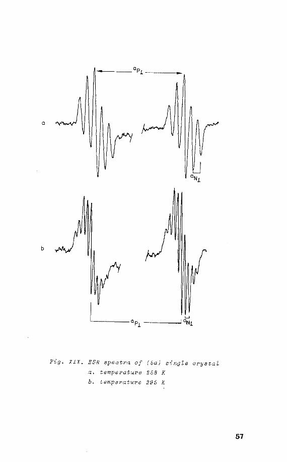
a
b
Fig. XIX. ESR speatPa of (5a) single arystal
a. temperature 258 K
b. temperature 295 K
57
h.f.c. aN-iso of 24.8 G indicates4
a spin density of 0.05
in its 2s orbital. The small anisatrapie contribution (0.8
G) can be attributed to dipole-dipole interaction. From
this a TBP-e structure is derived with two apical nitrogen
atoms accounting for the observed high h.f.c. and two
equatorial nitrogens with small h.f.c. values (< 5 G). On
further raising of the temperature (to 295 K) an ESR spec
trum was obtained which consisted of the same ap and ap lj ~
values as found at low temperatures, and additional h.f.c.
due to four equivalent atoms aN 14.4 and aN lj ~
12.7 G. These changes in the ESR spectrum are reversible
as indicated by the appearance of the initial spectrum on
cooling. Therefore this phenomenon has to be attributed to
a rapid pairwise interconversion of the nitrogen ligands.
Additional evidence was obtained a single crystal ESR
study of (Sa). The ESR spectra of an X-irradiated single
crystal of (5) at room show that two identical
radicals with an angle between their
± 2° are present. These were
in the same way as found for the
components of 34
dependent
sample; at 295 K
four equivalent nitrogens were observed, whereas on cool-
ing to 258 K only two couplings appeared, with
the principal ap values at the same positions as
found at 295 K (see • XIX and • XX). Unfortunately
the single crystal became polycrystalline at 253 K, show-
ing the features of the powdered with enhanced
resolution.
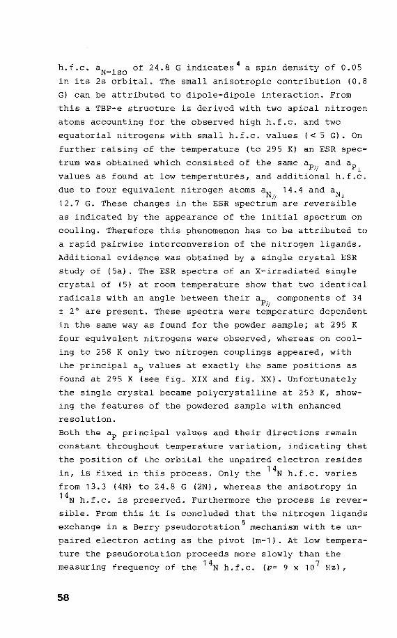
Bath the ap principal values and their directions remain
constant throughout temperature variation, indicating that
the position of the orbital the electron resides
in, is fixed in this process. Only the 14N h.f.c. varies
from 13.3 (4N) to 24.8 G (2N), whereas the anisotropy in 14
N h.f.c. is preserved. Purthermare the process is rever
sible. From this it is concluded that the nitrogen 1
exchange in a Berry pseudorotation 5 mechanism with te un
paired electron acting as the pivot (m-1). At low
ture the pseudorotatien proceeds more slowly than the
measuring frequency of the 14N h.f.c. (v= 9 x 10 7 Hz),
58
!GI
1 26 aN 258 K 25
24
14 aN 295 K
13
700
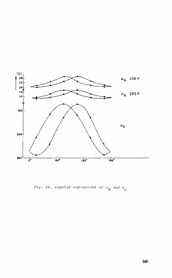
Fig. XX. AnguZar variations
59
while at room temperature the exchange frequency equals
this value, rendering the four nitrogens equivalent. From
this one calculates with equation (1)
V= e * - (L1G)
RT ( 1 )
v unimolecular rate constant
L1a* free energy of activation
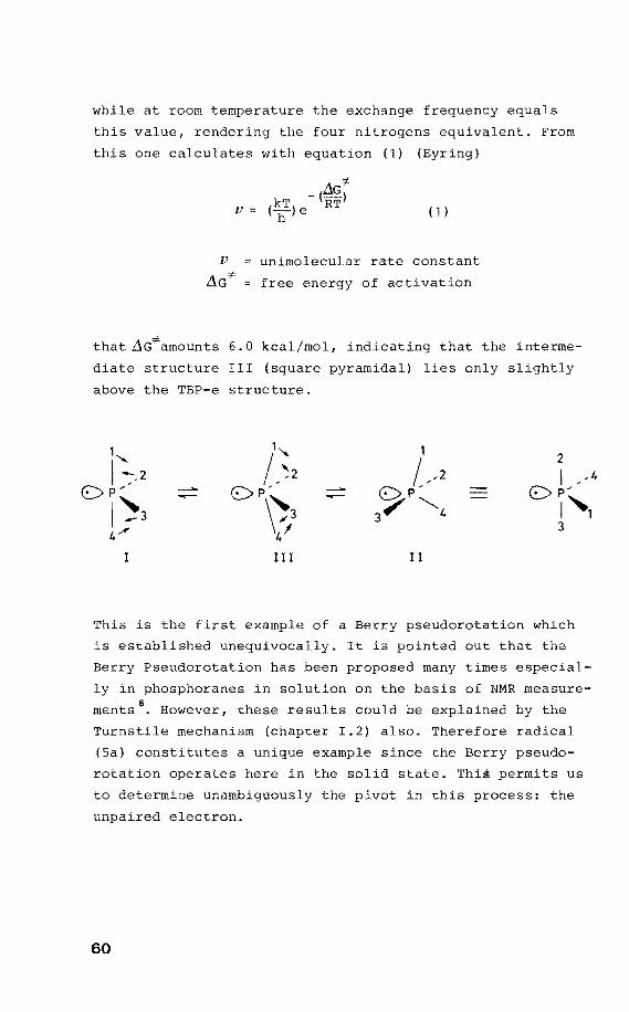
that L1G*amounts 6.0 kcal/mol, indicating that the interme
diate structure III (square pyramidal) lies only slightly
above the TBP-e structure.
,, I --:--2
OP" 1~3 4-'
I I I
This is the first of a Berry pseudorotation which
is established unequivocally. It is pointed out that the
Berry Pseudorotation has been proposed many times especial
ly in phosphoranes in solution on the basis of NMR measure
ments6. However, these results could be explained by the
Turnstile mechanism (chapter I.2) also. Therefore radical
(Sa) constitutes a unique example since the Berry pseudo
rotation operates here in the solid state. This permits us
to determine unambiguously the pivot in this process: the
unpaired electron.
60
IV.2 X-ray struoture determination
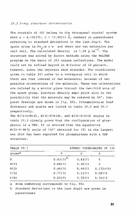
The crystals of (5) belong to the tetragonal crystal system 0
with a = 6.102(7), c 13.90(1) A, numbers in parentheses
referring to standard deviations in the last digit. The
space group is P4 2 /m n m and there are two molecules per
unit cell. The calculated density is 1.29 g cm- 3 . The
structure was solved by direct methods using the t-1ULTAN
program on the basis of 353 unique reflections. The model
could not be refined beyond an R-factor of 22 percent,
however, since the crystals show disorder. The coordinates
given in table IVJ refer to a tetragonal cell in which
there are four instead of two molecules, because of two
possible orientations of the molecule. These two orientations
are related by a mirror plane through the two-fold axis of
the space group. Electron density maps point also to the
possibility that the molecule may be rotated by 90°. Com
puter drawings are shown in fig. XXI. Intramolecular bond
distances and angles are listed in table IV.2 and IV.3
respectively.
The N(1)-P-N(2), N(3)-P-N(4), and N(1)-P-N(3) angles in
table IV.3 clearly prove that the contiguration of phos
phorus is a TBP. It is noticed that the equatorial
N(2)-P-N(1) angle of 145° obtained for (5) is the largest
one that has been reported for phosphoranes with a TBP
structure.
TABLE IV .1 ATOMIC COORDINATES OF (5)
atomsa x y z
p 0.43(1)b 0.43(1) 0
N ( 1) 0.68(1) 0.30(1) 0
N ( 3) 0.44(1) 0.44(1) 0.135(10)
c ( 5) 0.77(1) 0.23(1) 0. 08 ( 1)
c ( 6) 0.65(1) 0.35(1) 0.16(1)
a Atom numbering corresponds to fig. XXI
b Standard deviations in the last digit are given in
parentheses
61

Fig. XXI. ORTEP drawing of (SJ
62
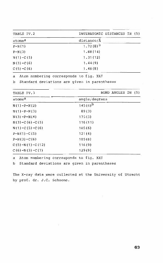
TABLE IV.2 INTERATOMIC DIST&~CES IN (5)
atornsa
P-N ( 1)
P-N(3)
N ( 1 > -c (5 >
N (3) -c (6)
c (5) -c (6)
1.88(14)
1.31 (12)
1. 44 (9)
1.48(8)
a Atorn nurnbering corresponds to fig. XXI
b Standard deviations are given in parentheses
TABLE IV.3 BOND ANGLES IN ( 5)
angle;degrees
N(1)-P-N(2) 145(6)
N ( 1 ) - P-N ( 3) 89 ( 3)
N(3)-P-N(4) 175(5)
N ( 31 -c < 6 > -c < 5 l 116(11)
N ( 1 l -c < 5 > -c < 6 > 105(6)
P-N(1)-C(5) 1 21 ( 4)
P-N ( 3) -C (6) 105(8)
C(5)-N(1)-C(12) 116 ( 9)
C(6)-N(3)-C(7) 129 (9)
a Atorn nurnbering corresponds to fig. XXI
b Standard deviations are given in parentheses
The X-ray data were collected at the University of Utrecht
by prof. dr. J.C. Schoone.
63
IV.J Discussion
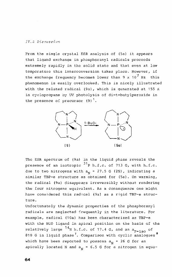
From the single crystal ESR analysis of (Sa) it appears
that ligand exchange in phosphoranyl radicals
extremely rapidly in the solid state and that even at low
temperature this interconversion takes place. However, if
the exchange frequency becomes lower than 9 x 10 7 Hz this
phenomenon is easily overlooked. This is nicely illustrated
with the related radical (9a), which is generated at 15S K
in cyclopropane by UV photolysis of di-t-butylperoxide in
the presence of precursor (9) 1•
t-BuO·
( 9) I 9ol
The ESR spectrum of (9a) in the liquid phase reveals the
presence of an isotropie 31 P h.f.c. of 713 G, with h.f.c.
due to two nitrogens with aN 27.5 G (2N), indicating a
similar TBP-e structure as obtained for (Sa). On warming,
the radical (9a) disappears irreversibly without rendering
the four nitrogens equivalent. As a consequence one might
have considered this radical (9a) as a rigid TBP-e struc
ture.
Unfortunately the dynamic properties of the phosphoranyl
radicals are neglected frequently in the literature. For
example, radical (10a) has been characterized as TBP-e
with the NCO ligand in apical position on the basis of the 14
relatively N h.f.c. of 17.4 G, and an aP-iso of
810 Gin liquid phase 7• Comparison with cyclic analogues 8
which have been reported to possess aN = 26 G for an
apically located N and aN 6.5 G for a nitrogen in equa-
64
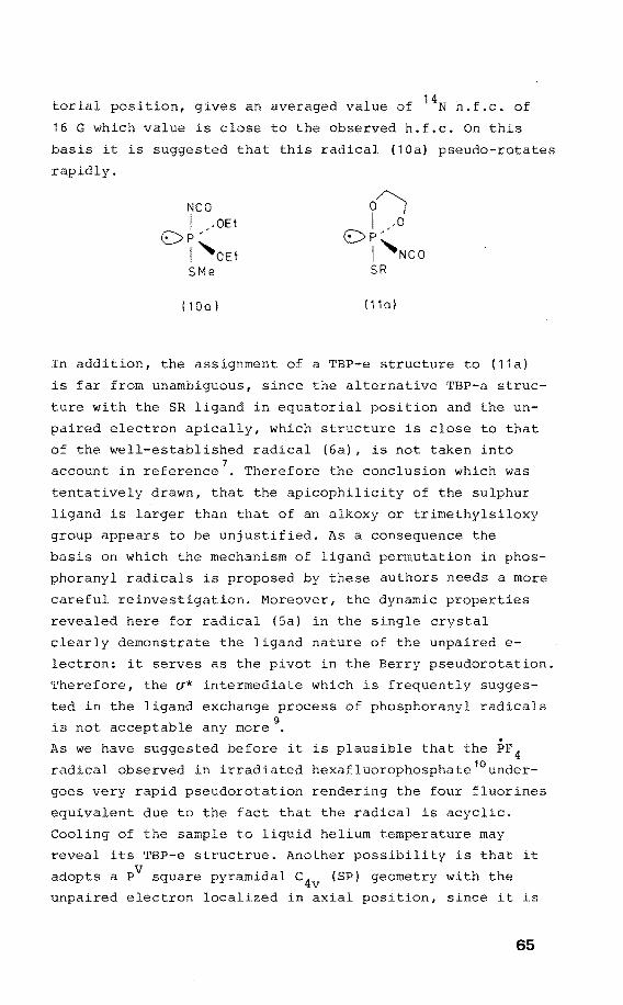
torial position, gives an averaged value of 14N h.f.c. of
16 G which value is close to the observed h.f.c. On this
basis it is suggested that this radical (10a) pseudo-rotates
rapidly.
NCO I ,-OEt
OP' I 'OEt SMe
( 1 Oa l ( 11 a)
In addition, the assignrnent of a TBP-e structure to (11a)
is far frorn unarnbiguous, since the alternative TBP-a struc
ture with the SR ligand in equatorial position and the un
paired electron apically, which structure is close to that
of the well-established radical (6a), is not taken into
account in reference7
• Therefore the conclusion which was
tentatively drawn, that the apicophilicity of the sulphur
ligand is larger than that of an alkoxy or trirnethylsiloxy
group appears to be unjustified. As a consequence the
basis on which the rnechanisrn of ligand perrnutation in phos
phoranyl radicals is proposed by these authors needs a more
careful reinvestigation. Moreover, the dynarnic properties
revealed here for radical (Sa) in the single crystal
clearly dernonstrate the ligand nature of the unpaired e
lectron: it serves as the pivot in the Berry pseudorotation.
Therefore, the a* interrnediate which is frequently sugges
ted in the ligand exchange process of phosphoranyl radicals
is not acceptable any more 9•
As we have suggested before it is plausible that the PF 4 radical observed in irradiated hexafluorophosphate 10 under
goes very rapid pseudorotation rendering the four fluorines
equivalent due to the fact that the radical is acyclic.
Cooling of the sample to liquid heliurn ternperature rnay
reveal its TBP-e structrue. Another possibility is that it
adopts a PV square pyrarnidal c4v (SP) geornetry with the
unpaired electron localized in axial position, since it is
65
inferred from the single crystal ESR study of (Sa) that
the SP structure is only slightly less stable (6.0 kcal/
mol) than the TBP-e structure. A ligand exchange process
accounts possibly also for the observed isotropie 31 P • 11
h.f.c. of -OPC1 3 in a single crystal of y-irradiated OPC1 3 •
It is apparent that the isotropie 31 P h.f.c. remains in
compatible with the results obtained here on phosphoranyl
radicals. Therefore it is suggested that the Cl3Po- radical
undergoes ligand exchange in such mode that the unpaired
electron is not the pivot. Since it is evident from the
analysis of the single crystal spectra of (Sa) that the
anisotropies of the ligand h.f.c. remain unaffected by the
pseudorotatien process, the alignment of the anisotropic
chlorine h.f.c. due to two apical chlorines does not simply
implicate that the Cl-P-Cl angle is 180°, but may deviate
dramatically from this value. The same applies for the
TBP-e radical PF 4 in irradiated PF 3 where a similar con-12
clusion was made about the axial F-P-F angle This is
clear from inspeetion of the angular dependenee of the
h.f.c. of the equatorlal fluorines of which the principal
h.f.c. are in disagreement with the proposed TBP-e struc
ture which implies an angle between aF and ap of 60°. ~ ~
66
and notes
1. J.E. Richman and T.J. Atkins, Tetrahedron Lett., 1978,
4333.
2. The structure determination of (5) was made by dr. A.
Koster of the department of Chemistry, while
the ORTEP drawing was obtained with assistance of dr.
G.J. Visser of the Computing Centre at Eindhoven Uni
versity of Technology, The Netherlands.
3. J.H.H. Hamerlinck, P.H.H. Hermkens, P. Schipper and
H.M. Buck, J. Chem. Soc., Chem. Commun., 1981, 358.
4. P.W. Atkins and M.C.R. Symons, "The Structure of Inor
ganic Radicals", Elsevier, Amsterdam, 1967.
5. R.S. Berry, J. Chem. Phys., 1960, 32, 933.
6. R. Luckenbach, "Dynamic of Pentaco-or-
dinated Phosphorus and Related Elements", G. Thieme,
Stuttgart, 1973.
7. J.R.M. Giles and B.P. Roberts, J. Chem. Soc., Perkin
Trans. II, 1981, 1211.
8. J.A. Baban and B.P. Roberts, J. Chem. Soc., Chem.
Commun., 1979, 537.
9. R.S. Hay and B.P. Roberts, J. Chem. Soc., Perkin Trans.
II, 1978, 770.
10. P.W. Atkins and M.C.R. Symons, J. Chem. Soc., 1964,
4363.
11. T. Gillbro and F. Williams, J. Am. Chem. Soc., 1974,
96, 5032.
12. A. Hasegawa, K. Ohnishi, K. Sogabe, and M. Miura, Mol.
Phys., 1975, 30, 1367.
67
CHAPTER V
Intra:molecular electron trans:fer
in phosphoranyl radicals
V.l Introduetion
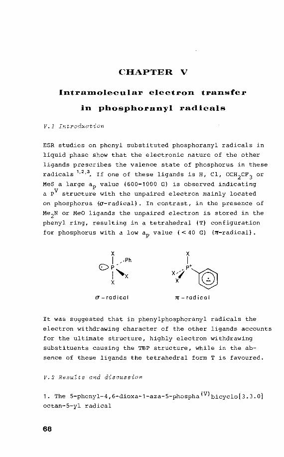
ESR studies on phenyl substituted phosphoranyl radicals in
liquid phase show that the electronic nature of the other
ligands prescribes the valenee state of phosphorus in these
radicals 1•2
•3
• If one of these ligands is H, Cl, OCH2cF 3 or
MeS a large ap value (600-1000 G) is observed indicating
a PV structure with the unpaired electron mainly located
on phosphorus (a-radical). In contrast, in the presence of
Me 2N or MeO ligands the unpaired electron is stored in the
phenyl ring, resulting in a tetrabedral (T) contiguration
for phosphorus with a low ap value ( < 40 G) (n-radical).
a-radical n-radical
It was suggested that in phenylphosphoranyl radicals the
electron withdrawing character of the other ligands accounts
for the ultimate structure, highly electron withdrawing
substituents causing the TBP structure, while in the ab
sence of these ligands the tetrabedral form T is favoured.
V.2 Results and discuesion
1. The 5-phenyl-4,6-dioxa-1-aza-5-phospha(V)bicyclo[3.3.0]
octan-5-yl radical
68
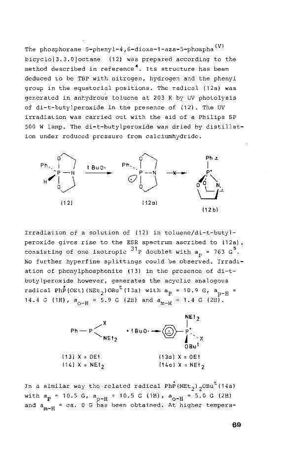
The phosphorane 5-phenyl-4 1 fi-dioxa-1-aza-5-phospha(V)
bicyclo[3.3.0Joctane (12) was prepared according to the
methad described in reference 4. Its structure has been
deduced to be TBP with nitrogen, hydragen and the phenyl
group in the equatorlal positions. The radical (12a) was
generated in anhydrous toluene at 203 K by UV photolysis
of di-t-butylperoxide in the presence of (12). The UV
irradiation was carried out with the aid of a Philips SP
500 w lamp. The di-t-butylperoxide was dried by distillat
ion under reduced pressure from calciumhydride.
Ph,_ 17 Ph, __ l7 Ph~
t Bu 0· I 'p -N P-N ~ p+
H~b~ <!7b~ ~'""-0 0\_ N, u~
11 2) ( 12o) (12 b)
Irradiation of a salution of (12) in toluene/di-t-butyl
peroxide gives rise to the ESR spectrum ascribed to (12a) 1
consisting of one isotropie 31 P doublet with ap = 763 G5
No further hyperfine splittings could be observed. Irradi
ation of phenylphosphonite (13) in the presence of di-t
butylperoxide however 1 generates the acyclic analogous
radical PhP(OEt) (NEt 2 )0But(13a) withap 10.9 G1 ap-H
14.4 G (1H) 1 a0
_H = 5.9 G (2H) and am-H 1.4 G (2H).
(13)X=0Et
114) X:: NEtz
NEtz
I + t BuO· --IQ'- P:_
'e./- j 'x OBut
(13o)X::OEt
l14ol x = NEt 2
In a similar way the related radical PhP(NEt 2 ) 20But(14a)
with ap = 10.5 G, ap-H = 10.5 G (1H) 1 a0
_H = 5.0 G (2H)
and am-H = ca. 0 G has been obtained. At higher tempera-
69
ture (235 K) the t-butylradical has been observed.
The phosphorus h.f.c. of (12a) tagether with the TBP struc
ture of its precursor indicates a TBP-e structure for (12a)
with the unpaired electron on phosphorus in equatorial
position. From the linewidth, aN is estimated less than
5 G. In contrast the h.f.c. values observed for the acyclic
analogous radicals indicate the unpaired electron to be
delocalized on the phenylring, indicating a tetrahedral
configuration of phosphorus as was earlier observed for
PhP(OMe)2But. Apparently the initially formed TBP structure
underwent stereoisomerization to the tetrahedral form 6•
The TBP-e structure of (12a) is stabilized by the two
five-membered rings, which span apical-equatorial positions.
Thus the energy of (12a) is lowered with respect to its
acyclic analogue. On the other hand, the rings will in
crease the energy of the phosphoniurn structure (12b) by
enhanced ring strain and crowding in comparison with the
acyclic species7
• This results in the formation of a
stable TBP-e structure. Therefore it is demonstrated with
(12a) that the valenee statePV on phosphoranyl radicals
in salution can be imposed by incorporation of phosphorus
within a suitably chosen framework.
2. The 3,3,3',3'-tetramethyl-1,1'-spirobi(3H-2,1-benzoxa
phosphvole)-1-yl radical
The phosphorane 3,3,3',3'-tetrarnethyl-1,1 '-spirobi(3H-2,1-
benzoxaphosphole) (15) was prepared according to the methad
described in reference 8• Its structure is inferred as TBP
with hydragen and the phenyl rings located equatorially.
UV irradiation was performed in toluene at 203 K in the
presence of di-t-butylperoxide. A powdered sample of (15)
was X-irradiated at 77 K. The phosphoniurn tetrafluoroborate
(16) was obtained in a similar way to that given in refe
rence9 for the analogous phosphoniumtrifluoromethanesulpho
nate salt by using an 50% salution of fluoroboric acid in
water instead of trifluoromethane sulphonic acid. Electra
lysis of the phosphonium salt was achieved at 228 K in a
flat cell using an acetonitrile solution.
70
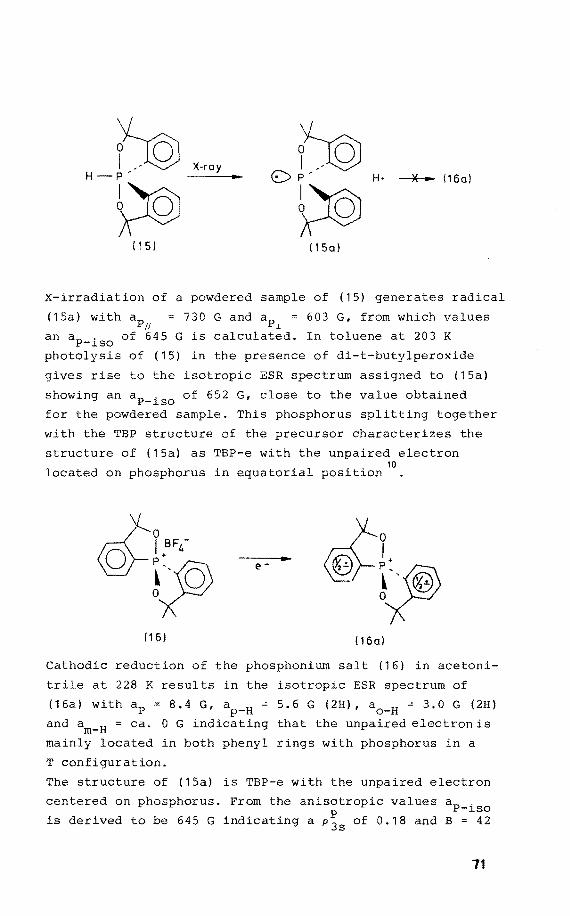
f© X-ray {Jg
H-P' 0 p' H· -----*---- ( 1 6 a I
~ ~ ( 151 ( 1 Sa l
X-irradiation of a powdered sample of (15) generates radical
(15a) with ap11
730 G and ap.L 603 G, from which values
an aP-iso of 645 G is calculated. In toluene at 203 K
photolysis of (15) in the presence of di-t-butylperoxide
gives rise to the isotropie ESR spectrum assigned to (15a)
showing an aP-iso of 652 G, close to the value obtained
for the powdered sample. This phosphorus splitting tagether
with the TBP structure of the precursor characterizes the
structure of (15a) as TBP-e with the unpaired electron 10
located on phosphorus in equatorial position
(16) 116a)
Cathadie reduction of the phosphonium salt (16) in acetoni
trile at 228 K results in the isotropie ESR spectrum of
(16a) withap 8.4 G, ap-H ~ 5.6 G (2H), a0
_H = 3.0 G (2H)
and am-H ca. 0 G indicating that the unpaired electron is
mainly located in both phenyl rings with phosphorus in a
T configuration.
The structure of (15a) is TBP-e with the unpaired electron
centered on phosphorus. From the anisotropic values aP-iso
is derived to be 645 G indicating a p~s of 0.18 and B = 42
71
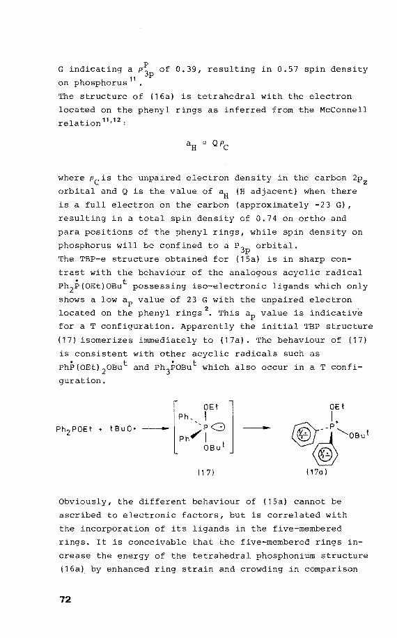
G indicating a p~ of 0.39, resulting in 0.57 spin density 11 p
on phosphorus
The structure of (16a) is tetrahadral with the electron
located on the phenyl rings as inferred from the McConnell
relation 11•12
:
where Pc is the unpaired electron density in the carbon 2p2
orbital and Q is the value of aH (H adjacent) when there
is a full electron on the carbon (approximately -23 G),
resulting in a totalspin density of 0.74 on ortho and
para positions of the phenyl rings, while spin density on
phosphorus will be confined to a P 3p orbital.
The TBP-e structure obtained for (15a) is in sharp con
trast with the behaviour of the analogous acyclic radical
Ph2P(OEt)OBut possessing iso-electronic ligands which only
shows a low ap value of 23 G with the unpaired electron
located on the phenyl rings 2• This value is indicative
for a T configuration. Apparently the initial TBP structure
(17) isomerizes immediately to (17a). The behaviour of (17)
is consistent with ether acyclic radicals such as
PhP(OEt) 20But and Ph3POBut which also occur in aT confi
guration.
[
OEt j Ph,,, I --- PG Ph.,-!
OBut --
I 1 7) l17a I
Obviously, the different behaviour of (15a) cannot be
ascribed to electrooie factors, but is correlated with
the incorporation of its ligands in the five-membered
rings. It is conceivable that the five-membered rings in
crease the energy of the tetrahedral phosphonium structure
(16a) by enhanced ring strain and crewding in camparisen
72
with the acyclic analogous radical (17a) 5 • For structure
(16a), however, strain effects are probably not dominant,
since this radical could be generated by cathadie reduction
of phosphonium salt (16) in acetonitrile at 228 K. It is
known that the stability of PV TBP structures is greatly
enhanced by bridging apical and equatorial positions. So
precursor (15) is surprisingly stable and furthermore
shows no pseudorotatien at room temperature 8, which be
haviour is in sharp contrast with acyclic analogues. Si
milarly, the harrier of stereo-isomerization of (15a) to
(16a) is increased compared with the analogous acyclic
phenylphosphoranyl radical (17) to (17a). Apparently, the V factors which determine pseudorotatien of the P phosphor-
anes (Muetterties' rules) 7 are similar to those deterroin
ing the stereo-isomerization of the corresponding TPB
phenylphosphoranyl radicals (15a) and (17). Moreover this
observation is consistent with the high ap value (600 -2
1000 G) for acyclic phenylphosphoranyl radicals on intro-
duetion of halogen, CF 3CH 2o, H or MeS ligands, which are
highly apicophilic, i.e. these ligands preferentially
occupy apical sites in the TBP PV structure, thus suppress-10
ing pseudorotatien and consequently electron transfer .
The question remains open whether these acyclic phosphoranyl
PV intermediates are TBP-e or TBP-a structures.
73
Heferences and notes
1. G. Boekestein, E.H.J.M. Jansen, and H.M. Buck, J. Chern.
Soc., Chern. Cornrnun., 1974, 118.
2. A.G. Davies, M.J. Parrott, and B.P. Roberts, J. Chern.
Soc., Chern. Cornrnun., 1974, 973.
3. J.M.F. van Dijk, J.F.M. Pennings, and H.M. Buck, J.
Am. Chern. Soc., 1975, 97, 4836.
4. D. Houalla, T. Mouheich, M. Sanchez, and R. Wolf,
Phosphorus, 1975, 5, 229.
5. J.H.H. Harnerlinck, P. Schipper and H.M. Buck, J. Chern.
Soc., Chern. Comrnun., 1979, 350.
6. W.G. Bentrude, Phosphorus and Sulfur, 1977, 3, 109.
7. R.F. Hudson and c. Brown, Acc. Chern. Res., 1972, 5,
204.
8. I. Granoth and J.C. Martin, J. Am. Chern. Soc., 1979,
1011 4623,
9. I. Granoth and J.C. Martin, J. Am. Chern. Soc., 1979,
1011 4618,
10. J.H.H. Harnerlinck, P. Schipper and H.M. Buck, J. Chern.
Soc., Chern. Comrnun., 1980, 975.
11. P.W. Atkins and M.C.R. Syrnons, "The Structure of Inor
ganic Radicals", Elsevier, Amsterdam, 1967.
12. A. Carrington and A.D. McLachlan, "Introduction to
Magnetic Resonance", Harper and Row, New York, 1969.
13. G. Boekestein, Thesis Eindhoven, 1975.
74
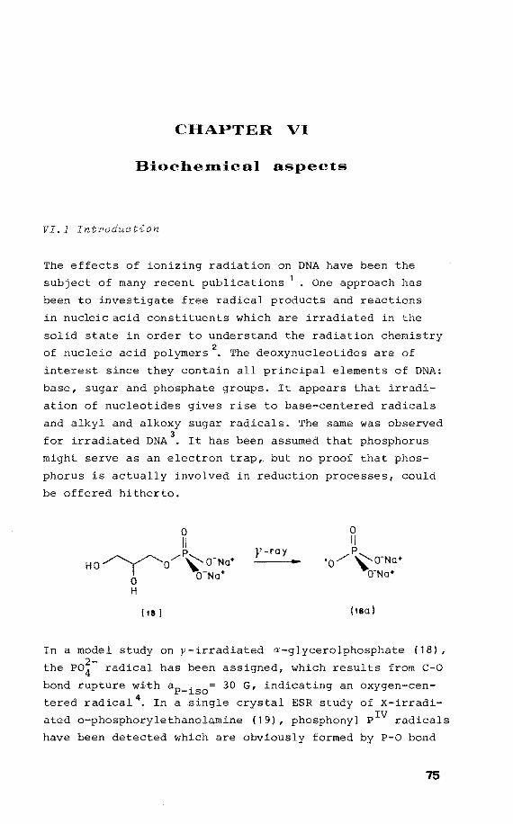
CHAPTER VI
Biochemica) aspects
VI.l Introduetion
The effects of ionizing radiation on DNA have been the
subject of many recent publications 1 • One approach has
been to investigate free radical products and reactions
in nucleicacid constituents which are irradiated in the
solid state in order to understand the radiation chemistry
of nucleic acid polymers2
• The deoxynucleotides are of
interest since they contain all principal elements of DNA:
base, sugar and phosphate groups. It appears that irradi
ation of nucleotides gives rise to base-centered radicals
and alkyl and alkoxy sugar radicals. The same was observed
for irradiated DNA3
• It has been assumed that phosphorus
might serve as an electron trap, but no proof that phos
phorus is actually involved in reduction processes, could
be affered hitherto.
y-ray
( 18) (18a I
In a model study on y-irradiated a-glycerolphosphate (18),
the PO~- radical has been assigned, which results from c-o bond rupture with aP-iso= 30 G, indicating an oxygen-cen
tered radical 4. In a .single crystal ESR study of X-irradi
ated o-phosphorylethanolamine (19), phosphonyl PIV radicals
have been detected which are obviously formed by P-O bond
75
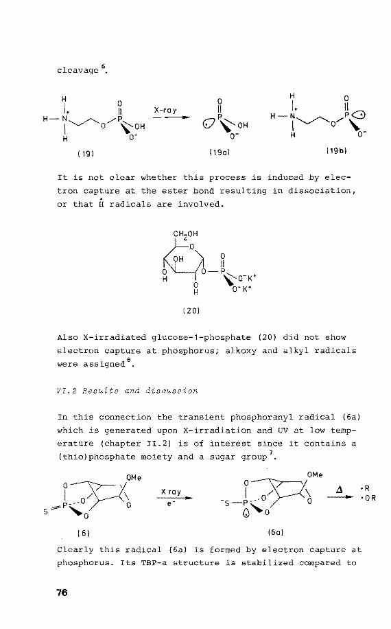
cleavage 5 •
H 0 I. 11 X-ray
0 11 p H
H 0
I+ 11 /""":\ N P......_:., H-N_ ./'-... /p I "'-.../ '0 ~OH
H o-0 '\:'oH
o- ~~o'' H o-
! 191 (19al !19bl
It is not clear whether this process is induced by elec-
tron capture at the ester bond
or that H radicals are involved.
( 20)
in dissociation,
Also X-irradiated glucose-1-phosphate (20) did not show
electron capture at phosphorus; alkoxy and alkyl radicals
were assigned 6 •
VI.2 ResuZts and disoussion
In this conneetion the transient phosphoranyl radical (6a)
which is generated upon X-irradiation and UV at low temp
erature (chapter II.2) is of interest since it contains a 7 (thio)phosphate moiety and a sugar group .
OMe
J::P6 s ""'o
x
(6) (6a)
•R •OR
Clearly this radical (6a) is formed by electron capture at
phosphorus. Its TBP-a structure is stabilized compared to
76
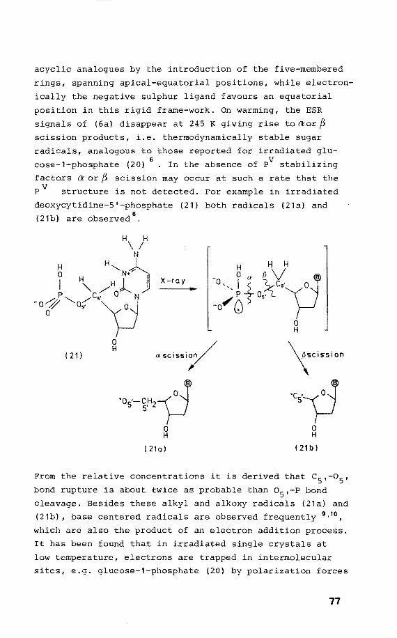
acyclic analogues by the introduetion of the five-membered
rings, spanning apical-equatorial positions, while electron
ically the negative sulphur ligand faveurs an equatorial
position in this rigid frame-work. On warming, the ESR
signals of (6a) disappear at 245 K giving rise to aor f3 scission products, i.e. thermodynamically stable sugar
radicals, analogous to those reported for irradiated glu
cose-1-phosphate (20) 6
• In the absence of PV stabilizing
factors a or f3 scission may occur at such a rate that the
P V structure is not detected. For example in irradiated
deoxycytidine-5'-phosphate (21) bath radicals (21a) and 8
(21b) are observed .
·os-~·"'~ 0 H
( 21a l
0 H
! 21 bI
From the relative concentrations it is derived that c5
,-o5 ,
bondrupture is about twice as probable than o5
,-P bond
cleavage. Besides these alkyl and alkoxy radicals (21a) and
(21b), base centered radicals are observed frequently 9 •10 ,
which are also the product of an electron addition process.
It has been found that in irradiated single crystals at
low temperature, electrans are trapped in intermolecular
sites, e.g. glucose-1-phosphate (20) by polarization farces
77
arising form nearby hydroxy groups 6
• At warming their
ESR signals disappear irreversibly. It is suggested now
that as a result, the phosphate moiety may be attacked by
electrons released from suitably situated holes, generating
a phosphoranyl radical anion which decomposes rapidly by
a and/or~scission in the absence of stabilizing factors
(i.e. five-membered rings, suitably chosen ligands), re
sulting in the usually detected alkyl and alkoxy radicals.
This mechanism, which is supported by the results obtained
for (6a), has to be checked on nucleotides, and this may
be achieved by 'fast response ESR'. In this way the ESR
spectra are recorded several microseconds after a UV laser 11
pulse . With this technique it should be possible to de-
tect the transient phosphoranyl PV radical before it is
destroyed by cleavage processes.
78
rences and notes
1. M. Dizdaroglu, C. von Sonntag, and D. Schulte-Frohlinde,
J. Am. Chem. Soc., 1975, 9?, 2277.
2. D.M. Close and W.A. Bernhard, J. Chem. Phys., 1979,
?0, 210.
3. For up-to-date reviews on the subject, see the series
"Electron Spin Resonance" (Specialist Periodical Re
ports), The Royal Society of Chemistry, London.
4. B. Rakvin and J.N. Herak, J. Chem. Phys., 1981, ?4,
2091 .
5. G.W. Fouse and W.A. Bernhard, J. Chem. Phys., 1979,
?0, 1667.
6. S.E. Locher and H.C. Box, J. Chem. Phys., 1980, ?2,
828.
7. J.H.H. Hamerlinck, P. Schipper and H.M. Buck, J. Chem.
Phys., 1982, in press.
8. D. Krilov, A. Velenik, and J.N. Herak, J. Chem. Phys.,
1978, 69, 2420.
9. J.N. Herak, D. Krilov, C.A. McDowell, J. Magn. Reson.,
1976, 23, 1.
10. D.M. Close, G.W. Fouse, and W.A. Bernhard, J. Chem.
Phys., 1977,66,4689.
11. L. Kevan and R.N. Schwartz, "Time Domain Electron Spin
Resonance", Wiley-Interscience, New York, 1979.
79
AppendÎx
1. Optiaal examination of an orthorhombia arystal
Light which vibrates in a plane perpendicular to the di
rection of propagation,is plane polarized light. A
izing microscope enables us to examine crystals
polarized light and to abserve the effect,the
on the plane of polarization. The analyzer
of the microscope each have the ability to
which passes through them. The analyzer usually has its
polarization direction perpendicular to that of the
izer, and when a crystal is observed in this way, the
crystal is said to be observed between crossed
Light which enters an optically anisatrapie
solved into two components vibrating in mutually
is re-
cular planes, i.e. double refraction occurs. If an anisa
tropie crystal is rotated on the microscope stage between
crossed polars, the light passing through it is only ex
tinguished in certain special orientations, each position
of extinction being separated by an angle of 90° from the
adjacent extinction positions. A relationship can be
deduced between the positions at which a crystal extinguishes
and the optie axes of the crystal. An optie axis is a
direction in the crystal in which there is no double re
fraction. Orthorhombic crystals have two optie axes from
which the directions of the crystallographic axes are
deduced. Identification of these axes is then per-
formed by the rotating crystal method.
2. The rotating arystaZ methad
This technique is very similar to the Debye-Scherrer (D-S)
method, in fact the same camera and monochromatic radiation
80
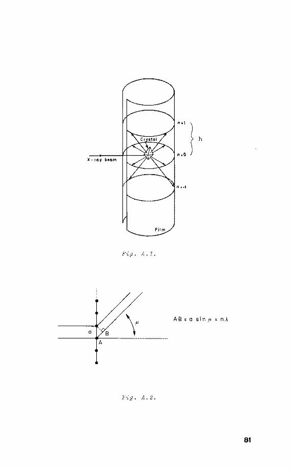
"., \
~ h
n.o X-ray beam
. A. 1.
a
• A. 2.
81
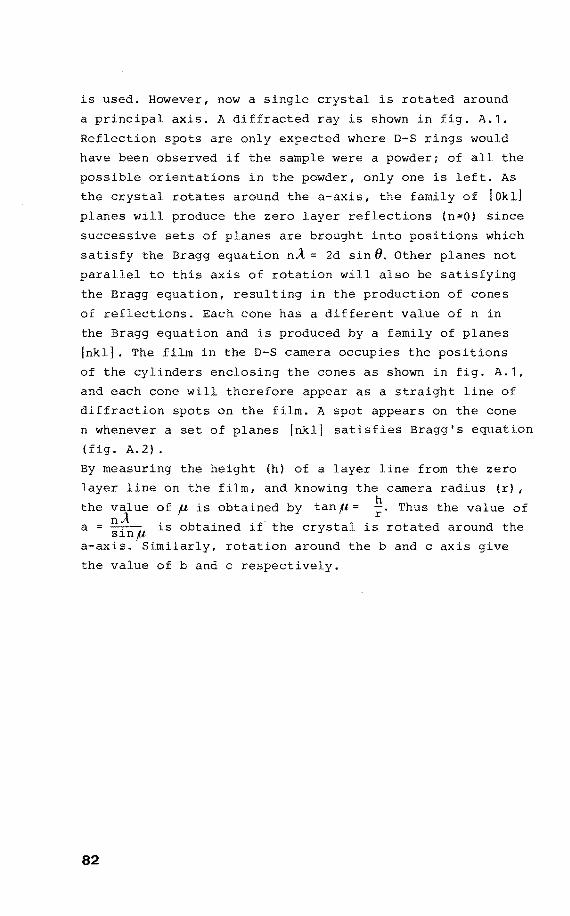
is used. However, now a single crystal is rotated around
a principal axis. A diffracted ray is shown in fig. A.1.
Reflection spots are only expected where D-S rings would
have been observed if the sample were a powderi of all the
possible orientations in the powder, only one is left. As
the crystal rotates around the a-axis, the family of [Okl]
planes will produce the zero layer reflections (n=O) since
successive sets of planes are brought into positions which
satisfy the Bragg equation nÀ 2d sin 8. Other planes nat
parallel to this axis of rotatien will also be satisfying
the Bragg equation, resulting in the production of cones
of reflections. Each cone has a different value of n in
the Bragg equation and is produced by a family of planes
[nkl] . The film in the D-S camera accupies the positions
of the cylinders enclosing the cones as shown in . A.1,
and each cone will therefore appear as a straight line of
diffraction spots on the film. A spot appears on the cone
n whenever a set of planes [nkl] satisfies Bragg's equation
(fig. A.2).
By measuring the height (h) of a layer line from the zero
layer line on the film, and knowing the camera radius (r) , h
the value of ft is obtained by tanp r" Thus the value of nÀ . b . d a sinp 1s o ta1ne if the crystal is rotated around the
a-axis. Similarly, rotatien around the b and c axis give
the value of b and c respectively.
82
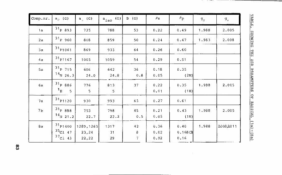
Comp.nr. a,; (G) aJ. ( G) a. ~so
(G)
1a 31p 893 735 788
2a 31p 960 808 859
3a 31 P1061 869 I 933
4a 31 P1167 1005 1059
Sa 31p 715 606 642 14N 26.3 24.0 24.8
6a 31p 886 776 813 1H 5 5 5
7a 31 P1120 930 993
7b 31p 888 753 798 14N 21 . 2 22.7 22.2
8a 31 P1400 1289,1263 1317 35Cl 4 7 23,24 31 3 7
Cl 43 22,22 29
B (G) Ps
53 0.22
50 0.24
64 0.26
54 0.29 I 36 0. 18
0.8 0.05
37 0.22
0.01
63 0.27
45 0.21
0.5 0.05
42 0.36
8 0.02
7 0.02
Pp
0.49
0.47
0.60
0.51
0.35
( 2N)
0.35
( 1 H)
0.61
0.43
( 1 N)
0.40
0. 1 6(1 Cl)
0. 1 6
g//
1. 988
1 . 983
1. 988
1. 988
1. 988
gj_
2.005
2.008
2.005
2.005
2.008,2011
00 PI
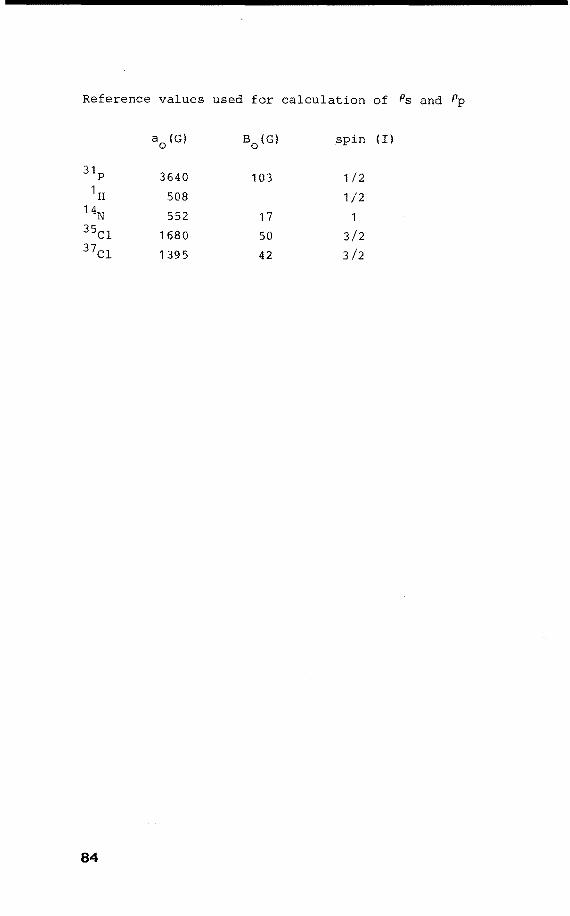
Reference values used for calculation of Ps and Pp
a0
(G) B0
(G) spin (I)
31p 3640 103 1/2 1H 508 1/2
14N 552 17 1 35Cl 1680 50 3/2 37c1 1 395 42 3/2
84
Summary
This thesis deals with the structure and the dynamic be
haviour of phosphoranyl radicals. These radicals result
from the interaction of ionizing radiation and organophos
phorus compounds, in which process selective homolytic
bond cleavage or electron addition takes place. When single
crystals are irradiated, it appears that the radicals re
tain the original orientation of the parent molecules. In
this way eentres are formed which are ideally arranged for
Electron Spin Resonance (ESR) spectroscopy. The anisatrapie
ESR spectra contain information about the 3s and 3p charac
ter of phosphorus and the direction of the hybrid orbital
in which the unpaired electron is located.
The main characteristic of the unpaired electron is its
ligand nature: it can occupy an equatorial (e) as well as
an apical (a) site in the trigonal bipyramidal (TBP)
structure of phosphorus. Since bath the TBP-e and TBP-a
contiguration are formed in one sample of HP(OCH2CH2 J 3N+BF~ upon X-irradiabion, comparison of the two stereo-isoroers is
possible. It appears that the amount of s character in the
apical bands is reduced in comparison with the equatorial
bands, in accordance with the observed differences in re
activity between apical and equatorial sites in the TBP
structure. Besides these TBP-a and TBP-e structures, the
octahedral geometry of ClP(o2c6H4 J 2 is established here,
with the unpaired electron and chlorine in the axial axis.
Intramolecular ligand reorganization in phosphoranyl ra
dicals is studied in a single crystal, which reveals the
exact mode of permutation: a Berry pseudorotatien with the
unpaired electron acting as the pivot. It is found that the
intermediate square pyramidal (SP) geometry in this process
85

lies only slightly above the TBP-e structure. Furthermore
it is demonstrated that the factors which determine pseudo
rotatien are similar to those determining intramolecular
electron transfer. In this process the initial a-radical
undergoes stereo-isomerization to a n-radical.
Electron addition to phosphorus in the thiophosphate 0,0,0-
triester of ~-D upon UV or X-irradiation
generates the radical anion in which phosphorus appears to
be in a TBP-a configuration. According to the behaviour of
this model compound a new mechanism is proposed for strand
break in DNA upon irradiation: initial electron attack at
phosphorus generates a phosphoranyl radical which subse
quently produces alkyl and alkoxy radicals by ~ and/or a scission processes. This mechanism accounts for the reaction
products which are observed in irradiated nucleo-
tides, i.e. alkyl and alkoxy radicals.
86
Samenvatting
In dit proefschrift worden de structuur en het dynamisch
gedrag van fosforanylradicalen beschreven. Deze radicalen
worden gevormd door de interactie van ioniserende straling
met organofosforverbindingen, waarbij selectief bindingen
verbroken worden of electronadditie optreedt. Bij bestra
ling van éénkristallen blijkt, dat de radicalen de oor
spronkelijke oriëntatie behouden van de moleculen waaruit
ze zijn ontstaan. De op deze manier gerangschikte radica
len zijn met electronenspinresonantie (ESR) spectroscopie
bestudeerd. De anisotrope ESR spectra leveren informatie
op over zowel de richting als de hybridisatie van de orbi
tal die het ongepaarde electron bevat.
Kenmerkend voor het ongepaarde electron is het ligand ka
rakter ervan: het kan een equatoriale (e) of apicale (a)
positie innemen in de trigonale bipiramidale (TBP) struc
tuur van fosfor. Aangezien beide vormen, TBP-e en ~BP-a
voorkomen in HP(OCH 2CH2 ) 3N+BF~ bij röntgenbestraling, is
het mogelijk de twee stereo-isomeren met elkaar te verge
lijken. Het blijkt dat de 3s bijdrage in de apicale bin
ding geringer is dan die in de equatoriale binding. Dit
komt overeen met de verschillen in reactiviteit tussen de
apicale en equatoriale plaatsen in fosforanen. Behalve deze
TBP-e en TBP-a configuraties is de structuur van
ClP(02c6H4 J 2 bepaald. Hierin is fosfor 6-waardig met het
chlooratoom en het ongepaarde electron in de as van de
octaëder.
Intramoleculaire liganduitwisseling in fosforanylradicalen
is bestudeerd in een é.énkristal, waardoor de permutatie
precies kon worden vastgesteld: een Berry pseudorotatie
met het ongepaarde electron als pivot. Het blijkt dat het
87
intermediair in dit proces, een vierkant piramidale struc
tuur met het ongepaarde electron apicaal, in energie slechts
weinig boven de TBP-e structuur ligt. Verder wordt aange
toond dat de factoren welke pseudorotatie bepalen, ook in
tramoleculaire electrenenoverdracht bewerkstelligen. In
dit proces vindt stereo-isomerizatie plaats van een aradicaal naar een n-radicaal. Bij UV of röntgenbestraling
van de thiofosfaat 0,0,0-triëster van ~-D-ribopyranoside
vindt electronadditie plaats aan fosfor, waarbij het radi
caal anion wordt gevormd. De structuur hiervan is TBP-a.
Aan de hand van het gedrag van dit modelsysteem is een
nieuw mechanisme opgesteld voor ketenbreuk in DNA onder
invloed van ioniserende straling: electronadditie aan fos
for genereert een fosforanylradicaal dat vervolgens disso
cieert met als reactieproducten alkyl en alkoxy radicalen.
Met dit mechanisme kunnen de reactieprodukten welke ont
staan bij bestraling van nucleotiden verklaard worden.
88
CurriculuiD vitae
De auteur van dit proefschrift werd geboren op 17 juli
1952 te Bandung (Indonesië). Na het behalen van het di
ploma gymnasium-p aan het Sint Miehiellyceum te Geleen
in 1970, werd in hetzelfde jaar begonnen met de studie
aan de Technische Hogeschool te Eindhoven. Het afstudeer
werk werd verricht bij de vakgroep Organische Chemie on-
der van prof. dr. H.M. Buck en dr. ir. W.G.
Voncken. In januari 1976 werd het ingenieursexamen met
lof
Na het vervullen van de militaire dienstplicht werd van
af mei 1978 tot mei 1982 in dienst van de Nederlandse
Stichting voor Zuiver Wetenschappelijk Onderzoek het on
derzoek, beschreven in dit proefschrift, uitgevoerd onder
leiding van prof. dr. H.M. Buck.
89
Dank~oord
Gedurende het onderzoek heb ik van velen positieve im
pulsen ontvangen ter stimulering van mijn onderzoek. Zo
wel voor de morele als technische ondersteuning dank ik
eenieder die aan de totstandkoming van dit proefschrift
heeft bijgedragen.
The work described in this thesis was supported by the
Netherlands Foundation for Chemical Research (S.O.N.)
with financial aid from the Netherlands Organization for
the Advancement of Pure Research (Z.W.O.)
90
STELLINGEN
behorend bij het proefschrift van
J.H.H. Harnerlinck
Eindhoven, 14 mei 1982
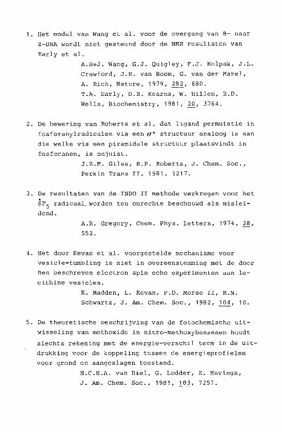
1. Het model van Wang et al. voor de overgang van B- naar
Z-DNA wordt niet gesteund door de NMR resultaten van
Early et al.
A.H~J. Wang, G.J. Quigley, F.J. Kolpak, J.L.
Crawford, J.H. van Boom, G. van der Marel,
A. Rich, Nature, 1979, 282, 680.
T.A. Early, D.R. Kearns, w. Rillen, R.D.
Wells, Biochemistry, 1981, 20, 3764.
2. De bewering van Roberts et al. dat ligand permutatie in
fosforanylradicalen via een a* structuur analoog is aan
die welke via een piramidale structuur plaatsvindt in
fosforanen, is onjuist.
J.R.M. Giles, B.P. Roberts, J. Chem. Soc.,
Perkin Trans II, 1981, 1217.
3. De resultaten van de INDO II methode verkregen voor het
SF5 radicaal,worden ten onrechte beschouwd als mislei
dend.
A.R. Gregory, Chem. Phys. Letters, 1974, 28,
552.
4. Het door Kevan et al. voorgestelde mechanisme voor
vesicle-tumbling is niet in overeenstemming met de door
hen beschreven electron spin echo experimenten aan le
cithine vesicles.
K. Madden, L. Kevan, P.D. Morse II, R.N.
Schwartz, J. Am. Chem. Soc., 1982, 104, 10.
5. De theoretische beschrijving van de fotochemische uit
wisseling van methoxide in nitro-methoxybenzenen houdt
slechts rekening met de energie-verschil term in de uit
drukking voor de koppeling tussen de energieprofielen
voor grond en aangeslagen toestand.
H.C.H.A. van Riel, G. Lodder, E. Havinga,
J. Am. Chem. Soc., 1981, 103, 7257.
6. Het meten van de 14N T1
waarden en de JCN koppeling is
niet voldoende om de verandering in de aggregatietoestand
van cholinefosfolipiden te volgen, aangezien geen reke
ning wordt gehouden met het 1H - 13c ontkoppelvermogen.
R. Murari, W.J. Baumann, J. Am. Chem. Soc.,
1981, 1 1238,
7. Het gebruik van de term microgolf indien straling met
een golflengte van enkele centimeters bedoeld wordt, is
misleidend.
8. Het feit dat wolken organisch materiaal in de melkweg
voorkomen met een massa die de totale massa van de aarde
verre overtreft, en de ontdekking van celstructuren in
meteorieten welke ouder z dan de aarde, laat toe te
veronderstellen dat het leven niet op aarde is ontstaan.
9. Het gebruiken van een bindingsstreepje om de positie
van het ongepaarde electron in de structuurformule van
fosforanylradicalen aan te geven dient ten stelligste
te worden afgeraden.
B.P. Roberts, J.C. Scaiano, J. Chem. Soc.,
Perkin Trans II, 1981, 905.
10. Het verdient aanbeveling om op auto's te vermelden dat
gebruik ervan schadelijk is voor de gezondheid.
11. In het kader van de in te voeren trimesterindeling aan
universiteiten en hogescholen verdient het aanbeveling
behalve de datum van opening van het academisch jaar
ook de sluiting ervan aan te geven.