Subcortical volumes across the lifespan: Data from 18,605 ...
18
RESEARCH ARTICLE Subcortical volumes across the lifespan: Data from 18,605 healthy individuals aged 3–90 years Danai Dima 1,2 | Amirhossein Modabbernia 3 | Efstathios Papachristou 4 | Gaelle E. Doucet 5 | Ingrid Agartz 6,7,8 | Moji Aghajani 9,10 | Theophilus N. Akudjedu 11,12 | Anton Albajes-Eizagirre 13,14 | Dag Alnæs 6,15 | Kathryn I. Alpert 16 | Micael Andersson 17 | Nancy C. Andreasen 18 | Ole A. Andreassen 6 | Philip Asherson 19 | Tobias Banaschewski 20 | Nuria Bargallo 21,22 | Sarah Baumeister 20 | Ramona Baur-Streubel 23 | Alessandro Bertolino 24 | Aurora Bonvino 24 | Dorret I. Boomsma 25 | Stefan Borgwardt 26 | Josiane Bourque 27 | Daniel Brandeis 20 | Alan Breier 28 | Henry Brodaty 29 | Rachel M. Brouwer 30 | Jan K. Buitelaar 31,32,33 | Geraldo F. Busatto 34 | Randy L. Buckner 35,36 | Vincent Calhoun 37 | Erick J. Canales-Rodríguez 13,14 | Dara M. Cannon 12 | Xavier Caseras 38 | Francisco X. Castellanos 39 | Simon Cervenka 8,40 | Tiffany M. Chaim-Avancini 34 | Christopher R. K. Ching 41 | Victoria Chubar 42 | Vincent P. Clark 43,44 | Patricia Conrod 45 | Annette Conzelmann 46 | Benedicto Crespo-Facorro 14,47 | Fabrice Crivello 48 | Eveline A. Crone 49,50 | Anders M. Dale 51 | Christopher Davey 52 | Eco J. C. de Geus 25 | Lieuwe de Haan 53 | Greig I. de Zubicaray 54 | Anouk den Braber 25 | Erin W. Dickie 55,56 | Annabella Di Giorgio 57 | Nhat Trung Doan 6 | Erlend S. Dørum 6,58,59 | Stefan Ehrlich 60,61 | Susanne Erk 62 | Thomas Espeseth 58,63 | Helena Fatouros-Bergman 8,40 | Simon E. Fisher 33,64 | Jean-Paul Fouche 65 | Barbara Franke 33,66,67 | Thomas Frodl 68 | Paola Fuentes-Claramonte 13,14 | David C. Glahn 69 | Ian H. Gotlib 70 | Hans-Jörgen Grabe 71,72 | Oliver Grimm 73 | Nynke A. Groenewold 65,74 | Dominik Grotegerd 75 | Oliver Gruber 76 | Patricia Gruner 77,78 | Rachel E. Gur 27,79,80 | Ruben C. Gur 27,79,80 | Ben J. Harrison 81 | Catharine A. Hartman 82 | † Members of the Karolinska Schizophrenia Project (KaSP): Göran Engberg 1 , Sophie Erhardt 1 , Lilly Schwieler 1 , Funda Orhan 1 , Anna Malmqvist 1 , Mikael Hedberg 1 , Lars Farde 2 , Simon Cervenka 2 , Lena Flyckt 5 , Karin Collste 2 , Pauliina Ikonen 2 , Fredrik Piehl 3 , Ingrid Agartz 4,5 1 Department of Physiology and Pharmacology, Karolinska Institute, Sweden; 2 Department of Clinical Neuroscience, Center for Psychiatry Research, Karolinska Institutet, Sweden; 3 Neuroimmunology Unit, Department of Clinical Neuroscience, Karolinska Institutet, Sweden; 4 NORMENT, Division of Mental Health and Addiction, KG Jebsen Centre for Psychosis Research, University of Oslo and Department of Psychiatric Research, Diakonhjemmet Hospital, Norway; 5 Center for Psychiatric Research, Department of Clinical Neuroscience, Karolinska Institutet, Sweden. Received: 22 June 2020 Revised: 27 November 2020 Accepted: 6 December 2020 DOI: 10.1002/hbm.25320 This is an open access article under the terms of the Creative Commons Attribution License, which permits use, distribution and reproduction in any medium, provided the original work is properly cited. © 2021 The Authors. Human Brain Mapping published by Wiley Periodicals LLC. Hum Brain Mapp. 2021;1–18. wileyonlinelibrary.com/journal/hbm 1
Transcript of Subcortical volumes across the lifespan: Data from 18,605 ...
R E S E A R CH A R T I C L E
Subcortical volumes across the lifespan: Data from 18,605healthy individuals aged 3–90 years
Danai Dima1,2 | Amirhossein Modabbernia3 | Efstathios Papachristou4 |
Gaelle E. Doucet5 | Ingrid Agartz6,7,8 | Moji Aghajani9,10 |
Theophilus N. Akudjedu11,12 | Anton Albajes-Eizagirre13,14 | Dag Alnæs6,15 |
Kathryn I. Alpert16 | Micael Andersson17 | Nancy C. Andreasen18 |
Ole A. Andreassen6 | Philip Asherson19 | Tobias Banaschewski20 |
Nuria Bargallo21,22 | Sarah Baumeister20 | Ramona Baur-Streubel23 |
Alessandro Bertolino24 | Aurora Bonvino24 | Dorret I. Boomsma25 |
Stefan Borgwardt26 | Josiane Bourque27 | Daniel Brandeis20 | Alan Breier28 |
Henry Brodaty29 | Rachel M. Brouwer30 | Jan K. Buitelaar31,32,33 |
Geraldo F. Busatto34 | Randy L. Buckner35,36 | Vincent Calhoun37 |
Erick J. Canales-Rodríguez13,14 | Dara M. Cannon12 | Xavier Caseras38 |
Francisco X. Castellanos39 | Simon Cervenka8,40 | Tiffany M. Chaim-Avancini34 |
Christopher R. K. Ching41 | Victoria Chubar42 | Vincent P. Clark43,44 |
Patricia Conrod45 | Annette Conzelmann46 | Benedicto Crespo-Facorro14,47 |
Fabrice Crivello48 | Eveline A. Crone49,50 | Anders M. Dale51 |
Christopher Davey52 | Eco J. C. de Geus25 | Lieuwe de Haan53 |
Greig I. de Zubicaray54 | Anouk den Braber25 | Erin W. Dickie55,56 |
Annabella Di Giorgio57 | Nhat Trung Doan6 | Erlend S. Dørum6,58,59 |
Stefan Ehrlich60,61 | Susanne Erk62 | Thomas Espeseth58,63 |
Helena Fatouros-Bergman8,40 | Simon E. Fisher33,64 | Jean-Paul Fouche65 |
Barbara Franke33,66,67 | Thomas Frodl68 | Paola Fuentes-Claramonte13,14 |
David C. Glahn69 | Ian H. Gotlib70 | Hans-Jörgen Grabe71,72 |
Oliver Grimm73 | Nynke A. Groenewold65,74 | Dominik Grotegerd75 |
Oliver Gruber76 | Patricia Gruner77,78 | Rachel E. Gur27,79,80 |
Ruben C. Gur27,79,80 | Ben J. Harrison81 | Catharine A. Hartman82 |
†Members of the Karolinska Schizophrenia Project (KaSP): Göran Engberg1, Sophie Erhardt1, Lilly Schwieler1, Funda Orhan1, Anna Malmqvist1, Mikael Hedberg1, Lars Farde2, Simon Cervenka2, Lena
Flyckt5, Karin Collste2, Pauliina Ikonen2, Fredrik Piehl3, Ingrid Agartz4,5
1Department of Physiology and Pharmacology, Karolinska Institute, Sweden; 2Department of Clinical Neuroscience, Center for Psychiatry Research, Karolinska Institutet, Sweden;3Neuroimmunology Unit, Department of Clinical Neuroscience, Karolinska Institutet, Sweden; 4NORMENT, Division of Mental Health and Addiction, KG Jebsen Centre for Psychosis Research,
University of Oslo and Department of Psychiatric Research, Diakonhjemmet Hospital, Norway; 5Center for Psychiatric Research, Department of Clinical Neuroscience, Karolinska Institutet,
Sweden.
Received: 22 June 2020 Revised: 27 November 2020 Accepted: 6 December 2020
DOI: 10.1002/hbm.25320
This is an open access article under the terms of the Creative Commons Attribution License, which permits use, distribution and reproduction in any medium,
provided the original work is properly cited.
© 2021 The Authors. Human Brain Mapping published by Wiley Periodicals LLC.
Hum Brain Mapp. 2021;1–18. wileyonlinelibrary.com/journal/hbm 1
Sean N. Hatton83 | Andreas Heinz62 | Dirk J. Heslenfeld84 | Derrek P. Hibar85 |
Ian B. Hickie83 | Beng-Choon Ho18 | Pieter J. Hoekstra86 | Sarah Hohmann20 |
Avram J. Holmes87 | Martine Hoogman33,66 | Norbert Hosten88 |
Fleur M. Howells65,74 | Hilleke E. Hulshoff Pol30 | Chaim Huyser89 |
Neda Jahanshad41 | Anthony James90 | Terry L. Jernigan91 | Jiyang Jiang29 |
Erik G. Jönsson6,8,40 | John A. Joska65 | Rene Kahn3 | Andrew Kalnin92 |
Ryota Kanai93 | Marieke Klein33,66,94 | Tatyana P. Klyushnik95 |
Laura Koenders53 | Sanne Koops30 | Bernd Krämer76 | Jonna Kuntsi19 |
Jim Lagopoulos96 | Luisa Lázaro97,14 | Irina Lebedeva95 | Won Hee Lee3 |
Klaus-Peter Lesch98 | Christine Lochner99 | Marise W. J. Machielsen53 |
Sophie Maingault48 | Nicholas G. Martin100 | Ignacio Martínez-Zalacaín14,101 |
David Mataix-Cols8,40 | Bernard Mazoyer48 | Colm McDonald12 |
Brenna C. McDonald28 | Andrew M. McIntosh102 | Katie L. McMahon103 |
Genevieve McPhilemy12 | José M. Menchón14,101 | Sarah E. Medland100 |
Andreas Meyer-Lindenberg104 | Jilly Naaijen32,33 | Pablo Najt12 |
Tomohiro Nakao105 | Jan E. Nordvik106 | Lars Nyberg17,107 | Jaap Oosterlaan108 |
Víctor Ortiz-García de la Foz14,109,110 | Yannis Paloyelis2 | Paul Pauli23,111 |
Giulio Pergola24 | Edith Pomarol-Clotet13,14 | Maria J. Portella13,112 |
Steven G. Potkin113 | Joaquim Radua8,22,114 | Andreas Reif73 | Daniel A. Rinker6 |
Joshua L. Roffman36 | Pedro G. P. Rosa34 | Matthew D. Sacchet115 |
Perminder S. Sachdev29 | Raymond Salvador13 | Pascual Sánchez-Juan109,116 |
Salvador Sarró13 | Theodore D. Satterthwaite27 | Andrew J. Saykin28 |
Mauricio H. Serpa34 | Lianne Schmaal117,118 | Knut Schnell119 |
Gunter Schumann19,120 | Kang Sim121 | Jordan W. Smoller122 | Iris Sommer123 |
Carles Soriano-Mas14,101 | Dan J. Stein99 | Lachlan T. Strike124 |
Suzanne C. Swagerman25 | Christian K. Tamnes6,7,125 | Henk S. Temmingh65 |
Sophia I. Thomopoulos41 | Alexander S. Tomyshev95 |
Diana Tordesillas-Gutiérrez13,126 | Julian N. Trollor29 | Jessica A. Turner127 |
Anne Uhlmann65 | Odile A. van den Heuvel9 | Dennis van den Meer6,15,128 |
Nic J. A. van der Wee129,130 | Neeltje E. M. van Haren131 | Dennis van't Ent25 |
Theo G. M. van Erp132,113,133 | Ilya M. Veer62 | Dick J. Veltman9 |
Aristotle Voineskos55,56 | Henry Völzke133,134,135 | Henrik Walter62 |
Esther Walton136 | Lei Wang137 | Yang Wang138 | Thomas H. Wassink18 |
Bernd Weber139 | Wei Wen29 | John D. West28 | Lars T. Westlye58 |
Heather Whalley102 | Lara M. Wierenga140 | Steven C. R. Williams2 |
Katharina Wittfeld71,72 | Daniel H. Wolf27 | Amanda Worker2 |
Margaret J. Wright124 | Kun Yang141 | Yulyia Yoncheva142 |
2 DIMA ET AL.
Marcus V. Zanetti34,143 | Georg C. Ziegler144 | Paul M. Thompson41 |
Sophia Frangou3,145 | Karolinska Schizophrenia Project (KaSP)
1Department of Psychology, School of Arts and Social Sciences, City University of London, London, UK
2Department of Neuroimaging, Institute of Psychiatry, Psychology and Neuroscience, King's College London, London, UK
3Department of Psychiatry, Icahn School of Medicine at Mount Sinai, New York, New York
4Psychology and Human Development, Institute of Education, University College London, London, UK
5Boys Town National Research Hospital, Omaha, Nebraska
6Norwegian Centre for Mental Disorders Research (NORMENT), Institute of Clinical Medicine, University of Oslo, Oslo, Norway
7Department of Psychiatric Research, Diakonhjemmet Hospital, Oslo, Norway
8Centre for Psychiatric Research, Department of Clinical Neuroscience, Karolinska Institutet, Stockholm, Sweden
9Department of Psychiatry, Amsterdam University Medical Centre, Location VUmc, Amsterdam, Netherlands
10Institute of Education & Child Studies, Section Forensic Family & Youth Care, Leiden University, Netherlands
11Institute of Medical Imaging and Visualisation, Department of Medical Science and Public Health, Faculty of Health and Social Sciences, Bournemouth University,
Poole, UK
12Clinical Neuroimaging Laboratory, Centre for Neuroimaging and Cognitive Genomics and NCBES Galway Neuroscience Centre, National University of Ireland,
Dublin, Ireland
13FIDMAG Germanes Hospitalàries, Madrid, Spain
14Mental Health Research Networking Center (CIBERSAM), Madrid, Spain
15Division of Mental Health and Addiction, Institute of Clinical Medicine, University of Oslo, Oslo, Norway
16Radiologics, Inc, Chicago, Illinois
17Department of Integrative Medical Biology, Umeå University, Umeå, Sweden
18Department of Psychiatry, Carver College of Medicine, The University of Iowa, Iowa City, Iowa
19Social, Genetic and Developmental Psychiatry Centre, Institute of Psychiatry, Psychology and Neuroscience, King's College London, London, UK
20Department of Child and Adolescent Psychiatry and Psychotherapy, Central Institute of Mental Health, Heidelberg University, Mannheim, Germany
21Imaging Diagnostic Centre, Hospital Clinic, Barcelona University Clinic, Barcelona, Spain
22August Pi i Sunyer Biomedical Research Institut (IDIBAPS), Barcelona, Spain
23Department of Psychology, Biological Psychology, Clinical Psychology and Psychotherapy, University of Würzburg, Wurzburg, Germany
24Department of Basic Medical Science, Neuroscience and Sense Organs, University of Bari Aldo Moro, Bari, Italy
25Department of Biological Psychology, Vrije Universiteit, Amsterdam, Netherlands
26Department of Psychiatry & Psychotherapy, University of Lübeck, Lubeck, Germany
27Department of Psychiatry, University of Pennsylvania, Philadelphia, Pennsylvania
28Department of Radiology and Imaging Sciences, Indiana University School of Medicine, Indianapolis, Indiana
29Centre for Healthy Brain Ageing, School of Psychiatry, University of New South Wales, Sydney, Australia
30Rudolf Magnus Institute of Neuroscience, University Medical Center Utrecht, Utrecht, Netherlands
31Donders Center of Medical Neurosciences, Radboud University, Nijmegen, Netherlands
32Donders Centre for Cognitive Neuroimaging, Radboud University, Nijmegen, Netherlands
33Donders Institute for Brain, Cognition and Behaviour, Radboud University, Nijmegen, Netherlands
34Laboratory of Psychiatric Neuroimaging, Departamento e Instituto de Psiquiatria, Hospital das Clinicas HCFMUSP, Faculdade de Medicina, Universidade de S~ao
Paulo, S~ao Paulo, Brazil
35Department of Psychology, Center for Brain Science, Harvard University, Cambridge, Massachusetts
36Department of Psychiatry, Massachusetts General Hospital, Boston, Massachusetts
37Tri-Institutional Center for Translational Research in Neuroimaging and Data Science (TReNDS), Georgia State University, Georgia Institute of Technology, Emory
University, USA Neurology, Radiology, Psychiatry and Biomedical Engineering, Emory University, Atlanta, Georgia
38MRC Centre for Neuropsychiatric Genetics and Genomics, Cardiff University, Cardiff, UK
39Department of Child and Adolescent Psychiatry, New York University, New York, New York
40Stockholm Health Care Services, Stockholm Region, Stockholm, Sweden
41Imaging Genetics Center, Mark and Mary Stevens Neuroimaging and Informatics Institute, Keck School of Medicine, University of Southern California, Los Angeles,
California
42Department of Neuroscience, KU Leuven, Mind-Body Research Group, Leuven, Belgium
DIMA ET AL. 3
43Department of Psychology, University of New Mexico, Albuquerque, New Mexico
44Mind Research Network, Albuquerque, New Mexico
45Department of Psychiatry, Université de Montréal, Montreal, Canada
46Department of Child and Adolescent Psychiatry, Psychosomatics and Psychotherapy, University of Tübingen, Tubingen, Germany
47HU Virgen del Rocio, IBiS, University of Sevilla, Sevilla, Spain
48Groupe d'Imagerie Neurofonctionnelle, Institut des Maladies Neurodégénératives, UMR5293, Université de Bordeaux, Talence, France
49Erasmus School of Social and Behavioural Sciences, Erasmus University Rotterdam, Rotterdam, Netherlands
50Faculteit der Sociale Wetenschappen, Instituut Psychologie, Universiteit Leiden, Leiden, Netherlands
51Center for Multimodal Imaging and Genetics, Department of Neuroscience and Department of Radiology, University of California-San Diego, La Jolla, California
52Department of Psychiatry, University of Melbourne, Melbourne, Australia
53Academisch Medisch Centrum, Universiteit van Amsterdam, Amsterdam, Netherlands
54Faculty of Health, Institute of Health and Biomedical Innovation, Queensland University of Technology, Brisbane, Australia
55Kimel Family Translational Imaging Genetics Laboratory, Campbell Family Mental Health Research Institute, CAMH, Toronto, Canada
56Department of Psychiatry, University of Toronto, Toronto, Canada
57Biological Psychiatry Lab, Fondazione IRCCS Casa Sollievo della Sofferenza, San Giovanni Rotondo (FG), Italy
58Department of Psychology, University of Oslo, Oslo, Norway
59Sunnaas Rehabilitation Hospital HT, Nesodden, Norway
60Division of Psychological and Social Medicine and Developmental Neurosciences, Technische Universität Dresden, Dresden, Germany
61Faculty of Medicine, Universitätsklinikum Carl Gustav Carus an der TU Dresden, Dresden, Germany
62Division of Mind and Brain Research, Department of Psychiatry and Psychotherapy, Charité-Universitätsmedizin Berlin, Berlin, Germany
63Bjørknes College, Oslo, Norway
64Language and Genetics Department, Max Planck Institute for Psycholinguistics, Nijmegen, Netherlands
65Department of Psychiatry and Mental Health, University of Cape Town, Rondebosch, South Africa
66Department of Human Genetics, Radboud University Medical Center, Nijmegen, Netherlands
67Department of Psychiatry, Radboud University Medical Center, Nijmegen, Netherlands
68Department of Psychiatry and Psychotherapy, Otto von Guericke University Magdeburg, Magdeburg, Germany
69Department of Psychiatry, Tommy Fuss Center for Neuropsychiatric Disease Research Boston Children's Hospital, Harvard Medical School, Boston, Massachusetts
70Department of Psychology, Stanford University, Stanford, California
71Department of Psychiatry and Psychotherapy, University Medicine Greifswald, University of Greifswald, Greifswald, Germany
72German Center for Neurodegenerative Diseases (DZNE), Site Rostock/Greifswald, Greifswald, Germany
73Department for Psychiatry, Psychosomatics and Psychotherapy, Universitätsklinikum Frankfurt, Goethe Universitat, Frankfurt, Germany
74Neuroscience Institute, University of Cape Town, Rondebosch, South Africa
75Department of Psychiatry and Psychotherapy, University of Münster, Munster, Germany
76Section for Experimental Psychopathology and Neuroimaging, Department of General Psychiatry, Heidelberg University, Heidelberg, Germany
77Department of Psychiatry, Yale University, New Haven, Connecticut
78Learning Based Recovery Center, VA Connecticut Health System, New Haven, Connecticut
79Lifespan Brain Institute, Perelman School of Medicine, University of Pennsylvania, Philadelphia, Pennsylvania
80Children's Hospital of Philadelphia, University of Pennsylvania, Philadelphia, Pennsylvania
81Melbourne Neuropsychiatry Center, University of Melbourne, Melbourne, Australia
82Interdisciplinary Center Psychopathology and Emotion regulation, University Medical Center Groningen, University of Groningen, Groningen, Netherlands
83Brain and Mind Centre, University of Sydney, Sydney, Australia
84Departments of Experimental and Clinical Psychology, Vrije Universiteit Amsterdam, Amsterdam, Netherlands
85Personalized Healthcare, Genentech, Inc, South San Francisco, California
86Department of Psychiatry, University Medical Center Groningen, University of Groningen, Groningen, Netherlands
87Department of Psychology, Yale University, New Haven, Connecticut
88Norbert Institute of Diagnostic Radiology and Neuroradiology, University Medicine Greifswald, University of Greifswald, Greifswald, Germany
89Bascule, Academic Centre for Children and Adolescent Psychiatry, Duivendrecht, Netherlands
90Department of Psychiatry, Oxford University, Oxford, UK
91Center for Human Development, Departments of Cognitive Science, Psychiatry, and Radiology, University of California, San Diego, California
4 DIMA ET AL.
92Department of Radiology, Ohio State University College of Medicine, Columbus, Ohio
93Department of Neuroinformatics, Araya, Inc, Tokyo, Japan
94Department of Psychiatry, University of California San Diego, La Jolla, California
95Mental Health Research Center, Russian Academy of Medical Sciences, Moskva, Russia
96Sunshine Coast Mind and Neuroscience, Thompson Institute, University of the Sunshine Coast, Sunshine Coast, Australia
97Department of Child and Adolescent Psychiatry and Psychology, Hospital Clinic, University of Barcelona, Barcelona, Spain
98Department of Psychiatry, Psychosomatics and Psychotherapy, Julius-Maximilians Universität Würzburg, Wurzburg, Germany
99SA MRC Unit on Risk and Resilience in Mental Disorders, Department of Psychiatry, Stellenbosch University, Stellenbosch, South Africa
100Queensland Institute of Medical Research, Berghofer Medical Research Institute, Brisbane, Australia
101Department of Psychiatry, Bellvitge University Hospital-IDIBELL, University of Barcelona, Barcelona, Spain
102Division of Psychiatry, University of Edinburgh, Edinburgh, UK
103School of Clinical Sciences, Institute of Health and Biomedical Innovation, Queensland University of Technology, Brisbane, Australia
104Department of Psychiatry and Psychotherapy, Central Institute of Mental Health, Heidelberg University, Heidelberg, Germany
105Department of Clinical Medicine, Kyushu University, Kyushu, Japan
106CatoSenteret Rehabilitation Hospital, Son, Norway
107Department of Radiation Sciences, Umeå Center for Functional Brain Imaging, Umeå University, Umeå, Sweden
108Department of Clinical Neuropsychology, Amsterdam University Medical Centre, Vrije Universiteit Amsterdam, Amsterdam, Netherlands
109Department of Psychiatry, University Hospital “Marques de Valdecilla”, Instituto de Investigación Valdecilla (IDIVAL), Santander, Spain
110Instituto de Salud Carlos III, Madrid, Spain
111Centre of Mental Health, University of Würzburg, Wurzburg, Germany
112Department of Psychiatry, Hospital de la Santa Creu i Sant Pau, Institut d'Investigació Biomèdica Sant Pau, Universitat Autònoma de Barcelona, Barcelona, Spain
113Department of Psychiatry, University of California at Irvine, Irvine, California
114Department of Psychosis Studies, Institute of Psychiatry, Psychology & Neuroscience, King's College London, London, UK
115Center for Depression, Anxiety, and Stress Research, McLean Hospital, Harvard Medical School, Boston, Massachusetts
116Centro de Investigacion Biomedica en Red en Enfermedades Neurodegenerativas (CIBERNED), Madrid, Spain
117Orygen, The National Centre of Excellence in Youth Mental Health, Parkville, Australia
118Centre for Youth Mental Health, The University of Melbourne, Melbourne, Australia
119Department of Psychiatry and Psychotherapy, University Medical Center Göttingen, Göttingen, Germany
120Centre for Population Neuroscience and Precision Medicine, Institute of Psychiatry, Psychology & Neuroscience, King's College London, London, UK
121Institute of Mental Health, Singapore, Singapore
122Center for Genomic Medicine, Massachusetts General Hospital, Boston, Massachusetts
123Department of Biomedical Sciences of Cells and Systems, Rijksuniversiteit Groningen, University Medical Center Groningen, Göttingen, Netherlands
124Queensland Brain Institute, University of Queensland, Brisbane, Australia
125PROMENTA Research Center, Department of Psychology, University of Oslo, Oslo, Norway
126Neuroimaging Unit, Technological Facilities, Valdecilla Biomedical Research Institute IDIVAL, Santander, Spain
127College of Arts and Sciences, Georgia State University, Atlanta, Georgia
128School of Mental Health and Neuroscience, Faculty of Health, Medicine and Life Sciences, Maastricht University, Maastricht, Netherlands
129Department of Psychiatry, Leiden University Medical Center, Leiden, Netherlands
130Leiden Institute for Brain and Cognition, Leiden, Netherlands
131Department of Child and Adolescent Psychiatry/Psychology, Erasmus University Medical Center, Sophia Children's Hospital, Rotterdam, The Netherlands
132Center for the Neurobiology of Learning and Memory, University of California Irvine, Irvine, California
133Institute of Community Medicine, University Medicine, Greifswald, University of Greifswald, Greifswald, Germany
134German Centre for Cardiovascular Research (DZHK), partner site Greifswald, Greifswald, Germany
135German Center for Diabetes Research (DZD), partner site Greifswald, Greifswald, Germany
136Department of Psychology, University of Bath, Bath, UK
137Department of Psychiatry and Behavioral Sciences, Feinberg School of Medicine, Northwestern University, Chicago, Illinois
138Department of Radiology, Medical College of Wisconsin, Milwaukee, Wisconsin
139Institute for Experimental Epileptology and Cognition Research, University of Bonn, Bonn, Germany
140Developmental and Educational Psychology Unit, Institute of Psychology, Leiden University, Leiden, Netherlands
141National High Magnetic Field Laboratory, Florida State University, Tallahassee, Florida
DIMA ET AL. 5
142Department of Child and Adolescent Psychiatry, Child Study Center, NYU Langone Health, New York, New York
143Instituto de Ensino e Pesquisa, Hospital Sírio-Libanês, S~ao Paulo, Brazil
144Division of Molecular Psychiatry, Center of Mental Health, University of Würzburg, Wurzburg, Germany
145Department of Psychiatry, Djavad Mowafaghian Centre for Brain Health, University of British Columbia, Vancouver, Canada
Correspondence
Sophia Frangou, Department of Psychiatry,
Icahn School of Medicine at Mount Sinai, 1425
Madison Avenue, New York, New York 10029,
USA.
Email: [email protected]
Danai Dima, University of London,
Northampton Square, London EC1V 0HB, UK.
Email: [email protected]
Funding information
National Institute of Mental Health, Grant/
Award Numbers: MH104284, MH116147,
R01MH113619, R01 MH090553,
R01MH117014, R01MH042191; Karolinska
Institutet; Stockholm County Council;
Southern and Eastern Norway Regional Health
Authority; German Centre for Cardiovascular
Research; DZHK; Siemens Healthineers;
University of Queensland; US National
Institute of Child Health and Human
Development, Grant/Award Number:
RO1HD050735; Australian National Health
and Medical Research Council; Eunice
Kennedy Shriver National Institute of Child
Health & Human Development; National
Institute on Drug Abuse, Grant/Award
Numbers: UL1 TR000153, 1 U24
RR025736-01, 1 U24 RR021992; Brain Injury
Fund Research Grant Program; Indiana State
Department of Health Spinal Cord; Parents
Against Childhood Epilepsy; Epilepsy Therapy
Project, Fight Against Childhood Epilepsy and
Seizures; Epilepsy Foundation; American
Epilepsy Society; Knut and Alice Wallenberg
Foundation; European Community's Horizon
2020 Programme; Vici Innovation Program;
NWO Brain & Cognition Excellence Program;
Netherlands Organization for Scientific
Research; Hersenstichting Nederland;
Netherlands Organization for Health Research
and Development; Geestkracht Programme of
the Dutch Health Research Council, Grant/
Award Number: 10-000-1001; Universiteit
Utrecht; FP7 Ideas: European Research
Council; Nederlandse Organisatie voor
Wetenschappelijk Onderzoek; National Center
for Advancing Translational Sciences; National
Institutes of Health; National Center for
Research Resources; Consortium grant, Grant/
Award Number: U54 EB020403; U.S. National
Institutes of Health, Grant/Award Numbers:
R01 CA101318, P30 AG10133, R01
AG19771; Medical Research Council, Grant/
Award Number: G0500092; Fundación
Instituto de Investigación Marqués de
Valdecilla, Grant/Award Numbers: API07/011,
NCT02534363, NCT0235832; Instituto de
Salud Carlos III, Grant/Award Numbers:
PI14/00918, PI14/00639, PI060507,
Abstract
Age has a major effect on brain volume. However, the normative studies available are
constrained by small sample sizes, restricted age coverage and significant methodo-
logical variability. These limitations introduce inconsistencies and may obscure or dis-
tort the lifespan trajectories of brain morphometry. In response, we capitalized on the
resources of the Enhancing Neuroimaging Genetics through Meta-Analysis
(ENIGMA) Consortium to examine age-related trajectories inferred from cross-
sectional measures of the ventricles, the basal ganglia (caudate, putamen, pallidum,
and nucleus accumbens), the thalamus, hippocampus and amygdala using magnetic
resonance imaging data obtained from 18,605 individuals aged 3–90 years. All sub-
cortical structure volumes were at their maximum value early in life. The volume of
the basal ganglia showed a monotonic negative association with age thereafter; there
was no significant association between age and the volumes of the thalamus, amyg-
dala and the hippocampus (with some degree of decline in thalamus) until the sixth
decade of life after which they also showed a steep negative association with age.
The lateral ventricles showed continuous enlargement throughout the lifespan. Age
was positively associated with inter-individual variability in the hippocampus and
amygdala and the lateral ventricles. These results were robust to potential con-
founders and could be used to examine the functional significance of deviations from
typical age-related morphometric patterns.
K E YWORD S
brain morphometry, ENIGMA, longitudinal trajectories, multisite
6 DIMA ET AL.
PI050427, PI020499; the Research Council,
Grant/Award Number: 223273; South Eastern
Norway Regional Health Authority, Grant/
Award Numbers: 2017-112, 2019107;
Swedish Research Council; European
Community's Seventh Framework Programme,
Grant/Award Number: 602450; King's College
London; South London and Maudsley NHS
Foundation Trust; Biomedical Research Centre;
National Institute for Health Research
1 | INTRODUCTION
Over the last 20 years, studies using structural magnetic resonance
imaging (MRI) have confirmed that brain morphometric measures
change with age. In general, whole brain, global and regional gray mat-
ter volumes increase during development and decrease with aging
(Brain Development Cooperative Group, 2012; Driscoll et al., 2009;
Fotenos, Snyder, Girton, Morris, & Buckner, 2005; Good et al., 2001;
Pfefferbaum et al., 2013; Pomponio et al., 2019; Raz et al., 2005;
Raznahan et al., 2014; Resnick, Pham, Kraut, Zonderman, &
Davatzikos, 2003; Walhovd et al., 2011). However, most published
studies are constrained by small sample sizes, restricted age coverage
and methodological variability. These limitations introduce inconsis-
tencies and may obscure or distort the lifespan trajectories of brain
structures. To address these limitations, we formed the Lifespan
Working group of the Enhancing Neuroimaging Genetics through
Meta-Analysis (ENIGMA) Consortium (Thompson et al., 2014, 2017)
to perform large-scale analyses of brain morphometric data extracted
from MRI images using standardized protocols and unified quality
control procedures, harmonized and validated across all participating
sites.
Here we focus on ventricular, striatal (caudate, putamen, nucleus
accumbens), pallidal, thalamic, hippocampal and amygdala volumes.
Subcortical structures are crucial for normal cognitive and emotional
adaptation (Grossberg, 2009). The striatum and pallidum (together
referred to as basal ganglia) are best known for their role in action
selection and movement coordination (Calabresi, Picconi, Tozzi,
Ghiglieri, & Di Filippo, 2014) but they are also involved in other
aspects of cognition particularly memory, inhibitory control, reward
and salience processing (Chudasama & Robbins, 2006; Richard, Cas-
tro, Difeliceantonio, Robinson, & Berridge, 2013; Scimeca &
Badre, 2012; Tremblay, Worbe, Thobois, Sgambato-Faure, &
Féger, 2015). The role of the hippocampus has been most clearly
defined in connection to declarative memory (Eichenbaum, 2004;
Shohamy & Turk-Browne, 2013) while the amygdala has been histori-
cally linked to affect processing (Kober et al., 2008). The thalamus is
centrally located in the brain and acts as a key hub for the integration
of motor and sensory information with higher-order functions
(Sherman, 2005; Zhang, Snyder, Shimony, Fox, & Raichle, 2010). The
role of subcortical structures extends beyond normal cognition
because changes in the volume of these regions have been reliably
identified in developmental (Ecker, Bookheimer, & Murphy, 2015;
Krain & Castellanos, 2006), psychiatric (Hibar et al., 2016; Kempton
et al., 2011; Schmaal et al., 2016; van Erp et al., 2016) and degenera-
tive disorders (Risacher et al., 2009).
Using data from 18,605 individuals aged 3–90 years from the
ENIGMA Lifespan working group we delineated the association
between age and subcortical volumes from early to late life in order to
(a) identify periods of volume change or stability, (b) provide normative,
age-adjusted centile curves of subcortical volumes and (c) quantify
inter-individual variability in subcortical volumes which is considered a
major source of inter-study differences (Dickie et al., 2013; Raz,
Ghisletta, Rodrigue, Kennedy, & Lindenberger, 2010).
2 | MATERIALS AND METHODS
2.1 | Study samples
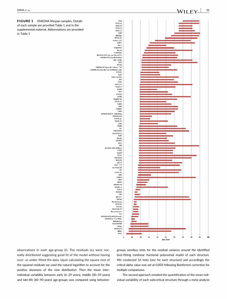
The study data derive from 88 samples comprising 18,605 healthy
participants, aged 3–90 years, with near equal representation of men
and women (48% and 52%) (Table 1, Figure 1). At the time of scan-
ning, participating individuals were screened to exclude the presence
of mental disorders, cognitive impairment or significant medical mor-
bidity. Details of the screening process and eligibility criteria for each
research group are shown in Table S1).
2.2 | Neuroimaging
Detailed information on scanner vendor, magnet strength and acquisi-
tion parameters for each sample are presented in Table S1. For each
sample, the intracranial volume (ICV) and the volume of the basal
ganglia (caudate, putamen, pallidum, nucleus accumbens), thalamus,
hippocampus, amygdala and lateral ventricles were extracted using
FreeSurfer (http://surfer.nmr.mgh.harvard.edu) from high-resolution
T1-weighted MRI brain scans (Fischl, 2012; Fischl et al., 2002). Prior
to data pooling, images were visually inspected at each site to exclude
participants whose scans were improperly segmented. After merging
the samples, only individuals with complete data were included out-
liers were identified and excluded using Mahalanobis distances. All
analyses described below were repeated for ICV-unadjusted volumet-
ric measures which yielded identical results and are only presented as
a separate supplement.
Approximately 20% of the samples had a multi-scanner design.
During data harmonization the scanner was modeled as a site. In each
DIMA ET AL. 7
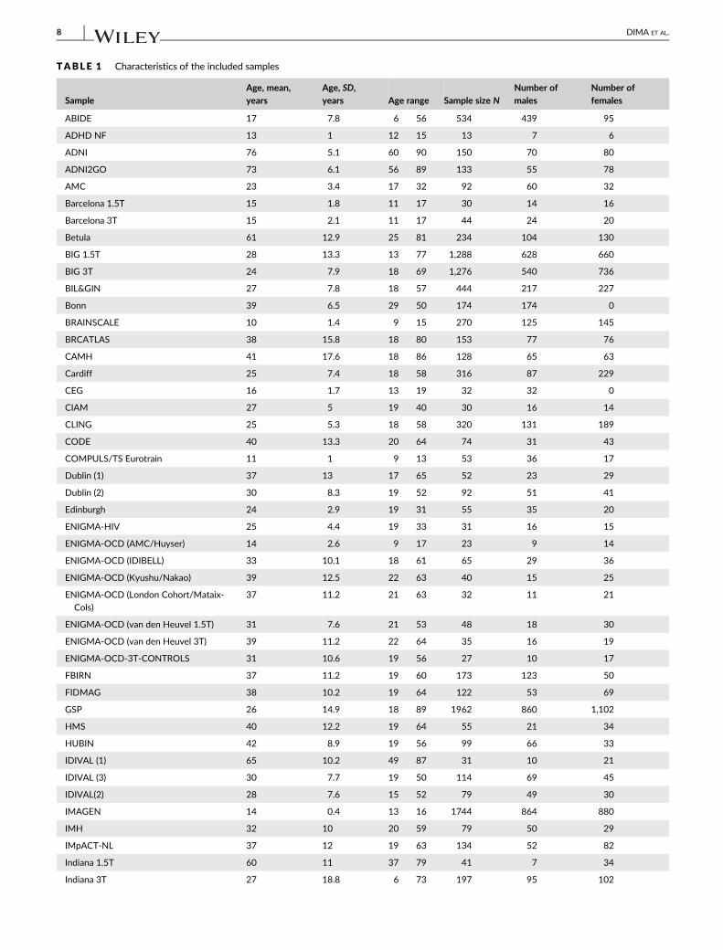
TABLE 1 Characteristics of the included samples
Sample
Age, mean,
years
Age, SD,
years Age range Sample size N
Number of
males
Number of
females
ABIDE 17 7.8 6 56 534 439 95
ADHD NF 13 1 12 15 13 7 6
ADNI 76 5.1 60 90 150 70 80
ADNI2GO 73 6.1 56 89 133 55 78
AMC 23 3.4 17 32 92 60 32
Barcelona 1.5T 15 1.8 11 17 30 14 16
Barcelona 3T 15 2.1 11 17 44 24 20
Betula 61 12.9 25 81 234 104 130
BIG 1.5T 28 13.3 13 77 1,288 628 660
BIG 3T 24 7.9 18 69 1,276 540 736
BIL&GIN 27 7.8 18 57 444 217 227
Bonn 39 6.5 29 50 174 174 0
BRAINSCALE 10 1.4 9 15 270 125 145
BRCATLAS 38 15.8 18 80 153 77 76
CAMH 41 17.6 18 86 128 65 63
Cardiff 25 7.4 18 58 316 87 229
CEG 16 1.7 13 19 32 32 0
CIAM 27 5 19 40 30 16 14
CLING 25 5.3 18 58 320 131 189
CODE 40 13.3 20 64 74 31 43
COMPULS/TS Eurotrain 11 1 9 13 53 36 17
Dublin (1) 37 13 17 65 52 23 29
Dublin (2) 30 8.3 19 52 92 51 41
Edinburgh 24 2.9 19 31 55 35 20
ENIGMA-HIV 25 4.4 19 33 31 16 15
ENIGMA-OCD (AMC/Huyser) 14 2.6 9 17 23 9 14
ENIGMA-OCD (IDIBELL) 33 10.1 18 61 65 29 36
ENIGMA-OCD (Kyushu/Nakao) 39 12.5 22 63 40 15 25
ENIGMA-OCD (London Cohort/Mataix-
Cols)
37 11.2 21 63 32 11 21
ENIGMA-OCD (van den Heuvel 1.5T) 31 7.6 21 53 48 18 30
ENIGMA-OCD (van den Heuvel 3T) 39 11.2 22 64 35 16 19
ENIGMA-OCD-3T-CONTROLS 31 10.6 19 56 27 10 17
FBIRN 37 11.2 19 60 173 123 50
FIDMAG 38 10.2 19 64 122 53 69
GSP 26 14.9 18 89 1962 860 1,102
HMS 40 12.2 19 64 55 21 34
HUBIN 42 8.9 19 56 99 66 33
IDIVAL (1) 65 10.2 49 87 31 10 21
IDIVAL (3) 30 7.7 19 50 114 69 45
IDIVAL(2) 28 7.6 15 52 79 49 30
IMAGEN 14 0.4 13 16 1744 864 880
IMH 32 10 20 59 79 50 29
IMpACT-NL 37 12 19 63 134 52 82
Indiana 1.5T 60 11 37 79 41 7 34
Indiana 3T 27 18.8 6 73 197 95 102
8 DIMA ET AL.

TABLE 1 (Continued)
SampleAge, mean,years
Age, SD,years Age range Sample size N
Number ofmales
Number offemales
Johns Hopkins 44 12.5 20 65 87 41 46
KaSP 27 5.7 20 43 32 15 17
Leiden 17 4.8 8 29 565 274 291
MAS 78 4.5 70 89 361 137 224
MCIC 33 12 18 60 93 63 30
Melbourne 20 3 15 26 102 54 48
METHCT 27 7.3 18 53 62 48 14
MHRC 22 2.9 16 28 52 52 0
Moods 33 9.8 18 51 310 146 164
NCNG 50 16.7 19 79 311 92 219
NESDA 40 9.8 21 56 65 22 43
NeuroIMAGE 17 3.7 8 29 376 172 204
Neuroventure 14 0.6 12 15 137 62 75
NTR (1) 15 1.4 11 18 34 11 23
NTR (2) 34 10.3 19 57 105 39 66
NTR (3) 30 5.9 20 42 29 11 18
NU 41 18.8 17 68 15 1 14
NUIG 37 11.5 18 58 89 50 39
NYU 31 8.7 19 52 51 31 20
OATS (1) 71 5.3 65 84 94 27 67
OATS (2) 68 4.4 65 81 33 13 20
OATS (3) 69 4.3 65 81 128 44 84
OATS (4) 70 4.6 65 89 95 23 72
OLIN 36 12.8 21 87 594 236 358
Oxford 16 1.4 14 19 38 18 20
PING 12 4.9 3 21 518 271 247
QTIM 23 3.4 16 30 342 112 230
Sao Paolo 1 27 5.8 17 43 69 45 24
Sao Paolo 3 30 8.1 18 50 83 44 39
SCORE 25 4.3 19 39 44 17 27
SHIP 2 55 12.3 31 84 368 206 162
SHIP TREND 50 13.9 21 81 788 439 349
StagedDep 47 8 27 59 84 20 64
Stanford 37 10.7 19 61 54 20 34
STROKEMRI 42 21.3 18 77 47 17 30
Sydney 37 21.1 12 79 147 58 89
TOP 35 9.8 18 73 296 155 141
Tuebingen 40 12.1 24 61 53 24 29
UMC Utrecht 1.5T 32 12.1 17 66 289 171 118
UMCU 3T 45 15.2 19 81 109 52 57
UNIBA 27 8.7 18 63 130 66 64
UPENN 36 13.6 16 85 185 85 100
Yale 14 2.2 10 18 23 12 11
Total 31 18.4 3 90 18,605 8,980 9,625
DIMA ET AL. 9
site, the intracranial volume (Figure S1) was used to adjust the subcortical
volumes via a formula based on the analysis of the covariance approach:
“adjusted volume = raw volume – b × (ICV – mean ICV)”, where b is the
slope of regression of a region of interest volume on ICV (Raz et al., 2005).
The values of the subcortical volumes were then harmonized between
sites using the ComBat method in R (Fortin et al., 2017, 2018; Radua
et al., 2020). Originally developed to adjust for batch effect in genetic
studies, ComBat uses an empirical Bayes to adjust for inter-site variability
in the data, while preserving variability related to the variables of interest.
2.3 | Fractional polynomial regression analyses
The effect of age on each ICV- and site-adjusted subcortical volume was
modeled using high order fractional polynomial regression (Royston &
Altman, 1994; Sauerbrei, Meier-Hirmer, Benner, & Royston, 2006) in
each hemisphere. Because the effect of site (scanner and Freesurfer ver-
sion) was adjusted using ComBat, we only included sex as a covariate in
the regression models. Fractional polynomial regression is currently con-
sidered the most advantageous modeling strategy for continuous vari-
ables (Moore, Hanley, Turgeon, & Lavoie, 2011) as it allows testing for a
wider range of trajectory shapes than conventional lower-order polyno-
mials (e.g., linear or quadratic) and for multiple turning points (Royston &
Altman, 1994; Royston, Ambler, & Sauerbrei, 1999). For each subcortical
structure, the best model was obtained by comparing competing models
of up to three power combinations. The powers used to identify the best
fitting model were −2, −1, −0.5, 0.5, 1, 2, 3 and the natural logarithm
(ln) function. The optimal model describing the association between age
and each of the volumes was selected as the lowest degree model based
on the partial F-test (if linear) or the likelihood-ratio test. To avoid over-
fitting at ages with more data points, we used the stricter .01 level of
significance as the cut-off for each respective likelihood-ratio tests,
rather than adding powers, until the .05 level was reached. For ease of
interpretation we centered the volume of each structure so that the
intercept of a fractional polynomial was represented as the effect at zero
for sex. Fractional polynomial regression models were fitted using Stata/
IC software v.13.1 (Stata Corp., College Station, TX). Standard errors
were also adjusted for the effect of site in the FP regression.
We conducted two supplemental analyses: (a) we specified addi-
tional FP models separately for males and females and, (b) we calcu-
lated Pearson's correlation coefficient between subcortical volumes
and age in the early (6–29 years), middle (30–59 years), and late-life
(60–90 years) age-group. The results of these analyses have been
included in the supplemental material.
2.4 | Inter-individual variability
Inter-individual variability was assessed using two complimentary
approaches. First, for each subcortical structure we compared the
early (6–29 years), middle (30–59 years) and late-life (60–90 years)
age-groups in terms of their mean inter-individual variability; these
groups were defined following conventional notions regarding periods
of development, midlife and aging. The variance of each structure in
each age-group was calculated as
ln
P ffiffiffiffiffie2i
q
nt
0@
1A
where e represents the residual variance of each individual (i) around
the nonlinear best fitting regression line, and n the number of
Abbreviations: ABIDE = Autism Brain Imaging Data Exchange; ADNI = Alzheimer's Disease Neuroimaging Initiative; ADNI2GO = ADNI-GO and ADNI-2;
ADHD-NF = Attention Deficit Hyperactivity Disorder-Neurofeedback Study; AMC = Amsterdam Medisch Centrum; Basel = University of Basel;
Barcelona = University of Barcelona; Betula = Swedish longitudinal study on aging, memory, and dementia; BIG = Brain Imaging Genetics; BIL&GIN = a
multimodal multidimensional database for investigating hemispheric specialization; Bonn = University of Bonn; BrainSCALE = Brain Structure and
Cognition: an Adolescence Longitudinal twin study; CAMH = Centre for Addiction and Mental Health; Cardiff = Cardiff University; CEG = Cognitive-
experimental and Genetic study of ADHD and Control Sibling Pairs; CIAM = Cortical Inhibition and Attentional Modulation study; CLiNG = Clinical
Neuroscience Göttingen; CODE = formerly Cognitive Behavioral Analysis System of Psychotherapy (CBASP) study; Dublin = Trinity College Dublin;
Edinburgh = The University of Edinburgh; ENIGMA-HIV = Enhancing NeuroImaging Genetics through Meta-Analysis-Human Immunodeficiency Virus
Working Group; ENIGMA-OCD = Enhancing NeuroImaging Genetics through Meta-Analysis- Obsessive Compulsive Disorder Working Group;
FBIRN = Function Biomedical Informatics Research Network; FIDMAG = Fundación para la Investigación y Docencia Maria Angustias Giménez;
GSP = Brain Genomics Superstruct Project; HMS = Homburg Multidiagnosis Study; HUBIN = Human Brain Informatics; IDIVAL = Valdecilla Biomedical
Research Institute; IMAGEN = the IMAGEN Consortium; IMH=Institute of Mental Health, Singapore; IMpACT = The International Multicentre persistent
ADHD Genetics Collaboration; Indiana = Indiana University School of Medicine; Johns Hopkins = Johns Hopkins University; KaSP = The Karolinska
Schizophrenia Project; Leiden = Leiden University; MAS = Memory and Ageing Study; MCIC = MIND Clinical Imaging Consortium formed by the Mental
Illness and Neuroscience Discovery (MIND) Institute now the Mind Research Network; Melbourne = University of Melbourne; Meth-CT = study of
methamphetamine users, University of Cape Town; MHRC = Mental Health Research Center; Muenster = Muenster University; N = number;
NESDA = The Netherlands Study of Depression and Anxiety; NeuroIMAGE = Dutch part of the International Multicenter ADHD Genetics (IMAGE) study;
Neuroventure: the imaging part of the Co-Venture Trial funded by the Canadian Institutes of Health Research (CIHR); NCNG = Norwegian Cognitive
NeuroGenetics sample; NTR = Netherlands Twin Register; NU = Northwestern University; NUIG = National University of Ireland Galway; NYU = New
York University; OATS = Older Australian Twins Study; Olin = Olin Neuropsychiatric Research Center; Oxford = Oxford University; QTIM = Queensland
Twin Imaging; Sao Paulo = University of Sao Paulo; SCORE = University of Basel Study; SHIP-2 and SHIP TREND = Study of Health in Pomerania; Staged-
Dep = Stages of Depression Study; Stanford = Stanford University; StrokeMRI = Stroke Magnetic Resonance Imaging; Sydney = University of Sydney;
TOP = Tematisk Område Psykoser (Thematically Organized Psychosis Research); TS-EUROTRAIN = European-Wide Investigation and Training Network on
the Etiology and Pathophysiology of Gilles de la Tourette Syndrome; Tuebingen = University of Tuebingen; UMCU = Universitair Medisch Centrum
Utrecht; UNIBA = University of Bari Aldo Moro; UPENN = University of Pennsylvania; Yale = Yale University.
10 DIMA ET AL.
observations in each age-group (t). The residuals (ei) were nor-
mally distributed suggesting good fit of the model without having
over- or under-fitted the data. Upon calculating the square root of
the squared residuals we used the natural logarithm to account for the
positive skewness of the new distribution. Then the mean inter-
individual variability between early (6–29 years), middle (30–59 years)
and late-life (60–90 years) age-groups was compared using between-
groups omnibus tests for the residual variance around the identified
best-fitting nonlinear fractional polynomial model of each structure.
We conducted 16 tests (one for each structure) and accordingly the
critical alpha value was set at 0.003 following Bonferroni correction for
multiple comparisons.
The second approach entailed the quantification of the mean indi-
vidual variability of each subcortical structure through a meta-analysis
F IGURE 1 ENIGMA lifespan samples. Detailsof each sample are provided Table 1 and in thesupplemental material. Abbreviations are providedin Table 1
DIMA ET AL. 11
of the SD of the adjusted volumes according to the method proposed
by Senior, Gosby, Lu, Simpson, and Raubenheimer (2016).
2.5 | Centile curves
Reference curves for each structure by sex and hemisphere were pro-
duced from ICV- and site-adjusted volumes as normalized growth centiles
using the parametric Lambda (λ), Mu (μ), Sigma (σ) (LMS) method (Cole &
Green, 1992) implemented using the Generalized Additive Models for
Location, Scale and Shape (GAMLSS) in R (http://cran.r-project.org/web/
packages/gamlss/index.html) (Rigby & Stasinopoulos, 2005; Stasinopoulos
& Rigby, 2007). LMS allows for the estimation of the distribution at each
covariate value after a suitable transformation and is summarized using
three smoothing parameters, the Box-Cox power λ, the mean μ and the
coefficient of variation σ. GAMLSS uses an iterative maximum (penalized)
likelihood estimation method to estimate λ, μ and σ as well as distribution
dependent smoothing parameters and provides optimal values for effec-
tive degrees of freedom (edf) for every parameter (Indrayan, 2014). This
procedure minimizes the Generalized Akaike Information Criterion (GAIC)
goodness of fit index; smaller GAIC values indicate better fit of the model
to the data. GAMLSS is a flexible way to derive normalized centile curves
as it allows each curve to have its own number of edf while overcoming
biased estimates resulting from skewed data
3 | RESULTS
3.1 | Fractional polynomial regression analyses
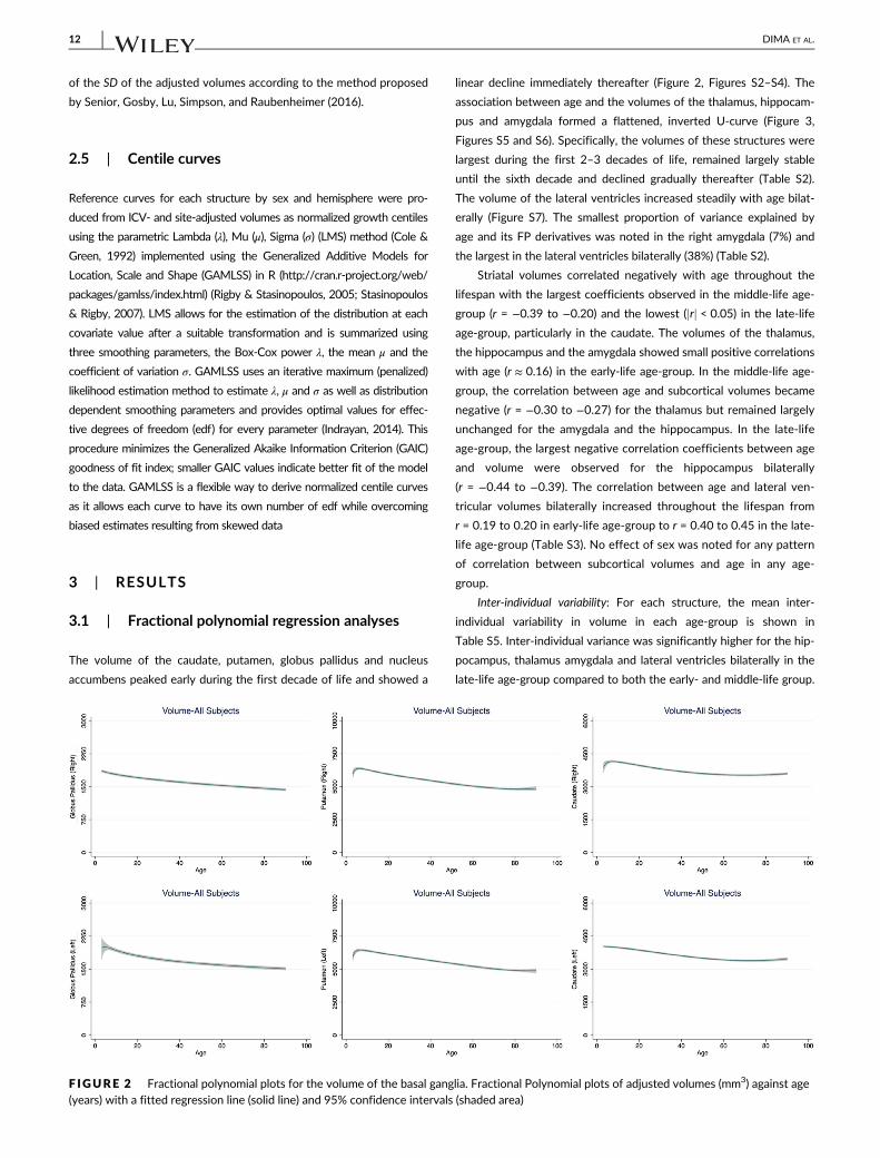
The volume of the caudate, putamen, globus pallidus and nucleus
accumbens peaked early during the first decade of life and showed a
linear decline immediately thereafter (Figure 2, Figures S2–S4). The
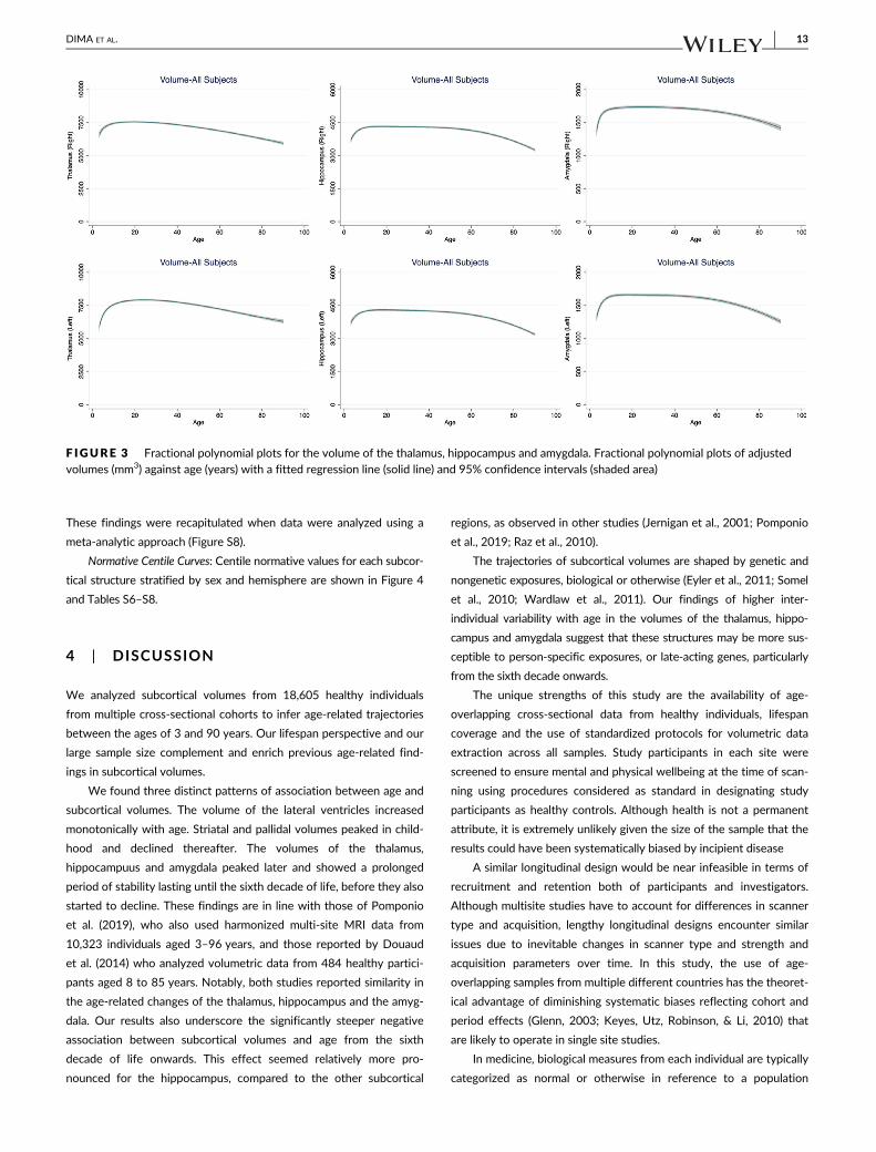
association between age and the volumes of the thalamus, hippocam-
pus and amygdala formed a flattened, inverted U-curve (Figure 3,
Figures S5 and S6). Specifically, the volumes of these structures were
largest during the first 2–3 decades of life, remained largely stable
until the sixth decade and declined gradually thereafter (Table S2).
The volume of the lateral ventricles increased steadily with age bilat-
erally (Figure S7). The smallest proportion of variance explained by
age and its FP derivatives was noted in the right amygdala (7%) and
the largest in the lateral ventricles bilaterally (38%) (Table S2).
Striatal volumes correlated negatively with age throughout the
lifespan with the largest coefficients observed in the middle-life age-
group (r = −0.39 to −0.20) and the lowest (jrj < 0.05) in the late-life
age-group, particularly in the caudate. The volumes of the thalamus,
the hippocampus and the amygdala showed small positive correlations
with age (r ≈ 0.16) in the early-life age-group. In the middle-life age-
group, the correlation between age and subcortical volumes became
negative (r = −0.30 to −0.27) for the thalamus but remained largely
unchanged for the amygdala and the hippocampus. In the late-life
age-group, the largest negative correlation coefficients between age
and volume were observed for the hippocampus bilaterally
(r = −0.44 to −0.39). The correlation between age and lateral ven-
tricular volumes bilaterally increased throughout the lifespan from
r = 0.19 to 0.20 in early-life age-group to r = 0.40 to 0.45 in the late-
life age-group (Table S3). No effect of sex was noted for any pattern
of correlation between subcortical volumes and age in any age-
group.
Inter-individual variability: For each structure, the mean inter-
individual variability in volume in each age-group is shown in
Table S5. Inter-individual variance was significantly higher for the hip-
pocampus, thalamus amygdala and lateral ventricles bilaterally in the
late-life age-group compared to both the early- and middle-life group.
F IGURE 2 Fractional polynomial plots for the volume of the basal ganglia. Fractional Polynomial plots of adjusted volumes (mm3) against age(years) with a fitted regression line (solid line) and 95% confidence intervals (shaded area)
12 DIMA ET AL.
These findings were recapitulated when data were analyzed using a
meta-analytic approach (Figure S8).
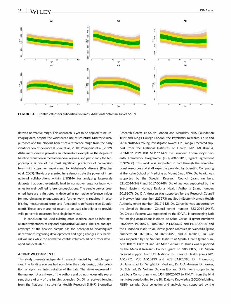
Normative Centile Curves: Centile normative values for each subcor-
tical structure stratified by sex and hemisphere are shown in Figure 4
and Tables S6–S8.
4 | DISCUSSION
We analyzed subcortical volumes from 18,605 healthy individuals
from multiple cross-sectional cohorts to infer age-related trajectories
between the ages of 3 and 90 years. Our lifespan perspective and our
large sample size complement and enrich previous age-related find-
ings in subcortical volumes.
We found three distinct patterns of association between age and
subcortical volumes. The volume of the lateral ventricles increased
monotonically with age. Striatal and pallidal volumes peaked in child-
hood and declined thereafter. The volumes of the thalamus,
hippocampuus and amygdala peaked later and showed a prolonged
period of stability lasting until the sixth decade of life, before they also
started to decline. These findings are in line with those of Pomponio
et al. (2019), who also used harmonized multi-site MRI data from
10,323 individuals aged 3–96 years, and those reported by Douaud
et al. (2014) who analyzed volumetric data from 484 healthy partici-
pants aged 8 to 85 years. Notably, both studies reported similarity in
the age-related changes of the thalamus, hippocampus and the amyg-
dala. Our results also underscore the significantly steeper negative
association between subcortical volumes and age from the sixth
decade of life onwards. This effect seemed relatively more pro-
nounced for the hippocampus, compared to the other subcortical
regions, as observed in other studies (Jernigan et al., 2001; Pomponio
et al., 2019; Raz et al., 2010).
The trajectories of subcortical volumes are shaped by genetic and
nongenetic exposures, biological or otherwise (Eyler et al., 2011; Somel
et al., 2010; Wardlaw et al., 2011). Our findings of higher inter-
individual variability with age in the volumes of the thalamus, hippo-
campus and amygdala suggest that these structures may be more sus-
ceptible to person-specific exposures, or late-acting genes, particularly
from the sixth decade onwards.
The unique strengths of this study are the availability of age-
overlapping cross-sectional data from healthy individuals, lifespan
coverage and the use of standardized protocols for volumetric data
extraction across all samples. Study participants in each site were
screened to ensure mental and physical wellbeing at the time of scan-
ning using procedures considered as standard in designating study
participants as healthy controls. Although health is not a permanent
attribute, it is extremely unlikely given the size of the sample that the
results could have been systematically biased by incipient disease
A similar longitudinal design would be near infeasible in terms of
recruitment and retention both of participants and investigators.
Although multisite studies have to account for differences in scanner
type and acquisition, lengthy longitudinal designs encounter similar
issues due to inevitable changes in scanner type and strength and
acquisition parameters over time. In this study, the use of age-
overlapping samples from multiple different countries has the theoret-
ical advantage of diminishing systematic biases reflecting cohort and
period effects (Glenn, 2003; Keyes, Utz, Robinson, & Li, 2010) that
are likely to operate in single site studies.
In medicine, biological measures from each individual are typically
categorized as normal or otherwise in reference to a population
F IGURE 3 Fractional polynomial plots for the volume of the thalamus, hippocampus and amygdala. Fractional polynomial plots of adjustedvolumes (mm3) against age (years) with a fitted regression line (solid line) and 95% confidence intervals (shaded area)
DIMA ET AL. 13
derived normative range. This approach is yet to be applied to neuro-
imaging data, despite the widespread use of structural MRI for clinical
purposes and the obvious benefit of a reference range from the early
identification of deviance (Dickie et al., 2013; Pomponio et al., 2019).
Alzheimer's disease provides an informative example as the degree of
baseline reduction in medial temporal regions, and particularly the hip-
pocampus, is one of the most significant predictors of conversion
from mild cognitive impairment to Alzheimer's disease (Risacher
et al., 2009). The data presented here demonstrate the power of inter-
national collaborations within ENIGMA for analyzing large-scale
datasets that could eventually lead to normative range for brain vol-
umes for well-defined reference populations. The centile curves pres-
ented here are a first-step in developing normative reference values
for neuroimaging phenotypes and further work is required in esta-
blishing measurement error and functional significance (see Supple-
ment). These curves are not meant to be used clinically or to provide
valid percentile measures for a single individual.
In conclusion, we used existing cross-sectional data to infer age-
related trajectories of regional subcortical volumes. The size and age-
coverage of the analysis sample has the potential to disambiguate
uncertainties regarding developmental and aging changes in subcorti-
cal volumes while the normative centile values could be further devel-
oped and evaluated.
ACKNOWLEDGMENTS
This study presents independent research funded by multiple agen-
cies. The funding sources had no role in the study design, data collec-
tion, analysis, and interpretation of the data. The views expressed in
the manuscript are those of the authors and do not necessarily repre-
sent those of any of the funding agencies. Dr. Dima received funding
from the National Institute for Health Research (NIHR) Biomedical
Research Centre at South London and Maudsley NHS Foundation
Trust and King's College London, the Psychiatry Research Trust and
2014 NARSAD Young Investigator Award. Dr. Frangou received sup-
port from the National Institutes of Health (R01 MH104284,
R01MH113619, R01 MH116147), the European Community's Sev-
enth Framework Programme (FP7/2007–2013) (grant agreement
n�602450). This work was supported in part through the computa-
tional resources and staff expertise provided by Scientific Computing
at the Icahn School of Medicine at Mount Sinai, USA. Dr. Agartz was
supported by the Swedish Research Council (grant numbers:
521-2014-3487 and 2017-00949). Dr. Alnæs was supported by the
South Eastern Norway Regional Health Authority (grant number:
2019107). Dr. O Andreasen was supported by the Research Council
of Norway (grant number: 223273) and South-Eastern Norway Health
Authority (grant number: 2017-112). Dr. Cervenka was supported by
the Swedish Research Council (grant number 523-2014-3467).
Dr. Crespo-Facorro was supported by the IDIVAL Neuroimaging Unit
for imaging acquisition; Instituto de Salud Carlos III (grant numbers:
PI020499, PI050427, PI060507, PI14/00639 and PI14/00918) and
the Fundación Instituto de Investigación Marqués de Valdecilla (grant
numbers: NCT0235832, NCT02534363, and API07/011). Dr. Gur
was supported by the National Institute of Mental Health (grant num-
bers: R01MH042191 and R01MH117014). Dr. James was supported
by the Medical Research Council (grant no G0500092). Dr. Saykin
received support from U.S. National Institutes of Health grants R01
AG19771, P30 AG10133 and R01 CA101318. Dr. Thompson,
Dr. Jahanshad, Dr. Wright, Dr. Medland, Dr. O Andreasen, Dr. Rinker,
Dr. Schmaal, Dr. Veltam, Dr. van Erp, and D.P.H. were supported in
part by a Consortium grant (U54 EB020403 to P.M.T.) from the NIH
Institutes contributing to the Big Data to Knowledge (BD2K) Initiative.
FBIRN sample: Data collection and analysis was supported by the
F IGURE 4 Centile values for subcortical volumes; Additional details in Tables S6-S9
14 DIMA ET AL.
National Center for Research Resources at the National Institutes of
Health (grant numbers: NIH 1 U24 RR021992 (Function Biomedical
Informatics Research Network) and NIH 1 U24 RR025736-01
(Biomedical Informatics Research Network Coordinating Center;
http://www.birncommunity.org). FBIRN data was processed by the
UCI High Performance Computing cluster supported by the National
Center for Research Resources and the National Center for Advancing
Translational Sciences, National Institutes of Health, through Grant
UL1 TR000153. Brainscale: This work was supported by Nederlandse
Organisatie voor Wetenschappelijk Onderzoek (NWO 51.02.061 to
H.H., NWO 51.02.062 to D.B., NWO- NIHC Programs of excellence
433-09-220 to H.H., NWO-MagW 480-04-004 to D.B., and
NWO/SPI 56-464-14192 to D.B.); FP7 Ideas: European Research
Council (ERC-230374 to D.B.); and Universiteit Utrecht (High Poten-
tial Grant to H.H.). UMCU-1.5T: This study is partially funded through
the Geestkracht Programme of the Dutch Health Research Council
(Zon-Mw, grant No 10-000-1001), and matching funds from partici-
pating pharmaceutical companies (Lundbeck, AstraZeneca, Eli Lilly,
Janssen Cilag) and universities and mental health care organizations
(Amsterdam: Academic Psychiatric Centre of the Academic Medical
Center and the mental health institutions: GGZ Ingeest, Arkin, Dijk en
Duin, GGZ Rivierduinen, Erasmus Medical Centre, GGZ Noord Hol-
land Noord. Groningen: University Medical Center Groningen and the
mental health institutions: Lentis, GGZ Friesland, GGZ Drenthe, Dim-
ence, Mediant, GGNet Warnsveld, Yulius Dordrecht and Parnassia
psycho-medical center The Hague. Maastricht: Maastricht University
Medical Centre and the mental health institutions: GGzE, GGZ
Breburg, GGZ Oost-Brabant, Vincent van Gogh voor Geestelijke
Gezondheid, Mondriaan, Virenze riagg, Zuyderland GGZ, MET ggz,
Universitair Centrum Sint-Jozef Kortenberg, CAPRI University of Ant-
werp, PC Ziekeren Sint-Truiden, PZ Sancta Maria Sint-Truiden, GGZ
Overpelt, OPZ Rekem. Utrecht: University Medical Center Utrecht
and the mental health institutions Altrecht, GGZ Centraal and Delta.).
UMCU-3T: This study was supported by NIMH grant number: R01
MH090553 (to RAO). The NIMH had no further role in study design,
in the collection, analysis and interpretation of the data, in the writing
of the report, and in the decision to submit the paper for publication.
Netherlands Twin Register: Funding was obtained from the Nether-
lands Organization for Scientific Research (NWO) and The Netherlands
Organization for Health Research and Development (ZonMW) grants
904-61-090, 985-10-002, 912-10-020, 904-61-193,480-04-004,
463-06-001, 451-04-034, 400-05-717, 400-07-080, 31160008,
016-115-035, 481-08-011, 056-32-010, 911-09-032, 024-001-003,
480-15-001/674, Center for Medical Systems Biology (CSMB, NWO
Genomics), Biobanking and Biomolecular Resources Research Infra-
structure (BBMRI-NL, 184.021.007 and 184.033.111); Spinozapremie
(NWO- 56-464-14192), and the Neuroscience Amsterdam research
institute (former NCA). The BIG database, established in Nijmegen in
2007, is now part of Cognomics, a joint initiative by researchers of the
Donders Centre for Cognitive Neuroimaging, the Human Genetics and
Cognitive Neuroscience departments of the Radboud University Medi-
cal Centre, and the Max Planck Institute for Psycholinguistics. The
Cognomics Initiative is supported by the participating departments and
centers and by external grants, including grants from the Biobanking
and Biomolecular Resources Research Infrastructure (Netherlands)
(BBMRI-NL) and the Hersenstichting Nederland. The authors also
acknowledge grants supporting their work from the Netherlands Orga-
nization for Scientific Research (NWO), that is, the NWO Brain & Cog-
nition Excellence Program (grant 433-09-229), the Vici Innovation
Program (grant 016-130-669 to BF) and #91619115. Additional sup-
port is received from the European Community's Seventh Framework
Programme (FP7/2007–2013) under grant agreements n� 602805
(Aggressotype), n� 603016 (MATRICS), n� 602450 (IMAGEMEND), and
n� 278948 (TACTICS), and from the European Community's Horizon
2020 Programme (H2020/2014–2020) under grant agreements n�
643051 (MiND) and n� 667302 (CoCA). Betula sample: Data collection
for the BETULA sample was supported by a grant from Knut and Alice
Wallenberg Foundation (KAW); the Freesurfer segmentations were
performed on resources provided by the Swedish National Infrastruc-
ture for Computing (SNIC) at HPC2N in Umeå, Sweden. Indiana sample:
This sample was supported in part by grants to BCM from Siemens
Medical Solutions, from the members of the Partnership for Pediatric
Epilepsy Research, which includes the American Epilepsy Society, the
Epilepsy Foundation, the Epilepsy Therapy Project, Fight Against Child-
hood Epilepsy and Seizures (F.A.C.E.S.), and Parents Against Childhood
Epilepsy (P.A.C.E.), from the Indiana State Department of Health Spinal
Cord and Brain Injury Fund Research Grant Program, and by a Project
Development Team within the ICTSI NIH/NCRR Grant Number
RR025761. MHRC study: It was supported in part by RFBR grant
20-013-00748. PING study: Data collection and sharing for the Pediat-
ric Imaging, Neurocognition and Genetics (PING) Study (National Insti-
tutes of Health Grant RC2DA029475) were funded by the National
Institute on Drug Abuse and the Eunice Kennedy Shriver National Insti-
tute of Child Health & Human Development. A full list of PING investi-
gators is at http://pingstudy.ucsd.edu/investigators.html. QTIM sample:
The authors are grateful to the twins for their generosity of time and
willingness to participate in our study and thank the many research
assistants, radiographers, and other staff at QIMR Berghofer Medical
Research Institute and the Centre for Advanced Imaging, University of
Queensland. QTIM was funded by the Australian National Health and
Medical Research Council (Project Grants No. 496682 and 1009064)
and US National Institute of Child Health and Human Development
(RO1HD050735). Lachlan Strike was supported by a University of
Queensland PhD scholarship. Study of Health in Pomerania (SHIP): this
is part of the Community Medicine Research net (CMR) (http://www.
medizin.uni-greifswald.de/icm) of the University Medicine Greifswald,
which is supported by the German Federal State of Mecklenburg- West
Pomerania. MRI scans in SHIP and SHIP-TREND have been supported
by a joint grant from Siemens Healthineers, Erlangen, Germany and the
Federal State of Mecklenburg-West Pomerania. This study was further
supported by the DZHK (German Centre for Cardiovascular Research),
the German Centre of Neurodegenerative Diseases (DZNE) and the
EU-JPND Funding for BRIDGET (FKZ:01ED1615). TOP study: this was
supported by the European Community's Seventh Framework Pro-
gramme (FP7/2007–2013), grant agreement n�602450. The Southern
and Eastern Norway Regional Health Authority supported Lars
DIMA ET AL. 15
T. Westlye (grant no. 2014-097) and STROKEMRI (grant
no. 2013-054). HUBIN sample: HUBIN was supported by the Swedish
Research Council (K2007-62X-15077-04-1, K2008-62P-20597-01-3,
K2010-62X-15078-07-2, K2012-61X-15078-09-3), the regional agree-
ment on medical training and clinical research between Stockholm
County Council, and the Karolinska Institutet, and the Knut and Alice
Wallenberg Foundation. The BIG database: this was established in Nij-
megen in 2007, is now part of Cognomics, a joint initiative by
researchers of the Donders Centre for Cognitive Neuroimaging, the
Human Genetics and Cognitive Neuroscience departments of the
Radboud university medical centre, and the Max Planck Institute for
Psycholinguistics. The Cognomics Initiative is supported by the partici-
pating departments and centres and by external grants, including grants
from the Biobanking and Biomolecular Resources Research Infrastruc-
ture (Netherlands) (BBMRI-NL) and the Hersenstichting Nederland. The
authors also acknowledge grants supporting their work from the Neth-
erlands Organization for Scientific Research (NWO), that is, the NWO
Brain & Cognition Excellence Program (grant 433-09-229), the Vici
Innovation Program (grant 016-130-669 to BF) and #91619115. Addi-
tional support is received from the European Community's Seventh
Framework Programme (FP7/2007–2013) under grant agreements n�
602805 (Aggressotype), n� 603016 (MATRICS), n� 602450
(IMAGEMEND), and n� 278948 (TACTICS), and from the European
Community's Horizon 2020 Programme (H2020/2014–2020) under
grant agreements n� 643051 (MiND) and n� 667302 (CoCA).
CONFLICT OF INTEREST
H.-J. G.: Travel grants and speaker honoraria from Fresenius Medical
Care, Neuraxpharm, Servier and Janssen Cilag; research funding from
Fresenius Medical Care. O. A. A.: Consultant to HealthLytix, speaker
honorarium from Lundbeck. A. M. D.: Founder and member of the Sci-
entific Advisory Board CorTechs Labs, Inc where he holds equity;
member of the Scientific Advisory of Human Longevity Inc; research
grants with General Electric Healthcare.
DATA AVAILABILITY STATEMENT
The ENIGMA Lifespan Working Group welcomes expression of inter-
est from researchers in the field who wish to use the ENIGMA sam-
ples. Data sharing is possible subsequent to consent for the principal
investigators of the contributing datasets. Requests should be
directed to the corresponding authors.
ORCID
Danai Dima https://orcid.org/0000-0002-2598-0952
Gaelle E. Doucet https://orcid.org/0000-0003-4120-0474
Moji Aghajani https://orcid.org/0000-0003-2040-4881
Rachel M. Brouwer https://orcid.org/0000-0002-7466-1544
Christopher R. K. Ching https://orcid.org/0000-0003-2921-3408
Simon E. Fisher https://orcid.org/0000-0002-3132-1996
Thomas Frodl https://orcid.org/0000-0002-8113-6959
David C. Glahn https://orcid.org/0000-0002-4749-6977
Ian H. Gotlib https://orcid.org/0000-0002-3622-3199
Oliver Grimm https://orcid.org/0000-0002-0767-0301
Sean N. Hatton https://orcid.org/0000-0002-9149-8726
Martine Hoogman https://orcid.org/0000-0002-1261-7628
Hilleke E. Hulshoff Pol https://orcid.org/0000-0002-2038-5281
Bernd Krämer https://orcid.org/0000-0002-1145-9103
Sophia Frangou https://orcid.org/0000-0002-3210-6470
REFERENCES
Brain Development Cooperative Group. (2012). Total and regional brain
volumes in a population-based normative sample from 4 to 18 years:
The NIH MRI Study of Normal Brain Development. Cerebral Cortex,
22, 1–12.Calabresi, P., Picconi, B., Tozzi, A., Ghiglieri, V., & Di Filippo, M. (2014).
Direct and indirect pathways of basal ganglia: A critical reappraisal.
Nature Neuroscience, 17, 1022–1030.Chudasama, Y., & Robbins, T. W. (2006). Functions of frontostriatal sys-
tems in cognition: Comparative neuropsychopharmacological studies
in rats, monkeys and humans. Biological Psychology, 73, 19–38.Cole, T. J., & Green, P. J. (1992). Smoothing reference centile curves: The
LMS method and penalized likelihood. Statistics in Medicine, 11,
1305–1319.Dickie, D. A., Job, D. E., Gonzalez, D. R., Shenkin, S. D., Ahearn, T. S.,
Murray, A. D., & Wardlaw, J. M. (2013). Variance in brain volume with
advancing age: Implications for defining the limits of normality. PLoS
One, 8, e84093.
Douaud, G., Groves, A. R., Tamnes, C. K., Westlye, L. T., Duff, E. P.,
Engvig, A., … Johansen-Berg, H. (2014). A common brain network links
development, aging, and vulnerability to disease. Proceedings of the
National Academy of Sciences of the United States of America, 111,
17648–17653.Driscoll, I., Davatzikos, C., An, Y., Wu, X., Shen, D., Kraut, M., &
Resnick, S. M. (2009). Longitudinal pattern of regional brain volume
change differentiates normal aging from MCI. Neurology, 72,
1906–1913.Ecker, C., Bookheimer, S. Y., & Murphy, D. G. (2015). Neuroimaging in
autism spectrum disorder: Brain structure and function across the
lifespan. Lancet Neurology, 14, 1121–1134.Eichenbaum, H. (2004). Hippocampus: Cognitive processes and neural rep-
resentations that underlie declarative memory. Neuron, 44, 109–120.
Eyler, L. T., Prom-Wormley, E., Fennema-Notestine, C., Panizzon, M. S.,
Neale, M. C., Jernigan, T. L., … Kremen, W. S. (2011). Genetic patterns
of correlation among subcortical volumes in humans: Results from a
magnetic resonance imaging twin study. Human Brain Mapping, 32,
641–653.
Fischl, B. (2012). FreeSurfer. NeuroImage, 62, 774–781.
Fischl, B., Salat, D. H., Busa, E., Albert, M., Dieterich, M., Haselgrove, C., …Dale, A. M. (2002). Whole brain segmentation: Automated labeling of
neuroanatomical structures in the human brain. Neuron, 33, 341–355.
Fortin, J. P., Cullen, N., Sheline, Y. I., Taylor, W. D., Aselcioglu, I.,
Cook, P. A., … Shinohara, R. T. (2018). Harmonization of cortical thick-
ness measurements across scanners and sites. NeuroImage, 167,
104–120.
Fortin, J. P., Parker, D., Tunc, B., Watanabe, T., Elliott, M. A., Ruparel, K., …Shinohara, R. T. (2017). Harmonization of multi-site diffusion tensor
imaging data. NeuroImage, 161, 149–170.Fotenos, A. F., Snyder, A. Z., Girton, L. E., Morris, J. C., & Buckner, R. L.
(2005). Normative estimates of cross-sectional and longitudinal brain
volume decline in aging and AD. Neurology, 64, 1032–1039.
Glenn, N. D. (2003). Distinguishing age, period, and cohort effects. In J. T.
Mortimer & M. J. Shanahan (Eds.), Handbook of the life course
(pp. 465–476). New York: Springer US.
Good, C. D., Johnsrude, I. S., Ashburner, J., Henson, R. N., Friston, K. J., &
Frackowiak, R. S. (2001). A voxel-based morphometric study of ageing
in 465 normal adult human brains. NeuroImage, 14, 21–36.
16 DIMA ET AL.
Grossberg, S. (2009). Cortical and subcortical predictive dynamics and
learning during perception, cognition, emotion and action. Philosophical
Transactions of the Royal Society of London. Series B, Biological Sciences,
364, 1223–1234.Hibar, D. P., Westlye, L. T., van Erp, T. G., Rasmussen, J., Leonardo, C. D.,
Faskowitz, J., … Andreassen, O. A. (2016). Subcortical volumetric
abnormalities in bipolar disorder. Molecular Psychiatry, 21, 1710–1716.Indrayan, A. (2014). Demystifying LMS and BCPE methods of centile esti-
mation for growth and other health parameters. Indian Pediatrics, 51,
37–43.Jernigan, T. L., Archibald, S. L., Fenema-Notestine, C., Gamst, A. C.,
Stout, J. C., Bonner, J., & Hesselink, J. R. (2001). Effects of age on tis-
sues and regions of the cerebrum and cerebellum. Neurobiology of
Aging, 22, 581–594.Kempton, M. J., Salvador, Z., Munafò, M. R., Geddes, J. R., Simmons, A.,
Frangou, S., & Williams, S. C. (2011). Structural neuroimaging studies
in major depressive disorder. Meta-analysis and comparison with bipo-
lar disorder. Archives of General Psychiatry, 68, 675–690.Keyes, K. M., Utz, R. L., Robinson, W., & Li, G. (2010). What is a cohort
effect? Comparison of three statistical methods for modelling cohort
effects in obesity prevalence in the United States, 1971–2006. SocialScience & Medicine, 70, 1100–1108.
Kober, H., Barrett, L. F., Joseph, J., Bliss-Moreau, E., Lindquist, K., &
Wager, T. D. (2008). Functional grouping and cortical-subcortical inter-
actions in emotion: A meta-analysis of neuroimaging studies.
NeuroImage, 42, 998–1031.Krain, A. L., & Castellanos, F. X. (2006). Brain development and ADHD.
Clinical Psychology Review, 26, 433–444.Moore, L., Hanley, J. A., Turgeon, A. F., & Lavoie, A. (2011). A comparison
of generalized additive models to other common modeling strategies
for continuous covariates: Implications for risk adjustment. Journal of
Biometrics and Biostatistics, 2, 109.
Pfefferbaum, A., Rohlfing, T., Rosenbloom, M. J., Chu, W., Colrain, I. M., &
Sullivan, E. V. (2013). Variation in longitudinal trajectories of regional
brain volumes of healthy men and women (ages 10 to 85 years) mea-
sured with atlas-based parcellation of MRI. NeuroImage, 65, 176–193.Pomponio, R., Erus, G., Habes, M., Doshi, J., Srinivasan, D., Mamourian, E.,
… Davatzikos, C. (2019). Harmonization of large MRI datasets for the
analysis of brain imaging patterns throughout the lifespan. NeuroImage,
208, 116450. https://doi.org/10.1016/j.neuroimage.2019.116450
Radua, J., Vieta, E., Shinohara, R., Kochunov, P., Quidé, Y., Green, M. J., …Pineda-Zapata, J. (2020). Increased power by harmonizing structural
MRI site differences with the ComBat batch adjustment method in
ENIGMA. NeuroImage, 218, 116956. https://doi.org/10.1016/j.
neuroimage.2020.116956
Raz, N., Ghisletta, P., Rodrigue, K. M., Kennedy, K. M., & Lindenberger, U.
(2010). Trajectories of brain aging in middle-aged and older adults:
Regional and individual differences. NeuroImage, 51, 501–511.Raz, N., Lindenberger, U., Rodrigue, K. M., Kennedy, K. M., Head, D.,
Williamson, A., … Acker, J. D. (2005). Regional brain changes in aging
healthy adults: General trends, individual differences and modifiers.
Cerebral Cortex, 15, 1676–1689.Raznahan, A., Shaw, P. W., Lerch, J. P., Clasen, L. S., Greenstein, D.,
Berman, R., … Giedd, J. N. (2014). Longitudinal four-dimensional map-
ping of subcortical anatomy in human development. Proceedings of the
National Academy of Sciences of the United States of America, 111,
1592–1597.Resnick, S. M., Pham, D. L., Kraut, M. A., Zonderman, A. B., &
Davatzikos, C. (2003). Longitudinal magnetic resonance imaging stud-
ies of older adults: A shrinking brain. The Journal of Neuroscience, 23,
3295–3301.Richard, J. M., Castro, D. C., Difeliceantonio, A. G., Robinson, M. J., &
Berridge, K. C. (2013). Mapping brain circuits of reward and motiva-
tion: In the footsteps of Ann Kelley. Neuroscience and Biobehavioral
Reviews, 37, 1919–1931.
Rigby, R. A., & Stasinopoulos, D. M. (2005). Generalized additive models
for location, scale and shape. Applied Statistics, 54, 507–554.Risacher, S. L., Saykin, A. J., West, J. D., Shen, L., Firpi, H. A.,
McDonald, B. C., & Alzheimer's Disease Neuroimaging Initiative.
(2009). Baseline MRI predictors of conversion from MCI to probable
AD in the ADNI cohort. Current Alzheimer Research, 6, 347–361.Royston, P., & Altman, D. G. (1994). Regression using fractional polyno-
mials of continuous covariates: Parsimonious parametric modelling.
Applied Statistics, 43, 429–467.Royston, P., Ambler, G., & Sauerbrei, W. (1999). The use of fractional poly-
nomials to model continuous risk variables in epidemiology. Interna-
tional Journal of Epidemiology, 28, 964–974.Sauerbrei, W., Meier-Hirmer, C., Benner, A., & Royston, P. (2006). Multi-
variable regression model building by using fractional polynomials:
Description of SAS, STATA and R programs. Computational Statistics
and Data Analysis, 50, 3464–3485.Schmaal, L., Veltman, D. J., van Erp, T. G., Sämann, P. G., Frodl, T.,
Jahanshad, N., … Hibar, D. P. (2016). Subcortical brain alterations in
major depressive disorder: Findings from the ENIGMA major depres-
sive disorder working group. Molecular Psychiatry, 21, 806–812.Scimeca, J. M., & Badre, D. (2012). Striatal contributions to declarative
memory retrieval. Neuron, 75, 380–392.Senior, A. M., Gosby, A. K., Lu, J., Simpson, S. J., & Raubenheimer, D.
(2016). Meta-analysis of variance: An illustration comparing the effects
of two dietary interventions on variability in weight. Evolution, Medi-
cine, and Public Health, 2016, 244–255.Sherman, S. M. (2005). Thalamic relays and cortical functioning. Progress in
Brain Research, 149, 107–126.Shohamy, D., & Turk-Browne, N. B. (2013). Mechanisms for widespread
hippocampal involvement in cognition. Journal of Experimental Psychol-
ogy. General, 142, 1159–1170.Somel, M., Guo, S., Fu, N., Yan, Z., Hu, H. Y., Xu, Y., … Khaitovich, P.
(2010). MicroRNA, mRNA, and protein expression link development
and aging in human and macaque brain. Genome Research, 20,
1207–1218.Stasinopoulos, D. M., & Rigby, R. A. (2007). Generalized additive models
for location scale and shape (GAMLSS) in R. Journal of Statistical Soft-
ware, 23, 1–46.Thompson, P. M., Andreassen, O. A., Arias-Vasquez, A., Bearden, C. E.,
Boedhoe, P. S., Brouwer, R. M., … ENIGMA Consortium. (2017).
ENIGMA and the individual: Predicting factors that affect the brain in
35 countries worldwide. NeuroImage, 145, 389–408.Thompson, P. M., Stein, J. L., Medland, S. E., Hibar, D. P., Vasquez, A. A.,
Renteria, M. E., … Alzheimer's Disease Neuroimaging Initiative,
EPIGEN Consortium, IMAGEN Consortium, Saguenay Youth Study
(SYS) Group. (2014). The ENIGMA consortium: Large-scale collabora-
tive analyses of neuroimaging and genetic data. Brain Imaging and
Behavior, 8, 153–182.Tremblay, L., Worbe, Y., Thobois, S., Sgambato-Faure, V., & Féger, J. (2015).
Selective dysfunction of basal ganglia subterritories: From movement to
behavioral disorders. Movement Disorders, 30, 1155–1170.van Erp, T. G., Hibar, D. P., Rasmussen, J. M., Glahn, D. C., Pearlson, G. D.,
Andreassen, O. A., … Turner, J. A. (2016). Subcortical brain volume
abnormalities in 2028 individuals with schizophrenia and 2540 healthy
controls via the ENIGMA consortium. Molecular Psychiatry, 21,
547–553.Walhovd, K. B., Westlye, L. T., Amlien, I., Espeseth, T., Reinvang, I., Raz, N.,
… Fjell, A. M. (2011). Consistent neuroanatomical age-related volume
differences across multiple samples. Neurobiology of Aging, 32,
916–932.Wardlaw, J. M., Bastin, M. E., Valdés Hernández, M. C., Maniega, S. M.,
Royle, N. A., Morris, Z., … Deary, I. J. (2011). Brain aging, cognition in
youth and old age and vascular disease in the Lothian Birth Cohort
1936: Rationale, design and methodology of the imaging protocol.
International Journal of Stroke, 6, 547–559.
DIMA ET AL. 17

Zhang, D., Snyder, A. Z., Shimony, J. S., Fox, M. D., & Raichle, M. E. (2010).
Noninvasive functional and structural connectivity mapping of the
human thalamocortical system. Cerebral Cortex, 20, 1187–1194.
SUPPORTING INFORMATION
Additional supporting information may be found online in the
Supporting Information section at the end of this article.
How to cite this article: Dima D, Modabbernia A,
Papachristou E, et al. Subcortical volumes across the lifespan:
Data from 18,605 healthy individuals aged 3–90 years. Hum
Brain Mapp. 2021;1–18. https://doi.org/10.1002/hbm.25320
18 DIMA ET AL.