TESIS DOCTORAL LA INFLUENCIA DE LA DOBLE TAREA EN...
189
I TESIS DOCTORAL 2016 LA INFLUENCIA DE LA DOBLE TAREA EN EL PROCESAMIENTO DE LAS EXPRESIONES FACIALES EMOCIONALES: SU IMPACTO EN EL ENVEJECIMIENTO NORMAL Y EN LAS ENFERMEDADES DE ALZHEIMER Y PARKINSON Autora Carmen Teresa Casares Guillén Licenciada en Psicología Departamento de Psicología Básica II Facultad de Psicología Directora Dra. Beatriz García Rodríguez
Transcript of TESIS DOCTORAL LA INFLUENCIA DE LA DOBLE TAREA EN...
I
TESIS DOCTORAL
2016
LA INFLUENCIA DE LA DOBLE TAREA EN EL PROCESAMIENTO DE LAS
EXPRESIONES FACIALES EMOCIONALES: SU IMPACTO EN EL
ENVEJECIMIENTO NORMAL Y EN LAS ENFERMEDADES DE
ALZHEIMER Y PARKINSON
Autora
Carmen Teresa Casares Guillén
Licenciada en Psicología
Departamento de Psicología Básica II
Facultad de Psicología
Directora
Dra. Beatriz García Rodríguez

II
TESIS DOCTORAL
Departamento de Psicología Básica II
Facultad de Psicología
Universidad Nacional de Educación a Distancia
La influencia de la doble tarea en el procesamiento de las expresiones faciales
emocionales: su impacto en el envejecimiento normal y en las enfermedades de
Alzheimer y Parkinson.
Autora:
Carmen Teresa Casares Guillén. Licenciada en Psicología.
Directora:
Dra. Beatriz García Rodríguez

III
A quienes siempre permanecerán a mi lado.
A mi familia y amigos

IV
Agradecimientos
La presente tesis doctoral simboliza el final de una larga etapa repleta de trabajo y
cargada de satisfacciones. A lo largo de estos años, me llevo la recompensa de haber
crecido como profesional, gracias a personas que me han ayudado a caminar con
entusiasmo y constancia hacia mis objetivos. Ésta es una pequeña muestra de
agradecimiento para todas ellas.
En primer lugar, quiero agradecer a la Dra. Beatriz García Rodríguez, directora de la
tesis, darme la oportunidad de conocer este apasionante mundo. Gracias a ella, he tenido
la posibilidad de participar en un proyecto de investigación que ha supuesto para mí un
reto personal en muchos aspectos y del que he salido reforzada, sin lugar a duda. Junto a
ella, he aprendido a desenvolverme en el mundo de la investigación y a conocer todos
sus recovecos, a menudo, tan complejos y difíciles de desentrañar. Ha sido mi mentora,
en el significado más extenso de la palabra, y me ha sabido transmitir el valor de la
teoría y el pensamiento crítico como elementos fundamentales para el avance de la
ciencia. Por todo ello, la doy las gracias y espero que esta tesis represente un punto y
seguido en nuestro trabajo en común.
En segundo lugar, me gustaría dar las gracias a la Universidad de Würzburg
(Alemania) y, en especial, al Dr. Heiner Ellgring, catedrático emérito de la misma, el
cual nos ha aportado su gran experiencia en el complejo mundo de la cuantificación de
las expresiones faciales emocionales. Dar las gracias al Dr. Stefan M. Schulz, que fue la
persona encargada de hacerme una estancia muy agradable y con el que pude conocer in
situ el trabajo experimental mediante nuevas técnicas de neuroimagen, como la
resonancia magnética funcional. Gracias al Dr. Paul Pauli, Jefe del Departamento de
Psicología I, Psicología Biológica, Psicología Clínica y Psicoterapia, por facilitarme en

V
todo lo posible mi participación en las actividades del grupo y de la Universidad. Y por
último, dar las gracias a Christina Müller, quien fue mi compañera en la elaboración y
puesta marcha del experimento y que me ofreció su amistad durante ese tiempo. Guardo
un grato recuerdo de todos ellos y de una experiencia personal que fue muy
enriquecedora.
En tercer lugar, quiero hacer una mención especial a las instituciones que han
colaborado para realizar los diferentes estudios experimentales. Por un lado, al Hospital
Universitario La Paz (Madrid), donde se recogieron las muestras de pacientes de
Alzheimer. Dar las gracias a la Dra. Anna Frank por poner a nuestra disposición las
instalaciones y los pacientes que necesitábamos, así como a Clarissa Vincent, por su
participación y su ayuda. Por otro lado, dar las gracias al Hospital Universitario 12 de
Octubre (Madrid) el cual nos abrió sus puertas para la recogida de muestra de pacientes
de Parkinson. En concreto, dar las gracias al Dr. José Antonio Molina, a la Dra. Rosa
Jurado-Barba y a la Dra. Isabel Morales.
A su vez, también quiero agradecer su ayuda a la Dra. Marisa Delgado, quien me
facilitó la recogida de datos de los participantes mayores sanos, y así como a los
diversos centros de mayores de la Comunidad de Madrid, que me facilitaron las
instalaciones para la recoger los datos de la muestra.
También deseo hacer una mención especial a mis compañeros de la Universidad
Nacional de Educación a Distancia, al Departamento de Psicología Básica II, y en
especial a, Pilar Toril y a Raquel Rodríguez, quienes se han convertido, a su vez, en
muy buenas amigas. Gracias por vuestra ayuda, consejos y esos momentos de risa
juntas. Espero que sea así por muchos años.

VI
Por último, doy las gracias a quienes siempre permanecerán a mi lado. A mis padres,
a mi hermana, a mis tíos y especialmente a mis abuelos, que me acompañan junto a su
recuerdo. Y a ti Noel, mi compañero de vida, por ser la persona que cada día me saca
una sonrisa y coge mi mano con fuerza. Sin lugar a duda, vuestro apoyo ha sido, y sigue
siendo, el principal bastón en el que apoyarme para caminar por este complicado
sendero que es la vida. Gracias a vuestra ayuda y ánimo he conseguido objetivos que
nunca me hubiese imaginado. Gracias por vuestra confianza y por creer en mí, eso ha
sido el impulso que necesitaba en muchos momentos. Por todo ello, os dedico éste y
todos mis futuros logros personales, académicos y profesionales.
Del mismo modo, me gustaría recordar a mi familia elegida. A todos los amigos que
durante ésta y otras etapas anteriores, han estado junto a mí y me han dado su cariño y
apoyo de muy diferentes maneras. A los de siempre, a los incondicionales y a los que se
han ido incorporando a lo largo de los años, muchas gracias.
Por último, quiero mostrar mi especial agradecimiento a todas las personas que de
manera voluntaria y desinteresada han participado en los distintos estudios, y sin los
cuales no hubiese sido posible esta tesis. En la misma medida, dar gracias a sus
familiares, por comprender el valor que éste tipo de aportaciones tiene para el avance
científico sobre el envejecimiento y la enfermedad neurodegenerativa, y cuya finalidad
es la mejora de la calidad de vida de sus seres queridos.
Una vez más, gracias a todos por formar parte de algo tan importante en mi vida.

VII
La presente Tesis Doctoral se ha desarrollado dentro del Proyecto Alter: Modulación
cognitiva del procesamiento emocional: impacto en el envejecimiento y la demencia
perteneciente al Ministerio de Ciencia e Innovación (REF: PSI2009-13598-C02-01-02,
Programa PSIC), cuyo investigador principal (IP) ha sido la Dra. Beatriz García
Rodríguez.

VIII
Publicaciones
La presente Tesis Doctoral está basada en las publicaciones que se referencian a
continuación:
Casares Guillén, C.T., García-Rodríguez, B., Delgado, M., y Ellgring, H. (2016). Age-
related changes in the processing of emotional faces in a dual-task paradigm.
Experimental Aging Research, 42 (2), 129-143. [Estudio 1]. doi:
10.1080/0361073X.2016.1132819.
García-Rodríguez, B., Casares-Guillén, C. T., Molina, J. A., Rubio, G., Jurado-Barba,
R., Morales, I., y Ellgring, H. (2011). Efectos diferenciales de la doble tarea en
el procesamiento emocional en pacientes con enfermedad de Parkinson no
medicados. Revista de Neurología, 53(6), 329-336. [Estudio 2].
García-Rodríguez, B., Casares Guillén, C.T., Jurado Barba, R., Rubio Valladolid, G.,
Molina Arjona, J.A., y Ellgring, H. (2012). Visuo-spatial interference affects the
identification of emotional facial expressions in unmedicated Parkinson’s
patients. Journal of Neurological Sciences, 313, 13–16. [Estudio 3]. doi:
10.1016/j.jns.2011.09.041.
García-Rodríguez, B., Vincent, C., Casares Guillén, C.T., Ellgring, H., y Frank, A.
(2012). The effects of different attentional demands in the identification of
emotional facial expressions in Alzheimer´s disease. American Journal of
Alzheimer Disease & Other Dementias, 27 (7), 530-536. [Estudio 4]. doi:
10.1177/1533317512459797.

IX
Índice de contenidos
Listado de abreviaturas y símbolos .................................................................................. 1
Listado de figuras ............................................................................................................. 2
Resumen ........................................................................................................................... 3
Abstract ............................................................................................................................. 5
El proyecto Alter .............................................................................................................. 7
Planteamiento general y justificación ............................................................................... 8
Capítulo 1. Introducción ................................................................................................. 11
1.1. El procesamiento de la expresión facial emocional .................................................... 11
1.2. El procesamiento emocional en el envejecimiento sano y patológico ........................ 13
1.3. Teorías explicativas sobre procesamiento emocional en el envejecimiento ............... 16
1.4. El procesamiento de las emociones: automático versus controlado. El reconocimiento
de EFEs y el paradigma de la DT. ........................................................................................... 20
Capítulo 2. Objetivo principal e hipótesis ...................................................................... 26
2.1. Objetivo principal ............................................................................................................. 26
2.2. Hipótesis ........................................................................................................................... 27
Capítulo 3. Método ......................................................................................................... 28
3.1. Participantes .................................................................................................................... 28
3.2. Estímulos ......................................................................................................................... 29
3.3. Tareas ............................................................................................................................... 31
3.3.1.Tarea primaria: Identificación de EFEs ...................................................................... 31
3.3.2.Tareas secundarias: Bloques de Corsi y Recuerdo de Dígitos Inversos ..................... 32
3.4. Condiciones experimentales de DT ................................................................................. 33
X
3.4.1. DT con interferencia visual ....................................................................................... 33
3.4.2. DT con interferencia verbal ....................................................................................... 35
3.5. Procedimiento general ..................................................................................................... 36
Capítulo 4. [Estudio 1]. Cambios relacionados con la edad en el procesamiento de caras
emocionales en un paradigma de DT. ............................................................................ 38
Capítulo 5. [Estudio 2]. Efectos diferenciales de la DT en el procesamiento emocional
en pacientes con EP no medicados. ................................................................................ 65
Capítulo 6. [Estudio 3]. La interferencia de una tarea visoespacial sobre la identificación
de EFEs en pacientes con EP no medicados................................................................... 88
Capítulo 7. [Estudio 4]. El efecto de las demandas atencionales en la identificación de
las EFEs en la EA. ........................................................................................................ 106
Capítulo 8. Conclusiones .............................................................................................. 128
Capítulo 9. Discusión final ........................................................................................... 138
Capítulo 10. Conclusions .............................................................................................. 142
Capítulo 11. Final discussion ....................................................................................... 151
Referencias ................................................................................................................... 155
1
Listado de abreviaturas y símbolos
BC. Bloques de Corsi
BDS. Escala de Demencia de Blessed
BDR. Tarea de recuerdo de dígitos inversos
DT. Doble tarea
DTC. Costes de la doble tarea
EA. Enfermedad de Alzheimer
EFE. Expresión Facial Emocional
EP. Enfermedad de Parkinson
FACS. Facial Action Coding System
GDS. Escala de Depresión Geriátrica de Yesavage
MMSE. Mini-Mental State Examination
MSE: Error cuadrático de la media
MT: Memoria de trabajo
p: Significación
RD: Recuerdo de dígitos
p2: Tamaño del efecto
UA: Unidad de Acción Facial
2
Listado de figuras
Figura 1. Ejemplo de algunas de las UAs involucradas en la expresión de la alegría:
UA 6 y UA 12 (adaptado de Ekman y Friesen, 1978).
Figura 2. Ejemplo de los estímulos emocionales, de izquierda a derecha (fila 1: alegría,
enfado y miedo; fila 2: tristeza, sorpresa y asco).
Figura 3. Ejemplo de un ensayo de la tarea primaria: simple identificación de la EFE.
Figura 4. Ejemplo de un ensayo de la tarea secundaria de naturaleza visual: los Bloques
de Corsi.
Figura 5. Ejemplo de un ensayo de la DT durante la codificación con interferencia
visual.
Figura 6. Ejemplo de un ensayo de la DT durante la recuperación con interferencia
visual.

3
Resumen
El objetivo de la presente tesis ha sido investigar el papel modulador de los recursos
cognitivos en el procesamiento de las expresiones faciales emocionales (EFEs) y su
impacto en el envejecimiento normal y en la enfermedad neurodegenerativa.
En primer lugar, la cuestión que se discute es si el procesamiento emocional es un
tipo de procesamiento automático o controlado. Estudios recientes cuestionan dicha
automaticidad, y defienden la influencia cognitiva en el procesamiento emocional y
especialmente, en el procesamiento de las EFEs. Uno de los paradigmas más utilizados
en el estudio de la automaticidad es el de la doble tarea (DT), el cual nos permite
evaluar la automaticidad de cada una de las tareas cuando se ejecutan de manera
simultánea. El declive en dicha ejecución será una de las variables que mejor diferencia
entre envejecimiento normal y patológico. Así, en segundo lugar, nos proponemos
estudiar la influencia de la capacidad cognitiva en la identificación de EFEs y su
impacto en el envejecimiento normal, en la enfermedad de Alzheimer (EA) y en la
enfermedad de Parkinson (EP).
Los objetivos específicos a investigar han sido: (i) el momento del procesamiento de
la EFE donde aparecen mayores déficits de identificación (codificación o recuperación);
(ii) el papel de la naturaleza de la tarea secundaria (visual o verbal) durante dicho
procesamiento y; (iii) el impacto de cada una de estas condiciones en el envejecimiento
normal y en la enfermedad neurodegenerativa.
Para alcanzar estos objetivos se han llevado a cabo cuatro estudios experimentales en
los que participaron: 107 mayores sanos, 40 adultos jóvenes, 15 pacientes de EA, y 30
pacientes de EP. En cada uno de ellos, los participantes tuvieron que identificar 24
EFEs bajo diferentes condiciones experimentales de DT: codificación simultánea y
4
recuperación. En todos los experimentos la tarea primaria fue la identificación de EFEs
y la tarea secundaria fue de naturaleza visual (Bloques de Corsi) o de naturaleza verbal
(Recuerdo de Dígitos Inversos).
Los resultados evidencian que: (i) la interferencia generada por una tarea secundaria
durante el procesamiento de la EFE tiene unos costes en dicho procesamiento y puede
provocar un deterioro del mismo; (ii) los costes son más elevados cuando la
interferencia ocurre en el momento de codificación de la EFE; (iii) los costes son más
elevados cuando la tarea secundaria es de naturaleza visual y; (iv) estos fenómenos se
evidencian en todos los grupos de participantes aunque aparecen en mayor medida
durante el envejecimiento normal y de manera crítica en el envejecimiento patológico.
Así, parece que la capacidad cognitiva modula la capacidad de procesar EFEs y, por
tanto, el procesamiento de las caras emocionales depende, en parte, de los recursos
cognitivos que demanda la tarea. De este modo, se pone de relieve el carácter
controlado de dicho procesamiento en situaciones complejas, como las relaciones
sociales, donde los individuos tienen que atender a varias fuentes de información de
manera simultánea (como la EFE y el mensaje de su interlocutor).
A su vez, estos datos empíricos ponen de manifiesto las dificultades de comunicación
que pueden presentar las personas mayores y, especialmente, los pacientes
neurodegenerativos en sus relaciones interpersonales.

5
Abstract
The aim of this thesis was to investigate the modulatory role of cognitive resources
on emotional facial expressions (EFEs) processing and its impact on normal aging and
neurodegenerative disease.
First, the issue discussed is whether the emotional processing is a type of automatic
or controlled processing. Recent studies have questioned this automaticity and support
the cognitive influence on emotional processing especially in the EFEs processing. One
of the most used paradigms to study the automaticity is the dual task (DT), which
allows us to evaluate the automaticity of each task when are simultaneously executed.
The decline in this performance will be one of the best variables that difference between
normal and pathological aging. So, secondly, we propose to study the influence of
cognitive ability to identify EFEs and its impact in aging, Alzheimer's disease (AD) and
Parkinson's disease (PD).
The specific aims to investigate are: (i) the time of EFE processing (encoding or
retrieval) where identification deficits are higher (ii) the role of secondary task nature
(visual or verbal) during such processing and; (iii) the impact of each of these
conditions in the normal aging and neurodegenerative disease.
To achieve these aims have been carried out four experimental studies where have
participate: 107 healthy older adults, 40 young adults, 15 AD patients and 30 PD
patients. In each study, the participants had to identify 24 EFEs under different
experimental conditions DT: DT at encoding and DT at retrieval. In all experiments, the
primary task was the EFEs identification and the secondary task was visual in nature
(Corsi Blocks) or verbal in nature (Backward Digit Recall).

6
The results show that: (i) the interference generated by a secondary task during EFE
processing has costs in such processing and may cause a decline on it; (ii) the costs are
higher when the interference occurs at the time of EFE encoding; (iii) the costs are
higher when the secondary task has visual nature and; (iv) these phenomena are evident
in all groups of participants although appear greater extent during normal aging and
critically in pathological aging.
Thus, it appears that cognitive ability modulates the ability to process EFEs and
therefore the processing of emotional faces depends, in part, of cognitive resources
required by the task. Thus, it highlights the controlled nature of that processing in
complex situations, such as social relations, where people have to attend several sources
of information simultaneously (such as EFE and the message of the speaker).
Also, these empirical data show the communication difficulties that may present the
elderly and especially neurodegenerative patients in their interpersonal relationships.
7
El proyecto Alter
La presente tesis doctoral se enmarca dentro del denominado: “Proyecto Alter:
modulación cognitiva del procesamiento emocional y su impacto en el envejecimiento y
la demencia", financiado por el Ministerio de Ciencia e Innovación y cuyo investigador
principal (IP) ha sido la Dra. Beatriz García Rodríguez, directora de la presente tesis,
durante un periodo de 4 años (2009-2013) y en el cual la doctoranda ha estado
contratada como personal investigador, durante el periodo (2010-2013).
Se trata de un proyecto multidisciplinar y coordinado, que se ha realizado en
colaboración con otras instituciones, en concreto, con la Universidad de Würzburg
(Alemania), el Hospital Universitario La Paz de Madrid y el Hospital Universitario 12
de Octubre, lo que ha permitido integrar la investigación médica y neurocientífica de las
emociones en relación con los procesos psicológicos que están principalmente afectados
en el envejecimiento, en la EA y en la EP, así como en el Deterioro Cognitivo Leve.
Su finalidad ha sido investigar el papel modulador de los procesos cognitivos en el
procesamiento de EFEs y su afectación durante el envejecimiento y las enfermedades
neurodegenerativas, mediante una metodología experimental novedosa para el estudio
del procesamiento emocional, como es la DT. Desde un punto de vista teórico, plantea
una cuestión de gran relevancia científica en la actualidad como es la de la
automaticidad del procesamiento emocional. Desde el punto de vista práctico, aporta
datos que nos permiten conocer hasta qué punto el envejecimiento sano y la enfermedad
neurodegenerativa pueden deteriorar una de las principales funciones de las emociones,
la de la comunicación interpersonal.

8
Planteamiento general y justificación
En las últimas décadas, la Neurociencia Afectiva ha acumulado un robusto cuerpo de
conocimiento científico sobre las emociones, como consecuencia del progresivo
aumento de la investigación desde disciplinas como la Psicología, la Neurología, la
Biología, etc., y cuyo principal interés ha sido recabar conocimiento sobre diferentes
aspectos acerca de los principales circuitos cerebrales que subyacen a su
funcionamiento, su procesamiento y la relación con otras áreas cognitivas como la
memoria, la atención o la motivación, así como la experiencia emocional de cada
individuo (Dalgleish, 2004).
En línea con este creciente interés por el estudio de las emociones, la literatura
científica pone de relieve la interrelación existente en nuestro cerebro entre los procesos
cognitivos y emocionales (e.g. Pessoa, 2008, 2010), lo que nos lleva a pensar que el
procesamiento emocional depende, en parte, de la capacidad cognitiva del individuo, y
por tanto, podría verse afectado por el deterioro de esta capacidad que está asociado al
envejecimiento sano y a determinadas enfermedades neurodegenerativas. En este
sentido, y basándonos en investigaciones previas, sabemos que el procesamiento de las
EFEs sufre un declive tanto en el envejecimiento normal (e.g. Calder, et al., 2003;
Isaacowitz, et al., 2007; Keightley, Chiew, Winocur, y Grady, 2007; Keightley, Winocur,
Burianova, Honqwanishkul, y Grady, 2006; Phillips, MacLean, y Allen, 2002; Sullivan
y Ruffman, 2004; Suzuki, Hoshino, Shigemasu, y Kawamura, 2007; Wong, Cronin-
Golomb, y Neargarder, 2005; Zsoldos, Cousin, Klein-Koerkamp, Pichat, y Hot, 2016)
como en el envejecimiento patológico (e.g. Cadieux y Greve, 1997; Hargrave,
Maddock, y Stone, 2002; Kan, Kawamura, Hasegawa , Mochizuki , y Nakamura, 2002;
Klein-Koerkampa, Beaudoinb, Baciua, y Hota, 2012; Koff, Zaitchik, Montepare, y

9
Albert, 1999; Pell y Leonard, 2005; Sprengelmeyer, et al., 2003), especialmente para las
EFEs de valencia negativa.
En línea con nuestra posición teórica al respecto, una posible explicación a este
fenómeno ha sido dada desde el enfoque cognitivo (para una revisión ver Ruffman,
Henry, Livingstone, y Phillips, 2008), el cual entiende que el declive o deterioro
cognitivo que acontece durante el envejecimiento sano y especialmente en la
enfermedad neurodegenerativa sería, en parte, el responsable de los déficits observados
en el procesamiento emocional. Desde esta perspectiva, el procesamiento emocional
sería un proceso controlado y no automático, como se venía defendiendo anteriormente
(e.g. Öhman, 2002), y podría verse afectado, entre otras variables, por la complejidad de
la tarea (e.g. García-Rodríguez, Ellgring, Fusari, y Frank, 2009a; García-Rodríguez,
Fusari, y Ellgring, 2008; García-Rodríguez, Fusari, Rodríguez, Zurdo-Hernández, y
Ellgring, 2009b; García-Rodríguez, et al., 2011; Orgeta, y Phillips, 2008; Pessoa,
McKenna, Gutierrez, y Ungerleider, 2002; Pessoa, y Ungerleider, 2005; Phillips y
Henry, 2005; Phillips, Tunstall, y Channon, 2007). De este modo, entendemos que las
personas que padecen un mayor deterioro cognitivo tendrán peor rendimiento en la
identificación de EFEs en función de los recursos cognitivos que demande la tarea.
Así, considerando lo anteriormente expuesto, el objetivo principal de la tesis ha sido
investigar el papel modulador de los recursos cognitivos en el procesamiento de EFEs y
su impacto tanto en el envejecimiento normal como en el envejecimiento patológico. La
tesis doctoral está basada en una nueva metodología (García, et al., 2009a; García, et al.,
2011) para el estudio del procesamiento de EFEs, como es la DT, que nos permitirá
valorar los déficits en el procesamiento emocional en función de la demanda cognitiva
específica en la que se produce dicho procesamiento. De este modo, nos acercamos al
fenómeno emocional de una forma ecológica y, por tanto, similar a como acontece en

10
las situaciones sociales de la vida diaria, donde los individuos tienen que procesar varias
fuentes de información de manera simultánea.
En resumen, nuestros resultados contribuyen a una mejor comprensión de cómo
ocurre el procesamiento de las EFEs en contextos estimulares complejos. Además, nos
permiten explicar cómo se realiza dicho procesamiento cuando los recursos cognitivos
disponibles en el individuo no son suficientes, como ocurre en el envejecimiento normal
y en la enfermedad neurodegenerativa. Por tanto, se aporta a la Neurociencia Afectiva
un marco comprensivo sobre la relación entre el procesamiento de la EFE y otros
procesos cognitivos, que será útil dentro de la comunidad científica para avanzar a nivel
teórico en el estudio de las emociones y como consecuencia, en su vertiente aplicada.
11
Capítulo 1. Introducción
1.1. El procesamiento de la expresión facial emocional
El procesamiento emocional tiene un papel fundamental para el correcto
desenvolvimiento de los individuos en su entorno (Reeve, 1994), facilitando la
interacción con otras personas. Nuestra capacidad de expresar y reconocer las
emociones a través del rostro y/o el cuerpo, favorece una correcta comunicación y tiene
una función relevante para nuestra adaptación al medio y, a su vez, para la regulación de
la conducta personal y social.
En la actualidad, desde la Neurociencia Afectiva se ha investigado el procesamiento
de las EFEs de manera destacada frente a otros estímulos emocionales, por varias
razones:
1) Tienen un carácter universal. Estudios previos (Ekman y Friesen, 1971; Ekman,
1972; Izard, 1994) muestran la universalidad de las emociones básicas a través
de las diferentes culturas, aunque siempre exista cierta influencia de éstas. Estos
datos apoyan lo previamente defendido por Tomkins (1962) y mucho antes por
Darwin (1985[1872]).
2) Tienen un carácter innato, ya que constituyen en sí uno de los medios de
comunicación no verbal más relevante y básico desde que nacemos (Fridlund,
1997; Nelson y Haan, 1997).
Como consecuencia de lo anteriormente expuesto, la EFE constituye un estímulo
óptimo para su utilización en la investigación. Su carácter universal e innato, aporta al
investigador la seguridad de estar trabajando con un estímulo emocional alejado de
connotaciones culturales o de cualquier otra índole (Ekman y Oster, 1979). Como
consecuencia de ello, para su estudio dentro del laboratorio, Ekman y Friesen (1978)
crearon un sistema para medir la acción facial denominado Facial Action Coding

12
System (FACS). Hoy día, los estímulos del FACS se han convertido en un estándar a la
hora de examinar el reconocimiento del comportamiento facial, proporcionando
material validado y fiable para evaluar la identificación de EFEs y para facilitar la
comparación entre diferentes estudios. El FACS permite medir objetivamente cualquier
conducta expresada por el rostro y hacer una interpretación muy aproximada de dicha
expresión. Distingue entre 44 Unidades de Acción (UAs) definidas como las unidades
mínimas que se pueden separar anatómicamente, así como distinguir visualmente y que
están formadas a partir de la actividad de uno o más músculos faciales. Así, cada
expresión facial puede desglosarse en una o varias UAs. La Figura 1 muestra un
ejemplo de algunas de las UAs involucradas en la expresión de la alegría.
Figura 1. Ejemplo de algunas de las UAs involucradas en la expresión de la alegría: UA 6 y UA 12
(adaptado de Ekman y Friesen, 1978).
UA 6
Levantamiento de los
Carrillos
Músculo facial
Orbicular de los ojos,
porción orbitaris
UA 12
Estiramiento de las
comisuras de los labios
hacia atrás y hacia arriba
Cigomático mayor

13
1.2. El procesamiento emocional en el envejecimiento sano y patológico
En relación al envejecimiento sano, ha sido ampliamente investigado y descrito el
deterioro cognitivo que acontece con la edad y que implica déficits en la velocidad de
procesamiento, la memoria o la atención (Grady, 2008; Woodruff-Pak, 1997). Sin
embargo, en las últimas décadas, el estudio del procesamiento emocional ha generado
un gran interés, y especialmente, los efectos de la edad en el procesamiento de las
EFEs.
Un estudio pionero sobre procesamiento emocional en el envejecimiento (Malatesta,
Izard, Culver, y Nicolich, 1978) ya señaló que las personas mayores sanas identifican
peor las EFEs que los adultos jóvenes. Posteriormente, otros trabajos (e.g. Calder et al.,
2003; Ebner y Fischer, 2014; Isaacowitz et al., 2007; Mather, 2016; Moreno, Borod,
Welkowitz, y Alpert, 1993; Phillips, Slessor, Bailey, y Henry, 2014; Ruffman, et al.,
2008; Sarabia, Navas, Ellgring, y García-Rodríguez, 2016; Sullivan y Ruffman, 2004;
Suzuki et al., 2007; Ziaei, Burianova, Von Hippel, Ebner, Phillips, y Henry, 2016) han
confirmado la existencia de este efecto en la identificación de las EFEs en función de la
edad. En concreto, parece que las personas mayores identifican peor que las más
jóvenes las EFEs de valencia negativa, como el miedo, el enfado y la tristeza (e.g.
Calder, et al., 2003; Isaacowitz, et al., 2007; Keightley et al., 2006; Phillips et al., 2002;
Sullivan, y Ruffman, 2004; Suzuki, et al., 2007; Wong et al., 2005; para una revisión
ver Ziaei y Fischer, 2016).
Este fenómeno se ha podido constatar especialmente en el envejecimiento
patológico, sobre todo en las enfermedades de Alzheimer (EA) y de Parkinson (EP). En
relación a la EP, la literatura muestra que junto a los síntomas motores existen otros
síntomas de tipo cognitivo y emocional que acompañan a la enfermedad (Perón,
Dondaine, Le Jeune, Grandjean, y Vérin, 2012). Dichas alteraciones se relacionan con

14
la pérdida de neuronas dopaminérgicas y una deposición de los cuerpos de Lewy en el
sistema nigroestriatal, que rompe el correcto funcionamiento de los circuitos
frontoestriatales (Braak y Del Tredici, 2009, 2010), y que a medida que avanza la
enfermedad, originan alteraciones en el córtex dando lugar al deterioro de la actividad
cognitiva y emocional de los pacientes. Un reciente informe de Movement Disorder
Society (Litvan, et al., 2011) que analizó la literatura sobre la disfunción mental en la
EP, determinó que el deterioro cognitivo es común en los pacientes no dementes y que
está asociado con el incremento de la edad, la duración y la severidad de la enfermedad,
aunque las distintas investigaciones han revelado grandes discrepancias acerca del
alcance de los daños y su evolución (Barone, et al., 2011; Mindham y Hughes, 2000;
Zgaljardic, Borod, Foldi, y Mattis, 2003).
De manera simultánea al deterioro de las funciones mentales, los trastornos afectivos
y emocionales también son muy frecuentes en los pacientes de EP, incluso muchas
veces preceden a los primeros síntomas motores de la enfermedad (Ishihara y Brayne,
2006; Jacob, Gatto, Thompson, Bordelon, y Ritz, 2010; Shiba, et al., 2000; Weisskopf,
Chen, Schwarzschild, Kawachi, y Ascherio, 2003), siendo relativamente frecuentes
trastornos como la depresión y la ansiedad. A su vez, algunas investigaciones añaden al
patrón psicológico asociado a la EP, déficits en la comprensión o procesamiento de las
emociones en el transcurso de la enfermedad, especialmente de las EFEs, aunque los
resultados acerca de cómo dicha habilidad está afectada por la enfermedad no son
concluyentes, existiendo diversos trabajos experimentales que apuntan en distintas
direcciones (Dujardin, et al., 2004; Herrera, Cuetos, y Rodríguez Ferreiro, 2011; Kan, et
al., 2002; Lawerence, Goerendt, y Brooks, 2007; Marneweck, y Hammond, 2014;
Sprengelmeyer et al., 2003). Por ejemplo, algunos autores señalan que los pacientes
medicados tienen dificultades para procesar la emoción de asco (e.g. Suzuki, Hoshino,

15
Shigemasu, y Kawamura, 2006) así como otras emociones negativas (Biseul, et al.,
2005; Kawamura y Kobayakawa, 2009; Pell, y Leonard, 2003; Perón, et al., 2010). Sin
embargo, otros estudios no han podido corroborar estos hallazgos (Adolphs, Schul, y
Tranel, 1998; Pell y Leonard, 2005).
Trabajos recientes defienden la hipótesis de que los déficits en el procesamiento de
EFEs característicos de los pacientes de EP dependen del tipo de recursos cognitivos
que demanda la tarea (Assogna, Pontieri, Caltagirone, y Spalletta, 2008; Assogna, et al.,
2010; Clark, Neargarder, y Cronin-Golomb, 2008; Clark, Neargarder y Cronin-Golomb,
2010; Gray y Degnen, 2010; Garrido-Vásquez, Pell, Paulmann, Sehm, y Kotz, 2016;
Narme, Bonnet, Dubois, y Chaby, 2011; Wagenbreth, Wattenberg, Heinze, y Zaehle,
2016), como por ejemplo, recursos visoespaciales y/o atencionales, recursos afectados
desde el comienzo de la enfermedad. Según esta hipótesis, el deterioro cognitivo
característico de la enfermedad podría ser, en parte, el responsable de las dificultades
que manifiestan los pacientes para procesar la información de naturaleza afectiva.
En cuanto a la EA, los estudios revelan que de manera simultánea a los déficits
cognitivos tales como la pérdida de memoria, alteraciones en la orientación, desordenes
lingüísticos, etc., los pacientes presentan déficits en el procesamiento emocional, ya que
desde los primeros estadios de la enfermedad se encuentran dañadas estructuras
neurales relacionadas con este proceso tales como la amígdala, estructura íntimamente
ligada a las emociones (Cueânod, Denys, y Michot, 1993; JianPeng, PingLei, Rui,
HuiFang, 2012; Scott, Dekosky, y Scheff, 1991; Sergerie, Chochol y Armony, 2008).
De hecho, se ha encontrado evidencia de deterioro en el reconocimiento de EFEs en
pacientes con EA (e.g. Albert, Cohen, y Koff, 1991; Allender y Kaszniak, 1989;
Cadieux y Greve, 1997; Hargrave, et al., 2002; Kholer, et al., 2005; Klein-Koerkampa,
Beaudoinb, Baciua, y Hota, 2012; McLellan, Johnston, Dalrymple-Alford, y Porter,

16
2008; Roudier, Marcie y Grancher, 1998; Torres, Santos, Barroso de Sousa, y Simoes,
2015). Otros autores (Lavenú y Pasquier, 2005) apuntan a que los déficits en el
procesamiento de EFEs en los pacientes de EA pudieran deberse también a daños
corticales. Lavenú y Pasquier (2005) realizaron un estudio longitudinal durante tres
años sobre la percepción de emociones faciales. Los autores concluyeron la existencia
de deterioro en el reconocimiento de las EFEs en los pacientes con EA que va
aumentando con la progresión de la enfermedad y que estaría relacionado con las áreas
corticales implicadas.
Sin embargo, a pesar de los incuestionables daños cerebrales asociados a la
enfermedad, otros trabajos (Burnham y Hogervorst, 2004) defienden que las
dificultades para reconocer EFEs en los pacientes de EA estaría relacionada con el tipo
de tarea de identificación emocional y que los pacientes tendrían dificultad para
reconocer las emociones en situaciones más complejas que la simple identificación,
como el emparejamiento, lo que podría explicarse por una disfunción de tipo
visoespacial asociada a su deterioro cognitivo.
En resumen, la literatura muestra que durante el envejecimiento normal y patológico
se produce un deterioro en la capacidad para procesar emociones, especialmente en las
emociones negativas. Para explicar este fenómeno, desde diversos ámbitos se han
propuesto diferentes teorías explicativas.
1.3. Teorías explicativas sobre procesamiento emocional en el envejecimiento
Para explicar los déficits en el procesamiento emocional de emociones negativas que
acontecen durante el envejecimiento se han desarrollado tres enfoques teóricos distintos

17
(Ruffman, et al., 2008), que argumentan desde distintas posturas, los déficits
encontrados en el reconocimiento de EFEs.
El enfoque socioemocional
Este enfoque se desarrolla dentro de la denominada teoría de la Selectividad
Socioemocional (Carstensen, Fung, y Charles, 2003), y explica los cambios
emocionales a lo largo del ciclo vital. Esta teoría defiende que la edad lleva asociada
una disminución de las relaciones sociales, como parte de un proceso selectivo mediante
el cual las personas mayores descartan las relaciones irrelevantes y priorizan las más
significativas y positivas (e.g. Carstensen, Isaacowitz, y Charles, 1999; Carstensen, et
al., 2003; McConatha, Leone, y Amstrong, 1997; Pasupathi, Carstensen, Turk-Charles,
y Tsai, 1998; Phillips, et al., 2002; Mather, 2012), denominando a este fenómeno el
“efecto de positividad”. Así, la vida afectiva tiene mayor relevancia y esto tiene como
consecuencia que las personas mayores expresen con mayor frecuencia los afectos
positivos e inhiban los negativos. A pesar de que este enfoque explica los sesgos
asociados a la edad en el procesamiento de las emociones negativas, no ha aportado
evidencias claras acerca de las dificultades que muestran las personas mayores en la
tarea de identificar EFEs, sino que más bien se centra exclusivamente en los aspectos
subjetivos de las emociones.
El enfoque neuropsicológico
Este enfoque propone que los cambios en el procesamiento emocional de estímulos de
valencia negativa son consecuencia de los cambios neurológicos asociados al paso de
los años. Estudios neurológicos con técnicas de neuroimagen (para una revisión ver
Reuter-Lorenz, y Park, 2010) han puesto de manifiesto que existen diferencias
anatómicas entre los cerebros envejecidos (por encima de los 60 años) y los jóvenes

18
(entre 18 y 35 años). Por ejemplo, los surcos se hacen más prominentes con la edad, se
produce una reducción de las ramificaciones dendríticas, hay una menor concentración
de neurotransmisores como la dopamina y la acetilcolina, y también se produce una
reducción del flujo sanguíneo. Muchos trabajos (e.g. Adolphs, 2002a; Calder, et al.,
2003; Isaacowitz, et al., 2007; Phillips, et al., 2002; St. Jacques, Bessette-Symons, y
Cabeza, 2009; Sullivan y Ruffman, 2004; Suzuki, et al., 2007) proponen que las
diferencias encontradas en las personas mayores en el procesamiento de EFEs podrían
estar relacionadas con los cambios en los sistemas neurales que acontecen durante el
envejecimiento, especialmente en áreas responsables del procesamiento emocional
como son: la amígdala, el giro fusiforme, la corteza cingulada, la corteza orbitofrontal,
etc. Algunos estudios han podido comprobar la baja activación de la amígdala
(Gunning-Dixon, et al., 2003; Tessitore, et al., 2005), especialmente cuando identifican
emociones de valencia negativa (Iidaka, et al., 2002; Williams, et al., 2006) y otros que
relacionan la pérdida de volumen de la amígdala con las dificultades para reconocer las
expresiones faciales de miedo y tristeza (e.g. Wright, Wedig, Williams, Rauch, y Albert,
2006). De manera similar, la reducción del volumen y el declive del metabolismo en la
corteza cingulada anterior estaría en la base de los déficits para el reconocimiento de la
tristeza y el daño en la corteza orbitofrontal se relacionaría con un deterioro en la
identificación del enfado (Tisserand, Visser, van Boxtel, y Jolles, 2000; Tisserand, et
al., 2002). Otra explicación plausible y defendida recientemente es que en el
envejecimiento se establece un patrón distinto de conexiones entre distintas áreas
cerebrales cuando las personas mayores tienen que reconocer la expresión emocional
(Nashiro, Sakaki y Mather, 2012). Y a su vez, existe una activación diferencial en las
áreas implicadas en el reconocimiento de EFEs de valencia negativa o positiva
(Keightley, et al., 2007).

19
Sin embargo, aunque se han podido comprobar las bases neurológicas del
procesamiento emocional, los resultados no son concluyentes y se ha propuesto que los
déficits encontrados en personas mayores también sean debidos al tipo de operación
mental realizada en la tarea de identificación emocional (e.g. simple identificación,
discriminación, etc.) o a la complejidad del estímulo emocional. Así, se entiende que no
es lo mismo identificar una emoción en un rostro aislado que discriminar una emoción
específica entre dos rostros distintos. Esta última tarea, es una tarea más compleja y
podría verse afectada, en mayor medida, por los efectos del envejecimiento.
El enfoque cognitivo
Por último, la tercera explicación propuesta destaca la influencia de los procesos
cognitivos en el procesamiento emocional. Desde este punto de vista, el deterioro
cognitivo general que acontece durante el envejecimiento podría estar relacionado con
las dificultades en el procesamiento de EFEs. Una forma de investigar la relación entre
el deterioro cognitivo y el reconocimiento de EFEs es examinar la relativa dificultad de
las personas mayores para reconocer las distintas expresiones. Según este enfoque, el
procesamiento emocional es un proceso controlado y no automático, como se venía
defendiendo hace décadas (Öhman, 2002), y podría verse afectado, entre otras variables,
por la dificultad de la tarea (e.g. García-Rodríguez, et al., 2008; García-Rodríguez, et
al., 2009a; García-Rodríguez, et al., 2009b; García-Rodríguez, et al., 2011; Ortega, y
Phillips, 2008; Pessoa, et al., 2002; Pessoa, y Ungerleider, 2005; Phillips y Henry, 2005;
Phillips, et al., 2007). De este modo, las personas mayores tendrían peor rendimiento en
la identificación de EFEs en función de los recursos cognitivos que demandase la tarea,
de las características del estímulo (por ejemplo, la intensidad del mismo) o el tipo de
tarea (por ejemplo, simple identificación o identificación en DT, etc.). En resumen, el
procesamiento emocional por tanto, se vería afectado en el envejecimiento en la misma
20
medida que otros procesos cognitivos (Phillips y Henry, 2005), tales como la velocidad
de procesamiento (Salthouse, 1996), la memoria operativa (Craik y Byrd, 1982) o la
inhibición (Hasher, Stoltzfus, Zacks, y Rympa, 1991).
1.4. El procesamiento de las emociones: automático versus controlado. El
reconocimiento de EFEs y el paradigma de la DT.
Como hemos visto, la cuestión de si el procesamiento de las emociones se lleva a cabo
de manera automática (bottom-up) o controlada (top-down) por el individuo, es decir, la
automaticidad o no del procesamiento emocional, es en la actualidad uno de los
aspectos más controvertidos en el estudio de la emoción.
Tradicionalmente las respuestas emocionales se han entendido como reacciones muy
rápidas y, por tanto, automáticas, que permiten al individuo adaptarse con una máxima
eficacia a las demandas de su entorno. Esta función de supervivencia, tanto filogenética
como ontogenéticamente hablando, se ha ligado a la selección natural (Darwin, 1985
[1872]) y entiende las EFEs como una respuesta emocional de carácter innato y
universal que permite la transmisión de información relevante de forma rápida al resto
de individuos.
Dichas posturas han mantenido que las emociones forman parte de circuitos
cerebrales denominados la “low road” (Le Doux, 1996) los cuales participan en el
procesamiento de los estímulos emocionales sin la influencia cortical. Las áreas
implicadas en esta ruta realizarían un procesamiento automático de los estímulos
emocionales sin la necesidad de utilizar recursos atencionales (Globisch, Hamm,
Esteves, y Öhman, 1999; Öhman, Esteves, y Soares, 1995; Vuilleumier, Armony,
Driver, y Dolan, 2001; Vuilleumier y Righart, 2011).

21
En este sentido, ha sido de especial relevancia el papel otorgado a la amígdala
(Sergerie, et al., 2008), que ha sido la estructura subcortical más ampliamente
relacionada con la emoción, y entendida como el centro computacional primario para el
registro y proceso de los estímulos emocionales, tanto en animales como en humanos
(Le Doux, 1990). Es Le Doux (1986, 1987, 1996) quien plantea que las emociones
pueden ser activadas desde estructuras subcorticales, las cuales procesarían la
información de un modo rápido y automático, sin necesidad de que dicha información
sea procesada por el neocórtex, mediante una activación tálamo-amigdalar.
En la misma línea, Öhman (2002) destaca la importancia de la amígdala en el
procesamiento no consciente de estímulos que causan miedo. Así, la amígdala se
considera la principal estructura subcortical implicada en el procesamiento emocional
de las señales sensoriales, en la respuesta rápida a estímulos emocionales y en la
convergencia con la información del contexto. Los estudios con pacientes con
afectación cerebral amigdalina y con técnicas de neuroimagen así lo corroboran
(Sánchez-Navarro y Román, 2004). Sabemos que la amígdala recibe información
sensorial directamente del tálamo, mediante una vía tálamo-amigdalar que permite un
procesamiento rápido de estímulos negativos, peligrosos o desagradables, y que se
relaciona con estructuras ejecutivas motoras y autonómicas como el hipotálamo.
En relación al procesamiento de EFEs, se ha visto que la lesión amigdalina produce
una alteración en el reconocimiento de caras emocionales (Young et al.,1995),
reduciendo la capacidad de los pacientes para reconocer la emoción de miedo, así como
su intensidad en imágenes de caras humanas (para una revisión ver Adolphs, 2002a). De
igual modo, los estudios con técnicas de neuroimagen funcional muestran que la
amígdala se activa durante la respuesta de miedo condicionado (La Bar, Gatenby, Gore,
Le Doux, y Phelps, 1998), así como durante el procesamiento de caras emocionales en

22
función del tipo de emoción expresada por la cara (Morris et al., 1996). En general,
todos los estudios aportan evidencia de la participación de la amígdala en el
procesamiento de estímulos emocionales, especialmente en los de tipo aversivo.
En paralelo al estudio de la amígdala, otros autores (e.g. Damasio, 1998; Davidson,
1995, 2003; Davidson e Irwin, 1999; Davidson, Jackson y Kalin, 2000; Sánchez-
Navarro, Martínez-Selva y Román, 2005) han investigado el papel de la corteza
prefrontal en la conducta emocional, atribuyendo a esta región una función importante
relacionada tanto con la experiencia como con la expresión emocional.
A este respecto, Cummings (1985) ya señaló que en función de la región prefrontal
dañada se producirán diferentes alteraciones de tipo emocional. Así, las regiones
orbitofrontal o ventral y medial son las que más directamente se encuentran
relacionadas con la emoción, mientras que la región dorsolateral se encuentra más
relacionada con diferentes funciones cognitivas. En concreto, las regiones prefrontal
paralímbica y la corteza orbitofrontal (o prefrontal ventromedial) corrigen las respuestas
emocionales y modifican la conducta de acuerdo a la variación del entorno. Estas serían
las encargadas de regular las emociones, bajo el compendio de las normas sociales y el
aprendizaje previo y tomar así decisiones, así como para el correcto funcionamiento
emocional en situaciones sociales que requieren el procesamiento de estímulos
complejos (Damasio, 1997). Una lesión en el lóbulo frontal, por ejemplo, produce
graves alteraciones conductuales, desadaptación al entorno y como en diversos estudios
se ha corroborado, dificultades en el procesamiento emocional (Bechara, Damasio y
Damasio, 2000; Keane, Calder, Hodges y Young, 2002; Phillips, Drevets, Rauch, y
Lane, 2003). Es por ello, que se considera que la corteza prefrontal juega un papel
importante precisamente en el ajuste social de las emociones (Anderson, Bechara,
Damasio, Tranel y Damasio, 1999). En línea con esta idea, el nivel de desarrollo
23
alcanzado por los procesos afectivos en los seres humanos, ha llevado a cuestionarse la
automaticidad de las emociones en ciertos contextos sociales, donde se hace necesaria la
implicación de áreas corticales y, por tanto, de otros procesos cognitivos (Pessoa, 2013,
2015) como, por ejemplo, los procesos atencionales (para una revisión ver Yiend,
2010). Así, en contextos complejos, la identificación de la emoción se vuelve un
proceso controlado donde la tarea de identificación tiene lugar simultáneamente con
otras actividades y el sujeto debe dividir su atención (e.g. decodificar la EFE y
relacionarla con un mensaje verbal).
Uno de los paradigmas más habituales para evaluar la automaticidad de un proceso
es el paradigma de la DT, que mide la interferencia generada por una tarea secundaria
en la ejecución de una primaria, revelando que un proceso automático no se ve afectado
por el procesamiento dual. Se trata así, de una tarea útil para la investigación del
procesamiento de EFEs, ya que como hemos comentado anteriormente, la identificación
de la cara emocional se realiza de manera simultánea a otro tipo de información. Por
tanto, la forma de acercarnos al fenómeno, y mantener una mayor validez ecológica es
mediante este tipo de tarea dual, que simula al igual que ocurre en situaciones de la vida
real, la división de la atención entre la EFE y otras fuentes de información. Los estudios
de atención dividida (Baddeley, Della Sala, Papagno y Spinnler, 1997; Cocchini, Logie,
Della-Sala, McPherson, y Baddeley, 2002a; Cocchini, Logie, Della-Sala, y
MacPherson, 2002b; MacPherson, Della-Sala, Logie, y Wilcock, 2007) evalúan la
capacidad para llevar a cabo más de una tarea al mismo tiempo. Cuando tenemos que
atender de forma simultánea y continuada a más de un estímulo, parece que la atención
se distribuye entre las distintas tareas, y este ha sido el supuesto fundamental de buena
parte de los modelos teóricos atencionales actuales. Según estos modelos, el organismo
cuenta con una serie de recursos atencionales que se distribuyen en función de las

24
demandas exigidas en un momento determinado. Sin embargo, la capacidad para
atender simultáneamente a dos fuentes de información es limitada (Murdock, 1982).
Cuando el sujeto no es capaz de atender simultáneamente a las múltiples demandas del
ambiente, el fenómeno más típico que suele producirse es la interferencia. Este
fenómeno indica hasta qué punto el deterioro en la ejecución de una tarea se debe a la
demanda de atención de otra.
Así, desde este paradigma se asume que (Navon y Miller, 1987):
(1) El sistema cognitivo posee una serie de recursos atencionales distribuidos entre
las diversas operaciones y/o tareas concurrentes que el individuo debe realizar,
(2) Cuando las operaciones o tareas concurrentes demandan poca atención su
ejecución puede llevarse a cabo sin que exista apenas deterioro en la realización de
ninguna de ellas. Sin embargo, cuando las demandas atencionales exigidas por las tareas
concurrentes superan la capacidad disponible del sistema cognitivo se produce el
fenómeno de la interferencia,
(3) La interferencia se produce porque la cantidad de demandas exigidas para realizar
dos tareas es superior a la cantidad de recursos de que dispone el sujeto (Norman y
Bobrow, 1975).
Estudios con el paradigma de DT indican que existen variables que intervienen en el
procesamiento en DT. Por ejemplo, no todas las fases de la DT están afectadas en la
misma medida por la introducción de la tarea secundaria (Logie, Cocchini, Della Sala, y
Baddeley, 2004; Naveh-Benjamin, Craik, Gavrilescu, y Anderson, 2000). Los costes
asociados son mayores cuando la introducción de la tarea secundaria tiene lugar durante
la codificación de la tarea primaria, es decir, en tareas donde la codificación de los
estímulos es concurrente (Dolman, Roy, Dimeck, y Hall, 2000; Park, Puglisi, y Smith,
1986; Salthouse, Rogan, y Prill, 1984). Sin embargo, los resultados son menos

25
concluyentes cuando la tarea secundaria es introducida en el periodo de recuperación de
la tarea primaria (Anderson, Craik, y Naveh-Benjamin, 1998; Logie, Della Sala,
MacPherson, y Cooper, 2007; Naveh-Benjamin, Craik, Guez, y Dori, 1998; Naveh-
Benjamin, Craik, Guez, y Kreuger, 2005).
Por otro lado, respecto a la naturaleza del tipo de tarea (visual, auditiva, semántica,
etc.) se sabe que cuando las dos tareas son de la misma naturaleza, es decir, compiten
por los mismos recursos, existe una mayor interferencia que cuando son de diferente
naturaleza (Logie, et al., 2007).
Por tanto, desde un enfoque cognitivo, será relevante tener en cuenta este tipo de
variables a la hora de estudiar el procesamiento emocional en situaciones estimulares
complejas que demanden al individuo unos elevados recursos cognitivos. A su vez, en
la misma línea será interesante estudiar grupos de población que por sus características
propias tengan una merma cognitiva (como en el envejecimiento sano y patológico) y,
que permitan investigar como tiene lugar el procesamiento emocional cuando los
recursos son escasos.

26
Capítulo 2. Objetivo principal e hipótesis
2.1. Objetivo principal
El objetivo principal de la presente tesis ha sido investigar el papel modulador de los
recursos cognitivos en el procesamiento de EFEs y su impacto en el envejecimiento
normal y en la EA y EP. Para ello, se estudió la interferencia que genera la introducción
de una tarea secundaria en el procesamiento de la EFE, mediante un paradigma de DT.
En concreto, se investigaron las siguientes variables de DT:
1) El momento de la introducción de la tarea secundaria durante el procesamiento de
la EFE (durante la codificación o durante la recuperación).
2) La naturaleza de la tarea secundaria (naturaleza visual o naturaleza verbal).
Dichas variables de DT serán estudiadas en función de: Envejecimiento (mayores
sanos versus adultos jóvenes) y Salud/Enfermedad (pacientes de EA y EP versus
mayores sanos respectivamente).
En base a ello, los objetivos específicos a investigar han sido:
1) El momento del procesamiento de la EFE donde hay mayores déficits de
identificación (codificación o recuperación).
2) El papel de la naturaleza de la tarea secundaria (visual o verbal) durante dicho
procesamiento.
3) El impacto de cada una de estas condiciones en el envejecimiento normal y
en la enfermedad neurodegenerativa.
27
2.2. Hipótesis
La hipótesis general sostiene que el procesamiento de las EFEs se verá afectado por la
situación de DT en todos los grupos experimentales. Sin embargo, de manera específica
esperamos encontrar:
1) Mayores déficits en el procesamiento emocional cuando la tarea secundaria se
presente en el momento de codificación de la EFE.
2) Mayores déficits en el procesamiento emocional cuando la tarea secundaria sea
de naturaleza visual.
3) Mayores déficits en el procesamiento de EFEs en función del deterioro cognitivo
asociado al envejecimiento y a la enfermedad neurodegenerativa. Así,
esperamos que el procesamiento emocional se verá negativamente afectado, en
primer lugar, en los pacientes EA y EP, en segundo lugar, en los mayores sanos
y, por último, en los adultos jóvenes.
Para poder alcanzar estos objetivos y poner a prueba nuestras hipótesis se han
realizado cuatro estudios experimentales (estudios 1, 2, 3 y 4) que se desarrollan en los
capítulos (4, 5, 6 y 7, respectivamente) de la presente tesis. Cada estudio experimental
se corresponde con un artículo de investigación, referenciado en el apartado de
publicaciones.

28
Capítulo 3. Método
3.1. Participantes
La muestra ha estado formada por un total de 4 grupos, divididos de la siguiente forma:
(1) 107 mayores sanos (44 hombres, 63 mujeres), con un rango de edad entre 54 y
86 años. Los participantes mayores fueron reclutados en diversos Centros
Sociales de la Tercera Edad de la Comunidad de Madrid.
(2) 40 adultos jóvenes (20 hombres, 20 mujeres) con un rango de edad entre 18 y 31
años. Los adultos jóvenes participaron en el estudio de manera voluntaria y
fueron reclutados, en un Centro de Voluntariado Social de la Comunidad de
Madrid.
(3) 30 pacientes con Enfermedad de Parkinson (17 hombres, 13 mujeres), con un
rango de edad entre 49 y 86 años. Los pacientes de Parkinson fueron reclutados
a través del Hospital Universitario 12 de Octubre de Madrid.
(4) 15 pacientes con Enfermedad de Alzheimer (6 hombres, 9 mujeres), con un
rango de edad entre 65 y 85 años. Los pacientes de Alzheimer fueron reclutados
a través del Hospital Universitario la Paz de Madrid.
Los criterios de exclusión para todos los grupos fueron: (i) tener valores por debajo
de 25 en la prueba de Mini-Mental State Examination (MMSE, Folstein, Folstein, y
McHughs, 1975); (ii) padecer algún tipo de demencia, con valores superiores a 4 en la
Blessed Dementia Scale (BDS, Blessed, Tomlinson, y Roth, 1968); (iii) padecer
depresión, evaluada por tener valores superiores a 7 en la Geriatric Depression Scale
(GDS, Brink, Yesavage, Lum, Heersema, Adey, et al., 1982) y/o; (iv) tener algún tipo
de desorden psiquiátrico diagnosticado. Dichas pruebas fueron seleccionadas en virtud
de su fácil administración y su comprobada validez, en la detección de deterioro o

29
trastornos cognitivos (MMSE y BDS), así como de trastornos emocionales (GDS), en el
estudio del envejecimiento y la enfermedad neurodegenerativa.
En concreto, el MMSE evalúa brevemente el estado cognitivo del individuo entorno
a las siguientes áreas: orientación en el espacio, orientación en el tiempo, codificación,
atención y concentración, recuerdo, lenguaje y construcción visual. Por otro lado, la
BDS evalúa la capacidad para desarrollar actividades de la vida diaria, con el fin de
detectar cambios en los hábitos que sean sintomáticos de deterioro cognitivo. Por
último, la GDS nos permite detectar la posible existencia de sintomatología depresiva.
Se puede encontrar una descripción específica de los valores obtenidos por cada
grupo de participantes, en el capítulo correspondiente a cada estudio experimental.
3.2. Estímulos
Los estímulos (ver Figura 2) fueron diseñados con un programa (Poser 6 Program,
Curious Labs, Santa Cruz, CA) que sirve para generar rostros humanos virtuales y cuyas
expresiones se determinan por los cambios descritos en el manual Facial Action Coding
System (FACS, Ekman y Friesen, 1978; Ekman, Friesen, y Hager, 2002). Las caras
pertenecían a 4 actores virtuales diferentes (2 hombres y 2 mujeres). La codificación de
las expresiones fue realizada por un experto codificador del FACS (J. H. Ellgring)
activando los músculos faciales según su correspondiente Unidad de Acción Facial
(UA). Este conjunto de estímulos ha sido validado en investigaciones previas (García-
Rodríguez, et al., 2009a; García-Rodríguez, et al., 2011; Weyers, Mühlberger, Hefele, y
Pauli, 2006).

30
Figura 2. Ejemplo de los estímulos emocionales, de izquierda a derecha (fila 1: alegría, enfado y miedo;
fila 2: tristeza, sorpresa y asco).
A continuación, la Tabla 1 muestra la descripción de las UAs implicadas en cada una
de las emociones básicas, las cuales han sido utilizadas para la creación de los rostros
humanos virtuales usados en todos los estudios experimentales de la presente tesis.
Tabla 1. Descripción de los estímulos emocionales en función de sus UAs.

31
3.3. Tareas
3.3.1.Tarea primaria: Identificación de EFEs
Identificación de EFEs
Se trata de una tarea de elección múltiple. En la pantalla de un ordenador se presentan
un total de 24 rostros humanos virtuales, de uno en uno, y los sujetos deben decidir que
emoción es expresada por cada rostro eligiendo entre una de las seis emociones básicas:
alegría, tristeza, enfado, miedo, sorpresa y asco. Previo a la aparición de la EFE, se
presenta en la pantalla una cruz blanca sobre un fondo negro durante 1.200
milisegundos (ms) con el objeto de fijar la atención de los participantes. Después de una
pantalla en negro de 600 ms de duración, aparece una EFE durante 1.200 ms (ver Figura
3). A continuación, los participantes tienen que identificar la emoción que expresa cada
uno de los rostros. Durante todo el experimento tienen a su disposición una hoja con el
nombre de cada una de las seis emociones.
Figura 3. Ejemplo de un ensayo de la tarea primaria: simple identificación de la EFE.

32
3.3.2.Tareas secundarias: Bloques de Corsi y Recuerdo de Dígitos Inversos
Bloques de Corsi
La tarea de los Bloques de Corsi (BC) se ha utilizado para medir las habilidades de la
memoria visoespacial (e.g. Berch, Krikorian, y Huha, 1998; Kessels, Van Zandvoort,
Postma, Kappelle, y De Haan, 2000; Orsini, Schiappa, y Grossi, 1981; Vilkki y Holst,
1989). Previo a la aparición de la matriz de los BC, se presenta una cruz en la pantalla
durante 1.200 ms con el objeto de fijar la atención de los participantes y posteriormente
una pantalla en negro de 600 ms de duración. A continuación, aparece una matriz de 9
círculos colocados en posición horizontal y vertical (3 × 3), de los cuales siete son de
color blanco y dos de color rojo. En cada ensayo de 1.200 ms de duración, se presenta
una secuencia diferente de la localización de los círculos rojos, en la que aparece un
círculo rojo en una posición durante 600 ms y cuando desaparece éste, aparece el
segundo círculo con la misma duración pero en posición diferente (ver Figura 4). Los
participantes tienen que indicar la posición de estos círculos en una hoja de respuesta.
Figura 4. Ejemplo de un ensayo de la tarea secundaria de naturaleza visual: los Bloques de Corsi.

33
Recuerdo de Dígitos Inversos
La tarea de recuerdo dígitos (RD) inversos se ha utilizado para medir las habilidades de
memoria auditiva y la capacidad de la memoria de trabajo (Hedden, y Gabrieli, 2004;
Wechsler, 1939, 1981, 1997). A través del ordenador los participantes escuchan pares
de números que deben repetir en orden inverso. Previo a la aparición de los dígitos, se
presenta una cruz en la pantalla durante 1.200 ms con el objeto de fijar la atención de
los participantes y posteriormente una pantalla en negro de 600 ms de duración. A
continuación, se escuchan dos números que los participantes deben repetir en orden
inverso. Cada ensayo tiene 1.200 ms de duración, y en cada uno aparee una secuencia
de números nueva.
3.4. Condiciones experimentales de DT
3.4.1. DT con interferencia visual
La condición de DT con interferencia visual es creada mediante la combinación de la
tarea primaria (identificación de la EFE) y la tarea secundaría de los BC. Dicha
condición experimental se ha estudiado en dos momentos del procesamiento de la EFE,
la codificación y la recuperación. A continuación, se explica con detalle cada uno de
ellos.
Identificación en DT durante la codificación
De manera similar, en primer lugar aparece en la pantalla del ordenador una cruz blanca
sobre fondo negro para centrar la atención del sujeto durante 1.200 ms. A continuación,
se presenta de manera simultánea durante 1.200 ms, una EFE y un ensayo de la tarea de

34
BC, y posteriormente una pantalla en negro de 1.200ms. Los participantes tienen que
responder siempre en el mismo orden, primero a la secuencia de BC y a continuación, a
la EFE que habían reconocido. Esta condición experimental se ha trabajado en los
estudios 1, 2, 3 y 4.
Figura 5. Ejemplo de un ensayo de la DT durante la codificación con interferencia visual.
Identificación en DT durante la recuperación
De manera similar a la anterior condición experimental, en primer lugar aparece en la
pantalla negra una cruz blanca con el fin de centrar la atención del sujeto. A
continuación, se presenta una EFE durante 1.200 ms y después un ensayo de la tarea de
los BC, también de 1.200 ms de duración. Los participantes contestan en el mismo
orden que en la condición de codificación, primero a los BC y después a la EFE. Esta
condición experimental se ha trabajado en los estudios 1 y 2.

35
Figura 6. Ejemplo de un ensayo de la DT durante la recuperación con interferencia visual.
3.4.2. DT con interferencia verbal
La condición de DT con interferencia verbal es creada mediante la combinación de la
tarea primaria (identificación de la EFE) y la tarea secundaría de RD inversos. Al igual
que en la condición con interferencia visual, se ha estudiado en dos momentos del
procesamiento de la EFE, la codificación y la recuperación. A continuación, se explica
con detalle cada uno de ellos.

36
Identificación en DT durante la codificación
En primer lugar, aparece una cruz blanca en el centro de la pantalla del ordenador
durante 1.200 ms. A continuación, se presenta durante 1.200 ms una EFE y
simultáneamente a través del ordenador el sujeto escucha una secuencia de dos dígitos.
Después aparece una pantalla en negro de 1.200 ms de duración y posteriormente los
participantes tienen que responder siempre en el mismo orden, primero a la secuencia
de dígitos, repitiéndolos en orden inverso y a continuación, a la EFE. Esta condición
experimental se ha trabajado en los estudios 1 y 4.
Identificación en DT durante la recuperación
En primer lugar, aparece una cruz en el centro de la pantalla del ordenador a modo de
estímulo discriminativo. A continuación, se presenta una EFE durante 1.200 ms y
después una secuencia de dos dígitos, que son escuchados por el sujeto mientras aparece
en la pantalla del ordenador una pantalla en negro también de 1.200 ms de duración.
Luego los participantes contestan en el mismo orden que para la condición anterior. Esta
condición experimental se ha trabajado en el estudio 1.
3.5. Procedimiento general
Para realizar la recogida de datos cada participante tuvo que pasar por dos sesiones:
1ª Sesión
Evaluación previa. Para comprobar que los participantes podían participar en los
experimentos y cumplían los criterios expuestos en el apartado 3.1., se les realizó una
evaluación neuropsicológica mediante las siguientes pruebas: el MMSE, la escala de

37
demencia Blessed, y la escala de depresión GDS. Una vez fueron corregidas las pruebas
y se comprobó que los sujetos cumplían los criterios para participar en la investigación,
se les informó sobre las condiciones concretas de su participación. Todos los
participantes que aceptaron el compromiso de formar parte en alguno de los estudios,
firmaron un consentimiento informado aprobado por el Comité de Ética de la
Universidad Nacional de Educación a Distancia y fueron citados para realizar la
segunda sesión.
2 ª Sesión
Recogida de datos experimentales. La recogida de datos tuvo lugar siempre en una
habitación tranquila y sin ruido que nos habilitaron en cada uno de los centros y
hospitales donde fueron reclutados los participantes. A todos se les informó
explícitamente de que eran libres para abandonar el estudio en el momento que lo
creyeran oportuno. Para evitar efectos de aprendizaje, el orden de realización de las
distintas tareas y condiciones experimentales (simples y en DT) fue contrabalanceado.
Se dio a los participantes un descanso siempre que fue necesario para evitar los efectos
de la fatiga. Las respuestas de los participantes fueron registradas por la doctoranda y en
ningún momento se les dio feedback sobre los resultados obtenidos en las pruebas.

38
Capítulo 4. [Estudio 1]. Cambios relacionados con la edad en el procesamiento de
caras emocionales en un paradigma de DT.
Casares Guillén, C.T., García-Rodríguez, B., Delgado, M., y Ellgring, H. (2016). Age-
related changes in the processing of emotional faces in a dual-task paradigm.
Experimental Aging Research, 42 (2), 129-143.
39
ABSTRACT
Background/ Study Context: Age-related changes appear to affect the ability to
identify emotional facial expressions in dual-task conditions (i.e., while simultaneously
performing a second visual task). The level of interference generated by the secondary
task depends on the phase of emotional processing affected by the interference and the
nature of the secondary task. The aim of the present study was to investigate the effect
of these variables on age-related changes in the processing of emotional faces.
Methods: The identification of emotional facial expressions (EFEs) was assessed in a
dual-task paradigm using the following variables: (a) the phase during which
interference was applied (encoding vs. retrieval phase); and (b) the nature of the
interfering stimulus (visuospatial vs. verbal). The sample population consisted of 24
healthy aged adults (mean age = 75.38) and 40 younger adults (mean age = 26.90). The
accuracy of EFE identification was calculated for all experimental conditions.
Results: Consistent with our hypothesis, the performance of the older group was poorer
than that of the younger group in all experimental conditions. Dual-task performance
was poorer when the interference occurred during the encoding phase of emotional face
processing and when both tasks were of the same nature (i.e., when the experimental
condition was more demanding in terms of attention).
Conclusions: These results provide empirical evidence of age-related deficits in the
identification of emotional facial expressions, which may be partially explained by the
impairment of cognitive resources specific to this task. These findings may account for
the difficulties experienced by the elderly during social interactions that require the
concomitant processing of emotional and environmental information.

40
INTRODUCTION
In recent years, psychogerontological research has increasingly focused on age-related
changes in emotional processing, particularly in the processing of emotional facial
expressions (EFEs). Several studies have shown a decline in the identification of
emotions in normal aging (Ruffman, Henry, Livingstone, & Phillips, 2008; Sullivan &
Ruffman, 2004), especially when elderly people have to process faces of negative
valence, such as fear, anger, and sadness (e.g., Calder, et al., 2003; Isaacowitz, et al.,
2007; Phillips, MacLean, & Allen, 2002; Sullivan & Ruffman, 2004; Suzuki, Hoshino,
Shigemasu, & Kawamura, 2007).
A review of literature indicates that these age-related changes could be explained
from different theoretical approaches. The first one is the Theory of Socioemotional
Selectivity (for a review, see Carstensen, Fung, & Charles, 2003; Carstensen & Mikels,
2005), based on the idea that older people normally avoid or ignore negative emotions
in their social relations and, only keep those that are associated with positive affect.
However, this argument has not been able to satisfactorily explain the difficulties of
older people to properly process stimuli of negative valence, such as fear faces.
A second theoretical perspective argues that the decline in the identification of
emotions in older people is associated with neuropsychological changes that occur in
brain areas related to emotional processing, such as the amygdala and prefrontal cortex
(e.g., Adolphs, Tranel, Damasio, & Damasio, 1995; Adolphs, 2002a, 2002b; Baena,
Allen, Kaut, & Hall, 2010; Cacioppo, Berntson, Bechara, Tranel, & Hawkley, 2011;
Calder et al., 2003; Heberlein, Padon, Gilihan, Farah, & Fellows, 2008; Isaacowitz, et
al., 2007; Suzuki, Hoshino, Shigemasu, & Kawamura, 2007). Nevertheless, although
the neuropathological alterations underlying emotional behavior have been described in
discrete brain regions, the functional consequences remain unclear.
41
The third approach highlights the influence of cognitive processes in emotional
processing. Therefore, the general cognitive decline that occurs in aging could be
related to difficulties in the EFE processing. Several studies (Adolphs & Damasio,
2001; Barnard, Duke, Byrne, & Davidson, 2007; Storbeck & Clore, 2007) have
suggested that EFE processing in aging is a controlled process rather than an automatic
process (Öhman, 2002), which could be affected, among other variables, by cognitive
resources required by the task (e.g., García-Rodríguez, Ellgring, Fusari, & Frank, 2009;
Orgeta & Phillips, 2008; Pessoa, McKenna, Gutierrez & Ungerleider, 2002; Pessoa &
Ungerleider, 2005; Phillips & Henry, 2005; Phillips, Tunstall & Channon, 2007;
Pollock, Khoja, Kaut, Lien, & Allen, 2012; Verhaeghen & Cerella, 2002).
From a methodological point of view, automaticity has been investigated using the
dual task (DT) paradigm. DT performance indicates how the attentional demands of a
given task affect the performance of a competing task, the degree to which one task
interferes with another reflecting the cognitive demands of each task (Fisk & Schneider,
1983; Naveh-Benjamin, 1987; Naveh-Benjamin & Jonides, 1986; Schneider, Dumais, &
Shiffrin, 1984; Schneider & Shiffrin, 1977). Thus, if EFE processing is automatic, it
should be unaffected by the interference of a secondary task. Two of the most important
variables influencing interference are (a) the phase during which the interference occurs
(i.e., the point during the ongoing task at which the secondary task is introduced); and
(b) the nature of each of the two tasks. DT costs (DTCs) are generally higher when both
tasks are processed concurrently (encoding phase; Naveh-Benjamin, Craik, Gavrilescu,
& Anderson, 2000), although the results are less conclusive when the secondary task is
introduced during the period of retrieval of the primary task (retrieval phase; Logie,
Della Sala, MacPherson, & Cooper, 2007; Naveh-Benjamin, Craik, Guez, & Dori,
1998). Moreover, the degree of interference caused by two tasks that are similar in

42
nature (e.g., visuospatial/visuospatial) is greater than that caused by two different tasks
(e.g., visuospatial/verbal: Logie, Cocchini, Della Sala, & Baddeley, 2004; Logie, et al.,
2007).
Research has shown that when a visuospatial secondary task is introduced during the
encoding phase of EFE, healthy aged individuals (García-Rodríguez et al., 2009, 2011c)
and patients with neurodegenerative disorders (García-Rodríguez, et al., 2001a, 2011b;
García-Rodríguez, Vincent, Casares-Guillén, Ellgring, & Frank, 2012) show greater
deficits in performance, suggesting that these deficits can be partially explained by
impaired access to the cognitive resources required to complete the task at hand. The
basic argument is that EFE processing is a fundamentally social behavior that takes
place in an environmental context and in parallel with other activities. One of main
functions of EFE processing system is to accept or reject verbal messages during
interpersonal communication, which involves the simultaneous processing of
information from more than one source. The cognitive system is limited, as not all
stimuli can always be fully processed and attention must be divided between several
sources of information. As such, the ability to process EFEs depends partly on the
ability to share attentional resources and to simultaneously process two tasks. Although
the elderly have greater difficulty in processing EFEs while simultaneously performing
a competing visuospatial task (Ebner & Johnson, 2010; García-Rodríguez, et al., 2009,
2011c), the interfering effects of competing verbal tasks have not yet been studied to
date in this paradigm.
The aim of the present study was to investigate age-related changes in EFE
processing assessing two DT variables: (1) the phase during which interference
occurred (the encoding or retrieval phase); and (2) the nature of the interfering stimulus
(visuospatial or verbal). Specifically, we analyzed the ability of elderly and young

43
subjects to process EFEs in four experimental conditions: in conjunction with a
visuospatial DT during encoding or during retrieval; and in conjunction with a verbal
DT during encoding or during retrieval.
Based on our previous studies (García-Rodríguez, et al., 2009, 2011c), we
hypothesized that the older group would experience maximum interference when the
secondary task was (1) introduced during the encoding phase of EFE (i.e., when
required with process both tasks simultaneously); and (2) when it was visuospatial in
nature (i.e., when both tasks were of a similar nature).
METHODS
Participants
The participants in this study were divided into two groups: an older group of 24
volunteers (12 males and 12 females aged 69–86) recruited from several centers for the
aged in Madrid; and a group of 40 young adult volunteers (20 males and 20 females
aged 18–31 years) who were recruited from a nongovernmental social support
organization. None of the participants experienced any neurological disorders while the
study was carried out, nor were they receiving any treatment that could potentially
affect cognition. The inclusion criteria were as follows: (i) a cutoff score of ≥25 in the
Mini-Mental State Examination test (MMSE; Folstein, Folstein, & McHugh, 1975); (ii)
a scores of >4 on the Blessed Dementia Scale (BDS; Blessed, Tomlinson, & Roth,
1968); and (iii) a score of >7 on the 15-item version of the Geriatric Depression Scale
(GDS; Yesavage, Brink, & Rose, 1983). MMSE and BDS scores were used to exclude
older participants with possible dementia that could affect their performance in the
experimental tasks. Participants were excluded if any evidence of psychiatric disorders
was reported (e.g., treatment with psychotropic medications, psychiatric hospitalization,

44
or suicide attempts), or that of substance abuse or brain damage (e.g., history of stroke,
seizures, brain tumors). Table 1 shows the educational parameters and the
neuropsychological scores (mean and standard deviation) of the participants, broken
down by group.
Table 1. Demographic characteristics of the younger and older adult groups
As expected, the two groups showed significant age differences (U = 0, z = −6.66,
p < .05). The younger group had completed more years of education (U = 163, z=−4.74,
p < .05), whereas the older group showed greater cognitive impairment (MMSE; U =
140, z = −6.09, p < .05), although no signs of clinical dementia were observed (Iliffe et
al., 1990). Finally, the two groups differed significantly in the GDS score (U = 310,
z=−3.35, p < .05), suggesting that some of the aged participants had slightly higher
depression scores, although no clinical signs of depression were detected.

45
Experimental Tasks
Simple Tasks
EFE identification. The EFE identification task used here as the primary task has been
used in several previous studies (García-Rodríguez, et al., 2009, 2011a, 2011b, 2011c,
2012), allowing us to compare our results with previously published findings. In a
forced choice task, participants were presented with 24 EFEs, one at a time. They were
asked to identify the emotion that best described the target stimulus from a list of the six
basic emotions (happiness, sadness, anger, fear, surprise, and disgust). Each trial
commenced with the appearance of a 1200-ms fixation cross coupled to an acoustic
signal, after which a black screen was displayed for 600 ms. Subsequently, each EFE
was displayed for 1200 ms, again followed by a black screen for 1200 ms. Participants
were then asked to identify the EFE.
The stimuli consisted of 24 full-color pictures of faces expressing the six basic
emotions (4 pictures for each emotion). The order of the stimuli was randomized for
each participant, and no feedback was provided regarding the participants’ performance.
The stimuli were generated using a three-dimensional (3-D) imaging technique that
creates virtual human faces (Poser 6; Curious Labs, Santa Cruz, CA), based on the
surface facial changes described in the Facial Action Coding System handbook (FACS;
Ekman & Friesen, 1978; Ekman, Friesen, & Hager, 2002). The emotional expressions
were portrayed by four virtual actors (two male and two female avatars), and an
experienced FACS coder (J.H.E.) created the images by activating the virtual muscles
that constitute the individual action units (AUs) (see Figure 1). Electrophysiological
studies (Moser et al., 2007; Weyers, Muhlberger, Hefele, & Pauli, 2006) have shown
that the emotional facial expressions displayed by avatars produce electromyograph
responses and amygdalar activation comparable to those induced by real human
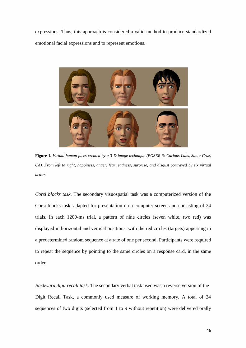
46
expressions. Thus, this approach is considered a valid method to produce standardized
emotional facial expressions and to represent emotions.
Figure 1. Virtual human faces created by a 3-D image technique (POSER 6: Curious Labs, Santa Cruz,
CA). From left to right, happiness, anger, fear, sadness, surprise, and disgust portrayed by six virtual
actors.
Corsi blocks task. The secondary visuospatial task was a computerized version of the
Corsi blocks task, adapted for presentation on a computer screen and consisting of 24
trials. In each 1200-ms trial, a pattern of nine circles (seven white, two red) was
displayed in horizontal and vertical positions, with the red circles (targets) appearing in
a predetermined random sequence at a rate of one per second. Participants were required
to repeat the sequence by pointing to the same circles on a response card, in the same
order.
Backward digit recall task. The secondary verbal task used was a reverse version of the
Digit Recall Task, a commonly used measure of working memory. A total of 24
sequences of two digits (selected from 1 to 9 without repetition) were delivered orally

47
by a recorded male human voice at a rate of one per second. Participants were required
to recall the digits in the reverse order of presentation.
Dual Tasks
EFE identification by all participants was tested in four experimental DT conditions: in
conjunction with a visuospatial DT during encoding or during retrieval; and in
conjunction with a verbal DT during encoding or during retrieval.
In the visuospatial DT during encoding condition, each EFE was displayed
simultaneously with a Corsi block trial for 1200 ms and after displaying a black screen
for 1200 ms, participants were required to first indicate the Corsi block sequence and
next to identify the EFE. In the visuospatial DT during retrieval condition, a 1200-ms
Corsi block trial was presented after a 1200-ms EFE presentation. The participant was
required to indicate the Corsi block sequence and then to identify the EFE. In both
protocols (DT during encoding and DT during retrieval), the interval between the onset
of EFE and the onset of the response was identical.
The procedure used to study verbal interference was similar to that used for
visuospatial interference but using the backward digit recall task as the secondary task.
Procedure
Participants were first given a brief description of the nature of the experiment and
asked to provide their written informed consent. All the research protocols involving
human subjects were first approved by the ethical board of the university (UNED). Each
participant was tested in all of the study conditions in two different sessions separated
by a 1-week interval. In the first session, participants were scored using the
neuropsychological scales and tested with the simple EFE identification task. In the
48
second session, they were tested in the visuospatial and verbal DT tasks. The order of
presentation of the DT tasks was balanced to avoid learning effects.
All participants were tested individually in a quiet room, and the experiment was
conducted using a 17-inch color display. Presentation of the stimuli and the data
recording were controlled using E-prime software, which was developed specifically for
this experiment. The instructions to the participants were presented on the screen before
each test condition, and the experimenter also described the tasks verbally. Participants
were asked to select the emotion that best described each face from a list of the six basic
emotions. The names of the six emotions were always visible, and the participants were
given as much time as they required to make their decision. Responses were recorded
by the experimenter.
RESULTS
First, a preliminary descriptive analysis was performed for baseline: simple EFE
identification task (Xolder = 13.63, SD = 4.16; Xyounger = 21.20, SD = 1.68), and the four
DT conditions: visuospatial DT during encoding (Xolder = 9.29, SD = 3.89;
Xyounger = 19.35, SD = 2.54); visuospatial DT during retrieval (Xolder = 11.75, SD = 3.05;
Xyounger = 20.73, SD = 2.16); verbal DT during encoding (Xolder = 11.42, SD = 3.79;
Xyounger = 20.75, SD = 1.99); and verbal DT during retrieval (Xolder = 12.46; SD = 3.16;
Xyounger = 21.03, SD = 1.79).
The analysis revealed a poorer performance in the older than in the younger group in
baseline as well as in DT conditions. To determine whether these differences could be
related to the variability in the performance of elderly population, the variables were
analyzed with the Kolmogorov-Smirnov (K-S) test in case the data vary significantly

49
from the pattern expected from a population with a normal distribution, and with the
Levene’s test to probe the homogeneity of variances across groups.
Baseline Analysis
Regarding simple EFE identification task, these preliminary analyses revealed that
performance was not distributed normally (z = 3.22, p < .05). Moreover, the Levene’s
test reached significance (F(1, 62) = 31.30, p < .05), meaning that the assumption of
homogeneity had to be rejected. Figure 2 shows the data distribution of EFE
identification task for each group.
Figure 2. Data distribution obtained for EFEs identification task.
Therefore, group differences were tested using a nonparametric test (Mann-Whitney
U test). The accuracy in the simple EFE identification task was determined to calculate
the baseline score for each group. The analysis revealed poorer performance in the older
than in the younger group in the simple EFE identification task (mean ranks = 13.5 and
43.9, respectively). Also, the Mann-Whitney U test revealed significant differences
between the performance of both groups (U = 24, z=−6.36, p < .05).

50
Dual-Task Analysis
Similarly to simple EFE identification task, the DT conditions were analyzed with the
K-S test and Levene’s test. The K-S test showed that data were not normally distributed:
visuospatial DT during encoding (z = 3.38, p < .05), visuospatial DT during retrieval
(z = 3.58, p < .05), verbal DT during encoding (z = 3.35, p < .05), and verbal DT during
retrieval (z = 3.58, p < .05). Also, Levene’s test showed that the variance was
significantly different between groups for all DT variables: visuospatial DT during
encoding (F(1, 62) = 6.58, p < .05), visuospatial DT during retrieval (F(1, 62) = 5.46,
p < .05), verbal DT during encoding (F(1, 62) = 15.64, p < .05), verbal DT during
retrieval (F(1, 62) = 7.13, p < .05). Figures 3 and 4 show the data distribution in DT
conditions for each group. Once again, we conducted the Mann-Whitney U tests. The
analysis revealed that older adults performed poorer than younger adults, as shown by
the mean ranks results of each variable (visuospatial DT during encoding = 44.1
vs. 13.7; visuospatial DT during retrieval = 44.16 vs. 13.06; verbal DT during encoding
= 43.88 vs. 13.54; verbal DT during retrieval = 44.31 vs. 12.81, respectively).
Fig. 3. Data distribution obtained for visuospatial DT conditions.
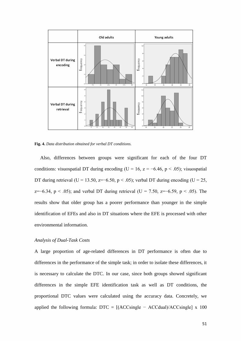
51
Fig. 4. Data distribution obtained for verbal DT conditions.
Also, differences between groups were significant for each of the four DT
conditions: visuospatial DT during encoding (U = 16, z = −6.46, p < .05); visuospatial
DT during retrieval (U = 13.50, z=−6.50, p < .05); verbal DT during encoding (U = 25,
z=−6.34, p < .05); and verbal DT during retrieval (U = 7.50, z=−6.59, p < .05). The
results show that older group has a poorer performance than younger in the simple
identification of EFEs and also in DT situations where the EFE is processed with other
environmental information.
Analysis of Dual-Task Costs
A large proportion of age-related differences in DT performance is often due to
differences in the performance of the simple task; in order to isolate these differences, it
is necessary to calculate the DTC. In our case, since both groups showed significant
differences in the simple EFE identification task as well as DT conditions, the
proportional DTC values were calculated using the accuracy data. Concretely, we
applied the following formula: DTC = [(ACCsingle − ACCdual)/ACCsingle] x 100

52
(Salthouse, Fristoe, Lineweaver, & Coon, 1995). This formula calculates the total
percentage DTC, which represents the cost due exclusively to DT, avoiding the
potential influence of differences associated with the simple task.
To calculate the DTC for each experimental condition, we created four new variables
(visuospatial DTC during encoding, visuospatial DTC during retrieval, verbal DTC
during encoding, verbal DTC during retrieval). Mann-Whitney U test revealed
significant differences between groups, in the EFEs identification, when the secondary
task was visuospatial in nature: visuospatial DTC during encoding (U = 226, z=−3.51,
p < .05); and visuospatial DTC during retrieval (U = 325, z=−2.16, p < .05).
However, when the secondary task was verbal in nature, we only observed
significant differences in performance when the DT was performed during encoding
(U = 277, z = −2.81, p < .05); and as such, the DTC for EFE identification was
significantly greater in the aged versus the young adult group. However, no significant
differences were observed between groups when the DT was performed during retrieval
(U = 373, z = −1.47, p > .05). These results could be explained by working memory
model (Baddeley & Della Sala, 1996; Baddeley & Hitch, 1974). First, as the
interference was generated by a verbal secondary task, both tasks were processed by
different slave systems (visuospatial sketchpad and phonological loop) and therefore
didn’t compete for the same type of resources, resulting in no significant differences
between groups in DT during the retrieval condition. However, encoding during DT
conditions resulted in higher DTC for older adults group, possibly because the
secondary task required central executive resources; i.e., both tasks were processed at
the same time, which didn’t happen in the retrieval condition.
Post Hoc analysis of Dual-Task Costs across emotions
Finally, a post hoc analysis was performed across emotions. Similarly to the analysis

53
above, we created four new variables (visuospatial DTC during encoding, visuospatial
DTC during retrieval, verbal DTC during encoding, verbal DTC during retrieval) for
each of the emotions. Table 2 shows the data of Mann-Whitney U test for each DTC
variable across emotions.
Table 2. Mann-Whitney U-test for each DTC variable across emotions.
As expected, the Mann-Whitney U test revealed higher DTC for older adults than
younger in all experimental conditions. These differences were statistically significant
for most of the emotion conditions. The greatest differences between groups occurred
during the visuospatial DT, where the DTCs were reflected by significant differences
between groups for all six emotions. By contrast, retrieval under the verbal DTC
condition showed the smallest differences between groups.
The performance was similar between groups in the identification of happiness and
anger except in the visuospatial DTC condition (i.e., when the cognitive resources
required were higher). By contrast, surprise, sadness, and fear were the emotions where
the differences between groups were the greatest.
DISCUSSION
The main goal of this present study was to investigate age-related changes in the
identification of emotional faces in different DT conditions. We studied the effects of
two DT variables: (a) the phase during which interference was applied (encoding or

54
retrieval); and (b) the nature of the interfering stimulus (visuospatial or verbal).
Specifically, we assessed the ability of the participants to process EFEs in four DT
conditions: visuospatial DT during encoding or during retrieval; and verbal DT during
encoding or during retrieval. In agreement with our hypothesis, we found that the
differences between older and young adult subjects were greatest (1) when the
secondary task was introduced during the encoding phase of EFE (i.e., when the
participants had to divide their attention in order to simultaneously process the EFE and
the secondary task); and (2) when both tasks were of a similar nature (i.e., when the
interference was generated by a visuospatial task).
The first observation is in line with previous findings, suggesting that the difficulties
experienced by the elderly in dividing their attention between tasks is due to limitations
imposed on central executive control that regulates attentional resources to
simultaneously coordinate two tasks (Salthouse, 2000). Indeed, this executive control
appears to be very sensitive to the effects of aging (Baddeley & Della Sala, 1996). As
mentioned above, emotional recognition often occurs in social situations in which it is
necessary to divide attention between emotional and nonemotional stimuli. Thus, in this
type of situation, the elderly may experience difficulties in interpersonal
communication.
The second observation is also in line with existing data that have demonstrated
increased interference in the DT paradigm when both tasks are similar in nature (Logie,
et al., 2004, 2007).
The third observation is deduced from post hoc analysis across emotions, suggesting
that older adults have selective difficulties when identifying some of the negative
emotions.

55
These data are in line with the previous literature that has reached a consensus about
the decline in the recognition of negative emotions. These studies, however, have not
produced consistent results across emotions (Calder, et al., 2003; Isaacowitz, et al.,
2007; Ruffman, et al., 2008; Sullivan & Ruffman, 2004; Suzuki, et al., 2007).
In general, our results are consistent with cognitive theory of emotional processing in
elderly (García-Rodríguez, et al., 2009, 2011c; Orgeta & Phillips, 2008; Pessoa, et al.,
2002; Phillips & Henry, 2005; Phillips, et al., 2007), which argues that deficits in EFE
identification are due, at least in part, to age-associated cognitive decline. Thus, the
elderly may experience difficulties in processing emotions in complex situations that are
cognitively demanding. As we have previously reported, these deficits in emotion
recognition are particularly marked in patients with cognitive impairment associated
with neurodegenerative disease or dementia (García-Rodríguez, et al., 2011a, 2011b,
2012). Also, the “positivity effect” could be explained by the cognitive approach, since
negative EFEs are more difficult to process than positive faces (Ekman & Friesen,
1978; Ekman, et al., 2002). Happy faces have different action units, which could result
in a better identification of them. However, this effect disappears when the
identification task overlaps the cognitive resources; therefore, easy and difficult faces
are poorly processed.
Whether EFE identification is an automatic or a controlled process remains the
subject of much debate. In the DT paradigm, an automatic process does not require
cognitive resources and thus should not be affected by the interference generated by a
concurrent task. By contrast, a controlled process consumes cognitive resources. Based
on our results, we conclude that the identification of emotional faces is not an automatic
process, particularly in complex situations in which EFE identification requires
cognitive resources. The conclusion that emotional identification is a controlled,

56
cognition-dependent process is in line with those of several other studies (Adolphs &
Damasio, 2001; Barnard, et al., 2007; Storbeck & Clore, 2007), pointing to a key role of
cognitive processes in emotional processing.
This finding suggests that the elderly experience greater difficulty in social contexts
in which emotional recognition occurs in tandem with the processing of other visual
stimuli (i.e., when the context demands the use of the same cognitive resources for
multiple tasks).
The present study offers new insight into the age-related changes in the processing of
emotional faces. Our findings further our understanding of the emotional lives of the
elderly and of the potential role of cognitive ability in modulating emotional processing
in this population, which may explain the difficulties experienced by the elderly in
performing some basic activities, such as identifying facial expressions. These
difficulties may have a significant impact on daily life, resulting in poorer social and
emotional function.
Finally, an important aspect of our study is the use of experimental tasks with a high
degree of ecological validity. EFE processing system is always active and ready to
process information and play a crucial communicative function (Ekman, 2003). Facial
expressions are used to confirm, contradict, or complete verbal messages, necessitating
dual processing of information on the part of the recipient. The use of the DT paradigm
to study the recognition of emotional faces constitutes a new approach with which to
analyze the complex mechanisms underlying EFE identification in a setting that closely
approximates to the real world.
In summary, our study provides experimental evidence of the difficulties experienced
by the elderly when attempting to attribute emotions to the facial expressions of others,

57
while simultaneously processing other environmental information. The functional
consequences of such impairment should be further investigated in order to improve the
emotional and social function of the elderly.
FUNDING
This research was funded by a grant from the Spanish Ministerio de Ciencia e
Innovación (Ref: PSI2009-13598-C02-01, Programa PSIC) to Beatriz García-
Rodríguez.
REFERENCES
Adolphs, R. (2002a). Neural systems for recognizing emotion. Current Opinion in
Neurobiology, 12, 169–177. doi: 10.1016/S0959-4388(02)00301-X
Adolphs, R. (2002b). Recognizing emotion from facial expressions: Psychological and
neurological mechanisms. Behavioral and Cognitive Neuroscience Reviews, 1,
21–62. doi: 10.1177/ 1534582302001001003
Adolphs, R., & Damasio, A. (2001). The interaction of affect and cognition: A
neurobiological perspective. In J. P. Forgas (Ed.), Handbook of affect and social
cognition (pp. 27–49). Mahwah, NJ: Lawrence Erlbaum Associates.
Adolphs, R., Tranel, D., Damasio, H., & Damasio, A. (1995). Fear and the human
amygdala. Journal of Neuroscience, 15, 5879–5891.
Baddeley, A. D., & Della Sala, S. (1996). Working memory and executive control.
Philosophical Transactions of the Royal Society of London, 351, 1397–1404.
doi: 10.1098/rstb.1996.0123
58
Baddeley, A. D., & Hitch, G. J. (1974). Working memory. In G. A. Bower (Ed.), Recent
advances in learning and motivation (pp. 47–89). New York: Academic Press.
Baena, E., Allen, P. A., Kaut, K. P., & Hall, R. J. (2010). On age differences in
prefrontal function: The importance of emotional/cognitive integration.
Neuropsychologia, 48, 319–333. doi: 10.1016/ j.neuropsychologia.2009.09.021
Barnard, P. J., Duke, D. J., Byrne, R. W., & Davidson, I. (2007). Differentiation in
cognitive and emotional meanings: An evolutionary analysis. Cognition &
Emotion, 21, 1155–1183. doi: 10.1080/ 02699930701437477
Blessed, G., Tomlinson, B. E., & Roth, M. (1968). The association between quantitative
measures of dementia and of senile change in the cerebral grey matter of elderly
subjects. The British Journal of Psychiatry, 114, 797–811. doi:
10.1192/bjp.114.512.797
Cacioppo, J. T., Berntson, G. G., Bechara, A., Tranel, D., & Hawkley, L. C. (2011).
Could an aging brain contribute to subjective well-being? The value added by a
social neuroscience perspective. In A. Todorov, S. T. Fiske, & D. A. Prentice
(Eds.), Social neuroscience: Toward understanding the underpinnings of the
social mind (pp. 249–262). New York, NY: Oxford University Press.
Calder, A. J., Keane, J., Manly, T., Sprengelmeyer, R., Scott, S., Nimmo-Smith, I., &
Young, A. W. (2003). Facial expression recognition across the adult life span.
Neuropsychologia, 41, 195–202. doi: 10.1016/S0028-3932(02)00149-5
Carstensen, L. L., Fung, H. H., & Charles, S. T. (2003). Socioemotional selectivity
theory and the regulation of emotion in the second half of life. Motivation and
Emotion, 27, 103–123. doi: 10.1023/ A:1024569803230

59
Carstensen, L. L., & Mikels, J. A. (2005). At the intersection of emotion and cognition.
Aging and the positivity effect. Current Directions in Psychological Science, 14,
117–121. doi: 10.1111/cdir.2005.14.issue-3
Ebner, N. C., y Johnson, M. K. (2010). Age-group differences in interference from
young and older emotional faces. Cognition & Emotion, 24, 1095–1116. doi:
10.1080/02699930903128395
Ekman, P. (2003). Emotions revealed. London, UK: Weidenfeld & Nicolson.
Ekman, P., & Friesen, W. V. (1978). The facial action coding system. Palo Alto, CA:
Consult Psychology Press.
Ekman, P., Friesen, W. V., & Hager, J. C. (2002). Facial action coding system –
investigator’s guide. Salt Lake City, UT: Research Nexus.
Fisk, A. D., & Schneider, W. (1983). Category and word search: Generalising search
principles to complex processing. Journal of Experimental Psychology:
Learning, Memory and Cognition, 10, 181–197.
Folstein, M. F., Folstein, S. E., & McHugh, P. R. (1975). “Mini-mental state”. A
practical method for grading the cognitive state of patients for the clinician.
Journal of Psychiatric Research, 12, 189–198. doi: 10.1016/0022-
3956(75)90026-6
García-Rodríguez, B., Ellgring, H., Fusari, A., & Frank, A. (2009). The role of
interference in identification of emotional facial expressions in normal ageing
and dementia. The European Journal of Cognitive Psychology. Special Issue on
Aging, Cognition and Neuroscience, 21, 428–444. doi: 10.1080/
09541440802475793

60
García-Rodríguez, B., Casares-Guillén, C., Jurado-Barba, R., Rubio, G., Molina, J. A.,
& Ellgring, H. (2011a). Visuo-spatial interference affects the identification of
emotional facial expressions unmedicated Parkinson’s patients. Journal of
Neurological Science, 313, 13–16. doi: 10.1016/ j.jns.2011.09.041
García-Rodríguez, B., Casares-Guillén, C. T., Molina, J. A., Rubio, G., Jurado-Barba,
R., Morales, I., & Ellgring, H. (2011b). Efectos diferenciales de la doble tarea en
el procesamiento emocional en pacientes con enfermedad de Parkinson no
medicados. Revista de Neurología, 53, 329–336.
García-Rodríguez, B., Fusari, A., Fernández-Guinea, S., Frank, A., Molina, J. A., &
Ellgring, H. (2011c). Decline of executive processes affects identification of
emotional facial expressions in aging. Current Aging Science, 4, 70–75. doi:
10.2174/1874609811104010070
García-Rodríguez, B., Vincent, C., Casares-Guillén, C. T., Ellgring, H., & Frank, A.
(2012). The effects of different attentional demands in the identification of
emotional facial expressions in Alzheimer’s disease. American Journal of
Alzheimer Disease & Other Dementias, 27, 530–536. doi: 10.1177/
1533317512459797
Heberlein, A. S., Padon, A. A., Gilihan, S. J., Farah, M. J., & Fellows, L. K. (2008).
Ventromedial frontal lobe plays a critical role in facial emotion recognition.
Journal Cognitive Neuroscience, 20, 721–733.
Iliffe, S., Booroff, A., Gallivan, S., Goldenberg, E., Morgan, P., & Haines, A. (1990).
Screening for cognitive impairment in the elderly using the mini-mental state
examination. The British Journal of General Practice, 40, 277–279.

61
Isaacowitz, D. M., Löckenhoff, C. E., Lane, R. D., Wright, R., Sechrest, L., Riedel, R.,
& Costa, P. T. (2007). Age differences in recognition of emotion in lexical
stimuli and facial expressions. Psychology and Aging, 22, 147–159. doi:
10.1037/0882-7974.22.1.147
Logie, R. H., Cocchini, G., Della Sala, S., & Baddeley, A.D. (2004). Is there a specific
executive capacity for dual task co-ordination? Evidence from Alzheimer’s
disease. Neuropsychology, 18, 504–513. doi: 10.1037/0894-4105.18.3.504
Logie, R. H., Della Sala, S., MacPherson, S. E., & Cooper, J. (2007). Dual task
demands on encoding and retrieval processes: Evidence from healthy adult
ageing. Cortex, 43, 159–169. doi: 10.1016/ S0010-9452(08)70453-2
Moser, E., Demtl, E., Robinson, S., Fink, B., Gur, R. C., & Grammer, K. (2007).
Amygdala activation at 3T in response to human and avatar facial expressions of
emotions. Journal of Neuroscience Methods, 161, 126–133. doi:
10.1016/j.jneumeth.2006.10.016
Naveh-Benjamin, M. (1987). Coding of spatial location information: An automatic
process?. Journal of Experimental Psychology: Learning Memory and
Cognition, 12, 378–386. doi: 10.1037/ 0278-7393.12.3.378
Naveh-Benjamin, M., Craik, F. I. M., Gavrilescu, D., & Anderson, N. D. (2000).
Asymmetry between encoding and retrieval processes: Evidence from divided
attention and a calibration analysis. Memory & Cognition, 28, 965–976. doi:
10.3758/BF03209344
Naveh-Benjamin, M., Craik, F. I., Guez, J., & Dori, H. (1998). Effects of divided
attention on encoding and retrieval processes in human memory: Further support

62
for an asymmetry. Journal of Experimental Psychology: Learning, Memory and
Cognition, 24, 1091–1104.
Naveh-Benjamin, M., & Jonides, J. (1986). On the automaticity of frequency coding:
Effects of competing task load, encoding strategy and intention. Journal of
Experimental Psychology: Learning, Memory and Cognition, 12, 378–386.
Öhman, A. (2002). Automaticity and the amygdala: Nonconscious responses to
emotional faces. Current Directions in Psychological Science, 11, 62–66. doi:
10.1111/1467-8721.00169
Orgeta, V., & Phillips, L. H. (2008). Effects of age and emotional intensity on the
recognition of facial emotion. Experimental Aging Research, 34, 63–79. doi:
10.1080/03610730701762047
Pessoa, L., McKenna, M., Gutierrez, E., & Ungerleider, L. G. (2002). Neural processing
of emotional faces requires attention. Proceedings of the National Academy of
Sciences of the United States of America, 99, 11458–11463. doi:
10.1073/pnas.172403899
Pessoa, L., & Ungerleider, L. G. (2005). Visual attention and emotional perception. In
L. Itti, G. Rees, & J. K. Tsotsos (Eds.), Neurobiology of attention (pp. 160–166).
San Diego, CA: Elsevier.
Phillips, L. H., & Henry, J. D. (2005). An evaluation of the frontal lobe theory of
cognitive aging. In J. Duncan, L. H. Phillips, & P. McLeod (Eds.), Measuring
the mind: Speed, control and age (pp. 191–216). London: Oxford University
Press.
63
Phillips, L. H., MacLean, R. D. J., & Allen, R. (2002). Age and the understanding of
emotions: Neuropsychological and sociocognitive perspectives. The Journals of
Gerontology Series B: Psychological Sciences and Social Sciences, 57, 526–
530. doi: 10.1093/geronb/57.6.P526
Phillips, L. H., Tunstall, M., & Channon, S. (2007). Exploring the role of working
memory in dynamic social cue decoding using dual task methodology. Journal
of Nonverbal Behavior, 31, 137–152. doi: 10.1007/s10919-007-0026-6
Pollock, J. W., Khoja, N., Kaut, K. P., Lien, M.-C., & Allen, P. A. (2012).
Electrophysiological evidence for adult age-related sparing and decrements in
emotion perception and attention. Frontiers in Integrative Neuroscience, 6, 1–
14. doi: 10.3389/fnint.2012.00060
Ruffman, T., Henry, J. D., Livingstone, V., & Phillips, L. H. (2008). A meta-analytic
review of emotion recognition and aging: Implications for neuropsychological
models of aging. Neuroscience & Biobehavioral Reviews, 32, 863–881. doi:
10.1016/j.neubiorev.2008.01.001
Salthouse, T. A. (2000). Steps toward the explanation of adult age differences in
cognition. In T. Perfect & E. Maylor (Eds.), Theoretical debate in cognitive
aging (pp. 19–49). London: Oxford University Press.
Salthouse, T. A., Fristoe, N. M., Lineweaver, T. T., & Coon, V. E. (1995). Aging of
attention: Does the ability to divide decline? Memory & Cognition, 23, 59–71.
doi: 10.3758/BF03210557

64
Schneider,W., Dumais, S. T., & Shiffrin, R. M. (1984). Automatic and control
processing and attention. In R. Parasunaman (Ed.), Varieties of attention (pp. 1–
27). New York: Academic Press.
Schneider, W., & Shiffrin, R. M. (1977). Controlled and automatic human information
processing: I. Detection, search, and attention. Psychological Review, 84, 1–66.
doi: 10.1037/0033-295X.84.1.1
Storbeck, J., & Clore, G. L. (2007). On the interdependence of cognition and emotion.
Cognition & Emotion, 21, 1212–1237. doi: 10.1080/02699930701438020
Sullivan, S., & Ruffman, T. (2004). Emotion recognition deficits in the elderly.
International Journal of Neuroscience, 114, 403–432. doi:
10.1080/00207450490270901
Suzuki, A., Hoshino, T., Shigemasu, K., & Kawamura, M. (2007). Decline or
improvement? Age related differences in facial expressions recognition.
Biological Psychology, 74, 75–84. doi: 10.1016/ j.biopsycho.2006.07.003
Verhaeghen, P., & Cerella, J. (2002). Aging, executive control, and attention: A review
of meta-analyses. Neuroscience and Biobehavioral Reviews, 26, 849–857.
Weyers, P., Muhlberger, A., Hefele, C., & Pauli, P. (2006). Electromyographic
responses to static and dynamic avatar emotional facial expressions.
Psychophysiology, 43, 450–453. doi: 10.1111/ j.1469-8986.2006.00451.x
Yesavage, J. A., Brink, T. L., & Rose, T. L. (1983). Development and validation of a
geriatric depression screening scale: A preliminary report. Journal of Psychiatric
Research, 17, 37–49. doi: 10.1016/ 0022-3956(82)90033-

65
Capítulo 5. [Estudio 2]. Efectos diferenciales de la DT en el procesamiento
emocional en pacientes con EP no medicados.
García-Rodríguez, B., Casares-Guillén, C. T., Molina, J. A., Rubio, G., Jurado-Barba,
R., Morales, I., y Ellgring, H. (2011). Efectos diferenciales de la doble tarea en el
procesamiento emocional en pacientes con enfermedad de Parkinson no medicados.
Revista de Neurología, 53(6), 329-336.

66
RESUMEN
Introducción. Trabajos previos han puesto de manifiesto que la correcta identificación
de las expresiones faciales emocionales (EFE) depende de los recursos cognitivos
disponibles. En este trabajo investigamos si la capacidad para identificar EFE en un
paradigma de doble tarea se ve afectada en la enfermedad de Parkinson (EP).
Objetivo. Investigar la interferencia que genera la introducción de una tarea secundaria
en el procesamiento de EFE durante las fases de codificación y recuperación de la
expresión facial en pacientes con EP no medicados.
Sujetos y métodos. Un total de 14 pacientes con EP de novo y 28 adultos sanos
identificaron 24 EFE bajo dos condiciones: codificación simultánea con una tarea
secundaria e introducción de la tarea secundaria entre el tiempo que media entre la
codificación de la primaria y el tiempo de latencia de la respuesta.
Resultados. Los resultados mostraron que los pacientes con EP identificaron las EFE
significativamente peor que los adultos sanos en la condición de codificación
simultánea. Por el contrario, no mostraron diferencias cuando la interferencia de la tarea
secundaria se produjo en la fase de recuperación de la información de la primaria.
Conclusiones. Los pacientes con EP muestran déficits específicos en el procesamiento
de EFE sólo cuando la tarea consume altos recursos de atención dividida, como ocurre
en las situaciones cotidianas.
Palabras clave. Atención dividida. Deterioro cognitivo. Enfermedad de Parkinson.
Expresión facial emocional. Interferencia visuoespacial. Paradigma de doble tarea.

67
ABSTRACT
Introduction. Previous research has shown that correct identification of emotional
facial expressions (EFE) depends on the cognitive resources that are available. In this
study, we examine whether the capacity to identify EFE in a dual task paradigm is
affected in Parkinson’s disease (PD).
Aim. To investigate the interference generated by introducing a secondary task in EFE
processing during the encoding and retrieval phases of facial expressions in non-
medicated PD patients.
Subjects and methods. A total of 14 patients de novo PD and 28 healthy adults
identified 24 EFE under two conditions: simultaneous encoding with a secondary task
and introduction of the secondary task between the time that spans the encoding of the
primary task and the response time latency.
Results. Results showed that identification of EFE by patients with PD was
significantly worse than by healthy adults in the simultaneous encoding condition. In
contrast, no differences were found when the interference of the secondary task took
place in the retrieval phase of the primary task.
Conclusions. PD patients only display specific deficits in EFE processing when the task
consumes high levels of divided attention resources, as occurs in everyday situations.
Key words. Cognitive impairment. Divided attention. Dual task paradigm. Emotional
facial expressions. Parkinson’s disease. Visuospatial interference.

68
INTRODUCCIÓN
En los últimos años, la neurociencia ha mostrado un creciente interés por investigar las
bases neurológicas del procesamiento o identificación de las expresiones faciales
emocionales (EFE) y por determinar en qué medida esta capacidad está deteriorada en
las enfermedades neurodegenerativas [1-3].
En relación con la enfermedad de Parkinson (EP), a pesar de que muchas
investigaciones han encontrado que los pacientes tienen déficits específicos en el
procesamiento de EFE, los resultados acerca de cómo la habilidad para identificar EFE
está afectada por la enfermedad no son concluyentes [4-7]. Por ejemplo, varios autores
señalan que los pacientes medicados tienen dificultades para procesar la emoción de
asco [8], así como otras emociones negativas [9-12], y que estos déficits están asociados
a la disfunción de los ganglios basales. Sin embargo, otros estudios no han podido
corroborar estos hallazgos [13,14]. Trabajos recientes defienden la hipótesis de que los
déficits en el procesamiento de EFE característicos de los pacientes de EP dependen del
tipo de recursos cognitivos que demanda la tarea [15-19], como, por ejemplo, recursos
visuo-espaciales o atencionales, recursos afectados desde el comienzo de la enfermedad.
Según esta hipótesis, el deterioro cognitivo característico de la enfermedad podría ser,
en parte, el responsable de las dificultades que manifiestan los pacientes para procesar la
información de naturaleza afectiva. Hay, fundamentalmente, dos estructuras cerebrales
vinculadas al procesamiento emocional: la amígdala [20] y la corteza prefrontal [21].
Muchos estudios neurológicos [22-24] han destacado el papel básico que desempeña la
amígdala en el procesamiento del miedo. Esta estructura cerebral proporciona una
detección automática y rápida del estímulo emocional, y prioriza el procesamiento de
estímulos externos, especialmente de valencia emocional negativa por su vinculación a
las funciones de supervivencia de los individuos. Sin embargo, se sabe que este

69
procesamiento amigdalar es insuficiente cuando se trata de estímulos emocionales
complejos o cuando el procesamiento ocurre en situaciones sociales donde hay que
coordinar al mismo tiempo varias fuentes de información [25-29]. En estos casos, es
importante el papel de la corteza prefrontal como el órgano responsable del correcto
funcionamiento de las funciones ejecutivas, proceso psicológico afectado desde los
primeros estadios de la EP [30-36].
Por funciones ejecutivas entendemos, entre otros, procesos como la toma de
decisiones, el aprendizaje de reglas de categorización o la ejecución de tareas, que
demandan recursos de atención dividida, habilidad necesaria para poder descodificar la
información emocional en contextos sociales, donde es necesario coordinar al mismo
tiempo diversas fuentes de información. Uno de los modelos más utilizados para evaluar
la atención dividida es el paradigma de la doble tarea (DT). La DT se basa en la
realización simultánea de dos tareas, de tal forma que el deterioro en la ejecución de una
tarea se considera un indicio de la demanda de atención de la otra. Mediante trabajos de
lesión se ha podido comprobar que la ejecución simultánea es un proceso muy sensible
a los efectos del envejecimiento, tanto normal como patológico [37,38]. Los estudios de
DT indican que no todos los tipos de doble procesamiento están afectados en la misma
medida por la introducción de la tarea secundaria. Las demandas mentales –es decir, los
costes– son mayores cuando las dos tareas se codifican simultáneamente [39,40], ya que
implica que hay que dividir las capacidades atencionales entre las dos fuentes de
información. Por el contrario, cuando la tarea secundaria se introduce en el período que
media entre la codificación de la primaria y el tiempo de latencia de la respuesta [41],
los costes en la ejecución de las dos tareas no son tan elevados. Esto es especialmente
crítico en el envejecimiento, probablemente porque en el primer caso la correcta
ejecución de las tareas depende fundamentalmente de la capacidad de la atención

70
dividida, mientras que en el segundo la correcta ejecución depende de la memoria [42-
45], proceso menos sensible al paso de los años. Parece que estos datos son sólidos
incluso cuando una de las tareas implicadas consiste en el procesamiento de
información emocional.
Trabajos previos [27,29] han puesto de relieve que la introducción de una tarea
secundaria afecta al procesamiento de las EFE, tanto en personas mayores como en
pacientes de Alzheimer, lo que indica que esta capacidad no implica un procesamiento
emocional automático (hipótesis muy aceptada por los investigadores) y, por tanto, que
es vulnerable al deterioro cognitivo. Sin embargo, y hasta donde nosotros sabemos, se
desconocen los efectos de la DT en el procesamiento de las expresiones faciales en
pacientes de EP. Siguiendo la hipótesis de que los déficits en el procesamiento de EFE
dependen en parte del tipo de tarea involucrada, el principal objetivo de este trabajo ha
sido investigar la interferencia que genera una tarea secundaria en el procesamiento de
EFE durante las fases de codificación y recuperación de la DT en un grupo de pacientes
de EP no medicados.
De acuerdo con los estudios antes mencionados, esperamos un rendimiento peor del
grupo de pacientes cuando la identificación de la EFE ocurre en la fase de codificación
que cuando ocurre en la de recuperación, es decir, cuando el recurso cognitivo
prioritario demandado por la tarea es la atención dividida.
Los resultados de este trabajo contribuirán a conocer hasta qué punto el deterioro del
procesamiento emocional covaría con los déficits cognitivos específicos de los pacientes
de EP y ayudarán a comprender mejor las consecuencias funcionales de la EP en la vida
afectiva de los pacientes.
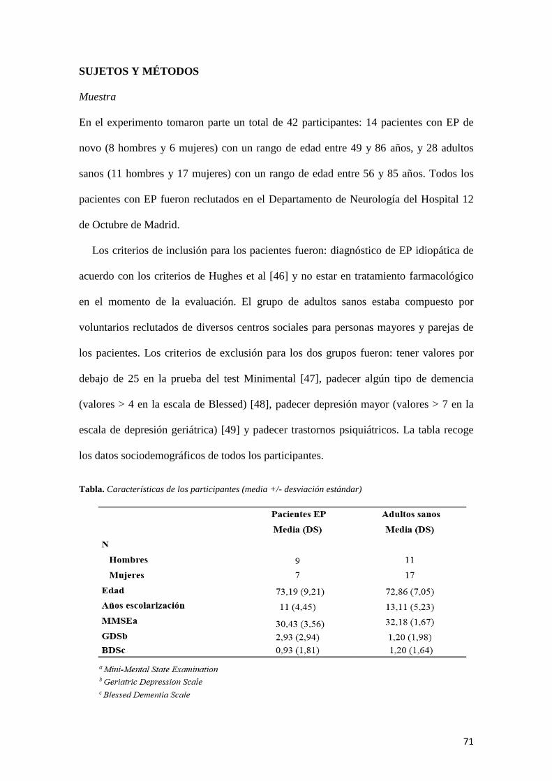
71
SUJETOS Y MÉTODOS
Muestra
En el experimento tomaron parte un total de 42 participantes: 14 pacientes con EP de
novo (8 hombres y 6 mujeres) con un rango de edad entre 49 y 86 años, y 28 adultos
sanos (11 hombres y 17 mujeres) con un rango de edad entre 56 y 85 años. Todos los
pacientes con EP fueron reclutados en el Departamento de Neurología del Hospital 12
de Octubre de Madrid.
Los criterios de inclusión para los pacientes fueron: diagnóstico de EP idiopática de
acuerdo con los criterios de Hughes et al [46] y no estar en tratamiento farmacológico
en el momento de la evaluación. El grupo de adultos sanos estaba compuesto por
voluntarios reclutados de diversos centros sociales para personas mayores y parejas de
los pacientes. Los criterios de exclusión para los dos grupos fueron: tener valores por
debajo de 25 en la prueba del test Minimental [47], padecer algún tipo de demencia
(valores > 4 en la escala de Blessed) [48], padecer depresión mayor (valores > 7 en la
escala de depresión geriátrica) [49] y padecer trastornos psiquiátricos. La tabla recoge
los datos sociodemográficos de todos los participantes.
Tabla. Características de los participantes (media +/- desviación estándar)
72
Los dos grupos fueron igualados en edad (t(40) = 3,53; p > 0,050), sexo (EP: χ² (1) =
0,28, p > 0,050; adultos sanos: χ² (1) = 1,28, p > 0,050) y años de escolarización (F(40) =
0,40; p > 0,050). Las puntuaciones en el test Minimental fueron significativamente
diferentes en los dos grupos (pacientes con EP, rango: 25 y 35; adultos sanos, rango: 29
y 35; F(40) = 7,35; p < 0,050) e indicaron que el grupo de pacientes tenía un ligero
deterioro cognitivo, pero no mostraron signos de demencia, ya que las puntuaciones en
la escala de Blessed no fueron estadísticamente significativas en los dos grupos (F(40) =
0,30; p > 0,050). Por último, encontramos diferencias marginalmente significativas en
relación con las puntuaciones en la escala de depresión (F(40) = 1,65; p < 0,050) que
revelan un estado de ánimo de los pacientes de EP más deprimido que los mayores
sanos, pero sin que se pueda hablar de depresión clínica ya que ningún paciente obtuvo
una puntuación superior a 7. Todos los participantes firmaron un consentimiento
informado aprobado por el Comité de Ética de la Universidad Nacional de Educación a
Distancia.
Tareas y procedimiento
Tarea primaria: identificación de expresiones faciales emocionales. En total, se
presentaron en la pantalla de un ordenador 24 estímulos que mostraban las seis
emociones básicas: alegría, tristeza, enfado, miedo, sorpresa y asco, expresadas por
cuatro modelos (dos hombres y dos mujeres). Los estímulos se diseñaron con un
programa (Poser 6 Program, Curious Labs, Santa Cruz, EE. UU.) que sirve para generar
rostros humanos virtuales y cuyas expresiones faciales se determinan por los cambios
descritos en el manual Facial Action Coding System (FACS) [50,51]. La codificación
de las emociones fue realizada por un experto codificador en el FACS (J.H.E.), que
activa los músculos según las correspondientes unidades de acción. Este conjunto de
73
estímulos se ha validado en investigaciones previas con personas mayores [29] y
pacientes con enfermedad de Alzheimer [27]. Antes de la aparición de la expresión
emocional y con el objetivo de fijar la atención de los participantes, se presentaba una
cruz en la pantalla durante 1.200 ms. Tras una pantalla en negro de 600 ms de duración,
aparecía la expresión emocional durante 1.200 ms. A continuación, los participantes
tenían que identificar la emoción que expresaba cada una de las caras, teniendo siempre
delante el nombre de las seis diferentes emociones. El orden de aparición de los
estímulos para cada participante fue aleatorio.
Tarea secundaria: Bloques de Corsi
La tarea de los bloques de Corsi se utiliza para medir las habilidades de la memoria
visuoespacial. En la pantalla del ordenador aparece una matriz de nueve círculos
colocados en posición horizontal y vertical (3 x 3), siete blancos y dos rojos. En cada
ensayo de 1.200 ms de duración se presentaba una secuencia diferente de la localización
de los círculos rojos (aparecía un círculo rojo en una posición durante 600 ms y, cuando
desaparecía, aparecía el segundo círculo con la misma duración pero en posición
diferente). Los participantes tenían que indicar la secuencia de los círculos en una hoja
de respuestas. El orden de las secuencias para cada participante fue aleatorizado. Un
requisito importante para el experimento fue que la dificultad de la tarea secundaria
resultase igual para los dos grupos. Decidimos utilizar la versión de dos círculos rojos
porque en pruebas previas al experimento comprobamos que no había diferencias
significativas entre un grupo pequeño de pacientes de EP y sus controles.

74
Procedimiento
La capacidad para identificar las expresiones se evaluó bajo dos condiciones: paradigma
de DT durante la codificación y paradigma de DT durante la recuperación. En la
primera condición se presentaban juntos durante 1.200 ms una EFE y un ensayo de la
tarea de los bloques de Corsi. A continuación aparecía una pantalla en negro de 1.200
ms de duración y después los participantes ten.an que responder siempre en el mismo
orden, primero a la secuencia de Corsi y a continuación a la EFE. En la segunda
condición, primero se presentaba una EFE durante 1.200 ms, después un ensayo de la
tarea de los bloques de Corsi (también de 1.200 ms de duración) y a continuación los
participantes contestaban en el mismo orden que para la primera condición. Con el
objetivo de evitar efectos de aprendizaje, se contrabalanceó el orden de presentación de
las dos condiciones. Las instrucciones se explicaron verbalmente y también aparec.an
en la pantalla del ordenador al inicio de cada una de las condiciones experimentales. Las
respuestas a las dos tareas fueron registradas por el experimentador.
Diseño experimental
En este estudio hemos utilizado un diseño factorial mixto, 2 (grupo: pacientes de EP y
adultos sanos) x 2 (tipo de presentación: DT durante la codificación y DT durante la
recuperación). El primer factor fue analizado como variable intersujetos, y el segundo,
intrasujetos –ANOVA (análisis de la varianza), SPSS v. 17.0–. La variable dependiente
fue el número de EFE identificadas correctamente. La puntuación máxima fue de 24
aciertos. El nivel de significación fue del 5%.

75
RESULTADOS
Análisis de la tarea primaria
Los resultados del ANOVA mostraron que el efecto principal para la comparación
intrasujetos del tipo de presentación (DT durante la codificación y DT durante la
recuperación) fue estadísticamente significativo (F(1,40) = 29,23; error de la raíz
cuadrada media, MSE = 201,52; p < 0,001; ηp2 = 0,422). La identificación de las EFE
fue más fácil en la condición DT durante la recuperación (media: 13,86) que en la
condición DT durante la codificación (media: 11,10) (Fig. 1).
Fig. 1. Porcentaje de respuestas correctas en la tarea de identificar EFEs en las dos condiciones
experimentales
La comparación entre grupos reveló un efecto de interacción estadísticamente
significativo entre grupo y tipo de presentación (F(1,40) = 29,23; MSE = 201,52;
p < 0,050; ηp2 = 0,143), lo que significa que los dos grupos tuvieron una ejecución
diferente dependiendo de la condición experimental. Para analizar estas diferencias
entre grupos, realizamos un análisis multivariante de la varianza (MANOVA) con las
puntuaciones obtenidas en las dos condiciones.
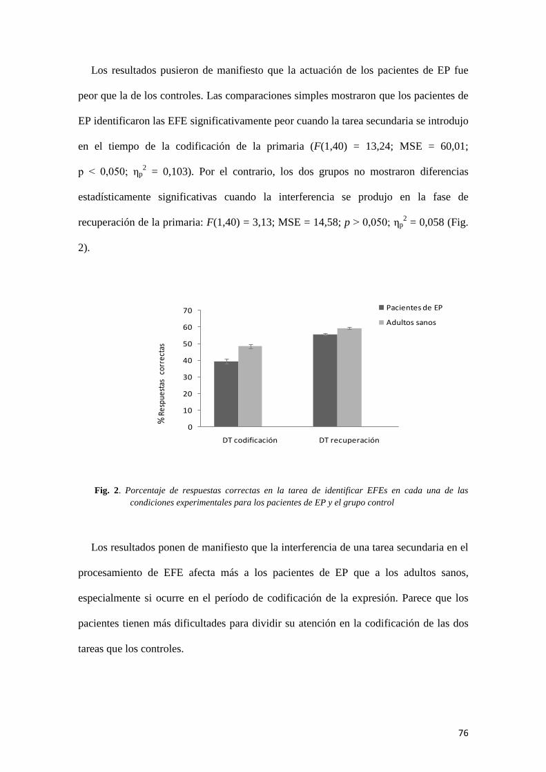
76
Los resultados pusieron de manifiesto que la actuación de los pacientes de EP fue
peor que la de los controles. Las comparaciones simples mostraron que los pacientes de
EP identificaron las EFE significativamente peor cuando la tarea secundaria se introdujo
en el tiempo de la codificación de la primaria (F(1,40) = 13,24; MSE = 60,01;
p < 0,050; ηp2 = 0,103). Por el contrario, los dos grupos no mostraron diferencias
estadísticamente significativas cuando la interferencia se produjo en la fase de
recuperación de la primaria: F(1,40) = 3,13; MSE = 14,58; p > 0,050; ηp2 = 0,058 (Fig.
2).
Fig. 2. Porcentaje de respuestas correctas en la tarea de identificar EFEs en cada una de las
condiciones experimentales para los pacientes de EP y el grupo control
Los resultados ponen de manifiesto que la interferencia de una tarea secundaria en el
procesamiento de EFE afecta más a los pacientes de EP que a los adultos sanos,
especialmente si ocurre en el período de codificación de la expresión. Parece que los
pacientes tienen más dificultades para dividir su atención en la codificación de las dos
tareas que los controles.
0
10
20
30
40
50
60
70
DT codificación DT recuperación
Pacientes de EP
Adultos sanos
% R
espu
esta
sco
rrec
tas

77
Para evaluar hasta qué punto estos déficits podr.an explicarse por la hipótesis de la
sobrecarga de la agenda visuoespacial [19], analizamos la actuación de ambos grupos en
la tarea de los bloques de Corsi en cada una de las condiciones experimentales.
Análisis de la tarea secundaria
Los porcentajes medios de respuesta han sido menores, en las dos condiciones, para el
grupo de pacientes de EP que para el grupo control (Fig. 3).
Fig. 3. Porcentaje de respuestas correctas en la tarea de los Bloques de Corsi en cada una de las
condiciones experimentales para los pacientes de EP y el grupo control
Para analizar si la actuación de ambos grupos fue estadísticamente significativa
realizamos un MANOVA con las puntuaciones obtenidas en las dos condiciones. Los
resultados muestran que los dos grupos han tenido una actuación estadísticamente
significativa tanto en la condición DT durante la codificación (F(1,40) = 29,29; MSE =
198,10; p < 0,001; ηp 2 = 0,423) como durante la recuperación (F(1,40) = 8,62; MSE =
138,85; p = 0,005; ηp 2 = 0,177). En este caso, los pacientes han tenido una peor
0
10
20
30
40
50
60
70
DT codificación DT recuperación
Pacientes de EP
Adultos sanos
% R
esp
ue
stas
corr
ect
as

78
actuación que los controles en las dos condiciones experimentales. En los pacientes de
EP, la tarea de los bloques de Corsi parece más vulnerable a los efectos de la DT que la
tarea de identificar EFE, que sólo se veía afectada por la DT en la condición de
codificación, pero no en la de la recuperación.
DISCUSIÓN
El objetivo del presente trabajo ha sido evaluar si los déficits en el procesamiento de
EFE en los pacientes de EP dependen de las demandas de la tarea. La hipótesis puesta a
prueba es que los pacientes de EP tienen mayores dificultades en el procesamiento de
EFE cuando el recurso cognitivo demandado es la atención dividida, recurso cognitivo
muy sensible a los efectos de la enfermedad. En concreto, hemos estudiado el papel de
la interferencia generada en el procesamiento de EFE por la introducción de una tarea
secundaria en dos condiciones de DT: en el momento de codificación y en el período
que media entre la codificación de la EFE y su respuesta. Los resultados obtenidos
ponen de manifiesto lo siguiente:
– Los pacientes tienen más dificultad que los adultos sanos en identificar EFE cuando
han de dividir su atención en dos tareas, en este caso de naturaleza visual, pero no
cuando el recurso cognitivo demandado para identificar la EFE correctamente es la
memoria.
– Los pacientes han tenido una peor actuación que los controles en la tarea de los
bloques de Corsi en cualquiera de las dos condiciones de la DT, es decir, cuando los
recursos demandados han sido la atención y la memoria. Estos resultados nos llevan a
tres conclusiones. En primer lugar, que los pacientes de EP tienen déficits desde el
comienzo de la enfermedad en el procesamiento de EFE en contextos complejos en los
que se requiere dividir la atención hacia diversas fuentes de información. Estos trabajos

79
coinciden con investigaciones previas que han señalado que los déficits en el
procesamiento de EFE en pacientes de EP dependen también de la carga cognitiva [15-
19].
Un aspecto relevante en este trabajo ha sido la utilización de tareas experimentales de
gran validez ecológica en el sentido de que los participantes tenían que codificar las
expresiones emocionales al mismo tiempo que procesaban otra información visual. Esto
es importante si consideramos que el sistema de procesamiento de EFE es un sistema
que esté siempre abierto o preparado para procesar de forma involuntaria [52], y que
una de sus funciones es la de aceptar o rechazar otro tipo de mensajes, lo que implica
obligatoriamente el procesamiento simultáneo de dos fuentes diferentes de información.
Este trabajo permite comprender los déficits reales que encuentran los pacientes en su
vida cotidiana cuando intentan integrar la información afectiva con su actividad
cognitiva habitual.
En segundo lugar, este trabajo pone de manifiesto que los déficits en la identificación
en DT ocurren especialmente cuando los pacientes tienen que codificar al mismo tiempo
la expresión facial y otra información, es decir, les cuesta dividir su atención para poder
procesar al mismo tiempo los dos estímulos. Por trabajos experimentales previos [53],
sabemos que los déficits en el doble procesamiento son mayores cuando coincide la
naturaleza perceptiva de las dos fuentes de información, es decir, las dos tareas
requieren procesamiento visual, como en nuestro caso. El análisis de la tarea secundaria
de los bloques de Corsi parece confirmar el patrón encontrado en la tarea primaria, ya
que pone de manifiesto que los pacientes de EP han tenido también más dificultades que
el grupo control, lo que parece lógico dada la naturaleza visuoespacial de la tarea
secundaria. Estos resultados están en línea con trabajos previos [19] que defienden la
hipótesis de que, en los pacientes de EP, las dificultades en el procesamiento de EFE

80
están relacionadas con los déficits visuoespaciales. Queda abierto a futuras
investigaciones comprobar si esta interferencia ocurriría también cuando la información
adicional a la EFE fuese de distinta naturaleza, por ejemplo, semántica.
En tercer lugar, un punto relevante que se deriva del trabajo es que cuando la
interferencia de la tarea secundaria ha tenido lugar en el período que media entre la
codificación de la primaria y su respuesta, los pacientes de EP no han mostrado
diferencias significativas con el grupo control, es decir, tienen relativamente preservada
esta facultad. Nos parece importante rese.ar este dato, pues los déficits en el
procesamiento de las EFE no parecen tan graves en la EP como en los pacientes con
enfermedad de Alzheimer, que han mostrado déficits en cualquiera de las condiciones
de DT [27].
Un aspecto destacable es que nuestros resultados apuntan a la hipótesis de que la
complejidad del estímulo por codificar podría ser una variable moduladora en la
capacidad para ejecutar en DT. Teniendo en cuenta que una de las cuestiones más
debatidas es si los pacientes de EP tienen dificultades en la identificación de las
emociones discretas o específicas, será interesante poder determinar si todas las
emociones básicas se ven igualmente afectadas por la capacidad de la atención dividida.
Es de esperar que las emociones más complejas estuvieran más afectadas que las más
simples. En relación con las EFE, no todas las emociones son igual de difíciles de
expresar. Mientras que la expresión de alegría sólo necesita tres unidades de acción o
movimientos musculares para poder expresarse, otras, como el miedo, necesitan al
menos cinco unidades de acción [50,51]. No hemos podido comprobar empíricamente
este punto porque en nuestro experimento el número de estímulos para cada EFE ha
sido relativamente bajo para evitar efectos de fatiga, especialmente en los pacientes de
EP. Pensamos que esta cuestión queda abierta a futuras investigaciones en las que se

81
estudie con una mayor muestra de estímulos emocionales hasta qué punto la
complejidad del estímulo es una variable relevante.
En resumen, queremos poner de manifiesto que nuestros resultados tienen un interés
teórico en el campo de la neurociencia afectiva. Como mencionamos al principio,
tradicionalmente se ha supuesto que el sustrato neurológico básico involucrado en el
procesamiento emocional era de naturaleza subcortical, basándose en la posible
naturaleza automática de éste. Sin embargo, una de las formas más habituales de evaluar
el grado de automaticidad de cualquier proceso psicológico es mediante el paradigma de
la DT. El supuesto básico de este paradigma es que si una tarea es automática, no se ve
afectada por su ejecución simultánea con otra. Nuestros datos indican que en los
pacientes con EP el procesamiento emocional de las EFE decae significativamente
cuando los estímulos emocionales se codifican junto con otra información, lo que
demuestra que el procesamiento emocional es probablemente un proceso controlado y
dependiente de los recursos cognitivos.
BIBLIOGRAFÍA
[1] LeDoux J. The emotional brain: the mysterious underpinnings of emotional life.
New York: Simon & Schuster; 1996.
[2] Garc.a-Rodr.guez B, Fusari A, Ellgring H. Procesamiento emocional de las
expresiones faciales en el envejecimiento normal y patol.gico. Rev Neurol 2008; 46:
609-17.
[3] Bucks RS, Radford SA. Emotion processing in Alzheimer’s disease. Aging Ment
Health 2004; 8: 22-32.
[4] Kan Y, Kawamura M, Hasegawa Y, Mochizuki S, Nakamura K. Recognition of
emotion from facial, prosodic and written verbal stimuli in Parkinson’s disease. Cortex

82
2002; 38: 623-30.
[5] Sprengelmeyer R, Young AW, Mahn K, Schroeder U, Woitalla D, Buttner T, et al.
Facial expression recognition in people with medicated and unmedicated Parkinson’s
disease. Neuropsychologia 2003; 41: 1047-57.
[6] Dujardin K, Blairy S, Defebvre L, Duhem S, Noel Y, Hess U, et al. Deficits in
decoding emotional facial expressions in Parkinson’s disease. Neuropsychologia 2004;
42: 239-50.
[7] Lawrence AD, Goerendt IK, Brooks DJ. Impaired recognition of facial expressions
of anger in Parkinson’s disease patients acutely withdrawn from dopamine replacement
therapy. Neuropsychologia 2007; 45: 65-74.
[8] Suzuki A, Hoshino T, Shigemasu K, Kawamura M. Disgust-specific impairment of
facial expression recognition in Parkinson’s disease. Brain 2006; 129: 707-17.
[9] Pell MD, Leonard CL. Processing emotional tone from speech in Parkinson’s
disease: a role for the basal ganglia. Cogn Affect Behav Neurosci 2003; 3: 275-88.
[10] Kawamura M, Kobayakawa M. Emotional impairment in Parkinson’s disease.
Parkinsonism Relat Disord 2009; 15: 47-52. www.neurologia.com Rev Neurol 2011; 53
(6): 329-336 335.
[11] Perón J, Biseul I, Leray E, Vicente S, Le Jeune F, Drapier S, et al. Subthalamic
nucleus stimulation affects fear and sadness recognition in Parkinson’s disease.
Neuropsychology 2010; 24: 1-8.
[12] Biseul I, Sauleau P, Haegelen C, Trebon P, Drapier D, Raoul S, et al. Fear
recognition is impaired by subthalamic nucleus stimulation in Parkinson’s disease.
Neuropsychologia 2005; 43: 1054-9.
[13] Adolphs R, Schul R, Tranel D. Intact recognition of facial emotion in Parkinson’s
disease. Neuropsychology 1998; 12: 253-8.

83
[14] Pell MD, Leonard CL. Facial expression decoding in early Parkinson’s disease.
Brain Res Cog Brain Res 2005; 23: 327-40.
[15] Assogna F, Pontieri FE, Caltagirone C, Spalletta G. The recognition of facial
emotion expressions in Parkinson’s disease. Eur Neuropsychopharmacol 2008; 18: 835-
48.
[16] Clark US, Neargarder S, Cronin-Golomb A. Specific impairments in the
recognition of emotional facial expressions in Parkinson’s disease. Neuropsychologia
2008; 46: 2300-9.
[17] Gray HM, Degnen LT. A meta-analysis of performance on emotion recognition
tasks in Parkinson’s disease. Neuropsychology 2010; 24: 176-91.
[18] Clark US, Neargarder S, Cronin-Golomb A. Visual exploration of emotional facial
expressions in Parkinson’s disease. Neuropsychologia 2010; 48: 1901-13.
[19] Assogna F, Pontieri FE, Cravello L, Peppe A, Pierantozzi M, Stefani A, et al.
Intensity-dependent facial emotion recognition and cognitive functions in Parkinson’s
disease. J Int Neuropsychol Soc 2010; 16: 867-76.
[20] Adolphs R. Fear, faces and the human amygdala. Curr Opin Neurobiol 2008; 18:
166-72.
[21] Heberlein AS, Padon, AA, Gilihan SJ, Farah MJ, Fellows LK. Ventromedial
frontal lobe plays a critical role in facial emotion recognition. J Cogn Neurosci 2008;
20: 721-33.
[22] Adolphs R, Tranel D, Damasio H, Damasio A. Impaired recognition of emotion in
facial expressions following bilateral damage to the human amygdala. Nature 1994;
372: 669-72.
[23] Calder AJ, Young AW, Rowland D, Perrett DI, Hodges JR, Etcoff NL. Facial
emotion recognition after bilateral amygdala damage-differentially severe impairment

84
of fear. Cognit Neuropsychol 1996; 13: 699-745.
[24] Sato W, Kubota Y, Okada T, Murai T, Yoshikawa S, Sengoku A. Seeing happy
emotion in fearful and angry faces-qualitative analysis of facial expression recognition
in a bilateral amygdaladamaged patient. Cortex 2002; 38: 727-42.
[25] Bechara A, Damasio H, Damasio A. Emotion, decision making and the
orbitofrontal cortex. Cereb Cortex 2000; 10: 295-307.
[26] Phillips ML, Drevets WC, Rauch SL, Lane R. Neurobiology of emotion perception
I: the neural basis of normal emotion perception. Biol Psychiatry 2003; 54: 504-14.
[27] García-Rodr.guez B, Ellgring H, Fusari A, Frank A. The role of interference in
identification of emotional facial expressions in normal ageing and dementia. Eur J
Cogn Psychol Aging Cogn Neurosci 2009; 21: 428-44.
[28] García-Rodríguez B, Fusari A, Rodríguez B, Zurdo-Hernández JM, Ellgring H.
Differential patterns of implicit emotional processing in Alzheimer’s disease and
healthy aging. J Alzheimers Dis 2009; 18: 541-51.
[29] García-Rodríguez B, Fusari A, Fernández-Guinea S, Frank A, Molina JA, Ellgring
H. Decline of executive processes affects identification of emotional facial expressions
in aging. Curr Aging Sci 2011; 4: 70-5.
[30] Lewis SJ, Dove A, Robbins TW, Barker RA, Owen AM. Cognitive impairments in
early Parkinson’s disease are accompanied by reductions in activity in frontostriatal
neural circuitry. J Neurosci 2003; 23: 6351-6.
[31] Monchi O, Petrides M, Doyon J, Postuma RB, Worsley K, Dagher A. Neural bases
of set-shifting deficits in Parkinson’s disease. J Neurosci 2004; 24: 702-10.
[32] Green J, McDonald WM, Viteck JL, Evatt M, Freeman A, Haber M, et al.
Cognitive impairments in advanced PD without dementia. Neurology 2002; 59: 1320-4.

85
[33] Brand M, Labudda K, Kalbe E, Hilker R, Emmans D, Fuchs G, et al. Decision-
making impairments in patients with Parkinson’s disease. Behav Neurol 2004; 15: 77-
85.
[34] Price AL. Explicit category learning in Parkinson’s disease: deficits related to
impaired rule generation and selection processes. Neuropsychology 2006; 20: 249-57.
[35] Filoteo JV, Maddox WT, Ing AD, Song DD. Characterizing rule-based category
learning deficits in patients with Parkinson’s disease. Neuropsychologia 2007; 45: 305-
20.
[36] Rodríguez-Constenla I, Cabo-López I, Bellas-Lamas P, Cebrián E. Trastornos
cognitivos y neuropsiquiátricos en la enfermedad de Parkinson. Rev Neurol 2010; 50
(Supl 2): S33-9.
[37] Hedden T, Gabrieli JD. Insights into the ageing mind: a view from cognitive
neuroscience. Nat Rev Neurosci 2004; 5: 87-96.
[38] Hallet M, Wu T. Dual task interference in Parkinson’s disease. Eur Neurol Rev
2009; 4: 34-7.
[39] Naveh-Benjamin M, Craik FI, Guez J, Dori H. Effects of divided attention on
encoding and retrieval processes in human memory: further support for an asymmetry. J
Exp Psychol Learn Mem Cognit 1998; 24: 1091-104.
[40] Logie RH, Cocchini G, Della Sala S, Baddeley AD. Is there a specific executive
capacity for dual task co-ordination? Evidence from Alzheimer’s disease.
Neuropsychology 2004; 18: 504-13.
[41] Naveh-Benjamin M, Craik FI, Gavrilescu D, Anderson ND. Asymmetry between
encoding and retrieval processes: evidence from divides attention and a calibration
analysis. Mem Cognit 2000; 28: 965-76.
86
[42] Salthouse TA, Rogan JD, Prill KA. Division of attention: age differences on a
visually presented memory task. Mem Cognit 1984; 12: 613-20.
[43] Logie RH, Della Sala S, MacPherson SE, Cooper J. Dual task demands on
encoding and retrieval processes: evidence from healthy adult ageing. Cortex 2007; 43:
159-69.
[44] Naveh-Benjamin M, Craik FI, Guez J, Kreuger S. Divided attention in younger and
older adults: effects of strategy and relatedness on memory performance and secondary
task costs. J Exp Psychol Learn Mem Cognit 2005; 31: 520-37.
[45] Anderson ND, Craik FI, Naveh-Benjamin M. The attentional demands of encoding
and retrieval in younger and older adults: I. Evidence from divided attention costs.
Psychol Aging 1998; 13: 405-23.
[46] Hughes AJ, Daniel E, Kilford L, Lees A. Accuracy of clinical diagnosis of
idiopathic Parkinson’s disease: a clinico-pathological study of 100 cases. J Neurol
Neurosurg Psychiatry 1992; 55: 181-4.
[47] Lobo A, Esquerra J, G.mez-Burgada F, Sala JM, Seva A. El mini-examen
cognoscitivo: un test sencillo y práctico para detectar alteraciones intelectuales en
pacientes médicos. Actas Luso Esp Neurol Psiquiatr Cienc Afines 1979; 3: 189-202.
[48] Blessed G, Tomlinson BE, Roth M. The association between quantitative measures
of dementia and of senile changes in grey matter of elderly subjects. Br J Psychiatry
1968; 114: 797-811.
[49] Sheikh JI, Yesavage JA. Geriatric Depression Scale (GDS): recent evidence and
development of a shorter version. In Brink TL, ed. Clinical gerontology: a guide to
assessment and intervention. New York: Haworth Press; 1986. p. 165-73.
[50] Ekman P, Friesen WV. The facial action coding system. Palo Alto, CA: Consult
Psychology Press; 1978.

87
[51] Ekman P, Friesen WV, Hager JC. Facial action coding system –investigator’s
guide. Salt Lake City, UT: Research Nexus; 2002.
[52] Ekman P. Emotions revealed. London: Weidenfeld & Nicolson; 2003.
[53] Salway AF, Logie RH. Visuospatial working memory, movement control and
executive demands. Br J Psychol 1995; 86: 253-69.
88
Capítulo 6. [Estudio 3]. La interferencia de una tarea visoespacial sobre la
identificación de EFEs en pacientes con EP no medicados.
García-Rodríguez, B., Casares Guillén, C.T., Jurado Barba, R., Rubio Valladolid, G.,
Molina Arjona, J.A., y Ellgring, H. (2012). Visuo-spatial interference affects the
identification of emotional facial expressions in unmedicated Parkinson’s patients.
Journal of Neurological Sciences, 313, 13–16.

89
ABSTRACT
There is evidence that visuo-spatial capacity can become overloaded when processing a
secondary visual task (Dual Task, DT), as occurs in daily life. Hence, we investigated
the influence of the visuo-spatial interference in the identification of emotional facial
expressions (EFEs) in early stages of Parkinson's disease (PD). We compared the
identification of 24 emotional faces that illustrate six basic emotions in, unmedicated
recently diagnosed PD patients (16) and healthy adults (20), under two different
conditions: a) simple EFE identification, and b) identification with a concurrent visuo-
spatial task (Corsi Blocks). EFE identification by PD patients was significantly worse
than that of healthy adults when combined with another visual stimulus

90
INTRODUCTION
Motor symptoms in Parkinson's disease (PD) [1] are usually accompanied by cognitive
impairments that include deficits in planning, sequencing and working memory [2–6],
all of which are tasks associated with the frontal lobe. Even at early stages of the
disease, deficits in cognitive functions may be evident in PD patients [7,8]. However,
the effects on the processing of emotional facial expressions (EFEs) remain
controversial. Deficits in EFE identification in PD patients, particularly in recognizing
disgust [9–14] and anger [15], have been attributed to basal ganglia [11,16] and insula
dysfunction [17–19]. By contrast, some studies have failed to identify deficits in
emotional processing in PD patients [20], indicating little consensus as to whether the
identification of EFEs per se is affected in PD patients or whether the deficits depend on
other factors, such as motor ability or cognitive competence. The capacity to identify
EFEs can be confounded with general cognitive abilities, such as attention [21–23],
working memory [24–27] and visuo-spatial skills. Indeed, it has been suggested that all
types of facial recognition, with or without emotional content, are dependent upon
visuo-spatial resources [28–32]. Like frontal lobe tasks, PD patients were found to have
visuo-perceptual disorders and difficulties in visual tracking tests have been confirmed
in PD patients [33,34], some of which may be attributed to deficits in visuo-spatial
memory [35–37]. Significantly, abnormalities in facial perception have been cited as
one of the sequelae of PD [38], suggesting that the underlying problem is a visuo-spatial
dysfunction [39].
In everyday life, facial expressions are normally perceived in conjunction with many
other visual stimuli, implying that EFEs are processed concurrently with other
interfering stimuli. As such, the ability to process EFEs depends partly on the ability to
share attention resources and to process two tasks simultaneously. Dual Task (DT)

91
paradigm studies indicate that the attention demands of the secondary task decrease
performance in the primary task, interfering with processing. Among other factors, the
cost or the amount of interference depends on the nature of both the tasks and when they
are both of the same nature, competition for the same resources results in greater
interference than when tasks are of a different nature [40]. Here we sought to compare
the effects of visuo-spatial interference on EFE processing in PD patients and healthy
adults. We hypothesized that this interference would be greater in PD patients and that
their ability to identify EFEs is impaired when visuo-spatial capacity is overloaded by
other visual stimuli.
METHODS
Participants
A total of 36 subjects participated in this study: 16 de novo outpatients with a clinical
diagnosis of idiopathic PD (9 males, 7 females: aged 49–86 years) and 20 healthy adults
(8 males, 12 females: aged 54–86 years). PD patients were diagnosed at the Neurology
Department of the Hospital 12 de Octubre (Madrid) and they all met the following
inclusion criteria: (i) a diagnosis of idiopathic PD on the basis of the UK Parkinson's
Disease Society Brain Bank [41]; and (ii) not yet receiving dopaminergic medication.
The severity of the disease corresponded to Hoehn and Yahr stages I–II [1] and
secondary causes of PD were excluded through MRI. The healthy adultswere recruited
fromvolunteers at a social Centre for the Aged in Madrid and fromthe partners of PD
patients with no indications of neurological disease.
The exclusion criteria for both groups were as follows: (i) dementia, as indicated by
the DSMIV clinical diagnostic criteria and scoresb25 on the Spanish version [42] of
Folstein's Mini-Mental State Examination [43] (maximum achievable score is 35 points,

92
cut-off score for dementia is b23/24 points); (ii) dementia, as indicated by scoresN4 on
the Blessed Dementia Scale [44]; (iii) major depression, as indicated by scoresN7 on the
15-item version of the Geriatric Depression Scale [45,46]; and (iv) any psychiatric
disorder. Background data for both groups of participants is summarized in Table 1.
Table 1. Participant characteristics
Both groups were matched with respect to age [t(34)=0.98, p=0.331], gender ratio [PD:
χ2(1)=1.00, p=0.317; healthy adults: χ
2(1)=0.80, p=0.371], years of education
[t(34)=−1.59, p=0.119], Mini-Mental State Examination [PD patients, range 23–35;
healthy adults, range 29–35; t(34)=−2.13, p=0.041], Blessed Scale [t(34)= 0.04, p=0.996]
and Geriatric Depression Scale [t(34)=1.60, p=0.122].
Tasks and procedure
Primary task: EFE identification
A forced-choice recognition task was used for EFE identification. Participants viewed
24 virtual faces on a computer screen that represented the six basic emotions (happiness,
sadness, surprise, anger, fear and disgust), as described previously [26, 27]. The
subjects were then asked to identify the emotion that best described the target stimulus

93
from a set of six possible responses. The order of the stimuli was randomized and the
emotions were represented by four virtual characters created by 3D imaging (two male
and two female avatars). The expressions were generated by an experienced Facial
Action Coding System [47, 48] coder (JHE) by activating the virtual muscles with
respect to single Action Units.
Each trial began with an acoustic signal, the tone “F” at a frequency of 500 Hz that
lasted 150 ms, and a 1200 ms white plus sign on a black screen as a visual signal
presented simultaneously with the acoustic signal. For 600 ms a black screen was
presented before the emotional stimuli were displayed within a 16Å~16 cm square for
1200 ms. After each stimulus, another black screen appeared for 600 ms before the
following EFE was presented. Participants were asked to identify the emotional
expression and the number of correct identifications was recorded as the dependent
variable. This score could vary between 0 and 4 within each emotion, and the maximum
total number of correct answers was 24.
Secondary task: Corsi Blocks
The Corsi Blocks task was employed as a secondary task. This task is commonly used
to assess visuo-spatial short-term memory performance in adults [e.g. 49], children [e.g.
50] and patients with neuropsychological deficits [e.g. 51]. The Corsi Blocks task
involves representing nine circles in a 3Å~3 horizontal and vertical array within a
16Å~16 cm square on a 17 in. computer monitor. In each 1200 ms trial, a specific
arrangement of the nine circles (7 white, 2 red) was displayed, with the two red circles
(targets) appearing in a predetermined sequence at a rate of one per second. Participants
are asked to mark the correct sequence of the red circles on an answer sheet. A key
prerequisite of this study was to ensure that a standard level of difficulty was
maintained in the Corsi Blocks task in order to equate the performance of the PD

94
patients and healthy adults. On the basis of prior tests with other samples of elderly
people, we decided to use the 2-circle condition to ensure that all participants could
solve the secondary task within the 600 ms time interval.
Conditions
EFE performance was tested under two conditions: a) simple EFE identification; and b)
EFE identification concurrent with the secondary task.
Procedure
At the beginning of the experiment, the nature of the study was explained to each
participant and written informed consent approved by the Ethical Committee of the
University of UNED was obtained. Participants were informed that they were free to
terminate their participation in the study at any point, although this did not occur. All
participants were tested individually in a quiet room. The experiment was conducted
using a 17 in. color display with stimulus presentation and data recording controlled by
the E-prime software developed for this experiment. To avoid learning effects, the order
in which the two conditions were carried out was balanced. The instructions were given
orally and presented on screen before each test condition. For the simple EFE
identification, participants were asked to select from the six emotions that best
described each expression. The emotions were visible and the participants were given as
much time as necessary to make their decision. In the Dual Task (DT) condition, the
EFE was displayed simultaneously with the Corsi Blocks for 1200 ms, after which
participants were required to indicate the sequence of the Corsi Blocks and identify the
EFE, in this order. In each display, the position (left or right) of the EFE and Corsi
Blocks was randomized (see Fig. 1). All responses were recorded by the experimenter.
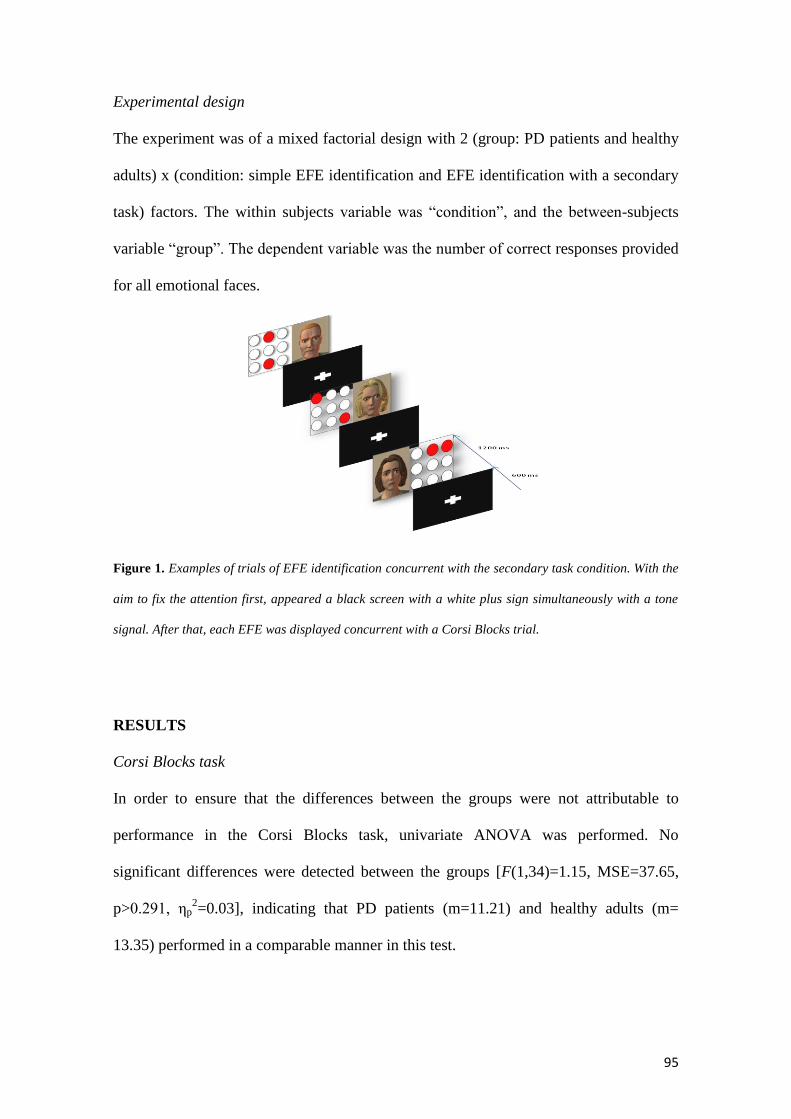
95
Experimental design
The experiment was of a mixed factorial design with 2 (group: PD patients and healthy
adults) x (condition: simple EFE identification and EFE identification with a secondary
task) factors. The within subjects variable was “condition”, and the between-subjects
variable “group”. The dependent variable was the number of correct responses provided
for all emotional faces.
Figure 1. Examples of trials of EFE identification concurrent with the secondary task condition. With the
aim to fix the attention first, appeared a black screen with a white plus sign simultaneously with a tone
signal. After that, each EFE was displayed concurrent with a Corsi Blocks trial.
RESULTS
Corsi Blocks task
In order to ensure that the differences between the groups were not attributable to
performance in the Corsi Blocks task, univariate ANOVA was performed. No
significant differences were detected between the groups [F(1,34)=1.15, MSE=37.65,
p>0.291, ηp2=0.03], indicating that PD patients (m=11.21) and healthy adults (m=
13.35) performed in a comparable manner in this test.

96
Identification of EFEs
The performance of the subjects in terms of the correct identification in both conditions
(simple EFE identification and EFE identification with secondary task) is summarized
for both groups in Fig. 2.
Figure 2. Accuracy rates for EFE identification (%) of PD patients and controls in the simple EFE
identification task and the EFE identification with a secondary task.
ANOVA revealed a significant main effect of condition [F(1,34)= 54.50,
MSE=72.90, p>0.001, ηp2=0.61], indicating that all participants identified more
emotional stimuli in the simple EFE identification condition (m=13.03) than when
required to perform a concurrent secondary task (m=11.11). Moreover, a significant
interaction was found between condition and group [F(1,34)=12.63, MSE=16.90,
p=0.001, ηp2=0.27]. To account for potential differences in DT performance due to
effects already present at baseline, proportional DT costs (DTC) were calculated:

97
DTC=((ACC single− ACC dual)/ACC single) x 100 [52]. The DTCs were analyzed by
univariate ANOVA, revealing a main effect of group [F(1,34)=12.75, MSE=7131.65,
p=0.001, ηp2=0.27], indicating that DTCs were significantly higher in PD patients
(~32%) than in healthy adults (~3.5%; Fig. 3).
Figure 3. Differences in the proportional Dual Task Costs (DTC%) for EFE identification by PD patients
and controls.
In conclusion, these results revealed no deficits in EFE identification in PD patients,
although EFE identification in a DT condition was significantly impaired in these
patients.
DISCUSSION
The effects of visuo-spatial interference on the ability to identify EFEs have been
examined in both non-medicated PD patients and healthy adults. As predicted, the
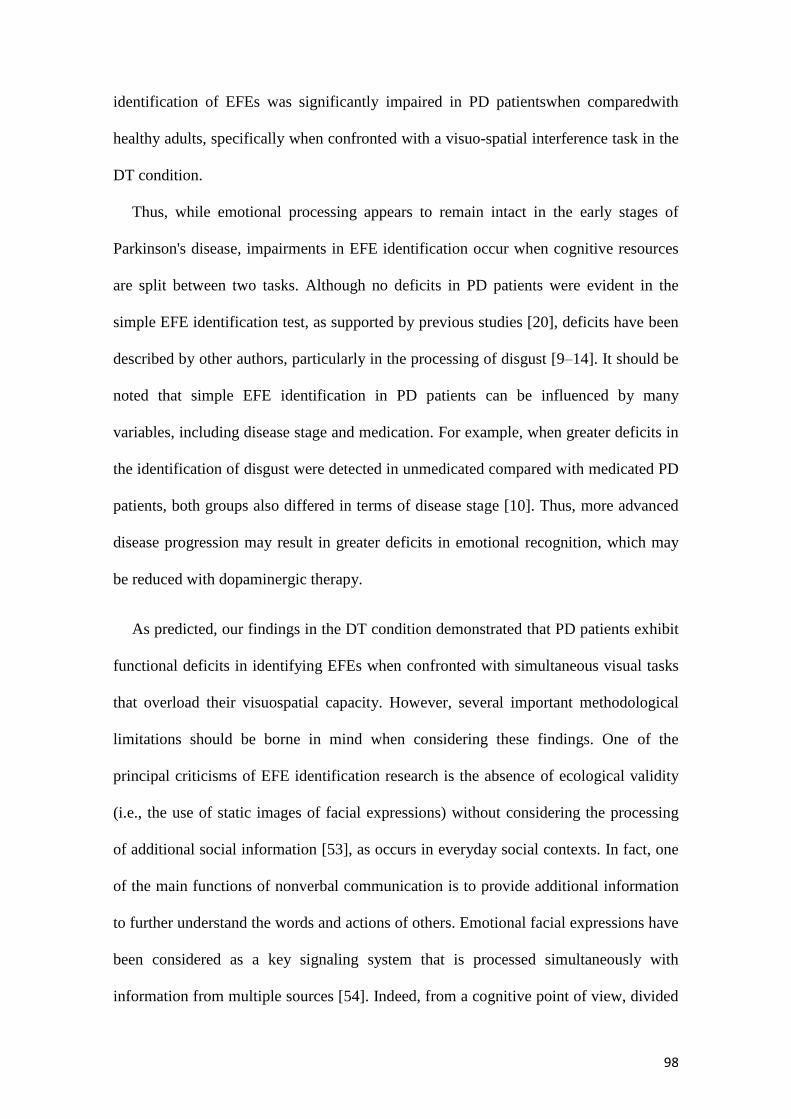
98
identification of EFEs was significantly impaired in PD patientswhen comparedwith
healthy adults, specifically when confronted with a visuo-spatial interference task in the
DT condition.
Thus, while emotional processing appears to remain intact in the early stages of
Parkinson's disease, impairments in EFE identification occur when cognitive resources
are split between two tasks. Although no deficits in PD patients were evident in the
simple EFE identification test, as supported by previous studies [20], deficits have been
described by other authors, particularly in the processing of disgust [9–14]. It should be
noted that simple EFE identification in PD patients can be influenced by many
variables, including disease stage and medication. For example, when greater deficits in
the identification of disgust were detected in unmedicated compared with medicated PD
patients, both groups also differed in terms of disease stage [10]. Thus, more advanced
disease progression may result in greater deficits in emotional recognition, which may
be reduced with dopaminergic therapy.
As predicted, our findings in the DT condition demonstrated that PD patients exhibit
functional deficits in identifying EFEs when confronted with simultaneous visual tasks
that overload their visuospatial capacity. However, several important methodological
limitations should be borne in mind when considering these findings. One of the
principal criticisms of EFE identification research is the absence of ecological validity
(i.e., the use of static images of facial expressions) without considering the processing
of additional social information [53], as occurs in everyday social contexts. In fact, one
of the main functions of nonverbal communication is to provide additional information
to further understand the words and actions of others. Emotional facial expressions have
been considered as a key signaling system that is processed simultaneously with
information from multiple sources [54]. Indeed, from a cognitive point of view, divided

99
attention plays an important role in this type of processing. Given that PD patients
exhibit attention deficits and problems in visual processing, the interference in our study
may have been particularly strong [33, 34] since two visuo-spatial tasks were used. This
may have led to cognitive overload in these patients that was reflected in a poorer
performance.
The findings in DT conditions support the deficits we identified previously when
normal elderly persons were compared with young adults [17]. However, this age
related effect was significantly augmented in PD patients, reflecting a stronger potential
interference associated with this disease. It is reasonable to assume that impaired frontal
lobe activity mediates this effect. From a theoretical point of view, the Dual Task
paradigm models the allocation of attention resources that are required to resolve two
tasks simultaneously. When one of the two tasks consumes more resources, the
execution of the other task is compromised, generating interference. Further research
will be necessary to determine whether this interference has the same effect when
primary and secondary tasks involve different modalities (e.g., a visuo-spatial emotional
task and a secondary auditory task). The present findings represent an important
contribution to our understanding of emotional processing in normal and pathological
aging. Specifically, we demonstrate that intact cognitive resources are required for
adequate emotional processing of stimuli that presumably represent universal facial
expressions of emotion.
From a practical point of view, these results may help better understand the affective
problems that arise in early Parkinson's disease.

100
ACKNOWLEDGMENTS
This research was funded by a grant from the Ministerio de Ciencia e Innovación, Spain
(Ref: PSI2009-13598-C02-01, Programa PSIC) awarded to Beatriz García-Rodríguez.
REFERENCES
[1] Hoehn M, Yahr M. Parkinsonism: onset, progression and mortality. Neurology
1967; 17(5):427–42.
[2] Taylor AE, Saint-Cyr JA, Lang AE. Frontal lobe dysfunction in Parkinson's disease:
the cortical focus of neostriatal outflow. Brain 1986; 109(5):845–83.
[3] Farina E, Cappa SF, Polimeni M, Magni E, Canesi M, Zecchinelli A, et al. Frontal
dysfunction in early Parkinson's disease. Acta Neurol Scand 1994; 90(1):34–8.
[4] Green J, McDonald WM, Vitek JL, Evatt M, Freeman A, Haber M, et al. Cognitive
impairments in advanced PD without dementia. Neurology 2002; 59(9):1320–4.
[5] Higginson CI, King DS, Levine D, Wheelock VL, Khamphay NO, Sigvardt KA.
The relationship between executive function and verbal memory in Parkinson's disease.
Brain Cogn 2003; 52(3): 343–52.
[6] Zgarljardic DJ, Borod JC, Foldi NS, Mattis P. A review of the cognitive and
behavioral sequelae of Parkinson's disease: relationship to frontostriatal circuitry. Cogn
Behav Neurol 2003; 16(4): 193–210.
[7] Lees AJ, Smith E. Cognitive deficits in the early stages of Parkinson's disease. Brain
1983; 106(2):257–70.
[8] Cooper JA, Sagar HJ, Jordan N, Harvey NS, Sullivan EV. Cognitive impairment in
early, untreated Parkinson's disease and its relationship to motor disability. Brain 1991;
114(5):2095–122.
[9] Kan Y, Kawamura M, Hasegawa Y, Mochizuki S, Nakamura K. Recognition of

101
emotion from facial, prosodic and written verbal stimuli in Parkinson's disease. Cortex
2002; 38(4):623–30.
[10] Sprengelmeyer R, Young AW, Mahn K, Schroeder U, Woitalla D, Buttner T, et al.
Facial expression recognition in people with medicated and unmedicated Parkinson's
disease. Neuropsychologia 2003; 41(8):1047–57.
[11] Dujardin K, Blairy S, Defebvre L, Duhem S, Noel Y, Hess U, et al. Deficits in
decoding emotional facial expressions in Parkinson's disease. Neuropsychologia 2004;
42(2):239–50.
[12] Suzuki A, Hoshino T, Shigemasu K, Kawamura M. Disgust-specific impairment of
facial expression recognition in Parkinson's disease. Brain 2006; 129:707–17.
[13] Assogna F, Pontieri FE, Caltagirone C, Spalletta G. The recognition of facial
emotion expressions in Parkinson's disease. Eur Neuropsychopharmacol 2008; 18:835–
48.
[14] Assogna F, Pontieri FE, Cravello L, Peppe A, Pierantozzi, Stefani A, et al.
Intensitydependent facial emotion recognition and cognitive functions in Parkinson's
disease. J Int Neuropsychol Soc 2010; 16:867–76.
[15] Lawerence AD, Goerendt IK, Brooks DJ. Impaired recognition of facial
expressions of anger in Parkinson's disease patients acutely withdrawn from dopamine
replacement therapy. Neuropsychologia 2007; 45:65–74.
[16] Phan KL, Wager T, Taylor SF, Liberzon I. Functional neuroanatomy of emotion: a
metaanalysis of emotion activation studies in PET and fMRI. Neuroimage 2002;
16:331–48.
[17] Phillips ML, Young AW, Senior C, Brammer M, Andrews C, Calder AJ, et al. A
specific neural substrate for perceiving facial expressions of disgust. Natura 1997;
389:495–8.

102
[18] Adolphs R, Tranel D, Damasio AR. Dissociable neural systems for recognizing
emotions. Brain Cogn 2003; 52:61–9.
[19] Wicker B, Keysers C, Plailly J, Royet JP, Gallese V, Rizzolatti G. Both of us
disgusted in my insula: the common neural basis of seeing and feeling disgust. Neuron
2003; 40:655–64.
[20] Adolphs R, Schul R, Tranel D. Intact recognition of facial emotion in Parkinson's
disease. Neuropsychology 1998; 12(2):253–8.
[21] Pessoa L, McKenna M, Gutierrez E, Ungerleider LG. Neural processing of
emotional faces requires attention. Proc Natl Acad Sci USA 2002; 99:11458–63.
[22] Pessoa L, Ungerleider LG. Visual attention and emotional perception. In: Itti L,
Rees G, Tsotsos JK, editors. Neurobiology of attention. San Diego, CA: Elsevier; 2005.
[23] García-Rodríguez B, Fusari A, Rodríguez B, Zurdo Hernández JM, Ellgring H.
Differential patterns of implicit emotional processing in Alzheimer's disease and healthy
aging. J Alzheimers Dis 2009; 18(3):541–51.
[24] Phillips LH, Tunstall M, Channon S. Exploring the role of working memory in
dynamic social cue decoding using dual task methodology. J Nonverbal Behav 2007;
31(2):137–52.
[25] Phillips LH, Channon S, Tunstall M, Hedenstrom A, Lyons K. The role of working
memory in decoding emotions. Emotion 2008; 8(2):184–91.
[26] García-Rodríguez B, Ellgring H, Fusari A, Frank A. The role of interference in
identification of emotional facial expressions in normal ageing and dementia. Eur J
Cogn Psychol Aging Cogn Neurosci 2009; 21(2–3):428–44.
[27] García-Rodríguez B, Fusari A, Fernández-Guinea S, Frank A, Molina JA, Ellgring
H. Decline of executive processes affects identification of emotional facial expressions
in aging. Curr Aging Sci 2011; 4:70–5.

103
[28] Buitelaar JK, van der Wees M, Swaab-Barneveld H, van der Gaag RJ. Verbal
memory and performance IQ predict theory of mind and emotion recognition ability in
children with autistic spectrum disorders and in psychiatric control children. J Child
Psychol Psychiatry 1999; 20:869–81.
[29] Haxby JV, Hoffman EA, Gobbini MI. The distributed human neural system for
face perception. Trends Cogn Sci 2000; 4:223–33.
[30] Maurer D, Le Grand R, Mondloch CJ. The many faces of configural processing.
Trends Cogn Sci 2002; 6:255–60.
[31] Curby KM, Gauthier I. A visual short-term memory advantage for faces. Psychon
Bull Rev 2007; 14:620–8.
[32] Morgan HM, Klein C, Boehm SG, Shapiro L, Linden DEJ. Working memory load
for faces modulates P300, N170, and N250r. J Cogn Neurosci 2008; 20(6):989–1002.
[33] Proctor F, Riklan M, Cooper IS, Teuber HL. Judgement of visual and postural
vertical by Parkinsonian patients. Neurology 1964; 14:287–93.
[34] Bowen FP, Hoehn MM, Yahr MD. Cerebral dominance in relation to tracking and
tapping performance in patients with Parkinsonism. Neurology 1972; 22:32–9.
[35] Horne DJ. Performance on delayed response tasks by patients with Parkinsonism. J
Neurol Neurosurg Psychiatry 1971; 34:192–4.
[36] Danta G, Hilton RC. Judgement of the visual vertical and horizontal in patients
with Parkinsonism. Neurology 1975; 25:43–7.
[37] Flowers KA. Visual ‘closed-loop’ and ‘open-loop’ characteristics of voluntary
movement in patients with Parkinsonism and intention tremor. Brain 1976; 99:269–310.
[38] Pell MD, Leonard CL. Facial expression decoding in early Parkinson's disease.
Cogn Brain Res 2005; 23:327–40.
[39] Cousins R, Hanley J, Davies A, Turnbull C, Playfer J. Understanding memory for

104
faces in Parkinson's disease: the role of configural processing. Neuropsychologia 2000;
38:837–47.
[40] Salway AFS, Logie RH. Visuospatial working memory, movement control and
executive demands. Br J Psychol 1995; 86:253–69.
[41] Hughes AJ, Daniel E, Kilford L, Lees A. Accuracy of clinical diagnosis of
idiopathic Parkinson's disease: a clinico-pathological study of 100 cases. J Neurol
Neurosurg Psychiatry 1992; 55:181–4.
[42] Lobo A, Esquerra J, Gómez Burgada F, Sala JM, Seva A. El Mini-Exámen
Cognoscitivo: un test sencillo y práctico para detectar alteraciones intelectuales en
pacientes médicos. Actas Luso Esp Neurol Psiquiatr 1979; 3:189–202.
[43] Folstein MF, Folstein SE, McHugh PR. “Mini-mental state”. A practical method
for grading the cognitive state of patients for the clinician. J Psychiatr Res 1975; 12 (3):
189–98.
[44] Blessed G, Tomlinson BE, Roth M. The association between quantitative measures
of dementia and of senile changes in grey matter of elderly subjects. Br J Psychiatry
1968; 114:797–811.
[45] Sheikh JI, Yesavage JA. Geriatric Depression Scale (GDS): recent evidence and
development of a shorter version. In: Brink TL, editor. Clinical gerontology: a guide to
assessment and intervention. New York: Haworth Press; 1986.
[46] Yesavage JA, Brink TL, Rose TL. Development and validation of a geriatric
depression screening scale: a preliminary report. J Psychiatry Res 1983; 17:37–49.
[47] Ekman P, Friesen WV. The facial action coding system. Palo Alto, California:
Consult Psychology Press; 1978.
[48] Ekman P, Friesen WV, Hager JC. Facial action coding system — investigator's
guide. Salt Lake City: Research Nexus; 2002.

105
[49] Smyth MM, Scholey KA. Determining spatial span: the role of movement time and
articulation rate. Q J Exp Psychol 1992; 45(A):479–501.
[50] Orsini A, Schiappa O, Grossi D. Sex and cultural differences in children's spatial
and verbal memory span. Percept Mot Skills 1981; 53(1):39–42.
[51] Vilkki J, Holst P. Deficient programming in spatial learning after frontal lobe
damage. Neuropsychologia 1989; 27:971–6.
[52] Salthouse TA, Fristoe NM, Lineweaver TT, Coon VE. Aging of attention: does the
ability to divide decline? Mem Cognit 1995; 23(1):59–71.
[53] Adolphs R. Recognizing emotion from facial expressions: psychological and
neurological mechanism. Behav Cogn Neurosci Rev 2002; 1:21–62.
[54] Ekman P. Emotions revealed. U.K.: Weidenfeld & Nicolson; 2003.

106
Capítulo 7. [Estudio 4]. El efecto de las demandas atencionales en la identificación
de las EFEs en la EA.
García-Rodríguez, B., Vincent, C., Casares Guillén, C.T., Ellgring, H., y Frank, A.
(2012). The effects of different attentional demands in the identification of emotional
facial expressions in Alzheimer´s disease. American Journal of Alzheimer Disease &
Other Dementias, 27, 530-536.
107
ABSTRACT
Alzheimer’s disease (AD) is characterized by the progressive impairment of mental and
emotional functions, including the processing of emotional facial expression (EFE).
Deficits in decoding EFE are relevant in social contexts in which information from 2 or
more sources may be processed simultaneously. To assess the role of contextual stimuli
on EFE processing in AD, we analyzed the ability of patients with AD and healthy
elderly adults to identify EFE when simultaneously performing another task.
Each of the 6 basic EFEs was presented to 15 patients with AD and 35 controls in a dual
task paradigm that is in parallel with a visuospatial or a semantic task. Results show that
the decoding of EFEs was impaired in patients with AD when they were simultaneously
processing additional visuospatial information, yet not when they were performed in
conjunction with a semantic task. These findings suggest that the capacity to interpret
emotional states is impaired in AD.
KEYWORDS
Alzheimer’s disease, emotional facial expressions, dual task performance,
visuospatial/semantic interference
108
INTRODUCTION
Alzheimer’s disease (AD) is a progressive neurodegenerative disorder that produces
severe deficits in cognitive function, affecting memory, planning, working memory,
attention, and communication. The deterioration of communication skills is directly
related to deficits in the nonverbal communication of emotions, including the ability to
recognize the emotional states of others1, and more specifically the ability to interpret
emotional facial expression (EFE).
The greatest known risk factor for AD is increasing age and indeed, the majority of
patients with AD are over 65 years of age. The impact of aging on the ability to
recognize EFE is well documented. Aging has been linked with impairment of the
ability to identify facial expressions of anger, sadness, and fear but not happiness,
surprise, or disgust2-4
. The accepted hypothesis is that in normal aging, these specific
deficits are induced by deterioration in brain areas that mediate emotional processing;
sadness is processed via the anterior and subcallosal cingulate5, while fear and disgust
are processed in the amygdala6,7
and basal ganglia,8,9
respectively. Structural and
morphological brain damage in patients with AD is more pervasive than that observed
in healthy aged adults, in these patients, affecting certain basic emotions more than
others10,11
. Several studies have described specific deficits in patients with AD in
identifying discrete emotional faces that is negative emotions12-14
, with the exception of
recognizing disgust, which is preserved15
. Importantly, results can vary depending on
the type of the EFE identification task employed. Thus, patients with AD exhibit
deficits in EFE decoding in Tasks that demand a high level of cognitive effort, such as
discrimination16
and matching,13
and these observations are supported by neuroimaging
studies17,18
demonstrating damage to the emotional processing areas of AD brains.
However, the origin and nature of this impairment remains unclear. When reviewing the

109
facial emotion recognition in AD19
several methodological inconsistencies in the
literature were highlighted, arguing that the identification of deficits is dependent on the
cognitive resources associated with each type of task (discriminating, matching, or
identifying EFEs). Indeed, it was concluded that patients with AD have no difficulties in
identifying particular EFEs, suggesting that studies in this area should employ tasks that
more closely approximate to basic social interactions.
In social contexts, nonverbally communicated emotions are processed constantly,
and they must be interpreted correctly during communication20
.This processing
provides additional information to facilitate the interpretation of other people’s words
and actions in everyday life. As interpersonal communication takes place in parallel
with other activities, information from various sources must be processed
simultaneously (eg, decoding EFEs while linking them to verbal information). This
continuous flow of information requires a network of processes that simultaneously
manipulate and evaluate information from multiple sources. As EFE processing depends
partly on the ability to split attention resources between at least 2 concurrent tasks,
emotional dual task (DT) paradigms offer the most accurate means of assessing the
processing of emotional stimuli that occurs in everyday life. The majority of studies
investigating deficits in divided attention employ DT paradigms in which 2 tasks are
performed individually or in combination. Studies of DT21,22
indicate that the division of
attention is associated with a decrement in task performance, particularly when both
tasks are of the same nature (eg, visual or semantic)23
. Our prediction that patients with
AD perform poorly in attention-demanding tasks is supported by an abundance of
behavioural and neurological data,24-30
demonstrating a disproportionate impairment in
the simultaneous performance of 2 tasks.

110
Given that successful communication and appropriate interactions are dependent on
the ability to detect and react to the affective state of others while processing
information from additional sources, it is important to determine whether these
fundamental processes are impaired in AD. As patients with AD exhibit deficits in the
simultaneous performance of 2 tasks,31,32
functional deficits in EFE identification
associated with this disease may influence the impaired interpersonal communication
observed in these patients in social contexts.
The main objective of this study was to investigate the role of context stimuli in the
interpretation of facial expressions by patients with AD. Specifically, we assessed the
capacity of patients with AD to identify EFEs while simultaneously performing a
visuospatial or semantic task. We predicted greater deficits in EFE identification in
patients with AD when required to simultaneously process additional information.
Moreover, we predicted greater DT impairment when EFE identification was performed
in parallel with a visuospatial rather than a semantic task (ie, when both tasks depend on
the same cognitive resources). This hypothesis was based on our previous studies of
emotional processing,33-36
which revealed that neurological patients performed worse
than healthy elderly adults in a DT paradigm, demonstrating that deficits in EFE
identification are due to pathological effects rather than those of aging.
METHODS
Participants
The AD patient group was comprised of 15 individuals (9 female and 6 male),
ranging in age from 65 to 85 years (mean, 76.87; standard deviation [SD], 6.64), who
were recruited from the outpatient practices of the Hospital Universitario La Paz in
Madrid. All patients had a diagnosis of probable AD according to National Institute of

111
Neurologic, Communicative Disorders and Stroke-Alzheimer’s Disease and Related
Disorders Association (NINCDS-ADRDA) criteria.37
Individuals with a history of
cerebrovascular accident, head injury, neurological disorders (other than AD),
cardiovascular disease, chronic alcoholism, and/or drug abuse were excluded from the
study. No participants were using neuroleptic medications, and all performed normally
in general medical and neurological screening tests.
Inclusion criteria also included normal corrected eyesight and a mild stage of the
disease process, as determined by the Mini-Mental State Examination38
(MMSE) and
the Blessed Dementia Scale39
(BDS). The lower cutoff for mild impairment was 18 out
of 30 and the mean MMSE score for participants with AD was 23.33 (SD, 3.37; range,
18-29). The control group was composed of 35 healthy adult volunteers (22 female and
13 male), from 66 to 85 years of age (mean, 73.86; SD, 3.94). The same criteria
described above were applied to the control participants, with an MMSE score of 24 or
higher as the cutoff for inclusion as a control participant to screen out individuals with
undiagnosed clinical impairments40,41
. The mean MMSE score for the control group was
29.31 (SD, 0.71; range, 27-30). Participants in the control group were recruited from a
variety of communitybased social organizations for the elderly individuals. An
additional exclusion criteria for dementia was a BDS score >4. A final exclusion
criterion for both groups was major depression, as indicated by scores >7 on the 15-item
version of the Geriatric Depression Scale (GDS),42,43
and a diagnosis of any psychiatric
disorder. Written informed consent was obtained from all participants or from
caregivers when appropriate. Background data for both groups are presented in Table 1.

112
Table 1. Demographic Characteristics of Alzheimer´s Disease (AD) and Control Group
The control and AD groups did not differ significantly in terms of age, t (48) = 1.99,
p > .050; gender ratio, AD: χ2 (1) = 0 .60, p > .05; Control group: χ
2 (1) = 2.31, p > .050;
or years of education, t (48) =- 2.00, p > .050; or GDS t (48) = 1.85, p = .050. As expected,
significant differences in MMSE, t (48) = -10.09, p > .000, and BDS scores, t (48) = 8.39,
p > .000, were observed between groups, demonstrating the impaired cognitive capacity
of the AD group with respect to controls.
Experimental Tasks
Emotional facial expression identification task. The EFE identification was performed
as described previously, thereby facilitating comparison with the present findings.33-36
In a forced-choice task, participants were presented 24 EFEs one at a time (4
representing each basic emotion: happiness, sadness, anger, fear, surprise, and disgust),
and they were asked to identify the emotion that best described the target stimulus. The
participants were provided with a response list with the names of the 6 emotions and
theywere given asmuch time as necessary tomake their decision. The stimuli were

113
generated using a 3-dimensional (3D) imaging technique (Poser 6, Curious Labs, Santa
Cruz, California), which creates virtual human faces whose expressions are determined
by the single action units described in the handbook of the Facial Action Coding System
(FACS).44,45 The emotions were portrayed by 4 virtual actors (2 male and 2 female
avatars) and the expressions were enacted by an experienced FACS coder (Johannes
Heiner Ellgring [JHE]), activating their virtualmuscles with respect to single action
units in the computer animation. These stimuli elicit facial electromyographical
reactions46
and amygdalar activation,47
and thus they represent a validmethod for
producing standardized expressions of emotions for the assessment of emotional
processing. The main dependent variable was accuracy (ie, the number of correct EFEs
identified). The total score ranged from0 to 4, with amaximumof 24 correct responses.
‘‘Correctness’’ was defined as an answer corresponding to the label given according to
the FacialAction Units.The chance level was considered as 1 correct answer for a single
emotion and 4 for the entire set of 24 items.
Visuospatial task (Corsi Blocks).
The Corsi Blocks (CB) task was used to create visuospatial interference. This task is
commonly used to assess short-term visuospatial memory in adults, children, and
patients with neuropsychological deficits. The CB task involves a representation of 9
circles in a 3 x 3 horizontal and vertical array. In each trial, a specific arrangement of
the 9 circles (7 white and 2 red) was displayed, with the 2 red circles (targets) appearing
in a predetermined sequence at a rate of 1 per second. Participants were asked to mark
the correct sequence of the red circles on an answer sheet.

114
Semantic task (Backward Digit Recall).
For semantic interference, we used a backward version of the digit recall task (BDR), a
commonly used measure of working memory. A total of 24 sequences of 2 digits
(selected from 1 to 9 without repetition) were presented aurally at a rate of 1 per second
from a recording of a male human voice. Participants were required to recall the digits
in the reverse order of presentation. To calculate any loss in DT performance, all
participants also performed the single tasks described above.
Dual Tasks
To experimentally simulate EFE processing in a social context, participants performed
an EFE identification task while simultaneously processing a visuospatial or semantic
task.
The EFE identification concurrent with a visuospatial task (EFE/ CB). Participants
performed the EFE identification task concurrent with a CB trial. A total of 24 trials
were conducted, each beginning with an acoustic signal, the tone ‘‘F’’ at a frequency of
500 Hz that lasted 150 ms. A visual signal was presented simultaneously, consisting of
a white plus sign on a black screen that remained visible for 1200 ms. Subsequently, a
black screen was displayed for 1200 ms beginning 600 ms prior to the EFE/CB trial.
Each test was followed by another black screen displayed for 600 ms before the
subsequent trial. Participants were required to indicate the sequence in the CB test and
identify the EFE, always in this order. In each trial, the order of the EFE and the
position (left or right) of the EFE and CB were randomized.

115
The EFE identification concurrent with a semantic task (EFE/ BDR). As in the EFE/CB
DT, each EFE was displayed simultaneously with a BDR trial for 1200 ms. After a
black screen was displayed for 600 ms, the participants were required to first indicate
the sequence of the BDR and to then identify the EFE, always in this order.
Procedure
Before beginning the experiment, the aims of the study were explained to each
participant and written informed consent was obtained. Participants were informed that
they were free to terminate their participation in the study at any point, although this did
not occur. The protocols for research involving human participants were approved by
the Ethical Committee of the University (UNED).
The trials were split into 2 different sessions. In the first session, participants
performed the single tasks and they were assessed using neuropsychological scales. The
DT trials were performed in the second session and the order in which the tasks were
presented was counterbalanced to avoid learning effects. All participants were tested
individually in a quiet room and the experiment was conducted using a 17-inch color
display. Stimulus presentation and data recording was controlled by E-prime software
developed for this experiment, and instructions were given verbally and presented
onscreen before each test condition. All responses were recorded by the experimenter.
Experimental Design
The experiment was of a mixed factorial design with 2 x 2 factors: group, AD patients
and controls; condition, EFE identification concurrent with a visuospatial task and EFE
identification concurrent with a semantic task. The withinsubjects variable was

116
‘‘condition’’ and the between-subjects variable was ‘‘group.’’ The dependent variable
was the number of correct responses provided for all EFEs.
RESULTS
The EFE Identification Tasks
A primary analysis compared the accuracy of patients with AD and control participants
in EFE identification in the 3e conditions tested. The number of EFEs correctly
identified revealed that patients with AD performed worse than healthy controls in the
simple EFE identification task (12.27 vs 15.29, respectively), as well as when EFE
identification was performed concurrent with a visuospatial task (9.47 vs 12.83,
respectively) or a semantic task (10.07 vs 13.43, respectively; Figure 1).
Figure 1. Accuracy of EFE identification (%) by patients with AD and control participants in the 3
experimental conditions. EFE indicates emotional facial expression; AD, Alzheimer´s disease.
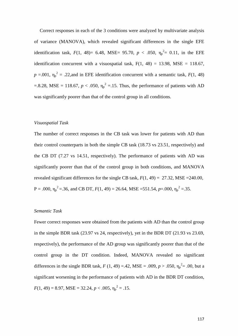
117
Correct responses in each of the 3 conditions were analyzed by multivariate analysis
of variance (MANOVA), which revealed significant differences in the single EFE
identification task, F(1, 48)= 6.48, MSE= 95.70, p < .050, ηp2= 0.11, in the EFE
identification concurrent with a visuospatial task, F(1, 48) = 13.98, MSE = 118.67,
p =.001, ηp2 = .22,and in EFE identification concurrent with a semantic task, F(1, 48)
=.8.28, MSE = 118.67, p < .050, ηp2 =.15. Thus, the performance of patients with AD
was significantly poorer than that of the control group in all conditions.
Visuospatial Task
The number of correct responses in the CB task was lower for patients with AD than
their control counterparts in both the simple CB task (18.73 vs 23.51, respectively) and
the CB DT (7.27 vs 14.51, respectively). The performance of patients with AD was
significantly poorer than that of the control group in both conditions, and MANOVA
revealed significant differences for the single CB task, F(1, 49) = 27.32, MSE =240.00,
P = .000, ηp2
=.36, and CB DT, F(1, 49) = 26.64, MSE =551.54, p=.000, ηp2 =.35.
Semantic Task
Fewer correct responses were obtained from the patients with AD than the control group
in the simple BDR task (23.97 vs 24, respectively), yet in the BDR DT (21.93 vs 23.69,
respectively), the performance of the AD group was significantly poorer than that of the
control group in the DT condition. Indeed, MANOVA revealed no significant
differences in the single BDR task, F (1, 49) =.42, MSE = .009, p > .050, ηp2= .00, but a
significant worsening in the performance of patients with AD in the BDR DT condition,
F(1, 49) = 8.97, MSE = 32.24, p < .005, ηp2 = .15.

118
Dual Task Performance
To determine whether the differences in DT performance can be attributed to effects
already present at baseline or whether they are due to combining the tasks, and to
account for the various strategies adopted when simultaneously performing 2 tasks, a
formula has been proposed to calculate an overall decrement score: µ= (1-[(Pm+
Pt)/2])*100, where µ is the combined DT score, Pm is the proportional decrease in
correct EFE responses between single (Xsingle) and DT (Xdual) conditions [(Xsingle-
Xdual)/Xsingle], and Pt is the equivalent proportional decrease in the scores of the
visuospatial task (CB) or the semantic task (BDR) task.48
Thus, a score of 100%
indicates no change in the DT score, while lower scores reflect a decrease in this value.
We calculated 2 new variables, for the DT combining EFE identification and a
visuospatial task (EFE/CB), and for EFE identification combined with a semantic task
(EFE/BDR). Analysis of these variables by MANOVA revealed larger decrement scores
in the EFE/CB, F(1, 49)= 7.77, MSE = 5709.72, p < .005, ηp2= .14, than the EFE/BDR
combination, F(1, 49) = 4.00, MSE= 850.51, p > .050, ηp2 =.08, with significant
differences between the 2 groups only in the former. When the EFE task was performed
in conjunction with the CB task, the observed decrease in accuracy was greater in the
AD group (47.44%) than in the control group (70.76%). However, when EFE
identification was performed in parallel with the BDR task, similar decreases in
accuracy were observed in AD and control groups (86.58% and 95.58%, respectively;
Figure 2).

119
Figure 2. Performance (%) for each DT combination of the AD and control groups. DT indicates dual
task; AD, Alzheimer´s disease.
In summary, our results show that EFE identification is impaired in patients with AD
as compared with controls, and that the performance of these patients decreases
significantly and disproportionately when tested while concurrently performing a
visuospatial but not a semantic task. These results suggest that patients with AD have
more difficulty integrating information relating to affective states with other
environmental stimuli, leading to difficulties in interpersonal communication.
DISCUSSION
The aim of the present study was to assess the role of context stimuli in the
interpretation of EFEs by patients with AD. We assessed the capacity of both groups to
identify EFEs while simultaneously processing other stimuli that occur during
interpersonal communication. The performance of the AD group was significantly
poorer than that of the control group in all experimental conditions, EFE identification
both alone and in the DT conditions. These findings demonstrate that patients with AD

120
have difficulties identifying EFEs even when not processing additional information. In
agreement with previous reports,5,49
these findings suggest that the specific
neurodegeneration associated with AD may provoke such deficits, that is pathological
alterations in the neural substrates underlying emotion processing impair EFE
recognition. Analyzing EFE identification in DT conditions revealed that as predicted,
patients with AD exhibited functional deficits in identifying emotional faces while
simultaneously processing other stimuli. Moreover, when facial emotional processing
was performed in parallel with another visuospatial task, competition for the same
resources resulted in greater DT decrements than when the concurrent task was of
semantic nature. This effect was especially evident in the AD patient group, suggesting
that emotional processing is impaired in patients with AD when performed in parallel
with a task of the same nature. This is probably an important factor in the social
communication deficits described in patients with AD. The present findings corroborate
those of our previous studies, 33-36
demonstrating that neurodegenerative diseases are
associated with deficits in emotional processing in complex stimuli contexts,
particularly when processing 2 tasks of the same nature. These results challenge the
proposed automaticity of emotional processing,50
and show that cognitive resources are
required to interpret the emotional content of stimuli, suggesting that emotional
identification is a nonautomatic process dependent on cognitive state. The basic
assumption of the DT paradigm is that automatic tasks are unaffected by simultaneous
execution of another task. In AD patients, EFE identification and visuospatial
processing were significantly impaired when performed concurrently, indicating that
both are controlled processes that are dependent on normal cognitive resources.
Moreover, in line with previous studies,24-26
we show that attentional control of
executive function declines in the early stages of AD and that the performance of 2
121
simultaneous tasks is markedly impaired, suggesting specific DTprocessing deficits. A
key aspect of the present study is the use of experimental tasks with significant
ecological validity. The facial emotional processing system is constantly activated and
ready,20
and one of the key functions of this system is to accept or reject other
messages, which involves the simultaneous processing of information from more than
one source. This study identifies some of the specific challenges experienced by patients
with AD when attempting to integrate affective information with normal cognitive
activity.
The present findings represent an important contribution to our understanding of
deficits in facial emotional recognition in with AD. Communication in everyday life
depends on correct perception and interpretation of different emotional and
nonemotional cues in a social context. Accurate interpretation of these cues plays an
important role in maintaining successful relationships and healthy psychological
function. The difficulties of AD patients in recognizing facial expressions may to
contribute to their impaired social behavior, resulting in poor communications and
interpersonal relations, or inappropriate social behavior. Furthermore, EFE recognition
may represent an important indicator of well-being and quality of life in these patients.
ACKNOWLEDGMENTS
This research was funded by a grant from the Ministerio de Ciencia e Innovación, Spain
(Ref: PSI2009-13598-C02-01, Programa PSIC) awarded to Beatriz García-Rodríguez.
Declaration of Conflicting Interests The authors declared no potential conflicts of
interest with respect to the research, authorship, and/or publication of this article.

122
REFERENCES
[1] Shimokawa A, Yatomi N, Anamizu S, et al. Influence of deteriorating ability of
emotional comprehension on interpersonal behavior in Alzheimer-type dementia. Brain
Cogn. 2001; 47(3): 423-433.
[2] Sullivan S, Ruffman T. Emotion recognition deficits in the elderly. Int J Neurosci.
2004; 114(3):403-432.
[3] Ruffman T, Henry JD, Livingstone V, Phillips LH. A metaanalytic review of
emotion recognition and aging: implications for neuropsychological models of aging.
Neurosci Biobehav Rev. 2008; 32(4):863-881.
[4] Suzuki A, Hoshino T, Shigemasu K, Kawamura M. Decline or improvement? Age-
related differences in facial expressions recognition. Biol Psychol. 2007; 74(1):75-84.
[5] Phan KL, Wager T, Taylor SF, Liberzon I. Functional neuroanatomy of emotion: a
meta-analysis of emotion activation studies in PET and fMRI. Neuroimage. 2002;
16(2):331-348.
[6] Vuilleumier P, Pourtois G. Distributed and interactive brain mechanisms during
emotion face perception: evidence from functional neuroimaging. Neuropsychologia.
2007; 45(1):174-194.
[7] Whalen PJ, Rauch SL, Etcoff NL, McInerney SC, Lee MB, Jenike MA. Masked
presentations of emotional facial expressions modulate amygdala activity without
explicit knowledge. J Neurosc. 1998; 18(1):411-418.
[8] Phillips ML, Williams LM, Heining, et al. Differential neural responses to overt and
covert presentations of facial expressions of fear and disgust. Neuroimage. 2004;
21(14):1484-1496.
[9] Sprengelmayer R, Schroeder U, Young AW, Epplen JT. Disgust in pre-clinical
Huntington’s disease: a longitudinal study. Neuropsychologia. 2006; 44(4):518-533.

123
[10] Adolphs R, Tranel D. Impaired judgments of sadness but not happiness following
bilateral amygdala damage. J Cognitive Neurosci. 2004; 16(3):453-462.
[11] Adolphs R, Tranel D, Hamann S, et al. Recognition of facial emotion in nine
individuals with bilateral amygdala damage. Neuropsychologia. 1999; 37(10):1111-
1117.
[12] Hargrave R, Maddock RJ, Stone V. Impaired recognition of facial expressions of
emotion in Alzheimer’s disease. J Neuropsychiatry Clin Neurosci. 2002; 14(1):64-71.
[13] Bucks RS, Radford SA. Emotion processing in Alzheimer’s disease. Aging Ment
Health. 2004; 8(3):222-232.
[14] Burnham H, Hogervorst E. Recognition of facial expressions of emotion by
patients with dementia of Alzheimer type. Dement Geriatr Cogn Disord. 2004;
18(1):75-79.
[15] Henry JD, Ruffman T, McDonald S, et al. Recognition of disgust is selectively
preserved in Alzheimer’s disease. Neuropsychologia. 2008; 46(5):1363-1370.
[16] Albert MS, Cohen C, Koff E. Perception of affect in patients with dementia of the
Alzheimer’s type. Arch Neurol. 1991; 48(8):791-795.
[17] Lavenu I, Pasquier F, Lebert F, Petit H, Van der Linden M. Perception of emotion
in frontotemporal dementia and Alzheimer disease. Alzheimer Dis Assoc Disord. 1999;
13(2):96-101.
[18] Rosen HJ, Wilson MR, Schauer GF, et al. Neuroanatomical correlates of impaired
recognition of emotion in dementia. Neuropsychologia. 2006; 44(3):365-373.
[19] McLellan T, Johnston L, Dalrymple-Alford J, Porter R. The recognition of facial
emotion in Alzheimer’s disease: a review of findings. Acta Neuropsychiatr. 2008; 20:
236-250.
[20] Ekman P. Emotions Revealed. New York, HY: Henry Holt; 2003

124
[21] Logie RH, Cocchini G, Della Sala S, Baddeley AD. Is there a specific executive
capacity for dual task co-ordination? Evidence from Alzheimers´disease.
Neuropsychology. 2004; 18(3):504-513.
[22] Naveh-Benjamin M, Craik FI, Gavrilescu D, Anderson ND. Asymmetry between
encoding and retrieval processes: evidence from divides attention and a calibration
analysis. Mem Cognit. 2000; 28(6):965-976.
[23] Salway AFS, Logie RH. Visuospatial working memory, movement control and
executive demands. Br J Psychol. 1995; 86(pt 2):253-269.
[24] Baddeley AD, Baddeley HA, Bucks RS, Wilcock GK. Attentional control in
Alzheimer’s disease. Brain. 2001; 124(pt 8):1492-1508.
[25] Baddeley AD, Bressi S, Della Sala S, Logie R, Spinnler H. The decline of working
memory in Alzheimer’s disease: A longitudinal study. Brain. 1991; 114(pt 6):2521-
2542.
[26] Parasuraman R, Haxby J. Attention and executive deficits in Alzheimer’s disease: a
review. Neuropsychol. 1993; 7:242-272.
[27] MacPherson SE, Della Sala S, Logie RH, Wilcock GK. Specific AD impairment in
concurrent performance of two memory task. Cortex. 2007; 43(7):858-865.
[28] Ramsden CM, Kinesella GJ, Ong B, Storey E. Performance of everyday actions in
mild Alzheimer’s disease. Neuropsychology. 2008; 22(1):17-26.
[29] Baddeley A, Della Sala S, Papagno C, Spinnler H. Dual-task performance in
dysexecutive and nondysexecutive patients with a frontal lesion. Neuropsychology.
1997; 11(2):187-194.
[30] Adcock RA, Constable RT, Gore JC, Goldman-Rakic PS. Functional
neuroanatomy of executive processes involved in dual-task performance. Proc Natl
Acad Sci USA. 2000; 97(7): 3567-3572.

125
[31] Crossley M, Hiscock M, Foreman JB. Dual-task performance in early stage
dementia: differential effects for automatized and effortful processing. J Clin Exp
Neuropsychol. 2004; 26:332-346.
[32] Sebastian MV, Menor J, Elosua MR. Attentional dysfunction of the central
executive in AD: evidence from dual task and perseveration errors. Cortex. 2006;
42(7):1015-1020.
[33] García-Rodríguez B, Ellgring H, Fusari A, Frank A. The role of interference in
identification of emotional facial expressions in normal ageing and dementia. Eur J Cog
Psychol Aging Cog Neurosci. 2009; 21(2-3):428-444.
[34] García-Rodríguez B, Fusari A, Fernández-Guinea S, Frank A, Molina JA, Ellgring
H. Decline of executive processes affects identification of emotional facial expressions
in aging. Curr Aging Sci. 2011; 4(1):70-75.
[35] García-Rodríguez B, Casares C, Jurado R, Rubio G, Molina JA, Ellgring H. Visuo-
spatial interference affects identification of emotional facial expressions in unmedicated
Parkinson’s patients. J Neurol Sci. 2012; 313(1-2):13-16.
[36] García-Rodríguez B, Casares C, Molina JA, et al. Differential effects of dual tasks
on emotional processing in non-medicated patients with Parkinson’s disease. Rev
Neurología. 2011; 53(6): 329-336.
[37] McKhann G, Drachman D, Folstein M, Katzman R, Price D, Stadlan EM. Clinical
diagnosis of Alzheimer’s disease: report of the NINCDS-ADRDA Work Group under
the auspices of Department of Health and Human Services Task Force on Alzheimer’s
disease. Neurology. 1984; 34(7):939-944.
[38] Folstein MF, Folstein SE, McHugh PR. ‘‘Mini-mental state.’’ A practical guide for
grading the cognitive state of patients for the clinician. J Psychiatr Res. 1975;
12(3):189-198.

126
[39] Blessed G, Tomlinson BE, RothM. The association between quantitative measures
of dementia and of senile changes in grey matter of elderly subjects. Brit J Psychiatr.
1968; 114(512):797-811.
[40] Cullum CM, Smernoff EN, Lord SE. Utility and psychometric properties of the
Mini-Mental Examination in healthy older adults. J Clin Exp Neuropsyc. 1991; 13: 88-
89.
[41] Galasko D, Klauber MR, Hofstetter R, Salmon D, Lasker B, Thal LJ. The Mini-
Mental State Examination in the early diagnosis of Alzheimer’s disease. Arch Neurol.
1990; 47(1):49-52.
[42] Sheikh JI, Yesavage JA. Geriatric Depression Scale (GDS): recent evidence and
development of a shorter version. In: Brink TL, ed. Clinical Gerontology: A Guide to
Assessment and Intervention. New York, NY: Haworth Press; 1986.
[43] Yesavage JA, Brink TL, Rose TL. Development and validation of a geriatric
depression screening scale: a preliminary report. J Psychiatry Res. 1983; 17(1):37-49.
[44] Ekman P, Friesen WV. The Facial Action Coding System. Palo Alto, CA: Consult
Psychology Press; 1978.
[45] Ekman P, Friesen WV, Hager JC. Facial Action Coding System— Investigator’s
Guide. Research nexus. Salt lake City, UT, 2002.
[46] Weyers P, Mu¨hlberger A, Hefele C, Pauli P. Electromyographic responses to
static and dynamic avatar emotional facial expressions. Psychophysiology. 2006;
43(5):450-453.
[47] Moser E, Demtl E, Robinson S, Fink B, Gur RC, Grammer K. Amygdala activation
at 3 T in response to human and avatar facial expressions of emotions. J Neurosci
Methods. 2007; 161(1): 126-133.
[48] Baddeley AD, Della Salla S, Colin G, Papagno C, Spinnler H. Testing central

127
executive functioning with a pencil-and-paper test. In: P. Rabbitt, ed. Methodology of
Frontal and Executive Functions. Hove, UK: Psychology Press; 1997:61-80.
[49] Norris CJ, Chen EE, Zhu DC, Small SL, Cacioppo JT. The interaction of social and
emotional processes in the brain. J Cogn Neurosci. 2004; 16(10):1818-1829.
[50] Öhman A. Automaticity and the amygdala: nonconscious responses to emotional
faces. Curr Dir Psychol Sci. 2002:11: 62-66.

128
Capítulo 8. Conclusiones
El objetivo principal de la tesis ha sido investigar el papel modulador de los recursos
cognitivos en el procesamiento de EFEs y su impacto en el envejecimiento normal y en
la EA y EP. Para poder alcanzar este objetivo, se han desarrollado cuatro estudios
experimentales en los que mediante un paradigma de DT se han estudiado las siguientes
variables: (i) el momento de la introducción de la tarea secundaria durante el
procesamiento de la EFE (codificación o recuperación) y, (ii) la naturaleza de la tarea
secundaria (visual o verbal).
La hipótesis general defendida asume que el procesamiento de las EFEs se vería
afectado por la situación de DT, en todos los grupos experimentales. A su vez, de
manera específica, esperábamos encontrar: (i) mayores déficits en el procesamiento de
EFEs cuando la tarea secundaria se presentara en el momento de codificación de la
EFE; (ii) mayores déficits en el procesamiento de EFEs cuando la tarea secundaria fuese
de naturaleza visual y; (iii) mayores déficits en el procesamiento de EFEs en función del
deterioro cognitivo asociado al envejecimiento normal y a la enfermedad
neurodegenerativa.
Tomados en conjunto, los datos obtenidos nos permiten confirmar dichas hipótesis, y
extraer conclusiones, por un lado, respecto al papel de la DT en el procesamiento de las
EFEs y, por otro, respecto su impacto en el envejecimiento sano y patológico.
En primer lugar, en relación al procesamiento de EFEs podemos concluir que:
1) La interferencia generada por una tarea secundaria durante el procesamiento de
EFEs, provoca unos costes en dicho procesamiento. Este fenómeno acontece en todos
los grupos experimentales, en mayor o menor medida, incluso en el de adultos jóvenes.

129
2) Los costes asociados a la interferencia de la tarea secundaria siempre son más
elevados cuando la interferencia ocurre en el momento de codificación de la EFE, es
decir, cuando ambas tareas se procesan de manera simultánea.
3) Los costes asociados a la interferencia de la tarea secundaria también son siempre
más elevados cuando dicha tarea es de naturaleza visual.
En consonancia con estudios cognitivos previos sobre DT (Baddeley, et al., 1997;
Cocchini, et al., 2002a; Cocchini, et al., 2002b; Cowan y Morey, 2007; Fougnie y
Marois, 2006; Fougnie y Marois, 2009), nuestros resultados muestran una disminución
en el rendimiento de los participantes como consecuencia de la interferencia generada
por la tarea secundaria. La existencia de costes debidos a la DT, nos permite afirmar que
el procesamiento de EFEs, en situaciones estimulares complejas, no tiene un carácter
tan automático como algunos autores habían supuesto (e.g. Öhman, 2002) y demanda la
intervención de recursos cognitivos como la memoria de trabajo (MT). Así, la correcta
ejecución de la DT se vincula con el correcto funcionamiento de la MT (Baddeley,
1986; Baddeley y Hitch, 1974), cuyo modelo de memoria nos permite explicar cómo
mantenemos y manipulamos la información durante un breve periodo de tiempo
mientras realizamos una o varias tareas simultáneamente. Cuando hablamos de DT, es
el ejecutivo central, elemento central en el modelo de Baddeley (1986), el que se
encarga de la coordinación de ambas tareas y la asignación de la cantidad necesaria de
recursos cognitivos a cada una de ellas.
Así, cuando ambas tareas son codificadas al mismo tiempo y, por tanto, requieren la
división de la atención entre la codificación de la EFE y la tarea secundaria, los costes
asociados son significativamente mayores, es decir, se produce una mayor interferencia
debido a que los recursos atencionales administrados por el ejecutivo central compiten

130
en el procesamiento simultáneo de ambas tareas. Sin embargo, cuando la codificación
no es simultánea y los recursos atencionales no compiten en el mismo momento
temporal, la ejecución no se ve afectada en la misma proporción. Este fenómeno ha sido
explicado por la limitación del ejecutivo central para gestionar una capacidad atencional
limitada (Baddeley, 1986; Fougnie y Marois, 2011; Pashler, 1994), y que se vería
sobrepasada por el solapamiento en la codificación de dos tareas concurrentes (Cowan y
Morey, 2007)
Del mismo modo, en cuanto a la naturaleza de la tarea secundaria, hemos
comprobado que existe una mayor interferencia cuando ambas tareas son de la misma
naturaleza, es decir, cuando la naturaleza de la tarea secundaria también es visual. Por el
contrario, cuando la tarea secundaria es de naturaleza verbal, la interferencia que genera
en el procesamiento de EFEs es menor, dado que ésta es una tarea visual y por tanto no
afecta en la misma medida la limitación propia de cada sistema de almacenamiento (el
bucle fonológico y/o la agenda visoespacial) de la MT. Estos datos coinciden con
trabajos previos donde se ha podido demostrar que los costes en DT son mayores
cuando las dos tareas son de la misma naturaleza sensorial (e.g. Fougnie y Marois,
2006; Salway y Logie, 1995) y compiten por el mismo tipo de recursos cognitivos. Una
explicación aportada desde los estudios de neuroimagen sugiere que cuando la tarea
demanda los mismos recursos cognitivos, hay una activación simultánea de las mismas
áreas del córtex, como el lóbulo frontal y parietal, que provocaría la interferencia y los
costes asociados a la DT (e.g. Klingberg, 1998).
En segundo lugar, respecto al impacto de la DT en el envejecimiento sano y
patológico, nuestros datos muestran que:

131
1) Las personas mayores sanas (Estudio 1) tuvieron un peor rendimiento que los adultos
jóvenes en todas las condiciones experimentales. Estas diferencias fueron significativas
en todos los casos, excepto cuando se estudiaron los costes de la condición de DT
durante la recuperación con interferencia verbal, es decir, cuando las dos tareas no
competían al mismo tiempo por los mismos recursos cognitivos. Por el contrario, la
mayor diferencia entre grupos apareció en la condición de DT durante la codificación
con interferencia visual, es decir, cuando tuvieron que dividir su atención para procesar
las dos tareas al mismo tiempo y además eran de la misma naturaleza. Estos resultados
nos sugieren que los déficits encontrados en el procesamiento de EFEs en mayores
sanos, podrían relacionarse con el declive cognitivo asociado a la edad.
2) Los pacientes de EP (Estudio 2 y Estudio 3), también presentaron dificultad para
reconocer las EFEs bajo situaciones de DT. Así, todos los participantes tuvieron peor
rendimiento en el procesamiento de EFEs cuando tenían que realizar de manera
simultánea la tarea visual, siendo significativamente peor su actuación que la de los
mayores sanos, cuando tuvieron que dividir su atención. Como sabemos (Kemps,
Szmalec, Vandierendonck y Crevits, 2005) los pacientes de EP tienen, entre otros
déficits cognitivos, dificultades para el manejo de la agenda visoespacial. Por ese
motivo, en el Estudio 2 para comprobar que los déficits encontrados no se debían a una
sobrecarga de la agenda visoespacial, además, se analizaron los datos de la tarea
secundaria. Los resultados mostraron que respecto a los mayores sanos, el rendimiento
de los pacientes de EP fue peor en la tarea de los BC, la cual se vio afectada
negativamente por la situación de DT. Resumiendo, parece que los pacientes con EP
muestran el mismo patrón de deterioro que las personas mayores, pero más acrecentado.
Este deterioro frente al grupo de mayores sanos podría relacionarse con los déficits
visoespaciales asociados a la enfermedad.

132
3) Por último y respecto a los pacientes de EA (Estudio 4), hay que señalar que tuvieron
un peor rendimiento respecto a los mayores sanos en todas las condiciones
experimentales, tanto en las simples como en las condiciones de DT. Para determinar
qué porcentaje del déficit era debido a la situación de DT, se analizaron los costes. Los
resultados de este análisis pusieron de manifiesto que los pacientes de EA tienen un
decremento significativo en su eficacia para realizar la tarea de identificar EFEs de
manera simultánea a la tarea visual. Sin embargo, no existieron diferencias
significativas en los costes cuando la identificación de EFEs se realizó junto a la tarea
verbal.
En resumen, los resultados respecto al envejecimiento sano y patológico apuntan en
la misma dirección de lo anteriormente expuesto, la elevada demanda de recursos
cognitivos por la situación de DT genera una pérdida en la eficacia en el procesamiento
de EFEs, lo que conlleva un rendimiento significativamente peor para las personas
mayores sanas respecto a los adultos jóvenes y para los pacientes de EA y EP en
relación a mayores cognitivamente sanos. En línea con nuestros estudios previos
(García-Rodríguez, et al., 2008; 2009a; 2009b; 2011), parece que el declive cognitivo
asociado a la edad y, en mayor medida, a la enfermedad neurodegenerativa podría estar
en la base de las dificultades encontradas para procesar EFEs en estos grupos de
participantes.
Estos resultados, nos abren la puerta a una discusión teórica centrada en el tipo de
déficit mostrado por nuestros participantes para procesar EFEs. Fundamentalmente,
podemos confirmar que dicho déficit está asociado a situaciones de atención dividida, y
por tanto, podría ser debido a las limitaciones del ejecutivo central, muy sensible al
deterioro del lóbulo frontal (Stuss y Alexander, 2000) causado, entre otros, por el
envejecimiento y la enfermedad neurodegenerativa (Baddeley, Bressi, Della Sala, Logie

133
y Spinnler, 1991; Baddeley y Della Sala, 1996). Así como, a las dificultades para la
regulación top-down por parte de la corteza prefrontal que afectaría, entre otros, al
funcionamiento de la memoria de trabajo (Gazzaley, 2013; Gazzaley y D´Exposito,
2007), y que se reflejaría en su ineficacia para coordinar dos tareas simultáneamente
(Salthouse, 2000; Salthouse, Fristoe, Lineweaver, y Coon, 1995). Esto se manifiesta de
manera destacada en la EP (Assogna, et al., 2008; Assogna, et al., 2010; Clark, et al.,
2008; Clark, et al., 2010; Gray y Degnen, 2010), donde el tipo de déficit cognitivo
asociado a la enfermedad podría estar en la base del deterioro en el procesamiento de las
EFEs. Parece que los pacientes ya desde el comienzo de la enfermedad (en este punto
me gustaría recordar que nuestros pacientes de EP estaban recién diagnosticados y
todavía sin medicar) pueden tener dificultad para reconocer EFEs, como consecuencia
de su déficit ejecutivo (Dalrymple-Alford, Kalders, y Jones, y Watson,1994; Kudlicka
Clare y Hindle, 2011), que les impide distribuir de manera adecuada los recursos
atencionales para reconocer la cara emocional y realizar la otra tarea visual. Y, por el
contrario, no presentarían un deterioro para reconocer las emociones, per se, sino para
hacerlo en contextos complejos que demandan un control ejecutivo por parte del
individuo. Otra interpretación teórica proviene de autores (e.g. Wu y Hallet, 2009) que
defienden que estos pacientes tienen su capacidad atencional relativamente preservada,
pero ejecutan las tareas menos automáticamente que los sujetos sanos. Según estos
autores, los pacientes usan más recursos atencionales para cada una de las tareas lo que
hace más difícil su coordinación simultánea. En este sentido, se ha abierto una línea de
investigación sobre la EP cuyo objetivo es investigar los déficits en el procesamiento
automático de la información, que aunque menos evidente, podrían ser los causantes de
la disfunción ejecutiva característica de estos pacientes. En línea con esta postura, varios
trabajos (Myers, et al., 2003; Osman, Wilkinson, Beigi, Castaneda, y Jahanshahi, 2008;

134
Soliveri, Brown, Jahanshahi, Caraceni, y Marsden, 1997; Swainson, et al., 2000) ya han
señalado que los pacientes de EP, presentan dificultades para aprender nuevos
automatismos, así como para conservar hábitos o conductas automáticas previamente
adquiridas, y que tendrían una relación directa con la severidad de la enfermedad. Sin
embargo y en nuestra opinión, esta explicación no es adecuada para interpretar nuestros
resultados, puesto que los datos apuntan claramente a que la habilidad de entender las
expresiones emocionales no es un proceso automático y que la eficacia depende de los
recursos cognitivos disponibles, fenómeno que aparece no sólo en los pacientes de EP
sino en los mayores cognitivamente sanos e incluso en los adultos jóvenes, que ven
afectado su rendimiento por la situación de DT.
Respecto a los pacientes de EA, hemos comprobado que tienen dificultades para
identificar EFEs incluso en situaciones donde la EFE aparece de manera aislada. Este
hecho podría explicarse por el mayor deterioro cognitivo asociado a los cambios
neuropatológicos típicos de esta enfermedad (Klein-Koerkampa, et al., 2012), que
limitarían en gran medida su capacidad para reconocer emociones. Así, en mayor
medida que los pacientes de EP, los pacientes de EA tienen comprometida su capacidad
para identificar EFEs en situaciones de DT (Baddeley, Baddeley, Bucks, y Wilcock,
2001; MacPherson, et al., 2007), lo que llevado al plano social y comunicativo,
dificultaría la codificación del estado emocional de su interlocutor, provocando un
deterioro en la vida socio-emocional de estos pacientes.
De manera alternativa a la posición teórica aquí asumida, se han dado dado otro tipo
de argumentaciones para explicar el deterioro que aparece en el procesamiento de EFEs
en el envejecimiento. Algunos autores defienden que las diferencias encontradas entre
los mayores sanos y los adultos jóvenes, no se explicarían por el peor funcionamiento
de estructuras como la corteza prefrontal (Mather, 2016) sino que se relacionan con la

135
utilización de diferentes tipos de estrategias cognitivas a la hora de procesar los
estímulos emocionales, tendiendo a la priorización de los estímulos positivos frente a
los negativos (Mather, 2012; Nashiro, 2012). Es decir, el denominado “efecto de
positividad” encontrado en la literatura (Carstensen et al., 2003) no sería causado por el
peor funcionamiento de estructuras relacionadas con el procesamiento de la emoción,
como la amígdala o la corteza prefrontal, sino por la mejora en la capacidad de
regulación de la actividad emocional (St. Jaques, et al., 2013), posiblemente
consecuencia de una compensación funcional del cerebro que aumenta la función de la
corteza prefrontal frente a la disminución de la actividad de la amígdala ante estímulos
negativos. Con los datos aportados por nuestros estudios no podemos posicionarnos al
respecto, ya que no hemos estudiado sistemáticamente el efecto de la valencia del
estímulo afectivo por diversas causas, como por ejemplo, la necesidad de incluir más
presentaciones en cada condición experimental ya que podrían inevitablemente
conducirnos a efectos de fatiga. Sin embargo, desde nuestra posición teórica
entendemos que las diferencias encontradas en el envejecimiento son consecuencia,
entre otros aspectos, de la complejidad de la tarea (Sassi, Campoy, Castillo, Inuggi, y
Fuentes, 2014) o el funcionamiento cognitivo del individuo (Labouvie-Vief, 2009). Es
más, pensamos que la composición estimular de cada expresión emocional podría
repercutir en la identificación de las emociones discretas y podría estar en la base de
dicho “efecto de positividad”. Como hemos comentado en la parte introductoria, el
FACS (Ekman y Friesen, 1978) representa el número de movimientos de los músculos
faciales (UAs) que contiene cada una de las emociones básicas. Desde el punto de vista
de los recursos cognitivos, consideramos un aspecto relevante el número de UAs que
contiene cada emoción, y que podría dificultar la tarea de identificar EFEs. Esto se

136
corresponde con el mayor número de UAs de las emociones negativas, y podría explicar
la peor ejecución en la identificación de emociones como el miedo o la tristeza.
En cualquier caso, el conjunto de datos aportados nos permiten concluir con bastante
contundencia que el procesamiento de las expresiones emocionales depende de los
recursos cognitivos disponibles y, por tanto, no es un procesamiento automático. Sin
embargo, en nuestra opinión, no se trata de una discusión dicotómica entre
procesamiento automático versus procesamiento controlado. No todos los estímulos
emocionales se procesan de la misma manera, sino que creemos que el procesamiento
emocional está en función de las características del estímulo emocional y de la
relevancia biológica que tenga para el individuo. Desde un punto de vista fisiológico,
por tanto, no se entiende una única vía subcortical en el procesamiento de las emociones
“low road” tradicionalmente relacionada con la amígdala, sino la existencia de varias
vías “many roads” que incluyen otras áreas corticales (Pessoa y Adolphs, 2010). Así,
según la inmediatez o no de la respuesta del individuo que demande cada situación
potencialmente emocional, será necesaria un tipo de respuesta u otro. Si nos
enfrentamos a estímulos que nos demandan una respuesta inmediata en el tiempo sería
probablemente imprescindible que procesásemos el estímulo de manera muy rápida, a
costa de ser poco precisos en nuestra respuesta (por ejemplo, el clásico ejemplo de
Öhman y la serpiente en el jardín). Situación distinta es por ejemplo, tener miedo a
padecer una enfermedad en el futuro. Aunque este pensamiento nos lleve a la acción,
por ejemplo, revisiones médicas, mejorar el estilo de vida, etc., esta situación no nos
demanda una reacción eficaz en milisegundos, por tanto, aquí esta emoción de miedo
surgiría de un procesamiento controlado de la información, donde los pensamientos o la
carga cognitiva relacionada con la experiencia previa de cada individuo jugaría un papel
esencial.
137
En línea con este punto de vista, se ha cuestionado (Pessoa, 2013, 2015) la
tradicional idea de un “modelo de procesamiento dual”, que diferencia entre áreas
cerebrales ligadas al procesamiento de las emociones y áreas cerebrales ligadas a otras
funciones cognitivas. Hoy por hoy, sabemos que la función de la amígdala va más allá
del procesamiento emocional y que participa de manera directa en una red cerebral
extensa, en la que trabaja junto a distintas áreas corticales como la corteza prefrontal, en
las interacciones cognitivo-emocionales de nuestro cerebro (Pessoa, 2010). Es más,
parece que la corteza prefrontal, no solo se relaciona con el procesamiento de estímulos
emocionales, o la toma de decisiones emocionales, sino que también se conecta de
manera directa en la identificación de EFEs cuando se procesan bajo una DT (Hermann,
Neueder, Troeller y Schulz, 2016). En este sentido, la corteza prefrontal podría jugar un
papel modulador en el procesamiento de EFEs, dependiendo del contexto y/o de la
complejidad de la tarea. Así, el nuevo acercamiento de la Neurociencia Afectiva a las
emociones pasa por entender nuestro cerebro como una red donde se coordinan áreas
que en el pasado se entendían como estructuras cerebrales individuales y que formaban
piezas compartimentadas de distintas áreas del cerebro. De este modo, parece que la
dicotomía entre procesamiento automático y procesamiento controlado es una
simplificación excesiva de nuestro funcionamiento cerebral y que, para ser aclarada
definitivamente, necesita continuar siendo investigada (Keren y Shul, 2009).

138
Capítulo 9. Discusión final
En suma, nuestros resultados aportan evidencia empírica de la interacción entre
procesamiento emocional y cognición. Tomados en su conjunto, nos posibilitan realizar
una serie de reflexiones finales en relación a la automaticidad de las emociones y su
impacto en el envejecimiento sano y patológico.
En primer lugar, hemos abierto una nueva línea de investigación en emoción que nos
permite conocer con mayor profundidad el funcionamiento de nuestra capacidad para
identificar expresiones emocionales. Como se ha comentado anteriormente, dado el
carácter universal de las EFEs (Ekman, 1972), éstas son el estímulo óptimo que nos
permite estudiar las emociones garantizando que dejamos a un lado cualquier
connotación de tipo ambiental, contextual o cultural. En este sentido, la tesis tiene una
finalidad teórica y los estudios experimentales que se aportan intentan arrojar luz a un
fenómeno complejo, como es la automaticidad o no del procesamiento emocional. Una
discusión añadida a este respecto, se encuentra en la voluntariedad que se le presume al
procesamiento emocional de las EFEs. Es cierto, tal y como ya señaló Ekman (2003)
que el procesamiento de EFEs es involuntario y se define como un sistema siempre
“on”, es decir, siempre estamos procesando las emociones de los otros. Sin embargo,
creemos que esto no deberíamos confundirlo con la automaticidad del procesamiento,
ya que hemos comprobado que cuando fallan los recursos cognitivos, como en el caso
de las personas mayores y especialmente en los pacientes de EA y EP, hay déficits en la
comprensión emocional de los rostros. Es decir, la falta de automaticidad que
entendemos tiene el procesamiento de EFEs en contextos de DT, no implica que
entendamos que el procesamiento de EFEs deba ser voluntario. Obviamente, está fuera
de nuestro control voluntario procesar las EFEs que están en nuestro contexto, ya que es
conocida y documentada la primacía que tienen los estímulos emocionales frente a los

139
neutrales para el ser humano (Bargh, Chaiken, Govender y Pratto, 1992; Bargh,
Chaiken, Raymond y Hymes, 1996; Zajonc, 2000). Sin embargo, si creemos
compatible, que el procesamiento de la EFE se pueda realizar de manera involuntaria y
a la vez requiera de recursos cognitivos para su correcta ejecución.
Por otro lado, entendemos que los resultados aquí encontrados no son
necesariamente generalizables a otro tipo de estímulos emocionales, como por ejemplo
la prosodia de un discurso, una determinada escena visual, etc. Si bien es cierto que las
dificultades encontradas en el procesamiento de EFEs en el envejecimiento (Ruffman, et
al., 2008) se han encontrado con otro tipo de estímulos afectivos, en nuestro caso,
creemos que sería necesaria la realización de nuevos estudios experimentales, que
pudieran confirmar nuestras hipótesis.
En segundo lugar, pensamos que esta tesis muestra una tecnología novedosa (García,
et al., 2009a; García, et al., 2011) en el estudio del procesamiento emocional y que
proporciona un valor añadido a la tesis y a los resultados que de ella se desprenden.
Estudiar las emociones bajo un paradigma de DT, nos ha permitido acercarnos al
fenómeno emocional desde una perspectiva más natural y ecológica (Isaacowitz, y
Stanley, 2011; Ruffman, 2011) y similar a tal y como acontece en las situaciones de la
vida real, donde los individuos procesan EFEs de manera concurrente a otra
información ambiental. A su vez, evita una de las principales críticas metodológicas que
se han hecho a trabajos experimentales sobre emociones, como ha sido la falta de
validez ecológica, por ejemplo, al trabajar con imágenes aisladas, sin considerar el
procesamiento de la información social adicional (Adolphs, 2002b).
140
En tercer lugar, este trabajo nos permite conocer los cambios que aparecen en la
habilidad de identificar emociones cuando se envejece y especialmente cuando el
envejecimiento conlleva severos daños cerebrales.
En este sentido, queremos poner de relieve las limitaciones que ha supuesto trabajar
con este tipo de muestra. Como era de esperar, los pacientes neurodegenerativos han
condicionado nuestro trabajo tanto en la obtención del número adecuado de
participantes para cada estudio como, a nivel metodológico, en la elaboración de las
tareas experimentales. En el caso de los pacientes de EP por ser pacientes recién
diagnosticados y no medicados, fue una muestra difícil de conseguir. En cuanto a los
pacientes de EA, la limitación surgió por el deterioro cognitivo asociado a su
enfermedad y su fatiga excesiva ante la situación experimental.
Sin embargo, a pesar de las limitaciones, este trabajo permite comprender los déficits
reales que encuentran las personas mayores y los pacientes neurodegenerativos en su
vida cotidiana cuando intentan integrar la información afectiva con su actividad
cognitiva habitual. La dificultad que muestran para percibir y reconocer adecuadamente
las emociones en los demás, les puede acarrear severas consecuencias en el plano
interpersonal. Así, dicho deterioro podría provocar una pérdida de la capacidad
comunicativa de los individuos, derivando en un peor funcionamiento social y
emocional, peor calidad de vida y que en casos extremos en su aislamiento social.
Desde un punto de vista aplicado, el conocimiento por parte de los profesionales y
las familias de este tipo de déficits les puede ayudar a enfocar su ayuda, en estos
aspectos, con el fin de mejorar la comunicación con estos pacientes y aminorar los
efectos del envejecimiento y/o de la enfermedad. Así, los resultados de la presente tesis
ponen de relieve la importancia de la evaluación e intervención de los procesos de

141
identificación emocional en mayores, pacientes EA y EP, así como en otras formas de
deterioro cognitivo como el Deterioro Cognitivo Leve, a menudo antesala de la
demencia. La finalidad sería el diseño de programas de intervención específicos para
mejorar su calidad comunicativa, desde una mejora de sus habilidades para reconocer
emociones, y generalizarlo a su vida cotidiana. Así mismo, se podría informar y dar
pautas a los familiares sobre la forma más óptima para relacionarse y dirigirse a ellos,
con el fin de tener una comunicación lo más satisfactoria posible.
Por último, queremos destacar la relevancia de nuestro trabajo de cara a futuras
investigaciones. Somos conscientes que el alcance teórico de nuestros resultados
entraña la necesidad de seguir investigando en esta misma dirección. En nuestra
opinión, se abre una nueva de línea de investigación que no ha de concluir aquí, sino
que creemos es el inicio de un nuevo camino en el estudio de las emociones. De manera
indiscutible, podemos concluir que la capacidad para identificar emociones, y en
concreto expresiones emocionales en el rostro, se ve afectada cuando la capacidad
cognitiva se encuentra comprometida y por tanto, no es un proceso automático. Sin
embargo, en el futuro deberemos poder responder a cuestiones que hemos planteado y
que todavía quedan sin contestar como, por ejemplo, la influencia en las distintas
emociones discretas. Del mismo modo, en el futuro deberemos ser capaces de incluir
nuestras argumentaciones teóricas dentro de teorías explicativas más amplias. Para ello,
nos será de utilidad la aportación de las técnicas de neuroimagen, que nos
proporcionarán información directa de los substratos cerebrales subyacentes a la
identificación de EFEs en situaciones estimulares complejas. La implicación de áreas
corticales en el procesamiento de EFEs, apoyaría los resultados expuestos en la presente
tesis y arrojaría luz en el debate abierto sobre la automaticidad del procesamiento
emocional y las interacciones cognitivo-emocionales.
142
Capítulo 10. Conclusions
The main aim of the thesis was to investigate the modulatory role of cognitive resources
on EFEs processing and its impact on normal aging, AD and PD. To achieve this goal,
had been developed four experimental studies in which, through a DT paradigm, have
been studied the following variables: (i) the time of the introduction of the secondary
task during EFE processing (encoding or retrieval) and (ii) the nature of the secondary
task (visual or verbal).
The general hypothesis defended assumes that EFEs processing would be affected by
the DT situation, in all experimental groups. As well, specifically, we expected to find:
(i) higher deficits in EFEs processing when the secondary task was presented at the time
of EFE encoding; (ii) higher deficits in EFEs processing when the secondary task had
visual nature and, (iii) higher deficits in EFEs processing according to the cognitive
decline associated with normal aging and neurodegenerative disease.
Overall, the data obtained allow us to confirm these hypotheses, and draw
conclusions on the one hand, regards the role of DT on EFEs processing and, on the
other hand, regarding their impact on the healthy and pathological aging.
First, in relation to the processing of EFEs we can conclude that:
1) The interference generated by a secondary task during EFEs processing, causes a cost
in such processing. This occurs in all experimental groups even in young adults group.
2) The costs associated with the secondary task interference, are higher when
interference occurs at the time of EFE encoding, that is, when both tasks are processed
simultaneously.
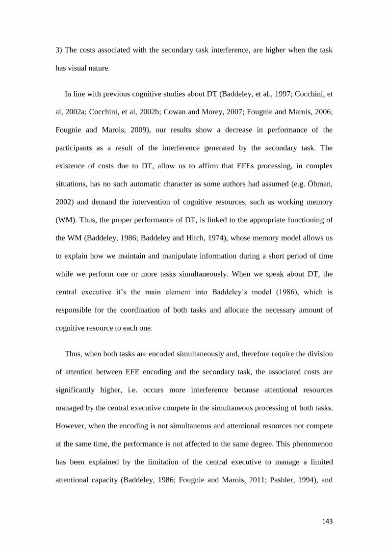
143
3) The costs associated with the secondary task interference, are higher when the task
has visual nature.
In line with previous cognitive studies about DT (Baddeley, et al., 1997; Cocchini, et
al, 2002a; Cocchini, et al, 2002b; Cowan and Morey, 2007; Fougnie and Marois, 2006;
Fougnie and Marois, 2009), our results show a decrease in performance of the
participants as a result of the interference generated by the secondary task. The
existence of costs due to DT, allow us to affirm that EFEs processing, in complex
situations, has no such automatic character as some authors had assumed (e.g. Öhman,
2002) and demand the intervention of cognitive resources, such as working memory
(WM). Thus, the proper performance of DT, is linked to the appropriate functioning of
the WM (Baddeley, 1986; Baddeley and Hitch, 1974), whose memory model allows us
to explain how we maintain and manipulate information during a short period of time
while we perform one or more tasks simultaneously. When we speak about DT, the
central executive it’s the main element into Baddeley´s model (1986), which is
responsible for the coordination of both tasks and allocate the necessary amount of
cognitive resource to each one.
Thus, when both tasks are encoded simultaneously and, therefore require the division
of attention between EFE encoding and the secondary task, the associated costs are
significantly higher, i.e. occurs more interference because attentional resources
managed by the central executive compete in the simultaneous processing of both tasks.
However, when the encoding is not simultaneous and attentional resources not compete
at the same time, the performance is not affected to the same degree. This phenomenon
has been explained by the limitation of the central executive to manage a limited
attentional capacity (Baddeley, 1986; Fougnie and Marois, 2011; Pashler, 1994), and
144
would be exceeded by the overlap to codifying two concurrent tasks (Cowan and
Morey, 2007).
Similarly, regards to the nature of the secondary task, we have found that there is a
greater interference when both tasks have the same nature, that is, when the nature of
the secondary task is visual. Conversely, when the secondary task has verbal nature the
interference generated on EFEs processing is less, since it´s a visual task and therefore
does not affect to the same extent the own limitation of each storage system
(phonological loop and / or visuospatial sketchpad) of WM. These data are consistent
with previous studies where it has been demonstrated that DT costs are greater when the
two tasks are of the same sensory nature (e.g. Fougnie and Marois, 2006; Salway and
Logie, 1995) and compete for the same type of cognitive resources. One explanation
given from neuroimaging studies suggests that when the task requires the same
cognitive resources, there is a simultaneous activation of the same areas of the cortex,
such as the frontal and parietal lobe, causing interference and the costs associated with
DT (e.g. Klingberg, 1998).
Secondly, regarding the impact of DT in healthy and pathological aging, our data
show that:
1) Healthy older adults (Study 1) performed worse than young adults in all experimental
conditions. These differences were significant in all cases, except when the costs of DT
condition were studied during retrieval phase with verbal interference, that is, when the
two tasks did not compete at the same time for the same cognitive resources. By
contrast, the biggest difference between groups appeared on the condition of DT during
encoding with visual interference, i.e., when they had to divide their attention to process
two tasks at the same time and also they were of the same nature. These results suggest

145
that the deficits found in older adults to process EFEs, might be related to cognitive
decline associated with age.
2) PD patients (Study 2 and Study 3), also had difficulty to recognize EFEs under DT
situations. Thus, all participants had worse performance in EFEs processing when they
had to simultaneously perform the visual task, and the performance was significantly
worse that healthy older adults when they had to divide their attention. As we know
(Kemps, Szmalec, Vandierendonck and Crevits, 2005) PD patients have, among other
cognitive deficits, difficulty to manage the visuospatial sketchpad. For this reason, in
Study 2, to verify that the deficits found were not due to an overload of visuospatial
sketchpad also were analysed the data of secondary task. The results showed that
compared to healthy older adults, PD patients performed worse in Corsi Blocks task,
which was affected by the DT situation. In sum, it seems that PD patients show the
same pattern of deterioration that older people, but more increased. This deterioration
compared to healthy older adults group could be related to visuospatial deficits
associated with the disease.
3) Finally, with regard to AD patients (Study 4), it should be noted that they had a
worse performance than healthy older adults, in all experimental conditions. This was
observed in the simple tasks and in the DT task conditions. To determine what
percentage of the deficit was due to the DT situation, the costs were analysed. The
results of this analysis showed that AD patients have a significant decrease to carry out
the EFE identification task simultaneously with the visual task. However, there were no
significant costs differences when the EFEs identification occurs with the verbal task.

146
In summary, the results regarding healthy and pathological aging, point in the same
direction of the above, the high demand for cognitive resources by the DT situation
generates a loss in EFEs processing efficacy, leading to a significant poor performance
for older adults compared to young adults and for patients with AD and PD in relation
to healthy older adults. In line with our previous studies (e.g. Garcia-Rodriguez, et al,
2008; 2009a; 2009b; 2011), it seems that the cognitive decline associated with age and,
to a greater extent, the neurodegenerative disease might be on the basis of the
difficulties found to process EFEs in these groups of participants.
These results, open the door to a theoretical discussion focused on the type of EFEs
identification deficit showed by our participants. Fundamentally, we can confirm that
this deficit is associated with situations of divided attention, and therefore could be due
to the limitations of central executive, very sensitive to a frontal lobe deterioration,
consequence, among others, by aging and neurodegenerative disease (Baddeley, 1991;
Baddeley and Della hall, 1996). As well as the difficulties for top-down regulation by
the prefrontal cortex that affect, among others, the functioning of WM (Gazzaley, 2013;
Gazzaley and D'Exposito, 2007), and which would be reflected in its inability to
coordinate two tasks simultaneously (Salthouse, 2000; Salthouse, Fristoe, Lineweaver,
and Coon, 1995). This is manifested prominently in the EP (Assogna, et al, 2008;
Assogna, et al, 2010; Clark et al, 2008; Clark, et al, 2010; Gray and Degnen, 2010),
where the type of cognitive deficit associated to the disease could be the basis of
deterioration in EFEs processing. It seems that patients from the onset of the disease (at
this point, I would like to recall that our PD patients were newly diagnosed and still
unmedicated) have difficulty recognizing EFEs, as a result of its executive deficit
(Dalrymple-Alford, Kalders and Jones, and Watson, 1994; Kudlicka Clare and Hindle,
2011), which prevents them from properly distribute attentional resources to recognize

147
the emotional face and perform other visual task. And, on the contrary, they would not
present a deterioration to recognize emotions, per se, but appear in contexts that demand
complex executive control by the individual. Another theoretical interpretation comes
from authors (Wu and Hallet, 2009) who argue that these patients have relatively
preserved their attentional capacity, but they execute the tasks less automatically than
healthy older adults. According to these authors, patients use more attentional resources
for each of the tasks making it more difficult simultaneous coordination. In this sense, it
has opened a research line on the EP which aims to investigate deficits in automatic
processing of information, although less evident, could be the basis of executive
dysfunction characteristic of these patients. In line with this view, several studies
(Myers, et al., 2003; Osman, Wilkinson, Beigi, Castaneda, and Jahanshahi, 2008;
Soliveri, Brown, Jahanshahi, Caraceni, and Marsden., 1997; Swainson, et al, 2000) they
have already indicated that PD patients, have difficulty learning new automatics process
as well as to preserve previously acquired habits or automatic behaviours, and they
would have a direct relationship with the severity of the disease. However, in our
opinion, this explanation is not appropriate to interpret our results, since the data clearly
suggest that the ability to understand the emotional expressions is not an automatic
process and that the effectiveness depends on the cognitive resources available,
phenomenon that appears not only in PD patients but in healthy older adults and even
young adults, who see their performance affected by the DT situation.
Regarding AD patients, we have found that they have difficulty identify EFEs even
in situations where the EFE appears in isolation. This could be explained by the greater
cognitive impairment associated with neuropathological changes typical of the disease
(Klein-Koerkampa, et al., 2012), which greatly limit their ability to recognize emotions.
Thus, a greater extent than PD patients, AD patients have compromised their ability to
148
identify EFEs in situations of DT (Baddeley, Baddeley, Bucks, and Wilcock., 2001;
MacPherson, et al, 2007), which led to social and communicative level, could difficult
the codification of the emotional state of others, causing a deterioration in the socio-
emotional life of these patients.
Alternatively, to our theoretical position, there have been other arguments given to
explain the deterioration on the EFEs processing in aging. Some authors argue that the
differences between healthy older adults and young adults, are not explained by the
worst performance of structures such as the prefrontal cortex (Mather, 2016) but are
relate to the use of different types of cognitive strategies when process emotional
stimuli and tend to prioritize positive stimuli compared to negative (Mather, 2012;
Nashiro, 2012). That is, the so-called the “positivity effect" found in the literature
(Carstensen, et al., 2003) would not be caused by the worst performance of structures
related to the processing of emotion, such as the amygdala or prefrontal cortex, but by
improving the ability of regulate the emotional activity (St. Jacques, et al., 2013),
possibly due to a brain functional compensation increases the prefrontal cortex function
against decreased activity amygdala to negative stimuli. With the information provided
in our studies, we can not position ourselves about it because we have not
systematically studied the effect of the valence of affective stimulus for various reasons,
such as the need to include more presentations in each experimental condition which
could lead to fatigue effects. However, our theoretical position understands the
differences in aging as a result, among other things, the complexity of the task (Sassi,
Campoy, Castle, Inuggi, and Fuentes, 2014) or cognitive functioning of the individual
(Labouvie-Vief, 2009). Moreover, we believe that the UAs composition of each
emotional expression could affect their identification and could be the basis of this
"positivity effect". As mentioned in the introductory part, in the FACS (Ekman and

149
Friesen, 1978) are represented the number of facial muscles movements that contains
each basic emotion, i.e. the UAs. From our point of view, we highlight the relevance of
the number of UAs of each basic emotion, which could increase the difficulty of the
EFE identification task. This corresponds to the largest number of UAs of negative
emotions, and could explain the worse performance in identifying emotions such as fear
or sadness.
In any case, the data provided allow us to conclude that the processing of emotional
expressions depends on cognitive resources and therefore is not an automatic process.
However, in our opinion, is not a dummy argument between automatic processing
versus controlled processing. Not all emotional stimuli are processed in the same way,
but we believe that the emotional processing is a function of the characteristics of
emotional stimulation and biological relevance it has for the individual. From a
physiological point of view, therefore, is not understood a single subcortical pathway in
processing emotions "low road" traditionally associated with the amygdala, but the
existence of several ways "many roads" including other cortical areas (Pessoa and
Adolphs, 2010). Thus, according to the immediacy or not the individual's response that
requires each potentially emotional situation, a response type or another it is necessary.
If we face stimuli that demand an immediate response would probably be necessary to
process the stimulus very quickly, though it may be inaccurate (e.g., the classic
Öhman´s example about the snake in the garden). Different situation is, for example, to
be afraid of having a disease in the future. Although this thought leads us to action, for
example, medical check-ups, improve our lifestyle, etc., this situation not demands us
an effective response in milliseconds, therefore here this emotion of fear arise from
processing of controlled information, where thoughts or cognitive burden related to the
previous experience of each individual play an essential role.

150
In line with our point of view, has been questioned (Pessoa, 2013, 2015) the
existence of a "dual processing model", that differentiate between brain areas linked to
emotional processing and brain areas linked to other cognitive functions. Today, we
know that the function of the amygdala goes beyond the emotional processing and
participates directly in an extensive brain network, where works with different cortical
areas such as the prefrontal cortex, in the cognitive-emotional interactions our brain
(Pessoa, 2010). Moreover, it seems that the prefrontal cortex, not only it is related to the
processing of emotional stimuli, or making emotional decisions, but also connects
directly in identifying EFEs when are processed under a DT (Hermann, Neueder,
Troeller and Schulz, 2016). In this sense, the prefrontal cortex may play a modulatory
role in EFEs processing, depending on the context and / or the complexity of the task.
Thus, the new approach of Affective Neuroscience to emotions involves understanding
the brain as a network where are coordinated areas that in the past were understood as
individual brain structures and compartmentalized pieces. Thus, it appears that the
dichotomy between automatic processing and controlled processing is an
oversimplification of our brain functioning and that to be definitively clarified needs to
continue being investigated (Keren and Shul, 2009).

151
Capítulo 11. Final discussion
In sum, our results provide empirical evidence of such interaction between cognition
and emotional processing. Thus, taken together, they allow us to perform final
reflections regarding the automaticity of emotions and their impact on the healthy and
pathological aging.
First, we have opened a new line of research in emotion that lets us know more
deeply our ability to identify emotional expressions. As discussed above, given the
universal character of EFEs (Ekman, 1972), these are the optimal stimulation that
allows us to study the emotions ensuring that we put aside any environmental,
contextual or cultural connotation. In this sense, the thesis has a theoretical purpose and
experimental studies have tried to shed light in a complex phenomenon, as is the
automaticity of emotional processing. An added argument in this respect, is in the
voluntariness that is presumed to emotional processing of EFEs. It is true, as already
noted Ekman (2003) that the EFEs processing is involuntary. Ekman (2003) defines it
as a system always “on”, that is, we are always processing the emotions of others.
However, we believe this should not be confused with automaticity, since we have
found that when cognitive resources fail, as in the case of the elderly and especially in
patients with AD and PD, there are deficits in emotional understanding faces. That is,
the lack of automaticity of EFEs processing in DT contexts, it does not mean that we
understand that EFEs processing should be voluntary. Obviously, it is outside of our
voluntary control the processing EFEs of our context, as it is known and documented
the importance the emotional stimuli compared to neutral for humans (Bargh, Chaiken,
Govender y Pratto, 1992; Bargh, Chaiken, Raymond y Hymes, 1996; Zajonc, 2000).
However, we supported that EFE processing can be performed involuntarily and
simultaneously requires cognitive resources for an accurate identification. On the other

152
hand, we understand that the results found here are not necessarily generalizable to
other emotional stimuli such as the prosody of a speech, a certain visual scene, etc.
While it is true that the difficulties found to the EFEs processing in aging have been
found with other affective stimuli (Ruffman et al., 2008), in our case, we believe that
would be necessary carry out new experimental studies to confirm our hypothesis
regards to DT.
Secondly, we think this thesis shows a new technology (Garcia, et al, 2009a; Garcia,
et al, 2011) to study the emotional processing and provides added value to the thesis and
the results that emerge. Study emotions under a paradigm of DT, it has allowed us to
approach the emotional phenomenon from a more natural and ecological perspective
(Isaacowitz, and Stanley, 2011; Ruffman, 2011) similar to happen in situations of real
life, where individuals processed the EFEs concurrently to other environmental
information. At the same time, it avoids the methodological criticisms that have been
made to experimental works on emotions, as the lack of ecological validity, for
example, when working with isolated images, without considering the processing of the
additional social information (Adolphs, 2002b).
Third, this thesis allows us to understand the changes in the ability to identify
emotions that occur when you get older and especially when aging leads to severe brain
damage. In this regard, we want highlight the limitations to work with this type of
sample. Predictably, the neurodegenerative patients have conditioned our studies, first,
in the number to be include in each sample, and secondly from a methodological level,
in the development of the experimental tasks. PD patients were newly diagnosed and
unmedicated and we had difficulties to obtain the sample. Regards to AD patients was
an impediment their cognitive impairment and therefore the excessive fatigue during the
experimental situations. However, despite the limitations this thesis provides insight
153
into the actual deficits that have elderly people and neurodegenerative patients in their
daily lives when they try to integrate the affective information with their usual cognitive
activity. The difficulty showed to perceive and adequately recognize emotions in others,
can have severe consequences on the interpersonal level. Thus, this deterioration could
result in loss of communicative ability of individuals, resulting in a worse social and
emotional functioning, poorer quality of life and in extreme cases to social isolation.
From an applied point of view, the knowledge by professionals and families of such
deficits can help them focus their aid on these aspects, in order to improve
communication with these patients and reduce the effects of aging and / or disease.
Thus, the results of this thesis highlight the importance of assessment and intervention
on emotional processes, in normal aging, EA and EP, as well as other forms of
cognitive impairment, as Mild Cognitive Impairment, normally the anteroom of
dementia. The purpose would be to design specific intervention programs to improve
their communication quality from improving their ability to recognize emotions, and
generalize it to their daily lives. Likewise, it could inform and give guidance to family
members on the most optimal way to relate and talk to them, in order to have a
successful communication as possible.
Finally, we would like to emphasize the importance of our work for future
researches. We are aware that the theoretical implications of our findings imply the need
for further research in this direction. In our view, a new line of research it is open and
not conclude here, and we believe that it is the beginning of a new path in the study of
emotions. Indisputably, we can conclude that the ability to identify emotions,
specifically emotional facial expressions, is affected when cognitive ability is
compromised and therefore is not an automatic process. However, in the future we will
have to respond to issues that we have risen and remain unanswered, such as the
154
influence on different discrete emotions. Similarly, in the future we will have to include
our theoretical arguments within broader explanatory theories. To do this, will be useful
the contribution of neuroimaging techniques, that will provide us direct information
about brain substrates that underlie the EFEs identification in complex situations. The
involvement of cortical areas in EFEs processing, would support the results presented in
this thesis and could shed light on the open debate about the automaticity of emotional
processing and cognitive-emotional interactions.
155
Referencias
Adolphs, R. (2002a). Neural systems for recognizing emotion. Current opinion in
Neurobiology, 12, 169-177.
Adolphs, R. (2002b). Recognizing emotion from facial expressions: psychological and
neurological mechanisms. Behavioral and Cognitive Neuroscience Reviews, 1,
21-61.
Adolphs, R., Schul, R., y Tranel, D. (1998). Intact recognition of facial emotion in
Parkinson's disease. Neuropsychology, 12(2), 253-258.
Albert, M.S., Cohen, C., y Koff, E. (1991). Perception of affect in patients with
dementia of the Alzheimer type. Archives of Neurology, 48, 791-795.
Allender, J., y Kaszniak, AW. (1989). Processing of emotional cues in patients with
dementia of the Alzheimer’s type. International Journal of Neuroscience, 46,
147-155.
Anderson, S.W., Bechara, A., Damasio, H., Tranel, D., y Damasio, A. (1999).
Impairment of social and moral behavior related to early damage in human
prefrontal cortex. Nature Neuroscience, 2(11), 1032-1037.
Anderson, N.D., Craik, F.I., y Naveh-Benjamin, M. (1998). The attentional demands of
encoding and retrieval in younger and older adults: I. Evidence from divided
attention costs. Psychology and Aging, 13, 405-423.
Assogna, F., Pontieri, F.E., Caltagirone, C., y Spalletta, G. (2008). The recognition of
facial emotion expressions in Parkinson’s disease. European
Neuropsychopharmacology, 18, 835-848.

156
Assogna, F., Pontieri, F.E., Cravello, L., Peppe, A., Pierantozzi, M., Stefani, A.,
…Spalletta, G. (2010). Intensity-dependent facial emotion recognition and
cognitive functions in Parkinson's disease. Journal of the International
Neuropsychological Society, 16(5), 867-876.
Baddeley, A.D. (1986). Working memory. Oxford: Oxford University Press.
Baddeley, A.D., Baddeley, H.A., Bucks, R.S., y Wilcock, G.K. (2001). Attentional
control in Alzheimer´s disease. Brain: A Journal of Neurology, 124 (8), 1492-
1508.
Baddeley, A.D., Bressi, S., Della Sala, S., Logie, R., y Spinnler, H. (1991). The decline
of working memory in Alzheimer's disease: a longitudinal study. Brain, 114,
2521–2542.
Baddeley, A.D., y Della-Sala, S. (1996). Working memory and executive control.
Philosophical Transactions of the Royal Society of London, 351, 1397-1404.
Baddeley, A.D., Della-Sala, S., Papagno, C., y Spinnler, H. (1997). Dual-task
performance in dysexecutive and nondysexecutive patients with a frontal lesion.
Neuropsychology, 11, 187-194.
Baddeley, A., y Hitch, G. (1974). Working memory. En G.A. Bower, The Psychology of
Learning and Motivation (pp. 47- 89). New York: Academic Press.
Bargh, J., Chaiken, S., Govender, R., y Pratto, F. (1992). The generality of the
automatic attitude activation effect. Journal of Personality and Social
Psychology, 62, 893-912.

157
Bargh, J., Chaiken, S., Raymond, P., y Hymes, C. (1996). The automatic evaluation
effect: Unconditional automatic attitude activation with a pronunciation task.
Journal of Experimental Social Psychology, 32, 104-128.
Barone P., Aarsland D., Burn D., Emre M., Kulisevsky J., y Weintraub D. (2011).
Cognitive Impairments in nondemented Parkinson´s disease. Movement
Disorders, 14(26), 2483-2495.
Bechara, A., Damasio, H., y Damasio, A. (2000). Emotion, decision making and the
orbitofrontal cortex. Cerebral Cortex, 10, 295-307.
Berch, D.B., Krikorian, R., y Huha, E.H. (1998). The Corsi Block-Tapping Task:
Methodological and theoretical considerations. Brain and Cognition, 38, 317-38.
Biseul, I., Sauleau, P., Haegelen, C., Trebon, P., Drapier, D., Raoul, S., …Vérin, M.
(2005). Fear recognition is impaired by subthalamic nucleus stimulation in
Parkinson’s disease. Neuropsychologia, 43, 1054-1059.
Blessed, G., Tomlinson, B.E., y Roth, M. (1968). The association between quantitative
measures of dementia and the senile changes in grey matter of elderly subjects.
British Journal of Psychiatry, 114, 797-811.
Braak, H., y Del Tredici, K. (2009). Neuroanatomy and pathology of sporadic
Parkinson´s disease. Advances in Anatomy, Embryology and Cell Biology, 201,
111-119.
Braak, H., y Del Tredici, K. (2010). Pathophysiology of sporadic Parkinson´s Disease.
Fortschritte der Neurologie-Psychiatrie, 78 Suppl 1, S2-4.

158
Brink, T.L., Yesavage, J.A., Lum, O., Heersema, P., Adey, M.B., y Rose, T.L. (1982).
Screening tests for geriatric depression. Clinical Gerontology, 1, 37-44.
Burnham, H., y Hogervorst, E. (2004). Recognition of facial expressions of emotion by
patients with dementia of the Alzheimer type. Dementia and Geriatric Cognitive
Disorders, 18, 75-79.
Cadieux, N.L., y Greve, K.W. (1997). Emotion processing in Alzheimer’s disease.
Journal of the International Neuropsychological Society, 3, 411-419.
Calder, A. J., Keane, J., Manly, T., Sprengelmeyer, R., Scott, S., Nimmo-Smith, I., y
Young, A.W. (2003). Facial expression recognition across the adult life span.
Neuropsychologia, 41, 195–202.
Carstensen, L.L., Fung, H.H., y Charles, S.T. (2003). Socioemotional selectivity theory
and the regulation of emotion in the second half of life. Motivation and Emotion,
27, 103-123.
Carstensen, L.L., Isaacowitz, D.M., y Charles, S.T. (1999). Taking time seriously: A
theory of socioemotional selectivity. American Psychologist, 54, 91-100.
Clark, U.S., Neargarder, S., y Cronin-Golomb, A. (2008). Specific impairments in the
recognition of emotional facial expressions in Parkinson’s Disease.
Neuropsychologia, 46(9), 2300-2309.
Clark, U.S., Neargarder, S., y Cronin-Golomb, A. (2010). Visual exploration of
emotional facial expressions in Parkinson´s disease. Neuropsychologia, 48,
1901-1913.

159
Cocchini, G., Logie, R., Della-Sala, S., McPherson, S., y Baddeley, A.D. (2002a).
Performing two memory tasks at the same time: little cost of concurrence.
Memory and Cognition, 30, 1086-95.
Cocchini, G., Logie, R. H., Della-Sala, S., y MacPherson, S. E. (2002b). Concurrent
performance of two memory tasks: Evidence from domain-specific working
memory systems. Memory and Cognition, 30(7), 1086-1095.
Cowan, N., y Morey, C.C. (2007). How can dual-task working memory retention limits
be investigated? Psychological Science, 18(8), 686–688.
Craik, F.I. M., y Byrd, M. (1982). Aging and cognitive deficits. The role of attentional
resources. En F.I.M. Craik y Trehub (Eds.). Aging and cognitive processes, (pp.
191-211). New York: Plenum Press.
Cueânod, C.A., Denys, A., y Michot, J.L. (1993). Amygdala atrophy in Alzheimer’s
disease. An in vivo magnetic resonance imaging study. Archives of Neurology,
50, 941-945.
Cummings, J.L. (1985). Clinical neuropsychiatry. New York: Grune&Stratton.
Dalgleish, T. (2004). The emotional brain. Nature, 5, 582-589.
Dalrymple-Alford, J.C., Kalders, A.S., Jones, R.D., y Watson, R.W. (1994). A central
executive deficit in patients with Parkinson’s disease. Journal of Neurology,
Neurosurgery & Psychiatry, 57, 360–367.
Damasio, A.R. (1997). Towards a neuropathology of emo- tion and mood. Nature, 386,
769-770.

160
Damasio, A.R. (1998). Emotion in the perspective of an integrated nervous system.
Brain Research Reviews, 26, 83-86.
Darwin, C. (1872). La expresión de las emociones en los animales y en el hombre.
Madrid: Editorial Alianza (1985).
Davidson, R.J. (1995) Cerebral asymmetry, emotion, and affective style. En R.J.
Davidson y K. Hugdahl (Eds.), Brain asymmetry (pp. 361-387). Cambridge: The
Mit Press.
Davidson, R.J. (2003). Affective neuroscience and psycho- physiology: toward a
synthesis. Psychophysiology, 40, 655-665.
Davidson, R.J. e Irwin, W. (1999). The functional neuro- anatomy of emotion and
affective style. Trends in Cognitive Science, 3, 11-21.
Davidson, R.J., Jackson, D.C. y Kalin, N.H. (2000). Emotion, plasticity, context, and
regulation: Perspectives from affective neuroscience. Psychological Bulletin,
126, 890-909.
Dolman, R., Roy, E.A., Dimeck, P.T., y Hall, C.R. (2000). Age, gesture span, and
dissociations among component subsystems of working memory. Brain and
Cognition, 43, 164-168.
Dujardin, K., Blairy, S., Defebvre, L., Duhem, S., Noel, Y., Hess, U., y Desteé, A.
(2004). Deficits in decoding emotional facial expressions in Parkinson’s disease.
Neuropsychologia, 42(2) 239-250.
Ebner, N.C. y Fischer, H. (2014). Emotion and aging: evidence from brain and
behavior. Frontiers in Psychology, 5, 1-6.

161
Ekman, P. (1972). Universals and cultural differences in facial expressions of emotion.
En: J. Cole (Ed.), Nebraska symposium on motivation (pp. 207-283). Lincoln:
University of Nebraska Press.
Ekman, P. (2003). Emotions Revealed. U.K: Weidenfeld & Nicolson.
Ekman, P. y Friesen, W.V. (1971). Constants across cultures in the face and emotion.
Journal of Personality and Social Psychology, 17, 124-129.
Ekman, P., y Friesen, W.V. (1978). The Facial Action Coding System. Palo Alto.
California: Consult Psychology Press.
Ekman, P., Friesen, W.V., y Hager, J.C. (2002). Facial Action Coding System –
Investigator’s Guide. Salt Lake City: Research Nexus.
Ekman, P., y Oster, H. (1979). Facial expressions of emotions. Annual Review of
Psychology, 30, 527-554.
Folstein, M.F., Folstein, S.E., y McHugh, P.R. (1975). "Mini-mental state". A practical
method for grading the cognitive state of patients for the clinician. Journal of
Psychiatric Research, 12(3), 189-198.
Fougnie, D., y Marois, R. (2006). Distinct capacity limits for attention and working
memory: Evidence from attentive tracking and visual working memory
paradigms. Psychological Science, 17(6), 526–534.
Fougnie, D., y Marois, R. (2009). Dual-task interference in visual working memory: a
limitation in storage capacity but not in encoding or retrieval. Attention,
Perception and Psychophysics, 71 (8), 1831-1841.

162
Fougnie, D., y Marois, R. (2011). What limits working memory capacity? Evidence for
modality-specific sources to the simultaneous storage of visual and auditory
arrays. Journal of Experimental Psychology Learning Memory and Cognition,
37 (6), 1329-1341.
Fridlund, A.J. (1997). The new ethology of human facial expressions. En A. J. Russell,
y J.M. Fernández-Dols (Eds.), The Psychology of facial expressions (pp.123-
167). New York: Cambridge Press.
García-Rodríguez, B., Ellgring, H., Fusari, A., y Frank, A. (2009a). The role of
interference in identification of emotional facial expressions in normal aging
and dementia. The European Journal of Cognitive Psychology. Special lssue on
Aging, cognition and neuroscience, 21(2/3), 428-444.
García-Rodríguez, B., Fusari, A. y Ellgring, H. (2008). Procesamiento emocional de las
expresiones faciales en el envejecimiento normal y patológico. Revista de
Neurología, 46, 609-617.
García-Rodríguez, B., Fusari, A. Fernández-Guinea, S., Frank, A., Molina, J.A., y
Ellgring, H. (2011). Decline of executive processes affects identification of
emotional facial expressions in aging. Current Aging Science, 4, 70-75.
García-Rodríguez, B., Fusari, A., Rodríguez, B., Zurdo-Hernández, J.M., y Ellgring, H.
(2009b). Differential patterns of implicit emotional processing in Alzheimer´s
disease and healthy Aging. Journal of Alzheimer´s disease, 18(3), 541-551.
Garrido-Vásquez, P., Pell, M., Paulmann, S., Sehm, B., y Kotz, S. (2016). Impaired
neural processing of dynamic faces in left-onset Parkinson´s disease.
Neuropsychología, 82, 123-133.

163
Gazzaley, A. (2013). Top-Down modulation deficit in the aging brain. An emerging
theory of cognitive aging. En D.T Stuss & R.T. Knight (Eds.), Principles of
frontal lobe function (pp. 593–608). Oxford University Press.
Gazzeley, A., y D´Exposito, M. (2007). Top-Down modulation and normal aging.
Annals of the New York Academy of Sciences, 1097, 67-83.
Globisch, J., Hamm, A. O., Esteves, F. y Öhman, A. (1999). Fear appears fast:
Temporal course of startle potentiation in animal fearful subjects.
Psychophysiology, 36, 66-75.
Grady, C. L. (2008). Cognitive neuroscience of aging. Annals of the New York Academy
of Sciences, 1124, 127-144.
Gray, H.M., y Degnen, L.T. (2010). A meta-analysis of performance on emotion
recognition tasks in Parkinson’s disease. Neuropsychology, 24(2), 176-191.
Gunning-Dixon F.M., Gur R.C., Perkins A.C., Schroeder L., Turner T., Turetsky B.I.,
… Gur, R.E. (2003). Age-related differences in brain activation during
emotional face processing. Neurobiology of Aging, 24, 285-295.
Hargrave, R., Maddock, R. J., y Stone, V. (2002). Impaired recognition of facial
expressions of emotion in Alzheimer’s disease. Journal of Neuropsychiatry and
Clinical Neurosciences, 14, 64-71.
Hasher, L., Stoltzfus, E., Zacks, R.T., y Rympa, B. (1991). Age and inhibition. Journal
of Experimental Psychology: Learning, Memory and Cognition, 17, 163-169.
Hedden, T., y Gabrieli, J.D. (2004). Insights into the ageing mind, a view from
cognitive neuroscience. Nature Reviews Neuroscience, 5, 87–96.
164
Herman, M., Neueder, D., Troeller, A.K., y Schulz, S. (2016). Simultaneous recording
of EEG and fNIRS during visuo-spatial and facial expression processing in a
dual task paradigm. International Journal of Psychophysiology, 109, 21-28.
Herrera, E., Cuetos, F., y Rodríguez Ferreiro, J. (2011). Emotion recognition
impairment in Parkinson´s disease patients without dementia. Journal of
Neurological Sciences, 310, 237-240.
Iidaka, T., Okada, T., Murata, T., Omori, M., Kosaka, H., Sadato, N., y Yonekura, Y.
(2002). Age-related differences in the medial temporal lobe responses to
emotional faces as revealed by fMRI. Hippocampus, 12, 352-362.
Isaacowitz, D. M., Lockenhoff, C. E., Lane, R. D., Wright, R., Sechrest, L., Riedel, R.,
y Costa, P.T. (2007). Age differences in recognition of emotion in lexical stimuli
and facial expressions. Psychology and Aging, 22, 147–159.
Isaacowitz, D. M., y Stanley, J. T. (2011). Bringing an ecological perspective to the
study of aging and recognition of emotional facial expressions: Past, current, and
future methods. Journal of Nonverbal Behavior, 35(4), 261-278.
Ishihara, L., y Brayne, C. (2006). A systematic review of depression and mental illness
preceding Parkinson´s disease. Acta Neurologica Scandinavica, 113, 211-220.
Izard, C. (1994). Innate and Universal Facial Expressions: Evidence from
Developmental and Cross-Cultural Research. Psychological Bulletin, 115, 288-
99.
Jacob, E.L., Gatto, N.M., Thompson, A., Bordelon, Y., y Ritz, B. (2010). Occurrence of
anxiety and depression prior to Parkinson´s disease. Parkinsonism and Related
Disorders, 16, 576-581.

165
JianPeng, Li., PingLei, P., Rui, H., y HuiFang, S. (2012). A meta-analysis of voxel-
based morphometry studies of white matter volume alterations in Alzheimer´s
disease. Neurosciences and Biobehavioral Reviews, 36, 757-763.
Kan, Y., Kawamura, M., Hasegawa, Y., Mochizuki, S., y Nakamura, K. (2002).
Recognition of emotion from facial, prosodic and written verbal stimuli in
Parkinson’s disease. Cortex, 38(4), 623-630.
Kawamura, M., y Kobayakawa, M. (2009). Emotional impairment in Parkinson’s
disease. Parkinsonism and Related Disorders, 15(1), 47-52.
Keane, J., Calder, A., Hodges, J., y Young, A. (2002). Face and emotion processing in
frontal variant frontotemporal dementia. Neuropsychologia, 40, 655-665.
Keightley, M.L., Chiew, K.S., Winocur, G., y Grady, C.L. (2007). Age-related
differences in brain activity underlying identification of emotional expressions in
faces. Social Cognitive and Affective Neuroscience, 2(4), 292–302.
Keightley, M.L., Winocur, G., Burianova, H., Honqwanishkul, D., y Grady, C.L.
(2006). Age effects on social cognition: Faces tell a different story. Psychology
and Aging, 21(3), 558-572.
Kemps, E., Szmalec, A., Vandierendonck, A., y Crevits, L. (2005). Visuo-spatial
processing in Parkinson’s disease: evidence for diminished visuo-spatial sketch
pad and central executive resources. Parkinsonism and Related Disorders, 11,
181-186.
Keren, G., y Schul, Y. (2009). Two is not always better than one: A critical evaluation
of two-system theories. Perspectives on psychological science, 4, 533–550.

166
Kessels, R.P.C., Van Zandvoort, M.J.E., Postma, A., Kappelle, L.J., y De Haan, E.H.F.
(2000). The Corsi Block-Tapping Task: Standardization and normative data.
Applied Neuropsychology, 7, 252-258.
Kholer, C.G., Gallagher, A., Bilker, W., Karlawish, J., Gur, R.E., y Clark, C.M. (2005).
Emotion-discrimination deficits in mild Alzheimer disease. The American
Journal of Geriatric Psychiatry, 13, 926-933.
Klein-Koerkampa, Y., Beaudoinb, M., Baciua, M., y Hota, P. (2012). Emotional
decoding abilities in Alzheimer´s disease: a meta-analysis. Journal of
Alzheimer´s disease, 32, 109-125.
Klingberg, T. (1998). Concurrent performance of two working memory tasks: potential
mechanisms of interference. Cerebral Cortex, 8, 593-601.
Koff, E., Zaitchik, D., Montepare, J., y Albert, M. S. (1999). Emotion processing in the
visual and auditory domains by patients with Alzheimer’s disease. Journal of the
International Neuropsychological Society, 5, 32-40.
Kudlicka, A., Clare, L., y Hindle, J.V. (2011). Executive functions in Parkinson's
disease: systematic review and meta-analysis. Movement disorders, 26(13),
2305-2315.
LaBar, K., Gatenby, J.C., Gore, J.C., Le Doux, J.E., y Phelps, E. (1998). Human
amygdala activation during conditioned fear acquisition and extinction: a mixed-
trial fMRI study. Neuron, 20, 937-945.
Labouvie-Vief, G. (2009). Dynamic integration theory: Emotion, cognition and
equilibrium in later life. En V. L. Bengtson, D. Gans, N. M. Putney, y M.
167
Silverstein (Eds.), Handbook of theory of aging (pp. 277–93). Nueva York:
Springer.
Lavenú, I. y Pasquier, F. (2005). Perception of emotion on faces in frontotemporal
Dementia and Alzheimer’s disease: A longitudinal study. Dementia and
Geriatric Cognitive Disorders, 19, 37-41.
Lawerence, A.D., Goerendt, I.K., y Brooks, D.J. (2007). Impaired recognition of facial
expressions of anger in Parkinson's disease patients acutely withdrawn from
dopamine replacement therapy. Neuropsychologia, 45, 65-74.
Le Doux, J. (1986). The neurobiology of emotion. En J.E. LeDoux, y W. Hirst (Eds.),
Mind and Brain: Dialogues in Cognitive Neuroscience (pp. 301-354).
Cambridge: Cambridge University Press.
Le Doux, J. (1987). Emotion. En V.B. Mountcastle, F. Plum, y S.R. Geiger (Eds.),
Handbook of Physiology. Sec. 1: The Nervous System, Vol. V, Higher Functions
of the Brain (pp. 419-455). Bethesda: American Physiological Society.
Le Doux, J. (1990). Cognitive-emotional interactions in the brain. Cognition and
Emotion, 3, 267-290.
Le Doux, J. (1996). The Emotional Brain. New York: Simon & Schuster.
Litvan, I., Aarsland, D., Adler, C.H., Goldman, J.G., Kulisevsky, J., Mollenhauer, B.,
… Weintraub, D. (2011). MDS task force on mild cognitive impairment in
Parkinson´s disease: critical review of PD-MCI. Movement Disorders, 26, 1814-
1824.
Logie, R.H., Cocchini, G., Della Sala, S., y Baddeley, A.D. (2004). Is there a specific

168
executive capacity for dual task co-ordination? Evidence from
Alzheimer´disease. Neuropsychology, 18, 504-513.
Logie, R.H., Della Sala, S., MacPherson, S.E., y Cooper, J. (2007). Dual task demands
on encoding and retrieval processes: evidence from healthy adult ageing. Cortex,
43, 159-169.
MacPherson, S. E., Della-Sala, S., Logie, R. H., y Wilcock, G. K. (2007). Specific AD
impairment in concurrent performance of two memory tasks. Cortex, 43, 858-
865.
Malatesta, C.Z., Izard, C.E., Culver, C., y Nicolich, M. (1978). Emotion
communication-skills in young, middle age, and older women. Psychology and
Aging, 2, 193-203.
Marneweck, M., y Hammond, G. (2014). Discriminating facial expressions of emotion
and its link with perceiving visual form in Parkison´s disease. Journal of
Neurological Science, 346, 149-155.
Mather, M. (2012). The emotion paradox in the aging brain. Annals of the New York
Academy of Sciences, 125 (1), 33–49.
Mather, M. (2016). The Affective Neuroscience of Aging. Annual Reviews of
Psychology, 67, 213-238.
McConatha, J.T., Leone, F.M., y Amstrong, J.M. (1997). Emotional control in
adulthood. Psychological Reports, 80, 499-507.
McLellan, T., Johnston, L., Dalrymple-Alford, J., y Porter, R. (2008). The recognition
of facial expressions of emotion in Alzheimer´s disease: a review of findings.
Acta Neuropsychiatrica, 20, 236-250.

169
Mindham, R.H.S, y Hughes, T.A. (2000). Cognitive impairment in Parkinson´s disease.
International Review of Psychiatry, 12, 281-289.
Moreno, C., Borod, J.C., Welkowitz, J., y Alpert, M. (1993). The perception of facial
emotion across the adult life span. Developmental Neuropsychology, 9, 305-314.
Morris, J.S., Frith, C.D., Perrett, D.I., Rowland D., Young, A.W., Calder A.J., y Dolan,
R.J. (1996). A differential neural response in the human amydala to fearful and
happy facial expressions. Nature, 383, 812-815.
Murdock, B.B. (1982). A Theory for the storage and retrieval of item and associative
information. Psychological Review, 89, 609-626.
Myers, C., Shohamy, D., Gluck, M., Grossman, S., Kluger, A., Ferris, S., … Schwartz,
R. (2003). Dissociating hippocampal vs. basal ganglia contributions to learning
and transfer. Journal of Cognitive Neuroscience, 15(2), 185–193.
Narme, P., Bonnet, A.M., Dubois, B., y Chaby, L. (2011). Understanding facial emotion
perception in Parkinson´s disease: the role of configural processing.
Neuropsycologia, 49, 3295-3302.
Nashiro, K., Sakaki, M., y Mather, M. (2012). Age differences in brain activity during
emotion processing: Reflections of age-related decline or increased emotion
regulation? Gerontology, 58, 156-163.
Naveh-Benjamin, M., Craik, F.I., Gavrilescu, D., y Anderson, N.D. (2000). Asymmetry
between encoding and retrieval processes: Evidence from divides attention and a
calibration analysis. Memory and Cognition, 28, 965-976.
Naveh-Benjamin, M., Craik, F.I., Guez, J., y Dori, H. (1998). Effects of divided
attention on encoding and retrieval processes in human memory: Further support

170
for an asymmetry. Journal of Experimental Psychology: Learning, Memory and
Cognition, 24, 1091-1104.
Naveh-Benjamin, M., Craik, F.I., Guez, J., y Kreuger, S. (2005). Divided attention in
younger and older adults: Effects of strategy and relatedness on memory
performance and secondary task costs. Journal of Experimental Psychology:
Learning, memory, and Cognition, 31, 520-537.
Navon, D. y Miller, J. (1987). Role of outcome conflict in dual task interference.
Journal of Experimental Psychology: Human Perception and Performance,
13(3), 435-448.
Nelson, C.A. y Haan, M. (1997). A neurobehavioral approach to the recognition of
facial expression in infancy. En J.A. Russell y J.M. Fernández-Dols (Eds.), The
Psychology of facial expressions (pp.187-199). New York: Cambridge Press.
Norman, D., y Bobrow. D. (1975). On data-limited and resource limited processes.
Cognitive Psychology, 7, 44-64.
Öhman, A. (2002). Automaticity and the amygdala: nonconscious responses to
emotional faces. Current Directions in Psychological Science, 11, 62-66.
Öhman, A., Esteves, F., y Soares, J.J.F. (1995). Preparedness and preattentive
associative learning: electrodermal conditioning to masked stimuli. Journal of
Psychophysiological, 9, 99-108.
Orgeta, V. y Phillips, L.H. (2008). Effects of Age and Emotional Intensity on the
Recognition of Facial Emotion. Experimental Aging Research, 34, 63-79.
Orsini, A., Schiappa, O., y Grossi, D. (1981). Sex and culture differences in children’s
spatial and verbal-memory span. Perceptual and Motor Skills, 53, 39-42.

171
Osman, M., Wilkinson, L., Beigi, M., Castaneda, C.S., y Jahanshahi, M. (2008).
Patients Parkison´s disease learn to control complex systems via procedural as
well as non-procedural learning. Neuropsychología, 46, 2355-2363.
Park, D.C., Puglisi, J.T., y Smith, A.D. (1986). Memory for pictures: Does an age-
related decline exist? Psychology and Aging, 1, 11-17.
Pashler, H. (1994). Dual-Task interference in simple tasks: data and theory.
Psychological Bulletin, 116 (2), 220-244.
Pasupathi, M., Carstensen, L.L., Turk-Charles, S., y Tsai, J. (1998). Emotion and aging.
En H. Friedman (Eds.), Encyclopedia of mental health (vol., 2, pp 91-101). San
Diego, CA: Academic Press.
Pell, M.D., y Leonard, C.L. (2003). Processing emotional tone from speech in
Parkinson's disease: a role for the basal ganglia. Cognitive, Affective, and
Behavioral Neuroscience, 3(4), 275-88.
Pell, M.D., y Leonard, C.L. (2005). Facial expression decoding in early Parkinson´s
disease. Cognitive Brain Research, 23, 327-340.
Perón, J., Biseul, I., Leray, E., Vicente, S., Le Jeune, F., Drapier, S.,…Vérin, M. (2010).
Subthalamic nucleus stimulation affects fear and sadness recognition in
Parkinson’s Disease. Neuropsychology, 24(1) 1-8.
Perón, J., Dondaine, T., Le Jeune, F., Grandjean D, y Vérin M. (2012). Emotional
processing in Parkinson´s disease: a systematic review. Movement disorders,
27(2), 186-199.
Pessoa, L. (2008). On the relationship between emotion and cognition. Nature Reviews
Neuroscience, 9(2), 148-158.
172
Pessoa, L. (2010). Emergent processes in cognitive-emotional interactions. Dialogues in
Clinical Neuroscience, 12(4), 433-448.
Pessoa, L. (2013). The cognitive-emotional brain. From interactions to integration. MIT
Press.
Pessoa, L. (2015). Précis on The Cognitive-Emotional Brain. Behavioral and Brain
Sciences, 1-66.
Pessoa, L., y Adolphs, R. (2010). Emotion processing and the amygdala: from a low
road to many roads of evaluating biological significance. Nature Reviews
Neuroscience,11 (11), 773-783.
Pessoa, L., McKenna, M., Gutierrez, E., y Ungerleider, L.G. (2002). Neural processing
of emotional faces requires attention. Proceedings of the National Academy of
Sciences USA, 99, 11458-11463.
Pessoa, L. y Ungerleider, L.G. (2005). Visual attention and emotional perception. En L.
Itti, G. Rees, y J.K. Tsotsos (Eds.), Neurobiology of attention (pp. 160-166)
San Diego, CA: Elsevier.
Phillips, M.L., Drevets, W.C., Rauch, S.L. y Lane, R. (2003). Neurobiology of emotion
perception I: the neural basis of normal emotion perception. Biological
Psychiatry, 54, 504-514.
Phillips, L.H., y Henry, J.D. (2005). An evaluation of the frontal lobe theory of
cognitive aging. En J. Duncan, L.H. Phillips, y P. McLeod (Eds.), Measuring the
Mind: Speed, Control and Age (pp. 191-216). Oxford: Oxford University Press.
Phillips, L.H., MacLean, R.D.J., y Allen, R. (2002). Aging and the understanding of
emotions: neuropsychological and sociocognitive perspectives. Journal of
173
Gerontology: Psychological Sciences and Social Sciences, 57, 526-530.
Phillips, L.H., Slessor, G., Bailey, P.E., y Henry, J.D. (2014). Older Adults ‟Perception
of Social and Emotional Cues”. En P. Verhaeghen, y C.K. Hertzog (Eds.),
Oxford Handbook of Emotion, Social Cognition, and Problem Solving in
Adulthood (pp. 9-25). New York: Oxford University Press.
Phillips, L.H., Tunstall, M., y Channon, S. (2007). Exploring the role of working
memory in dynamic social cue decoding using dual task methodology. Journal
of Nonverbal Behavior, 31(2), 137-152.
Reeve, J. (1994). Motivacion y Emocion. Madrid, Espana. Editorial McGraw-Hill.
Reuter-Lorenz, P.A., y Park, D.C. (2010). Human neuroscience and the aging mind: A
new look at old problems. The Journals of Gerontology Series B: Psychological
Sciences and Social Sciences, 65B, 405-415.
Roudier, M., Marcie, P., y Grancher, A.S. (1998). Discrimimination of facial identity
and emotions in Alzheimer’s Disease. Neurology Sciences, 154, 151-158.
Ruffman, T. (2011). Ecological validity and age-related change in emotion recognition.
Journal of Nonverbal Behavior, 35(4), 297-304.
Ruffman, T., Henry, J., Livingstone, V., y Phillips, L.H. (2008). A meta-analytic review
of emotion recognition and aging: Implications for neuropsychological models
of aging. Neuroscience and Biobehavioral Reviews, 32, 863-881.
Salthouse, T.A. (1996). The processing-speed theory of adult age differences in
cognition. Psychological Review, 103, 403-428.
174
Salthouse, T.A. (2000). Steps toward the explanation of adult age differences in
cognition. En T. Perfect, y E. Maylor (Eds.), Theoretical Debate in Cognitive
Aging (pp.19-49). London: Oxford University Press.
Salthouse, T.A., Fristoe, N.M., Lineweaver, T.T., y Coon, V.E. (1995). Aging of
attention: Does the ability to divide decline? Memory and Cognition, 23(1), 59-
71.
Salthouse, T.A., Rogan, J.D., y Prill, K.A. (1984). Division of attention: Age
differences on a visually presented memory task. Memory and Cognition, 12,
613-620.
Salway, A.F.S. y Logie, R.H. (1995). Visuospatial working memory, movement control
and executive demands. British Journal of Psychology, 86, 253-269.
Sánchez-Navarro, J.P., Martínez-Selva, J.M. y Román, F. (2005). Emotional response in
patients with frontal brain damage: Effects of affective valence and information
content. Behavioral Neuroscience, 119, 87-97.
Sánchez-Navarro, J.P., y Román, F. (2004). Amígdala, corteza prefrontal y
especialización hemisférica en la experiencia y expression emocional. Anales de
Psicología, 20, 223-240.
Sarabia, C., Navas, M.J., Ellgring, H., y García-Rodríguez, B. (2016). Skilful
communication: Emotional facial expressions recognition in very old adults.
International Journal of Nursing Studies, 54, 104-111.
Sassi, F., Campoy, G., Castillo, A., Inuggi A., y Fuentes, L. (2014). Task difficulty and
response complexity modulate affective priming by emotional facial
expressions. The Quartely Journal of Experimental Psychology, 67(5), 861–871.

175
Scott, S.A., Dekgiosky, T., y Scheff, S.W. (1991). Volumetric atrophy of the amygdala
in Alzheimer’s disease: qualitative serial reconstruction. Neurology, 41, 351-
356.
Sergerie, K., Chochol, C., y Armony, J.L. (2008). The role of the amydala in emotional
processing: a quantitative meta-analysis of functional neuroimaging studies.
Neuroscience and Biobehavioral Reviews, 32, 811-830.
Shiba, M., Bower, J.H., Maragonore, D.M., McDonnell, S.K., Peterson, B.J., Ahlskog,
J.E.…Rocca, W.A. (2000). Anxiety disorders and depressive disorders
preceding Parkinson´s disease: a case-control study. Movement Disorders, 15,
669-677.
Soliveri, P., Brown, R. G., Jahanshahi, M., Caraceni, T., y Marsden, C. D. (1997).
Learning manual pursuit tracking skills in patients with Parkinson’s disease.
Brain, 120, 1325–1337.
Sprengelmeyer, R., Young, A.W., Mahn, K., Schroeder, U., Woitalla, D., Buttner, T.,
… Przuntek, H. (2003). Facial expression recognition in people with medicated
and unmedicated Parkinson’s disease. Neuropsychologia, 41(8), 1047-1057.
St. Jacques, P.L., Bessette-Symons, B., y Cabeza, R. (2009). Functional neuroimaging
studies of aging and emotion: fronto-amygdalar differences during emotional
perception and episodic memory. Journal of the International
Neuropsychological Society, 15, 819–825.
St. Jacques, P. L., Winecoff, A., y Cabeza, R. (2013). Emotion and aging: linking neural
mechanisms to psychological theory. En J. Armony, Jorge y P. Vuilleumier

176
(Eds.), The Cambridge handbook of human affective neuroscience (pp. 635-
662). Cambridge University Press, Cambridge.
Stuss, D.T., y Alexander, G. (2000). Executive functions and the frontal lobes: A
conceptual view. Psychological Research, 63, 289-298.
Sullivan, S., y Ruffman, T. (2004). Emotion recognition deficits in the elderly.
International Journal of Neuroscience, 114, 403-432.
Suzuki. A., Hoshino, T., Shigemasu, K., y Kawamura, M. (2006). Disgust-specific
impairment of facial expression recognition in Parkinson´s disease. Brain, 129,
707-717.
Suzuki, A., Hoshino, T., Shigemasu, K., y Kawamura, M. (2007). Decline or
improvement? Age-related differences in facial expression recognition.
Biological Psychology, 74, 75–84.
Swainson, R., Rogers, R. D., Sahakian, B. J., Summers, B. A., Polkey, C. E., y Robbins,
T. W. (2000). Probabilistic learning and reversal deficits in patients with
Parkinson’s disease or frontal or temporal lobe lesions: Possible adverse effects
of dopaminergic medication. Neuropsychologia, 38(5), 596–612.
Tessitore, A., Hariri, A.R., Fera, F., Smith, W.G., Das, S., Weinberger, D.R., y Mattay,
V.S. (2005). Functional changes in the activity of brain regions underlying
emotion processing in the elderly. Psychiatry Research Neuroimaging, 139, 9-
18.
Tisserand, D.J., Pruessner, J.C., Arigita, E.J.S., van Boxtel, M.P.J., Evans, A.C., Jolles,
J., y Uylings, H.B.M. (2002). Regional frontal cortical volumes decrease

177
differentially in aging: an MRI study to compare volumetric approaches and
voxel-based morphometry. Neuroimage, 17, 657-669.
Tisserand, D.J., Visser, P.J., van Boxtel, M.P.J., y Jolles, J. (2000). The relation
between global and limbic brain volumes on MRI and cognitive performance in
healthy individuals across the age range. Neurobiology of Aging, 21, 569-576.
Tomkins, S.S. (1962). Affect. Imagery. Consciousness 1: The positive affects. Nueva
York: Springer-Verlag.
Torres, B., Santos, R.L., Barroso de Sousa, M., y Simoes, J. (2015). Facial expression
recognition in Alzheimer´s disease: a longitudinal study. Arquivos de
Neuropsiquiatría, 73(5), 383-389.
Vilkki, J., y Holst, P. (1989). Deficient programming in spatial learning after frontal
lobe damage. Neuropsychologia, 27, 971-976.
Vuilleumier, P., Armony, J.L., Driver, J., y Dolan, R.J. (2001). Effects of attention and
emotion on face processing in the human brain: An event-related fMRI study.
Neuronology, 30, 829-841.
Vuilleumier, P. y Righart, R. (2011). Attention and automaticity in processing facial ex-
pressions. En A. J. Calder, G. Rhodes Johnson M., y Haxby J. (Eds), The oxford
handbook of face perception (pp. 449-478). Oxford: Oxford University Press.
Wagenbreth, C., Wattenberg, L., Heinze, H.J., y Zaehle, T. (2016). Implicit and explicit
processing of emotional facial expressions in Parkison´s disease. Behavioural
Brain Research, 303, 182-190.

178
Wechsler, D. (1939). The Measurement of Adult Intelligence. Williams and Wilkins
(Eds.), Baltimore.
Wechsler, D. (1981). Manual for the Wechsler Adult Intelligence Scale-Revised
(WAIS-R). The Psychological Corporation, San Antonio, TX.
Wechsler, D. (1997). WAIS-III Administration and Scoring Manual. The Psychological
Corporation, San Antonio, TX.
Weisskopf, M.G., Chen, H., Schwarzschild, M.A., Kawachi, L., y Ascherio, A. (2003).
Prospective study of phobic anxiety and risk of Parkinson´s disease. Movement
Disorders, 18, 646-651.
Weyers, P., Mühlberger, A., Hefele, C., y Pauli, P. (2006). Electromyographic
responses to static and dynamic avatar emotional facial expressions.
Psychophysiology, 43, 450-453.
Williams, L.M., Brown, K.J., Palmer, D., Liddell, B.J., Kemp, A.H., Olivieri, G.,…
Gordon, E. (2006). The mellow years? –neural basis of improving emotional
stability over age. Journal of Neuroscience, 26, 6422-6430.
Wong, B., Cronin-Golomb, A., y Neargarder, S. (2005). Patterns of visual scanning as
predictors of emotion identification in normal aging. Neuropsychology, 19, 739-
749.
Woodruff-Pak, D.S. (1997). The neuropsychology of aging. Malden, MA: Blackwell.
Wright, C.L., Wedig, M.M., Williams, D., Rauch, S.L., y Albert, M.S. (2006). Novel
fearful faces activate the amygdala in healthy young adults and elderly adults.
Neurobiology of aging, 27, 361-374.

179
Wu T., y Hallett M. (2009). Dual Task Interference in Parkinson´s disease. US
Neurology, 5(1), 30-33.
Yiend, J. (2010). The effects of emotion on attention: a review of attentional processing
of emotional information. Cognition and Emotion, 24(1), 3-47.
Young, A.W., Aggleton, J.P., Hellawell, D.J., Johnson, M., Broks, P. y Hanley, J.R.
(1995). Face processing impairments after amygdalotomy. Brain, 118, 15-24.
Zajonc, R. (2000). Feeling and thinking: Closing the debate over the independence of
affect. En J.P. Forgas (Eds.), Feeling and thinking: The role of affect in social
cognition. Studies in emotion and social interaction (pp. 31-58). Nueva York:
Cambridge University Press.
Zgaljardic, D.J., Borod, J.C., Foldi, N.S., y Mattis, P. (2003). A review of the cognitive
and behavioral sequelae of Parkinson´s disease: relationship to frontostriatal
circuitry. Cognitive and Behavioral Neurology, 16, 193-210.
Ziaei, M., Burianova, H., Von Hippel, W., Ebner, N., Phillips, L.H., y Henry, J. (2016).
The impact of aging on the neural networks involved in gaze and emotional
processing. Neurobiology of Aging, 48, 182-194.
Ziaei, M. y Fischer, H. (2016). Emotion and Aging: The impact of emotion on attention,
memory, and face recognition in late adulthood (Chapter 13). En J. R. Absher y
J. Cloutier (Eds.), Neuroimaging Personality, Social Cognition, and Character
(pp. 259-278). Elsevier.
Zsoldos, I., Cousin, E., Klein-Koerkamp, Y., Pichat, C., y Hot, P. (2016). Age-related
differences in brain activity during implicit and explicit processing of fearful
facial expressions. Brain Research, 1650, 208-217.