Testicular characterization and spermatogenesis of the ...
18
RESEARCH ARTICLE Testicular characterization and spermatogenesis of the hematophagous bat Diphylla ecaudata Soraia Fonseca Marinho da Silva 1 , Carlos Henrique de Souza Silva 1 , Fernanda Carolina Ribeiro Dias 2 , Eugenia Cordero-Schmidt 3 , Juan Carlos Vargas-Mena 3 , Ingrid Gracielle Martins da Silva 4 , So ˆ nia Nair Ba ´o 4 , Thaı ´s Gomes de Carvalho 1 , Raimundo Fernandes de Arau ´ jo Ju ´ nior 1 , Carlos Eduardo Bezerra de Moura 5 , Fabiana Cristina Silveira Alves de Melo 6 , Se ´ rgio Luis Pinto da Matta 2 , Danielle Barbosa Morais ID 1 * 1 Department of Morphology, Federal University of Rio Grande do Norte-UFRN, Natal, Rio Grande do Norte, Brazil, 2 Department of General Biology, Federal University of Vic ¸ osa-UFV, Vic ¸ osa, Minas Gerais, Brazil, 3 Department of Ecology, Federal University of Rio Grande do Norte-UFRN, Natal, Rio Grande do Norte, Brazil, 4 Department of Cell Biology, University of Brası ´lia-UnB, Brası ´lia, Distrito Federal, Brazil, 5 Department of Animal Sciences, Federal Rural University of the Semi-Arid Region-UFERSA, Mossoro ´ , Rio Grande do Norte, Brazil, 6 Department of Animal Biology, Federal University of Vic ¸ osa-UFV, Vic ¸ osa, Minas Gerais, Brazil * [email protected] Abstract Diphylla ecaudata is a hematophagous bat endemic of South America, with food preference for bird blood. Given the lack of information about the reproductive activity of this species, this study aimed to describe the testicular morphology and histomorphometry of D. ecau- data in order to understand its reproductive biology, specially spermatogenesis. The ani- mals were collected in Lajes city, Rio Grande do Norte, Brazil. Following euthanasia, the testes were histologically processed for morphological, morphometric, ultrastructural and immunohistochemical analyses. Their average body weight was 24.64g, with a gonadoso- matic index of 0.49%, tubulesomatic index of 0.47%, and a total of 32.20m of seminiferous tubules per gram of testis. The pre-meiotic, meiotic, and post-meiotic phases accounted for 56.20%, 9.30%, and 34.50% of the seminiferous epithelium cycle, respectively. The ultra- structure of spermiogenesis was similar to that described in other mammals and the perfora- torium was not observed in the sperm. Androgen receptors were detected in Sertoli cell nuclei and Leydig cell cytoplasm, while aromatase enzyme was detected only in Sertoli cell nuclei. FGF2 and BCL-2 activities were detected in the cytoplasm of zygotene and pachy- tene primary spermatocytes, as well as round and elongated spermatids. D. ecaudata showed testicular pattern similar to other mammals and characteristics common to other bat species. This species stood out for its high efficiency of Sertoli cells, which presented high capacity to support germ cells, besides the highest sperm production rates among those already recorded. This study is the first step towards the knowledge of D. ecaudata repro- duction and the first description of its spermatogenesis. PLOS ONE | https://doi.org/10.1371/journal.pone.0226558 December 13, 2019 1 / 18 a1111111111 a1111111111 a1111111111 a1111111111 a1111111111 OPEN ACCESS Citation: Silva SFMd, Silva CHdS, Dias FCR, Cordero-Schmidt E, Vargas-Mena JC, Silva IGMd, et al. (2019) Testicular characterization and spermatogenesis of the hematophagous bat Diphylla ecaudata. PLoS ONE 14(12): e0226558. https://doi.org/10.1371/journal.pone.0226558 Editor: Rajakumar Anbazhagan, National Institute of Child Health and Human Development, UNITED STATES Received: September 22, 2019 Accepted: November 28, 2019 Published: December 13, 2019 Copyright: © 2019 Silva et al. This is an open access article distributed under the terms of the Creative Commons Attribution License, which permits unrestricted use, distribution, and reproduction in any medium, provided the original author and source are credited. Data Availability Statement: All relevant data are within the manuscript. Funding: The authors are thankful to CNPq (Conselho Nacional de Desenvolvimento Cientı ´fico e Tecnolo ´gico) for the financial support (Project 401467/2014-7: “Ecology and Conservation of Bats in Caatinga Potiguar”), and to CAPES (Coordenac ¸ão de Aperfeic ¸oamento de Pessoal de Nı ´vel Superior) for the scholarship provided to SFMS.
Transcript of Testicular characterization and spermatogenesis of the ...

RESEARCH ARTICLE
Testicular characterization and
spermatogenesis of the hematophagous bat
Diphylla ecaudata
Soraia Fonseca Marinho da Silva1, Carlos Henrique de Souza Silva1, Fernanda Carolina
Ribeiro Dias2, Eugenia Cordero-Schmidt3, Juan Carlos Vargas-Mena3, Ingrid Gracielle
Martins da Silva4, Sonia Nair Bao4, Thaıs Gomes de Carvalho1, Raimundo Fernandes
de Araujo Junior1, Carlos Eduardo Bezerra de Moura5, Fabiana Cristina Silveira Alves
de Melo6, Sergio Luis Pinto da Matta2, Danielle Barbosa MoraisID1*
1 Department of Morphology, Federal University of Rio Grande do Norte-UFRN, Natal, Rio Grande do Norte,
Brazil, 2 Department of General Biology, Federal University of Vicosa-UFV, Vicosa, Minas Gerais, Brazil,
3 Department of Ecology, Federal University of Rio Grande do Norte-UFRN, Natal, Rio Grande do Norte,
Brazil, 4 Department of Cell Biology, University of Brasılia-UnB, Brasılia, Distrito Federal, Brazil,
5 Department of Animal Sciences, Federal Rural University of the Semi-Arid Region-UFERSA, Mossoro, Rio
Grande do Norte, Brazil, 6 Department of Animal Biology, Federal University of Vicosa-UFV, Vicosa, Minas
Gerais, Brazil
Abstract
Diphylla ecaudata is a hematophagous bat endemic of South America, with food preference
for bird blood. Given the lack of information about the reproductive activity of this species,
this study aimed to describe the testicular morphology and histomorphometry of D. ecau-
data in order to understand its reproductive biology, specially spermatogenesis. The ani-
mals were collected in Lajes city, Rio Grande do Norte, Brazil. Following euthanasia, the
testes were histologically processed for morphological, morphometric, ultrastructural and
immunohistochemical analyses. Their average body weight was 24.64g, with a gonadoso-
matic index of 0.49%, tubulesomatic index of 0.47%, and a total of 32.20m of seminiferous
tubules per gram of testis. The pre-meiotic, meiotic, and post-meiotic phases accounted for
56.20%, 9.30%, and 34.50% of the seminiferous epithelium cycle, respectively. The ultra-
structure of spermiogenesis was similar to that described in other mammals and the perfora-
torium was not observed in the sperm. Androgen receptors were detected in Sertoli cell
nuclei and Leydig cell cytoplasm, while aromatase enzyme was detected only in Sertoli cell
nuclei. FGF2 and BCL-2 activities were detected in the cytoplasm of zygotene and pachy-
tene primary spermatocytes, as well as round and elongated spermatids. D. ecaudata
showed testicular pattern similar to other mammals and characteristics common to other bat
species. This species stood out for its high efficiency of Sertoli cells, which presented high
capacity to support germ cells, besides the highest sperm production rates among those
already recorded. This study is the first step towards the knowledge of D. ecaudata repro-
duction and the first description of its spermatogenesis.
PLOS ONE | https://doi.org/10.1371/journal.pone.0226558 December 13, 2019 1 / 18
a1111111111
a1111111111
a1111111111
a1111111111
a1111111111
OPEN ACCESS
Citation: Silva SFMd, Silva CHdS, Dias FCR,
Cordero-Schmidt E, Vargas-Mena JC, Silva IGMd,
et al. (2019) Testicular characterization and
spermatogenesis of the hematophagous bat
Diphylla ecaudata. PLoS ONE 14(12): e0226558.
https://doi.org/10.1371/journal.pone.0226558
Editor: Rajakumar Anbazhagan, National Institute
of Child Health and Human Development, UNITED
STATES
Received: September 22, 2019
Accepted: November 28, 2019
Published: December 13, 2019
Copyright: © 2019 Silva et al. This is an open
access article distributed under the terms of the
Creative Commons Attribution License, which
permits unrestricted use, distribution, and
reproduction in any medium, provided the original
author and source are credited.
Data Availability Statement: All relevant data are
within the manuscript.
Funding: The authors are thankful to CNPq
(Conselho Nacional de Desenvolvimento Cientıfico
e Tecnologico) for the financial support (Project
401467/2014-7: “Ecology and Conservation of Bats
in Caatinga Potiguar”), and to CAPES
(Coordenacão de Aperfeicoamento de Pessoal de
Nıvel Superior) for the scholarship provided to
SFMS.

Introduction
Diphylla ecaudata is a relatively rare species of hematophagous bat. In Rio Grande do Norte
state, Brazil, it was first recorded in 2017 [1]. This is the second most captured species of hema-
tophagous bats, following Desmodus rotundus. It does not cause major economic and epidemi-
ological impacts, mainly due to its feeding preference for the blood of birds [2, 3, 4]. However,
since the availability of wild prey for D. ecaudata was severely reduced in the Caatinga dry for-
ests, a highly modified biome that has been exposed to anthropic pressures and defaunation,
domestic birds became more accessible and abundant prey [5, 6]. This dietary flexibility associ-
ated with the scarcity of native birds resulted in the first human blood registration in the diet
of this species under natural conditions [7]. Thus, the effect of anthropogenic impacts on the
ecological balance of D. ecaudata also reflects in its medical-sanitary and economic relevance.
Therefore, it is important to understand the reproductive biology of the species aiming to max-
imize rational management actions.
The knowledge on D. ecaudata gametogenesis is extremely limited, and one factor that con-
tributes to the scarcity of studies on its reproduction is that this is a secretive species which has
a more restricted distribution when compared to other bats, especially those with a hematoph-
agous habit [4]. The few studies on D. ecaudata reproduction are based mainly on ecological
and behavioral studies of female. D. ecaudata, a polygynous species, has a gestation period of
approximately 5.5 months, with births occurring during spring and summer, which coincides
with the birth of domestic and native birds in Latin America [8]. Usually, only one animal per
litter is born and, occasionally, two offspring can be generated per year [9]. No studies were
found on the male reproductive activity of this species. Therefore, the present study aimed to
describe the morphology of the testes of D. ecaudata, as well as the testicular histomorphome-
try, in order to understand its reproductive biology and spermatogenesis. So, this study repre-
sents an extra effort to increase understanding of reproductive patterns in bats, specially D.
ecaudata, which could contribute to developing of conservational programs regarding this spe-
cies, face to the anthropogenic pressures on its natural area.
Materials and methods
Study area and animals collection
The animals were collected in Lajes city, Rio Grande do Norte, Brazil (05º42’00"S, 36º14’41"W),
in February (n = 1), July (n = 3) and September (n = 2) of 2017. This is a tropical area with a
warm and humid weather, without a clear distinction among the seasons of the year [10]. Usu-
ally are stablished only the dry season (September to February) and the rainy season (March to
August) [11]. The captures were authorized by the Chico Mendes Institute for Biodiversity Con-
servation (ICMBio, license number 55562–1). All experimental procedures were conducted in
accordance with the recommendations of the National Council for Animal Experimentation
Control (CONCEA). The protocol was approved by the Ethics Committee on Animal Use of
the Federal University of Rio Grande do Norte (CEUA UFRN, protocol number 056/2016). All
efforts were made to minimize animal suffering.
Six adult D. ecaudata males were captured at nightfall using mist nets at the entrance to the
abandoned ore galleries, which animals used as shelters. Adult animals were identified based
on the fusion of the epiphyseal cartilage of the fourth finger at the metacarpal-phalangeal junc-
tion [12].
The animals were transported in bags suitable for containment and transport of bats to
Natal city, Rio Grande do Norte, Brazil, and the euthanasia was performed on the same day.
The animals were anesthetized intraperitoneally (xylazine 50 mg/kg and ketamine 80 mg/kg),
Spermatogenesis of the vampire bat Diphylla ecaudata
PLOS ONE | https://doi.org/10.1371/journal.pone.0226558 December 13, 2019 2 / 18
Competing interests: The authors have declared
that no competing interests exist.

weighed and subsequently euthanized by deepening the anesthetic plane (xylazine 150 mg/kg
and ketamine 240 mg/kg).
Histological processing
One testis of each animal was fixed in Karnovsky solution [13] for 24 hours and histologically
processed for either morphological and morphometric analyses under light microscopy, or for
ultrastructural analysis, under transmission electron microscopy.
Testicular fragments were embedded in glycol methacrylate (Historesin, Leica), cut into 3-
μm sections using a rotatory microtome (Leica RM 2245), and stained with toluidine blue/
sodium borate 1% (Merck) for light microscopy analyses. For ultrastructural analysis, testicu-
lar fragments were post-fixed with 2% osmium tetroxide and 1.6% potassium ferricyanide in
0.2 M sodium cacodylate buffer, followed by staining in 0.5% aqueous solution of uranyl ace-
tate, overnight. Dehydration was performed in ethanol and acetone, followed by the addition
of embedding resin (Spur, Sigma-Aldrich1). Ultrathin sections were contrasted with uranyl
acetate and lead citrate and observed under a transmission electron microscope (JEOL 1011).
The other testis of each animal was fixed in 4% Paraformaldehyde, processed for embed-
ding in histological paraffin and destined for immunohistochemical analyses. Testicular sec-
tions with 4 μm thickness were obtained on signaled slides. The histological sections were
deparaffinized, rehydrated, washed in 0.3% Triton X-100 in phosphate buffer and incubated
with endogenous peroxidase (3% hydrogen peroxide). The sections were incubated overnight
at 4˚ C in the presence of primary antibodies (Santa Cruz Biotechnology) against pre-apoptotic
protein BCL-2 (1: 400), fibroblast growth factor FGF2 (1: 400), aromatase (1: 200), and andro-
gen receptor (1: 200). The sections were carefully rinsed with phosphate buffer and incubated
in the presence of secondary antibody streptavidin/HRP-conjugated (Biocare Medical) for 30
minutes. Immunoreactive cells were visualized by colorimetric detection following the proto-
col provided by the manufacturer (TrekAvidin-HRP Label + Kit Biocare Medical). The sec-
tions were counterstained with hematoxylin and the labeled positive areas were captured by a
photomicroscope (Nikon E200 LED).
Considering each used antibody, the number of positive cells per tubular cross section was
quantified in relation to the number of cells without immunostaining in an area of approxi-
mately 40,000 μm2. The following formula was used: [(number of marked cells / number of
unmarked cells) / number of analyzed sections].
Testicular morphometry
Both testes were weighted after fixation, using an analytical balance (BEL M214AIH). The
gonadosomatic index (GSI) was calculated by dividing the testes weight by body weight and
multiplying by 100, in order to quantify the investment in the testicles regarding the total body
mass.
Digital images were obtained using a light-field photomicroscope (Olympus BX-50 or BEL
Bio2/3 Eurekam 5.0) and analyzed using the Image-Pro Plus1 software. Then, the volumetric
proportions of all components of the seminiferous tubule (tunica propria, seminiferous epithe-
lium and lumen), intertubule and tunica albuginea were determined after counting 3,520
intersection points, per animal, in 10 square grids randomly placed over these digital images
(100x magnification). In order to obtaining their percentages, the counting obtained for each
element in each image was divided by the number of points scored, multiplying this value by
100. Seminiferous tubules and intertubule volumes were calculated by multiplying the testes’
weight by their respective percentages and dividing these values by 100 [14, 15]. Since the
mammalian testis density is around 1 [16], its weight was considered equal to the volume.
Spermatogenesis of the vampire bat Diphylla ecaudata
PLOS ONE | https://doi.org/10.1371/journal.pone.0226558 December 13, 2019 3 / 18

The tubulesomatic index (TSI) was calculated in order to quantify the investment in the
seminiferous tubules regarding the total body mass. It was obtained by dividing the tubular
volume by the body weight and multiplying the result by 100. The mean tubular diameter was
obtained by measuring 20 tubular cross-sections per animal, regardless the stage of the cycle.
These sections were also used to measure the height of the seminiferous epithelium, from the
tunica propria to the tubular lumen, taking two diametrically opposite measurements in each
cross section [14, 15].
The seminiferous tubule length (STL, in meters) per testis was estimated as follows:
STL = STV/ лR2 (STV = seminiferous tubule volume; лR2 = tubule area; R = tubular diameter/
2). The STL was divided by the testicular weight to calculate the length of the seminiferous
tubules per gram of testis (STL/g), to allow comparisons between different species [14, 15].
Coincident points (n = 1000) over the intertubular components were recorded: Leydig cell,
blood and lymphatic vessels, and connective tissue. The volumetric rates of these components
were also estimated (400x magnification). The percentage of these components in the intertu-
bule was estimated by multiplying the total number of points on each component by 100 and
dividing the obtained value by 1000. The percentage of these components in the testis was
obtained by multiplying the percentage of intertubule by the percentage of each component in
the intertubule and dividing the obtained value by 100. The volume of each intertubular com-
ponent in the testicular parenchyma was calculated by the following formula: (percentage of
each component in the testis x gonadal weight) / 100. The values were expressed in μL [14, 17].
The mean diameter of the Leydig cell was obtained after measuring 30 cells per animal,
selecting those with the most spherical nuclei and evident nucleoli. The nuclear volume was
obtained by using the formula 4/3 πR3, where R = nuclear diameter/2. The cytoplasmic volume
was estimated by multiplying the percentage of cytoplasm by the nuclear volume, divided by
the nuclear percentage. The single cell volume was estimated by adding the nuclear volume to
the cytoplasmatic volume. These values were expressed in μm3. The total volume occupied by
the Leydig cells in the testicular parenchyma was obtained by multiplying the percentage of
Leydig cells in the testis by the gonadal weight and dividing the obtained value by 100. The
number of Leydig cells per testis was estimated from the Leydig cell individual volumes and
the total volume occupied by these cells in the testicular parenchyma. This value was divided
by the gonadal weight to estimate the number of Leydig cells per gram of testis. The Leydigoso-
matic index (LSI), which quantifies the investment in Leydig cells to body mass, was estimated
by dividing the Leydig cell volume in the testicular parenchyma by the body weight and multi-
plying by 100 [14, 17].
Stages of the seminiferous epithelium cycle
The stages of the seminiferous epithelium cycle of D. ecaudata were characterized according to
the tubular morphology method [18], based on the shape and position of different germ cells
within the epithelium and on the occurrence of meiotic divisions. The relative frequency of
each stage described was taken after random characterization and counting of 200 cross sec-
tions of seminiferous tubules per animal [14, 19].
Cell counts and spermatogenic yield
The number of each cell type found at Stage 1 of the seminiferous epithelium cycle was esti-
mated by counting their nuclei (germ cells) or nucleoli (Sertoli cells) in 10 random tubular
cross sections per animal. Thirty nuclear diameters of type-A spermatogonia (SPTG A), pri-
mary spermatocytes in preleptotene/leptotene (PL/L), primary spermatocytes in pachytene
(PC), round spermatids (RS) and Sertoli cells (SC) nuclei were measured for each animal. The
Spermatogenesis of the vampire bat Diphylla ecaudata
PLOS ONE | https://doi.org/10.1371/journal.pone.0226558 December 13, 2019 4 / 18

results were corrected due to variations in the size of the cells and the section thickness, as
described by [20].
The intrinsic yield of spermatogenesis was calculated based on the ratio between corrected
germ cell numbers, in order to quantify spermatogenesis efficiency. The mitotic index (PL/L :
SPTG A) was calculated to determine the loss or degeneration that occurred during the sper-
matogonial phase; the meiotic index (RS : PC), so as to determine the efficiency of the meiotic
divisions; and the overall yield of spermatogenesis (RS : SPTG A) to quantify the efficiency of
the spermatogenic process [14, 19].
The total Sertoli cell support capacity was calculated using the sum of all germ cells types
divided by the number of Sertoli cells ((SPG A + PL/L + PC + RS) : SC). The total number of
Sertoli cells per testis was obtained by multiplying their corrected number by the tubular
length per testis (in μm) and dividing the result by the section thickness [14]. The obtained
results were divided by the testicular weight in order to calculate the number of Sertoli cells
per gram of testis [14, 19].
The cell loss in spermiogenesis was assumed to be nonsignificant [21] and the spermatic
reserve of the testis (SRT) was calculated on the basis of the round spermatid populations,
using the formula: SRT = (seminiferous tubule length / cut thickness) × corrected number of
round spermatids per cross-section [14, 15, 18].
Statistical analysis
The results were submitted to descriptive statistical analysis and the averages obtained were
expressed as mean ± standard deviation.
Results
Biometry and seminiferous tubule morphometry
Table 1 contains the mean values for biometry and volumetric proportions of testicular paren-
chyma components of D. ecaudata, as presented in Fig 1. The testicular parenchyma was
Table 1. Biometry and morphometry of the testicular components of Diphylla ecaudata. The data are reported as
mean ± standard deviation (SD) of the mean.
Parameters Mean ± SD (n = 6)
Body weight (g) 24.64 ± 1.45
Testes weight (g) 0.12 ± 0.04
Gonadosomatic Index (%) 0.49 ± 0.17
Tunica albuginea (%) 8.39 ± 1.27
Seminiferous tubules (%) 95.98 ± 0.92
Tunica propria (%) 4.49 ± 0.61
Epithelium (%) 63.37 ± 3.13
Lumen (%) 28.13 ± 3.44
Intertubule (%) 4.02 ± 0.92
Seminiferous tubules volume (mL) 0.12 ± 0.04
Tubulesomatic Index (%) 0.47 ± 0.16
Tubular diameter (μm) 195.09 ± 6.00
Epithelium height (μm) 44.26 ± 4.31
Seminiferous Tubules Length per testis (m) 3.90 ± 1.31
Seminiferous Tubules Length per gram of testis (m/g) 32.20 ± 2.00
https://doi.org/10.1371/journal.pone.0226558.t001
Spermatogenesis of the vampire bat Diphylla ecaudata
PLOS ONE | https://doi.org/10.1371/journal.pone.0226558 December 13, 2019 5 / 18

predominantly composed of seminiferous tubules, and the tubular compartment, mainly of
seminiferous epithelium.
Intertubular morphology and morphometry
Table 2 presents the histomorphometry of the intertubular compartment of D. ecaudata. This
compartment was predominantly composed of Leydig cells, followed by blood vessels, lym-
phatic vessels and connective tissue (Fig 1B). The occupation of intertubule and testicular
parenchyma by blood vessels was similar to that of lymphatic vessels, while the volume of
Fig 1. Cross sections of Diphylla ecaudata testis. TC: Tubular Compartment; SE: Seminiferous Epithelium; L: Lumen; Black arrow: tunica propria; IC:
Intertubular Compartment; �: Leydig cell nucleus; C: Leydig cell cytoplasm; White arrow: Connective tissue; : Lymphatic vessel;►:Blood vessel. Scale Bars: a:
30 μm, b: 10 μm.
https://doi.org/10.1371/journal.pone.0226558.g001
Table 2. Volumetric proportion (%) and volume of the intertubular compartment of Diphylla ecaudata. The data
are reported as mean ± standard deviation (SD) of the mean.
Parameters Mean ± SD (n = 6)
Intertubule volume (mL) 0.006±0.002
Percentage in the intertubule (%)
Leydig cells 48.45±14.31
Blood vessels 24.20±9.06
Lymphatic vessels 20.27±19.12
Connective tissue 7.08±3.25
Percentage in the testicular parenchyma (%)
Leydig cells 2.26±1.10
Blood vessels 1.12±0.61
Lymphatic vessels 0.97±0.94
Connective tissue 0.34±0.18
Volume per testicular parenchyma (μL)
Leydig cells 2.52±0.86
Blood vessels 1.23±0.38
Lymphatic vessels 1.42±1.67
Connective tissue 0.44±0.29
https://doi.org/10.1371/journal.pone.0226558.t002
Spermatogenesis of the vampire bat Diphylla ecaudata
PLOS ONE | https://doi.org/10.1371/journal.pone.0226558 December 13, 2019 6 / 18

lymphatic vessels by testicular parenchyma was greater than the volume of blood vessels. The
Leydig cell morphometry is presented in Table 3.
Stages of the seminiferous epithelium cycle (SEC)
Fig 2 shows the SEC of D. ecaudata, which is divided into eight stages, as described by Bernd-
ston (1977). Sertoli cells (SC) and type-A spermatogonia (SPG A) were found in all stages. Dur-
ing spermatogonial mitosis, type-A spermatogonia goes through transition to the intermediate
type, which was found at stage 6, while type-B spermatogonia was found at stage 7. The type B
spermatogonia originates the primary spermatocyte in preleptotene at stage 8 and this cell
begins the first meiotic division. The transition from preleptotene to leptotene occurs at stages 1
to 2, originating zygotene primary spermatocyte at stage 2. This spermatocyte was observed
until stage 4, which originated the pachytene primary spermatocyte. In this stage, the pachytene
spermatocyte originated the diplotene primary spermatocyte, thus finishing the first meiotic
division, followed by the second meiotic division and originating the secondary spermatocytes.
Since the second meiotic division is faster than the first, the secondary spermatocyte quickly
originated the round spermatids still at stage 4. The round spermatid begins the elongation pro-
cess only at stage 2 and can be found until the end of the current SEC, at the beginning of the
next cycle. Thus, elongated spermatids emerge from stage 3 and can be viewed up to stage 8.
While different spermatocyte generations can be seen at stages 1 to 8, only one spermatid
generation is observed at stages 1 to 3 and two generations at stages 5 to 8 (Figs 2 and 3). Stage
4 is characterized by diplotene primary spermatocyte division to originate secondary sper-
matocytes, which divide to produce round spermatids. The round spermatids begin elongation
at stage 2 and will reach the lumen at stage 8.
The process of spermatid elongation involves progressive reduction of the cytoplasmic area
concomitant with nuclear flattening, association of the acrosomal cap to the nuclear surface
and development of the sperm tail. In this sperm region, the cytoskeleton had an axial filament
composed of a central pair of microtubules surrounded by the fibrous sheath composed of 9
pairs of peripheral microtubules. No perforatorium occurrence was detected (Fig 4).
According to the frequency of each SEC stage in D. acaudata (Fig 3), the pre-meiotic (stages
1 to 3), meiotic (stage 4), and post-meiotic (stages 5 to 8) phases account for 56.20%, 9.30%,
and 34.50%, respectively.
Cell counts and spermatogenic yield
The corrected numbers of germ and Sertoli cells at stage 1 of SEC are described in Table 4. The
population of preleptotene/leptotene and pachytene primary spermatocytes were similar.
Table 3. Morphometry of the Leydig cell of Diphylla ecaudata. The data are reported as mean ± standard deviation
(SD) of the mean.
Parameters Mean ± SD (n = 6)
Nuclear diameter (μm) 13.32±2.31
Nuclear percentage (%) 26.97±7.41
Nuclear volume (μm3) 1335.73±758.16
Cytoplasmic percentage (%) 73.03±7.41
Cytoplasmic volume (μm3) 4305.02±3744.37
Leydic cell volume (μm3) 5640.75±4469.36
Number of Leydig cells per testis (x105) 5.78±3.05
Number of Leydig cells per gram of testis (x105) 47.82±16.64
Leydigosomatic index (%) 0.005±0.006
https://doi.org/10.1371/journal.pone.0226558.t003
Spermatogenesis of the vampire bat Diphylla ecaudata
PLOS ONE | https://doi.org/10.1371/journal.pone.0226558 December 13, 2019 7 / 18
Considering all the cells that composed the seminiferous epithelium in D. ecaudata at stage 1,
each Sertoli cell was able to support on average 30 germ cells.
Immunohistochemical analysis
Androgen receptors showed immunostaining in Sertoli cell nuclei (Fig 5A) and Leydig cell
cytoplasm (Fig 5B), while aromatase enzyme was detected only in Sertoli cell nuclei (Fig 5C).
Immunostaining for FGF2 (Fig 5D) and BCL-2 (Fig 5E) was detected in the zygotene and
pachytene primary spermatocytes, besides round spermatids (Table 5). The elongated sperma-
tids showed a discrete immunoreactivity, and only for FGF2 (Fig 5D).
Discussion
This study stands out for being the first to describe the spermatogenic process of the hairy-leg-
ged vampire bat D. ecaudata. The few studies on the reproduction of this species are based
mainly on behavioral aspects related to the females, while no studies on the gametogenesis of
males were found.
The difficulty in collecting these animals must be considered, as they have the most
restricted distribution among vampire bats, fast moving and dislocate quickly to other shelters
when disturbed [4]. So, due to the limited sample size, it wasn’t possible to infer on this study
about reproductive seasonality, and this study focuses on the testicular morphology and sper-
matogenic yields of this species.
Fig 2. Stages of the seminiferous epithelium cycle (SEC) of Diphylla ecaudata according to the Tubular Morphology Method. a: Stage I; b: Stage II; c: Stage
III; d: Stage IV; e: Stage V; f: Stage VI; g: Stage VII; h: Stage VIII. Sc: Sertoli cell nuclei; A: Type A spermatogonia; I: Intermediate spermatogonia; B: Type B
spermatogonia; PL/L: Preleptotene/Leptotene primary spermatocyte; Z: Zygotene primary spermatocyte; P: Pachytene primary spermatocyte; M: Meiotic
division; Ss: Secondary spermatocyte; Rs: Round spermatid; Es: Elongated spermatid; Rb: Residual body; L: Lumen. Scale bar: 20 μm.
https://doi.org/10.1371/journal.pone.0226558.g002
Spermatogenesis of the vampire bat Diphylla ecaudata
PLOS ONE | https://doi.org/10.1371/journal.pone.0226558 December 13, 2019 8 / 18

Biometry and seminiferous tubule morphometry
Diphylla ecaudata is the smallest species of hematophagous bat [4], weighing approximately 25
g. The two other known hematophagous bat species, Desmodus rotundus and Diaemus youngi,have an approximate body weight of 36–42 g [8, 14] and 30–38 g [22], respectively. The
gonadal weight of D. ecaudata was similar to that previously reported for other bat species [15,
23, 24].
The GSI and TSI found for D. ecaudata were also similar to those observed in other neo-
tropical bat species [14, 15, 24, 25]. As most of these species live in harem systems, consisting
of a dominant male and groups of 8 to 12 adult females [8, 26], it is required a greater invest-
ment in gonads when compared to monogamous animals, as observed in several mammals
[27, 28]. In bats, it was observed that species in which multiple males roost with multiple
females shows the largest relative testes, single-male/multi-female species are intermediate in
testes size, and the smallest relative testes occurs in single-male/single-female species [28, 29].
So, despite the association between testes size and mating systems in bats are multifactorial,
the GSI and TSI for D. ecaudata reinforce the literature data regarding a polygynic mating sys-
tem on this species, as well in other bats [15, 26].
The arrangement of testicular parenchyma and its percentages, with seminiferous tubules
and intertubule, were similar to those reported in other bats, such as D. rotundus [14].
Fig 3. Diagram of the spermatogenic process of Diphylla ecaudata representing the progression of the germ cells alongside the stages of the
seminiferous epithelium cycle and the frequency (%) of each stage. Each row corresponds to a generation of spermatogenic cells and each column
corresponds to a stage. Roman numbers indicate the cycles of cell division necessary to complete the spermatogenesis process. A: Type A spermatogonia; I:
Intermediate spermatogonia; B: Type B spermatogonia; PL/L: Preleptotene/Leptotene primary spermatocyte; Z: Zygotene primary spermatocyte; P: Pachytene
primary spermatocyte; D: Diplotene primary spermatocyte; Ss: Secondary spermatocyte; Rs: Round spermatid; Es: Elongated spermatid.
https://doi.org/10.1371/journal.pone.0226558.g003
Spermatogenesis of the vampire bat Diphylla ecaudata
PLOS ONE | https://doi.org/10.1371/journal.pone.0226558 December 13, 2019 9 / 18

However, these phylostomids of the subfamily Desmodontinae present a slightly higher per-
centage of testicular parenchyma than other individuals of the families Phyllostomidae and
Molossidae [15, 24]. The percentage of tubular compartment represented by seminiferous epi-
thelium in D. ecaudata was lower than that recorded for other bat species [14, 15, 24].
The tubular diameter (195.09 μm) was higher than that observed in other bats (137.50 μm
[30]; 139.54 μm [24]; 175.00 μm [31]; 188.04μm [14]), which is close to the value presented by
Fig 4. Ultrastructural aspects of spermiogenesis of Diphylla ecaudata. a: round spermatid without acrosomal cap, at stage 5 of seminiferous
epithelium cycle; b-d: spermatid elongation with different degrees of nuclear association of the acrosomal cap; e: elongated spermatids with
complete formation of the acrosome cap; f: transverse sections of the flagellum. �: nucleoli; White arrow: acrosome; M: mitochondria; FS: fibrous
sheath; AF: axial filament; MT: microtubule. Scale bars: a: 2 μm, b: 0.5 μm, c: 1 μm, d: 0.2 μm, e: 0.5 μm, f: 0.2 μm, detail: 0.2 μm.
https://doi.org/10.1371/journal.pone.0226558.g004
Table 4. Corrected number of germ and Sertoli cells per tubule cross section at stage 1 of the seminiferous epithe-
lium cycle (SEC) and spermatogenic indexes of Diphylla ecaudata. The data are reported as mean ± standard devia-
tion (SD) of the mean.
Parameters Mean ± SD (n = 6)
Sertoli cells 2.83±0.63
Type A spermatogonia 0.90±0.40
Preleptotene/leptotene primary spermatocytes 15.00±4.24
Pachytene primary spermatocytes 17.67±3.39
Round spermatids 49.33±10.51
Mitotic index 19.37±11.13
Meiotic index 2.81±0.38
Spermatogenic yield 67.03±41.19
Sertoli cell support capacity 30.16±7.26
Sertoli cell/testis (x105) 37.67±14.17
Sertoli cell/g of testis (x106) 59.44±13.36
Spermatic reserve/testis (x106) 69.36±40.06
Spermatic reserve/g of testis (x107) 103.65±21.67
https://doi.org/10.1371/journal.pone.0226558.t004
Spermatogenesis of the vampire bat Diphylla ecaudata
PLOS ONE | https://doi.org/10.1371/journal.pone.0226558 December 13, 2019 10 / 18
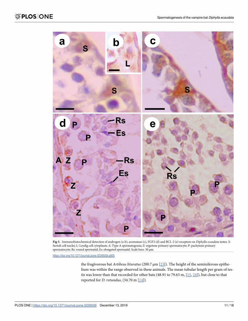
the frugivorous bat Artibeus lituratus (200.7 μm [23]). The height of the seminiferous epithe-
lium was within the range observed in these animals. The mean tubular length per gram of tes-
tis was lower than that recorded for other bats (48.91 to 79.63 m, [15, 24]), but close to that
reported for D. rotundus, (34.70 m [14]).
Fig 5. Immunohistochemical detection of androgen (a-b), aromatase (c), FGF2 (d) and BCL-2 (e) receptors on Diphylla ecaudata testes. S:
Sertoli cell nuclei; L: Leydig cell cytoplasm; A: Type A spermatogonia; Z: zygotene primary spermatocyte; P: pachytene primary
spermatocyte; Rs: round spermatid; Es: elongated spermatid. Scale bars: 30 μm.
https://doi.org/10.1371/journal.pone.0226558.g005
Spermatogenesis of the vampire bat Diphylla ecaudata
PLOS ONE | https://doi.org/10.1371/journal.pone.0226558 December 13, 2019 11 / 18

Intertubular morphology and morphometry
The intertubular compartment of D. ecaudata seemed morphologically similar to that described
for other mammals, consisting of Leydig cells, blood and lymphatic vessels, and connective tis-
sue. However, its percentage within the testicular parenchyma was the lowest recorded [17, 32,
33], which was similar to that found in the hematophagous bat D. rotundus [14].
Leydig cells were the main component of D. ecaudata intertubular compartment, which is
also observed in other bat species [14, 17, 33]. The largest investment in these cells is directly
related to the matting pattern of this species and its polygynic behavior [8]. Therefore, they
require greater androgenic investment when compared with monogamous species, such as the
crab-eating fox [34]. The number of Leydig cells per gram of testis found in D. ecaudata (47.82
x 105 cells) was lower than the observed for frugivorous bat S. lilium (11.3 x 10⁷ cells) [17] and
the insectivorous bat M. molossus (48.49 x 10⁶ cells) [33].
The Leydigosomatic index for D. ecaudata (0.005%) was smaller to that observed in other
bat species, whose average ranged from 0.015% to 0.04% [14, 35]. This index was close to that
found in other mammals, such as mice and ocelots, 0.007% and 0.0036% respectively [36, 37].
Both the nuclear diameter of Leydig cells and their volumes were larger than those found for
other bat species and other mammals [14, 17, 33, 38, 39]. This higher investment in Leydig cell
nuclear diameter and volume compared to its number suggest an alternative to guarantee the
concentration of testosterone to maintain the libido and ensure the protection of the harem.
Stages of the seminiferous epithelium cycle (SEC)
In D. ecautada, as well as in other bats and mammals, the SEC is divided into eight stages, as
described by Berndston [18]. Stage 2 was the most frequent, while in other species, stage 1 is
usually the most observed [14, 15, 19, 33, 35, 40, 41]. Zygotene primary spermatocytes emerged
only from stage 2, similarly to the observed in most mammals already studied. However, it dif-
fers from the observed in other bat species, such as insectivore Molossus molossus and frugi-
vore Sturnira lilium, in which these cells were found at stage 1 [15, 19, 32, 42]. The pachytene
primary spermatocyte is found at all stages, since this phase may last for hours, days or even
weeks, depending on the species [14, 19, 38, 39, 41].
Spermatogonia are present in all SEC stages due to their constant mitotic activity. Thus,
type A spermatogonia could be observed in all stages, as well as intermediate type at stage 6
and type B at stage 7, as reported in M. molossus, S. lilium, D. rotundus and Myotis levis bats
[14, 19, 43] and other mammals, such as domestic cat and mice [36, 38]. In guinea pig, how-
ever, intermediate spermatogonia were observed at stage 5, and type B spermatogonia, at
stages 6 and 7 [39].
The ultrastructural analysis of spermatids showed the acrosome formation caused by the
agglutination of the Golgi complex pro-acrosomal vesicles and adhesion to the nuclear surface,
Table 5. Expression frequency of the androgen receptor, aromatase, FGF 2 and BCL-2 of Diphylla ecaudata testes. The data are reported as mean ± standard devia-
tion (SD) of the mean.
Cell type Androgen Aromatase FGF 2 BCL-2
Sertoli cell 0.32 ± 0.18 1.00 ± 0.09 --- ---
Leydig cell 0.02 ± 0.04 --- --- ---
Zygotene primary spermatocyte --- --- 1.00±0.79 1.00±1.00
Pachytene primary spermatocyte --- --- 1.00±0.12 1.00±0.16
Round spermatid --- --- 1.00±0.17 1.00±0.00
Elongated spermatid --- --- 1.00±0.31 ---
https://doi.org/10.1371/journal.pone.0226558.t005
Spermatogenesis of the vampire bat Diphylla ecaudata
PLOS ONE | https://doi.org/10.1371/journal.pone.0226558 December 13, 2019 12 / 18

which occupies about two-thirds of the nucleus in mature mammalian sperm [44, 45]. No per-
foratorium was observed in the present study. This structure is related to sperm penetration
into the oocyte cytoplasm and is poorly developed or absent in several bat families [46–48].
The early stages of flagella formation were also evident in the region that will originate the
sperm tail. The axial filament and microtubules were observed in the tail end portion, and the
organization of the axial filament was lost along the length of the end piece. This pattern of
microtubule organization showed by D. ecaudata was similar to that found in other bat species
[49–52].
Cell counts and spermatogenic yield
While the germinative cell population at stage 1 of the SEC in D. ecaudata was similar to that
observed in other bat species, the amount of Sertoli cells was considerably smaller, which
reflected in a smaller number of these cells per gram of testis. Thus, while D. ecaudata showed
approximately 2.8 Sertoli cells per tubular cross-section at stage 1 and 59 x 106 Sertoli cells per
gram of testis, D. rotundus, M. molossus and S. lilium presented, respectively, 5.76, 8.48 and
8.51 Sertoli cells at stage 1 and 13.10 x 10⁷, 28.09 x 10⁷ and 22.31 x 10¹³ Sertoli cells per gram of
testis [14, 15, 24]. On the other hand, the support capacity of Sertoli cells was approximately 30
cells, which is higher than that observed in other mammals (range from 10 to 22 cells) [32, 42,
39, 53– 55] and indicates the higher efficiency of these cells in D. ecaudata.
The mitotic index of D. ecaudata (19.37%) was higher than that observed in D. rotundus(16.93% [14]), S. lilium (15.48% [24]) and M. molossus (13.76% [15]), while the meiotic index
and the spermatogenic yield were similar between these bat species. The sperm reserve per
gram of testis of D. eucadata (103.65 x 10⁷ cells) was considerably higher than that found in S.
lilium and M. molossus (range from 56.64 x 10⁷ to 76.52 x 10⁷ cells) and in other mammals
(range from 103.80 x 10⁶ to 165.90 x 10⁶ cells) [56, 57]. This index is calculated based on the
seminiferous tubule length and the round spermatid population, since cell loss during sper-
miogenesis is considered nonsignificant [21]. Thus, the round spermatid population is consid-
ered a safe parameter to determine the number of sperm produced [32]. This finding indicates
that D. ecaudata presents the highest sperm production rates among those already recorded.
Immunohistochemical analysis
This is the first study describing the expression of androgen receptors, aromatase, FGF2 and
BCL-2 in D. ecaudata testes, which provides knowledge about the cells responsive to these
important factors related to the spermatogenesis regulation.
D. ecaudata expressed androgen receptors more often in Sertoli cells than in Leydig cells.
Similarly, these receptors also showed more discrete expression in Leydig cells of A. lituratus,which indicates that this cell population is more regulated by estrogen than androgen [58].
Aromatase expression has been detected in Leydig cells, Sertoli cells, spermatocytes, sperma-
tids and sperm from mice, rats, sheep and horses [59–63]. However, in D. ecaudata its expres-
sion was observed only in Sertoli cells, while in the Myotis nigricans bat its expression was
observed in elongated spermatids, Sertoli and Leydig cells [64].
Fibroblast growth factors (FGFs) are polypeptides that act on cell proliferation, meiosis and
cell differentiation [65]. The FGF2 expression in D. ecaudata was observed in zygotene and
pachytene primary spermatocytes, as well as in round and elongated spermatids. However, in
other mammals, such as rodents, deer, cattle and humans, FGF2 was detected exclusively in
Leydig cells and spermatogonia [66–69]. The expression of the anti-apoptotic protein BCL-2
was similar to that presented by the rodent Lagostomus maximus, located in pachytene and
zygotene primary spermatocytes and round spermatids [70], which may be related to the
Spermatogenesis of the vampire bat Diphylla ecaudata
PLOS ONE | https://doi.org/10.1371/journal.pone.0226558 December 13, 2019 13 / 18
common occurrence of these cell types during the SEC, and to the maintenance of the epithe-
lium integrity throughout the cycle.
Conclusions
The main differences founded in the spermatogenic process of D. ecaudata were the lower per-
centage of tubular compartment represented by seminiferous epithelium and the lower tubular
length per gram of testis, when compared to other bats and other mammals. On the other
hand, differently to that found in other bats, the primary spermatocyte in zygotene emerged
only from stage 2 of the seminiferous epithelium and the amount of Sertoli cells was consider-
ably smaller in D. ecaudata, contrasting with a higher support capacity by these cells, and a
higher sperm reserve per gram of testis.
Therefore, D. ecaudata showed testicular pattern similar to that of other mammals and
characteristics common to other bat species, such as large investment in seminiferous tubules
and Leydig cells. Although it was expected that the testicular pattern was similar to that found
in other bat species, this species stood out for its high efficiency of Sertoli cells, which pre-
sented high capacity to support germ cells, and a high spermatic reserve of the testis. The
description of the D. ecaudata spermatogenic process is the first step to obtain knowledge of
the male’s reproduction. This information may be useful to correlate with female reproduction
and elaborate conservation plans to improve management and prevent the extinction of the
species.
Acknowledgments
The authors are thankful to Luã Barbalho de Macêdo (UFERSA) and Vinicius Garcia Barreto
(UFRN) for their assistance in the immunohistochemical staining protocols, to CNPq (Con-
selho Nacional de Desenvolvimento Cientıfico e Tecnologico) for the financial support (Proj-
ect 401467/2014-7: “Ecology and Conservation of Bats in Caatinga Potiguar”), and to CAPES
(Coordenacão de Aperfeicoamento de Pessoal de Nıvel Superior) for the scholarship provided
to SFMS.
Author Contributions
Conceptualization: Danielle Barbosa Morais.
Data curation: Soraia Fonseca Marinho da Silva, Danielle Barbosa Morais.
Formal analysis: Soraia Fonseca Marinho da Silva, Carlos Henrique de Souza Silva.
Funding acquisition: Danielle Barbosa Morais.
Investigation: Soraia Fonseca Marinho da Silva.
Methodology: Soraia Fonseca Marinho da Silva, Carlos Henrique de Souza Silva, Fernanda
Carolina Ribeiro Dias, Eugenia Cordero-Schmidt, Juan Carlos Vargas-Mena, Ingrid Gra-
cielle Martins da Silva, Sonia Nair Bao, Thaıs Gomes de Carvalho, Raimundo Fernandes de
Araujo Junior, Carlos Eduardo Bezerra de Moura, Sergio Luis Pinto da Matta, Danielle Bar-
bosa Morais.
Project administration: Danielle Barbosa Morais.
Resources: Sonia Nair Bao, Raimundo Fernandes de Araujo Junior, Carlos Eduardo Bezerra
de Moura, Sergio Luis Pinto da Matta.
Supervision: Danielle Barbosa Morais.
Spermatogenesis of the vampire bat Diphylla ecaudata
PLOS ONE | https://doi.org/10.1371/journal.pone.0226558 December 13, 2019 14 / 18

Writing – original draft: Soraia Fonseca Marinho da Silva.
Writing – review & editing: Soraia Fonseca Marinho da Silva, Fabiana Cristina Silveira Alves
de Melo, Danielle Barbosa Morais.
References1. Vargas-Mena JC, Cordero-Schmidt E, Bento DM, Rodrıguez-Herrera B, Medellın RA, Venticinque EM.
Divesity of cave dwelling bats in the tropical dry forest of Rio Grande do Norte, Brazil. Mastozool. Neo-
trop. 2018; 25(1): 199–212.
2. Greenhall A, Schmidt U, Joermann G. Diphylla ecaudata. Mammalian Species. 1984; 227: 1–3.
3. Uieda W. Perıodo de atividade alimentar e tipos de presa dos morcegos hematofagos (Phyllostomidae)
no Sudeste do Brasil. Rev Bras Biol. 1992; 52: 563–573.
4. Aguiar LMS. Subfamılia Desmodontinae. In: Reis NR, Peracchi AL, Pedro WA, Lima IP, editors. Morce-
gos do Brasil. Londrina: EDUEL; 2007. pp. 39–43.
5. Uieda W. Comportamento alimentar de morcegos hematofagos ao atacar aves, caprinos e suınos, em
condicões de cativeiro. PhD Thesis, Universidade Estadual de Campinas. 1994. Available from: http://
repositorio.unicamp.br/jspui/handle/REPOSIP/316256.
6. Leal IR, Tabarelli M, Silva JMC. Ecologia e conservacão da Caatinga. Recife: Ed. Universitaria da
UFPE; 2003.
7. Ito F, Bernard E, Torres RA. What is for dinner? First report of human blood in the diet of the hairy-leg-
ged vampire bat Diphylla ecaudata. Acta Chiropterol. 2016; 18(2): 2016.
8. Delpietro VHA, Russo RG. Observations of the common vampire bat (Desmodus rotundus) and the
hairy-legged vampire bat (Diphylla ecaudata) in captivity. Mammal Biol. 2002; 67: p. 65–78.
9. Texas Parks & Wildlife. "Hairy Legged Vampire" (On-line). 1994. Accessed on November 03, 2019 at
http://www.nsrl.ttu.edu/tmot1/diphecau.htm.
10. Araujo EHS, Martins TLF, Araujo VMD. Tratamento de dados climaticos para avaliacão do desem-
penho termico de edificacões em Natal—RN. Natal: Editora Universitaria–EDUFRN. 1998.
11. Lima Junior NB, Arandas MJG, Marinho KSN, Aguiar JFCA, Pontes ARM, Santos KRP. Histomorfome-
tria testicular do morcego Phyllostomus discolor (Chiroptera: Phyllostomidae) em areas de Mata Atlan-
tica de Pernambuco. Braz J Vet Res Anim Sci. 2014; 51(3): 263–270.
12. Kunz TH, Anthony ELP. Age estimation and post-natal growth in the bat Myotis lucifugus. J Mammal.
1982; 63:23–32.
13. Karnovsky MJ. A formaldehyde-glutaraldehyde fixative of high osmolarity for use in electron micros-
copy. J Cell Biol 1965; 27:137A.
14. Morais DB, Puga LCHP, Paula TAR, Freitas MBD, Matta SLP. The spermatogenic process of the com-
mon vampire bat Desmodus rotundus under a histomorphometric view. PLoS ONE. 2017; 12(3): 1–18.
15. Morais DB, Cupertino MC, Goulart LS, Freitas KM, Freitas MB, Paula TA, et al. Histomorphometric
evaluation of the Molossus molossus (Chiroptera, Molossidae) testis: The tubular compartment and
indices of sperm production. Anim Reprod Sci. 2013; 140: 268–278. https://doi.org/10.1016/j.
anireprosci.2013.06.003 PMID: 23845822
16. Johnson L, Petty CS, Neaves WB. A new approach to quantification of spermatogenesis and its applica-
tion to germinal cell attrition during human spermatogenesis. Biol Reprod. 1981; 25: 217–226. https://
doi.org/10.1095/biolreprod25.1.217 PMID: 6793101
17. Morais DB, Barros MS, Freitas MB, Paula TA, Matta SL. Histomorphometric characterization of the
intertubular compartment in the testes of the bat Sturnira lilium. Anim Reprod Sci. 2014; 147(3–4):180–
186. https://doi.org/10.1016/j.anireprosci.2014.03.008 PMID: 24793584
18. Berndtson WE. Methods for quantifying mammalian spermatogenesis: a review. J Anim Sci. 1977; 44
(5): 818–883. https://doi.org/10.2527/jas1977.445818x PMID: 324963
19. Morais DB, Paula TAR, Barros MS, Balarini MK, Freitas MB, Matta SLP. Stages and duration of the
seminiferous epithelium cycle in the bat Sturnira lilium (E. Geoffroy, 1810, Chiroptera: Phyllostomidae).
J Anat. 2013; 3: 372–379.
20. Amann RP, Almquist JO. Reproductive capacity of dairy bulls. VIII. Direct and indirect measurement of
testicular sperm production. J Dairy Sci. 1962; 45(6): 774–781.
21. Johnson L, Varner DD, Roberts ME, Smith Tl, Keillor GE, Scrutchfield WL. Efficiency of spermatogene-
sis: a comparative approach. Anim Reprod Sci. 2000; 60–61: 471–480. https://doi.org/10.1016/s0378-
4320(00)00108-1 PMID: 10844217
Spermatogenesis of the vampire bat Diphylla ecaudata
PLOS ONE | https://doi.org/10.1371/journal.pone.0226558 December 13, 2019 15 / 18

22. Aguiar LMS, Camargo WR, Portella AS. Occurrence of white-winged vampire bat, Diaemus youngi
(Mammalia, Chiroptera), in the Cerrado of Distrito Federal, Brazil. Rev Bras Zool. 2006; 23(3): 893–
896.
23. Duarte APG, Talamani AS. Reproduction of the large fruit-eating bat Artibeus lituratus (Chiroptera:
Phyllostomidae) in a Brazilian Atlantic forest area. Mammal Biol. 2010; 75: 320–325.
24. Morais DB, Barros MS, Paula TA, Freitas MB, Gomes ML, Matta SL. Evaluation of the cell population of
the seminiferous epithelium and spermatic indexes of the bat Sturnira lilium (Chiroptera: Phyllostomi-
dae). PLoS One 2014; 9(7):e101759. https://doi.org/10.1371/journal.pone.0101759 PMID: 25003782
25. Roy VK, Krishna A. Role of leptin in seasonal adiposity associated changes in testicular activity of ves-
pertilionid bat, Scotophilus heathi. Gen Comp Endocrinol. 2010; 168(1): 160–168. https://doi.org/10.
1016/j.ygcen.2010.04.023 PMID: 20450917
26. Racey PA, Entwistle AC. Life-history and reproductive strategies of bats. In: Crichton EG, Krutzsch PH,
editors. Reproductive biology of bats. London: Academic Press; 2000. pp. 364–367.
27. Kenagy GJ, Trombulak SC. Size and function of mammalian testes in relation to body size. J Mammal.
1986; 67(1):1–22.
28. Wilkinson G S, McCracken GF. PMID: Bats and balls: Sexual selection and sperm competition in bats.
In: Kunz TH, Fenton MB, editors. Bat Ecology. Chicago: University of Chicago Press; 2003. pp. 129–
155.
29. Lupold S, Tomkins JL, Simmons LW, Fitzpatrick JL. Female monopolization mediates the relationship
between pre- and postcopulatory sexual traits. Nat Commun. 2014; 5:3184. https://doi.org/10.1038/
ncomms4184 PMID: 24452310
30. Beguelini MR, Goes RM, Taboga SR, Morielle-Versute E. Two periods of total testicular regression are
peculiar events of the annual reproductive cycle of the black Myotis bat, Myotis nigricans (Chiroptera:
Vespertilionidae). Reprod Fertil Dev. 2014; 26(6): 834–846. https://doi.org/10.1071/RD13109 PMID:
23830483
31. Kurohmaru M, Saruwatari T, Kimura J, Mukohyama M, Watanabe G, Taya K, Hayashi Y. Seasonal
changes in spermatogenesis of the japanese lesser horseshoe bat, Rhinolophus cornutus from a mor-
phological viewpoint. Okijimas Folia Anat. 2002; 79(4): 93–100.
32. Franca LR, Russell LD. The testis of domestic mammals. In: Martinez-Garcia F, Regadera J, editors.
Male reproduction: a multidisciplinary overview. Madrid: Churchill Livingstone; 1998. pp. 197–219.
33. Morais DB, Oliveira LC, Cupertino MC, Freitas KM, Freitas MB, Paula TA, et al. Organization and sea-
sonal quantification of the intertubular compartment in the bat Molossus molossus (Pallas, 1776) testis.
Microsc Res Tech 2013; 76(1): 94–101. https://doi.org/10.1002/jemt.22141 PMID: 23077089
34. Caldeira BC, Paula TAR, Matta SLP, Balarini MK, Campos PKA. Morphometry of testis and seminifer-
ous tubules of the adult crab-eating fox (Cerdocyon thous, Linnaeus, 1766) adulto. Ceres 2010; 57(5):
569–575.
35. Paula TAR, Costa DS, Matta SLP. Avaliacão histologica quantitativa do testıculo de capivaras (Hydro-
choerus hydrochaeris) adultas. J Biosci. 2002; 18(1): 121–136.
36. Morais ACT, Barbosa LP, Neves MM, Matta SLP, Morais DB, Melo BES. Parametros morfofisiologicos
testiculares de camundongos (Mus musculus) suplementados com geleia real. Arqu Bras Med Vet Zoo-
tec. 2009; 61(1): 110–118.
37. Sarti P, Paula TAR, Polli GO, Deco-Souza T, Araujo GR. Morfofisiologia do tecido intertubular e das
celulas de Leydig de jaguatirica (Leopardus pardalis) adulta. Arq Bras Med Vet Zootec. 2011; 63
(5):1060–1065.
38. Franca LR, Godinho CL. Testis morphometry, seminiferous epithelium cycle length, and daily sperm
production in domestic cats (Felis catus). Biol Reprod. 2003; 68(5): 1554–1561. https://doi.org/10.
1095/biolreprod.102.010652 PMID: 12606460
39. Costa GMJ, Leal MC, Silva JV, Ferreira ACS, Guimarães DA, Franca LR Spermatogenic cycle length
and sperm production in a feral pig species (Collared Peccary, Tayassu tajacu). J Androl. 2010;
31:221–230. https://doi.org/10.2164/jandrol.109.008524 PMID: 19745218
40. Farias TO, Notini AA, Talamoni SA, Godinho HP. Testis morphometry and stages of the seminiferous
epithelium cycle in an epididymal sperm-storing neotropical vespertilionid, Myotis levis (Chiroptera).
Anat Histol Embryol. 2014; 44(5): 361–369. https://doi.org/10.1111/ahe.12148 PMID: 25258091
41. Silva SF, Vieira MEL, Freitas MB, Matta SLP, Morais DB. Duration of the seminiferous epithelium cycle
in the frugivorous bat Artibeus lituratus. Theriogenol 2019; In Press.
42. Costa KLC, Matta SLP, Gomes MLM, Paula TAR, Freitas KM, Carvalho FAR, et al. Histomorphometric
evaluation of the neotropical brown brocket deer Mazama gouazoubira testis, with an emphasis on cell
population indexes of spermatogenic yield. Anim Reprod Sci 2011; 127(3–4):202–212. https://doi.org/
10.1016/j.anireprosci.2011.07.016 PMID: 21889273
Spermatogenesis of the vampire bat Diphylla ecaudata
PLOS ONE | https://doi.org/10.1371/journal.pone.0226558 December 13, 2019 16 / 18

43. Morais DB, Paula TAR, Freitas KM, Matta SLP. Cycle of the seminiferous epithelium of the bat Molos-
sus molossus, characterized by tubular morphology and acrosomal development. Asian Pac J Reprod.
2012; 1(4): 303–307.
44. Christensen AK, Fawcett DW. The fine structure of testicular interstitial cells in mice. Am J Anat. 1966;
118: 551–571. https://doi.org/10.1002/aja.1001180214 PMID: 5331133
45. Holt WV, Moore HDM. Ultrastructural aspects of spermatogenesis in the common marmoset (Callithrix
jacchus). 1984; J Anat. 138: 175–188. PMID: 6423594
46. Fawcett DW, Ito S. The fine structure of bat spermatozoa. Am J Anat. 1965; 116: 567–610. https://doi.
org/10.1002/aja.1001160306 PMID: 14324688
47. Lee JH, Choi BJ, Son SW. Spermiogenesis in the Korean greater horseshoe bat, Rhinolophus ferrume-
quinum korai. Kor J Electron Microsc. 1992; 22: 97–117.
48. Phillips DM, Rasweiler JJ, Murwali F. Giant, accordioned sperm acrosomes of the greater bulldog bat,
Noctilio leporinus. Mol Reprod Dev. 1997; 48: 90–94. https://doi.org/10.1002/(SICI)1098-2795
(199709)48:1<90::AID-MRD11>3.0.CO;2-# PMID: 9266765
49. Beguelini MR, Taboga SR, Morielle-Versute E. Ultrastructural characteristics of spermatogenesis in
Pallas’s mastiff bat, Molossus molossus (Chiroptera: Molossidae). Microsc Res Tech. 2012; 75(7):
856–868. https://doi.org/10.1002/jemt.22005 PMID: 22253210
50. Beguelini MR, Taboga SR, Morielle-Versute E. Ultrastructural characteristics of the spermatogenesis
during the four phases of the annual reproductive cycle of the black myotis bat, Myotis nigricans (Chir-
optera: Vespertilionidae). Microsc Res Tech. 2013; 76(10): 1035–1049. https://doi.org/10.1002/jemt.
22264 PMID: 23857678
51. Beguelini MR, Bueno LM, Caun DL, Taboga SR, Morielle-Versute E. Ultrastructure of spermatogenesis
in the short-tailed fruit bat, Carollia perspicillata (Chiroptera: Phyllostomidae: Carollinae). J Morphol.
2014; 275(1):111–123. https://doi.org/10.1002/jmor.20202 PMID: 24142890
52. Bueno LM, Beguelini MR, Comelis MT, Taboga SR, Morielle-Versute E. Ultrastructure of spermatogen-
esis, spermatozoon and processes of testicular regression and recrudescence in Eptesicus furinalis
(Chiroptera: Vespertilionidae). Anim Reprod Sci. 2014; 148(3–4): 228–244. https://doi.org/10.1016/j.
anireprosci.2014.05.018 PMID: 24954586
53. Costa DS, Menezes CMC, Paula TAR. Spermatogenesis in white-lipped peccaries (Tayassu pecari).
Anim Reprod Sci. 2007; 98(3–4):322–334. https://doi.org/10.1016/j.anireprosci.2006.03.014 PMID:
16647229
54. Zhengwei Y, McLachlan RI, Bremmer WJ, Wreford NG. Quantitative (stereological) study of the normal
spermatogenesis in the adult monkey (Macaca fascicularis). J Androl. 1997; 18:681–687. PMID:
9432141
55. Zhengwei Y, Wreford NG, Royce P, Kretser D, McLachlan RI. Stereological evaluation of human sper-
matogenesis after suppression by testosterone treatment: heterogeneous pattern of spermatogenic
impairment. J Clin Endocrinol Metab. 1998; 83(4): 1284–1291. https://doi.org/10.1210/jcem.83.4.4724
PMID: 9543157
56. Bittencourt VL, Paula TAR, Matta SLP, Fonseca CC, Neves MTD, Costa MEL, et al. Avaliacão da popu-
lacão celular do epitelio seminıfero e ındices indicativos da producão espermatica, atraves de biopsia
testicular em lobo-guara (Chrysocyon brachyurus, IIiger 1811) adulto. Rev Bras Rep Anim. 2004; 28
(2): 108–113.
57. Azevedo MHF, Paula TAR, Balarini MK, Matta SLP, Peixoto JV, Guião Leite FL et al. Organization and
quantification of the elements in the intertubular space in the adult jaguar testis (Panthera onca, Lin-
naeus, 1758). Micron. 2008; 39(8): 1166–1170. https://doi.org/10.1016/j.micron.2008.05.005 PMID:
18602267
58. Oliveira RL, Oliveira AG, Mahecha GAB, Nogueira JC, Oliveira CA. Distribution of estrogen receptors
(ERα and ERβ) and androgen receptor in the testis of big fruit-eating bat Artibeus lituratus is cell- and
stage-specific and increases during gonadal regression. Gen Comp Endocrinol. 2009; 161(2): 283–
292. https://doi.org/10.1016/j.ygcen.2009.01.019 PMID: 19523379
59. Nitta H, Bunick D, Hess RA, Janulis L, Newton SC, Millette CF, et al. Germ cells of the mouse testis
express P450 aromatase. Endocrinol. 1993; 132: 396–1401.
60. Almadhidi J, Seralini GE, Fresnel J, Silberzachn P, Gaillard J. Immunohistochemical localization of
cytochrome P450 aromatase in equine gonads. J Histochem Cytochem. 1995; 43: 571–577. https://
doi.org/10.1177/43.6.7769228 PMID: 7769228
61. Bilińska B, Leśniak M, Schmalz B. Are ovine Leydig cells able to aromatize androgens? Reprod Fertil
Dev. 1997; 9(2): 193–199. https://doi.org/10.1071/r96038 PMID: 9208429
Spermatogenesis of the vampire bat Diphylla ecaudata
PLOS ONE | https://doi.org/10.1371/journal.pone.0226558 December 13, 2019 17 / 18
62. Levallet J, Bilińska B, Mittre H, Genissel C, Fresnel J, Carreau S. Expression and immunolocalization of
functional Cytochrome P450 aromatase in mature rat testicular cells. Biol Reprod. 1998; 58: 919–926.
https://doi.org/10.1095/biolreprod58.4.919 PMID: 9546721
63. Levallet J, Bilińska B, Mittre H, Genissel C, Fresnel J, Carreau S. Expression and immunolocalization of
functional Cytochrome P450 aromatase in mature rat testicular cells. Biol Reprod. 1998; 58(4): 919–
926. https://doi.org/10.1095/biolreprod58.4.919 PMID: 9546721
64. Beguelini MR, Falleiros LR, Goes RM, Rahal P, Morielle-Versute E, Taboga SR. Differential expression
of aromatase, estrogen receptor alpha and 17β-HSD associated with the processes of total testicular
regression and recrudescence in the bat Myotis nigricans (Chiroptera: Vespertilionidae). Gen Comp
Endocrinol. 2014; 201: 53–64. https://doi.org/10.1016/j.ygcen.2014.03.044 PMID: 24726986
65. Niederberger CS, Shubhada S, Kim SJ, Lamb DJ. Paracrine factors and the regulation of spermatogen-
esis. World J Urol. 1993; 11: 120–128. https://doi.org/10.1007/bf00182039 PMID: 7688256
66. Han IS, Sylvester SR, Kim KH, Schelling ME, Venkateswaran S, Blanckaert VD, et al. Basic fibroblast
growth factor is a testicular germ cell product which may regulate sertoli cell function. Mol Endocrinol.
1993; 7(7): 889–897. https://doi.org/10.1210/mend.7.7.8413313 PMID: 8413313
67. Steger K, Tetens F, Seitz J, Grothe C, Bergmann M. Localization of fibroblast growth factor 2 (FGF-2)
protein and the receptors FGFR 1–4 in normal human seminiferous epithelium. Histochem Cell Biol.
1998; 110(1): 57–62. https://doi.org/10.1007/s004180050265 PMID: 9681690
68. Wagener A, Blottner S, Goritz F, Streich WJ, Fickel J. Differential changes in expression of a and b
FGF, IGF-1 and -2, and TGF-alpha during seasonal growth and involution of roe deer testis. Growth
Factors. 2003; 21(2): 95–102. https://doi.org/10.1080/08977190310001621023 PMID: 14626357
69. Abd-Elmaksoud A, Vermehren M, Nutzel F, Habermann FA, Sinowatz F. Analysis of fibroblast growth
factor 2 (FGF2) gene transcription and protein distribution in the bovine testis. Growth Factors. 2005;
23(4): 295–301. https://doi.org/10.1080/08977190500233706 PMID: 16338792
70. Gonzalez CR, Muscarsel Isla ML, Vitullo AD. The balance between apoptosis and autophagy regulates
testis regression and recrudescence in the seasonal-breeding South American plains vizcacha, Lagos-
tomus maximus. PLoS One. 2018; 13(1):e0191126. https://doi.org/10.1371/journal.pone.0191126
PMID: 29385162
Spermatogenesis of the vampire bat Diphylla ecaudata
PLOS ONE | https://doi.org/10.1371/journal.pone.0226558 December 13, 2019 18 / 18