UNIVERSIDAD COMPLUTENSE DE MADRID - …eprints.ucm.es/38471/1/T37529.pdf · Alotipos de las...
182
UNIVERSIDAD COMPLUTENSE DE MADRID FACULTAD DE MEDICINA TESIS DOCTORAL Susceptibilidad al virus herpes simple tipo 1: contribución de complejos genéticos polimórficos relacionados con la citotoxicidad celular MEMORIA PARA OPTAR AL GRADO DE DOCTORA PRESENTADA POR Manuela Moraru Director Blas Carlos Vilches Ruiz Madrid, 2016 © Manuela Moraru, 2015
Transcript of UNIVERSIDAD COMPLUTENSE DE MADRID - …eprints.ucm.es/38471/1/T37529.pdf · Alotipos de las...

UNIVERSIDAD COMPLUTENSE DE MADRID
FACULTAD DE MEDICINA
TESIS DOCTORAL
Susceptibilidad al virus herpes simple tipo 1: contribución de
complejos genéticos polimórficos relacionados con la citotoxicidad
celular
MEMORIA PARA OPTAR AL GRADO DE DOCTORA
PRESENTADA POR
Manuela Moraru
Director
Blas Carlos Vilches Ruiz
Madrid, 2016
© Manuela Moraru, 2015

UNIVERSIDAD COMPLUTENSE DE MADRID
FACULTAD DE MEDICINA
DPTO. MICROBIOLOGÍA I
SUSCEPTIBILIDAD AL VIRUS HERPES SIMPLE TIPO 1:
CONTRIBUCIÓN DE COMPLEJOS GENÉTICOS
POLIMÓRFICOS RELACIONADOS CON LA
CITOTOXICIDAD CELULAR
MANUELA MORARU
DIRECTOR: BLAS CARLOS VILCHES RUIZ
MADRID, 2015


ABREVIATURAS


ADCC, Citotoxicidad Cellular Dependiente de Anticuerpos
ADN, Ácido Desoxirribonucleico
APC, Célula presentadora de antígenos
ApoE, Apoliproteína E
ARN, Ácido Ribonucleico
hCMV, Citomegalovirus humano
CBP, CREB binding protein
CTL, Linfocito T Citotóxico
CSSG-1, Cold Sore Susceptibility Gene 1
DC, Célula Dendritica
EBV, Virus de Epstein-Barr
FcγR, Receptor para el Fragmento cristalizable de la IgG
FcγRv, FcγR viral
FcεRIγ, Cadena γ del Receptor para el Fragmento cristalizable de la IgE, FcεRI
HCF, Host Cell Factor
HLA, Antígeno Leucocitario Humano
HIV, Virus de la Inmunodeficiencia Humana
HSE, Encefalitis Herpética
HSV, Virus Herpes Simple
Hve, Receptor para la Entrada de Herpesvirus
ICP, Infected Cell Protein
IE, Immediate Early
IFN, Interferón
Ig, Inmunoglobulina
IRF, Interferon Regulatory Factor
ITIM, Secuencia Inhibidora de Inmunoreceptores que contienen Tirosinas, Immunoreceptor Tyrosine-based Inhibition Motifs
ITAM, Secuencia Activadora de Inmunoreceptores que contienen Tirosinas, Immunoreceptor Tyrosine-based Activation Motifs
Jak, Quinasa de Jano
KIR, Killer-cell Ig-like Receptor
LAT, Transcrito Asociados a la Latencia, Latency Associated Transcript
LILR, Leukocyte Immunoglobulin-Like Receptor family
LRC, Complejo de Receptores Leucocitarios, Leukocyte Receptor Complex
MDA-5, Melanoma Differentiation-Associated protein 5
MHC, Complejo Principal de Histocompatibilidad
MLPA, amplificación múltiple de sondas dependiente de la ligación, Multiplex Ligation-dependent Probe Amplification
NCR, Receptor de la Citotoxicidad Natural
NEMO, NF-κB Essential Modulator
NF-κB, Factor Nuclear-κB
NK, Célula Citotóxica Natural
NKC, Complejo NK, NK Complex
PAMPs, Patrones Moleculares derivados de Patógenos
PCR, Reacción en Cadena de la Polimerasa
PCR-SSP, PCR con cebadores específicos de secuencia
PRR, Receptor de Reconocimiento de Patrones
RIG-1, Retinoic acid-Inducible Gene 1
SNP, Polimorfismo de un Solo Nucleótido
Stat, Signal Transducer and Activator of Transcription
TAP, Transportador de Péptidos Antigénicos
TBK1, TANK-Binding Kinase 1
TCR, Receptor de Células T
TLR, Toll-like Receptor
TNF, Factor de Necrosis Tumoral
TRAF, TNF Receptor Associated Factor
TRIF, TIR-domain-containing adapter-Inducing Interferon-β

AGRADECIMIENTOS


A Carlos, por confiar en mí, por su paciencia (que no es poco tratándose de mí), por descubrirme un
mundo que ignoraba, y sobre todo por iniciarme en el camino de la ciencia.
A los que me apoyaron y confiaron en mi durante mis primeros pasos de esta larga carrera, y que,
desgraciadamente ya no están para los nuevos alumnos, Verónica Dinu y Florin Draghia. Y al Dr.
Bâra, por descubrirme ese maravilloso mundo de la inmunología.
A Aresio, a Arantxa, a Luís, a María y especialmente a Charo de Pablo y a Fernando, por su cariño, su
sabiduría y por compartir con todos nosotros tantos misterios de la inmunología y… mucho más.
A Esperanza y a Charo Solís, a Mariví y a Palmira, a Helena y a Charo Martín, a Maria José y a
Matilde, a Isabel y especialmente a Marisa, por esa acogida tan cálida y por vuestro apego.
A Elisa, por todo lo que hemos compartido durante estos años y por ser “mis oidos” en innumerables
ocasiones. A David, por enseñarme que uno se puede ilusionar con algo tan abstracto como una
mutación y por ayudarme a saber que sí, en Rumanía realmente hay un trocito de la antigua Galicia ☺.
A Elvira, por apoyarme cuando más lo necesitaba.
A Marta y a Nata, por vuestro cariño y por ser… como sois; a Marlene, por el equilibrio que siempre ha
aportado al entorno; a Kelly, por su alegría y a Karina, por su franqueza. A Ana, a Rocío, a Ángel, a
Ana A.C.S.I. :P, a Vanesa, a David, a “las Irenes”, a Rodri, a Laura y también a María Cañizares, por
los buenos ratos durante los periodos de descanso diario. A Natalia por esos respiros tan bienvenidos
y… enriquecedores☺.
A mis compañeras y amigas que siempre han creído en mí Cristina, Alexa, Suza, y especialmente a Ana,
siempre tendrás un lugar en mi corazón. También a Ale, sin tu ayuda no habría sido posible seguir este
camino.
A mis padres, a mi a hermana y a mis a abuelos, por su apoyo incondicional; he llegado hasta aquí
gracias a vosotros. A Margarita y Ramón, “mi nueva familia” con la que nunca habría siquiera osado
soñar, sois mucho mas de lo que cualquiera pueda desear.
… y a Alberto, por contribuir a cada uno de los detalles de esta tesis pero sobre todo por estar siempre a
mi lado y… por todo lo demás ☺ .


ÍNDICE


Resumen ……………………………………………………………………………………… 1
Introducción ………………………………………………………………………………….. 7
1. El virus Herpes simple tipo 1 ……………………………………………………………... 7 1.1. El curso clínico de la infección por HSV-1 ……………………………………. 7 1.2. Estructura del HSV-1 ……………………………………………………………. 8 1.3. Estadios de la infección productiva ……………………………………………. 8 1.4. La infección latente …………………………………………………………….. 10
2. La Respuesta inmunitaria frente a HSV-1 …………………………………………….... 11 2.1. Producción de citoquinas en respuesta a patrones moleculares virales .... 11 2.2. Moléculas polimórficas en la interfaz entre la respuesta inmunitaria innata e adaptativa …………………………………………………………....... 13
2.2.1. El Complejo Principal de Histocompatibilidad ………………………. 13 2.2.2. Receptores de células NK para HLA de clase I …………………….. 18
2.2.2.1. Los KIR (Killer-cell Ig-like Receptors) …………………… 19 2.2.2.2. Los heterodímeros CD94/NKG2 …………………………. 24
2.2.3. Los receptores para el fragmento cristalizable de la IgG ………….. 27 2.2.4. Alotipos de las inmunoglobulinas …………………………………….. 32
2.3. La respuesta inmunitaria celular y humoral ………………………………….. 34 2.3.1. Las células NK ………………………………………………………….. 34 2.3.2. Los linfocitos T ………………………………………………………….. 36 2.3.3. La inmunidad humoral …………………………………………………. 38
3. Mecanismos de inmunoevasión del HSV-1 …………………………………………….. 38
Objetivos ………………………………………………………………………………………. 41
Publicaciones ………………………………………………………………………………… 45
1. Host genetic factors in susceptibility to Herpes simplex type 1 virus infection: Contribution of polymorphic genes at the interface of innate and adaptive immunity …………………………………………………………………………….. 48 2. Assessment of copy-number variation in the NKG2C receptor gene in a single-tube and characterization of a reference cell panel, using standard polymerase chain reaction …………………………………………………………………… 78 3. NK cells and immunoglobulins interplay in defense against Herpes simplex virus type 1: epistatic interaction of CD16A and IgG1 allotypes of variable affinity modulates antibody-dependent cellular cytotoxicity and susceptibility to clinical reactivation …………………………………………………………………………….. 88
Discusión …………………………………………………………………………………….. 116
1. La influencia del polimorfismo genético del sistema HLA …………………………..... 119 2. Las implicaciones de la variabilidad de los KIR y sus ligandos ………..………….…. 122 3. El receptor NKG2C ……………………………...………………………………..….…… 126 4. El polimorfismo genético de los complejos FCGR e IGH .……………..……………... 128
4.1. Los alotipos CD16A-158V y G1m3 ……………………………………….….. 128 4.2. El dimorfismo funcional CD32A-131H/R .................................................... 130 4.3. Los polimorfismos genéticos que condicionan la expresión de CD32B y CD32C en las células NK ……………………………………………….. 130
5. Otros polimorfismos genéticos del hospedador que modifican el curso clínico de la infección ……………………………………………………………………....... 133
Conclusiones ………………………………………………………………………………… 136
Bibliografía .………………………………………………………..…………………………. 140
Anexo …………………………………………………………..……………………………… 160


SUMMARY


Summary
- 1 -
Susceptibility to Herpes simplex virus type 1 – Contribution of
polymorphic host genetic complexes involved in cellular cytotoxicity
Background
Herpes Simplex virus type 1 (HSV-1) is a wide-spread human pathogen that infects
most adults in early life and establishes life-long latent infection in sensitive ganglia.
Quiescent HSV-1 can reactivate periodically in response to certain signals (UV-light
exposure, fever, stress) and produce recurrent disease, most often at the site of
primary infection. The clinical course of HSV-1 infection varies remarkably, from
asymptomatic virion excretion to patients with more than one clinically relevant episode
monthly. While the most common clinical picture of HSV-1 reactivation is herpes
labialis, a minority of the infected individuals can develop life-threatening episodes of
herpetic encephalitis, sepsis-like syndrome, eczema herpeticum, herpetic keratitis or
congenital disease. Immunosuppressed individuals are prone to these exacerbated or
frequent HSV-1 manifestations, but symptomatic reactivations also occur in many
otherwise healthy individuals. Susceptibility to clinically relevant HSV-1 reactivation is
thought to depend on the virus itself, environmental factors and host genetics.
HSV-1 (Herpesviridae family, Alphaherpesvirinae subfamily) is a large (150-200 nm),
spherical, DNA enveloped virus, whose genome includes more than eighty genes.
HSV-1 gene transcription follows a stepwise sequence, where three major gene groups
are distinguished: immediate early (IE), early and late genes. IE genes are transcribed
without prior HSV-1 protein synthesis, as their promoters exploit the host cell
transcriptional machinery. IE-gene encoded proteins promote the transcription of early
genes and a subset of late genes, which conduct virus DNA and structural proteins
synthesis in the productive stage of the infection. Though the classical definition of
HSV-1 latency implies viral genome retention in neurons without virion production,
there is increasing evidence of limited viral transcription and protein synthesis during
this quiescent stage.
Interferons, cytotoxic lymphocytes (mainly T lymphocytes and NK cells), dendritic cells
(DCs) and antibodies have all been involved in host immune response to HSV-1. Like
other Herpesvirus, HSV-1, in turn, developed a large array of immunoevasion
mechanisms, further confirming the importance of host immune control of the infection.
Examples include ICP47 binding to the transporter for antigenic peptides (TAP), thus
blocking HLA class I-mediated Ag presentation; and viral glycoproteins gE and gI
generation of a receptor for the Fc of IgG, which could protect the infected cells from
both complement- and cell-mediated lysis.
Summary
- 2 -
Goals and objectives
The goal of the present work was to assess host immunogenetic factors contribution to
the variable susceptibility to clinical HSV-1 infection.
1. To evaluate HLA polymorphism role in the outcome of HSV-1 infection.
2. To assess the impact of KIR and their ligand genetic diversity on the clinical
expression of HSV-1 infection.
3. To design an efficient method for the assessment of KLRC2 (NKG2C) copy-
number variation and to evaluate its contribution to the susceptibility to HSV-1
recurrent infection.
4. To explore whether FCGR and IGH genetic variation modifies the clinical
course of HSV-1 infection and the intensity of NK-cell mediated antibody-
dependent cellular cytotoxicity (ADCC) against opsonised, HSV-1 infected
targets.
Methodology and results
We performed genetic and functional studies on samples (DNA, sera and peripheral
blood mononuclear cells, PBMC) derived from 302 healthy individuals, of whom 164
had symptomatic HSV-1 infection and 138 were asymptomatic.
1. HLA polymorphism contribution to the clinical course of HSV-1 infection
Cytotoxic CD8+ T lymphocytes (CTL) recognize viral peptides presented by infected
cells and DCs on HLA class I molecules. Differentiation of antigen-specific CTLs and
generation of memory CD8+ T lymphocytes require functional CD4+ T helper cells,
which, in turn, depend on antigen presentation on HLA class II molecules and co-
stimulation by DCs. The extreme polymorphism of the HLA system conditions the
peptide repertoire of each individual and, eventually, the antigen presentation efficacy.
To assess whether this genetic diversity modifies the clinical course of HSV-1 infection,
we used PCR-based methods to analyse the HLA allele distribution in the different
subgroups of individuals included in this study.
Class II HLA-DRB1 allele frequencies had overall similar distributions among the
symptomatic and asymptomatic subgroups. In contrast, differences between the two
clinical subgroups were observed when HLA class I genes (HLA-A, B and C) were
compared. In particular, we found two significant associations, not previously
described; the presence of HLA-B*18 allele was increased in asymptomatics, whereas
HLA-C*15 allele was more frequently found in symptomatic individuals. Furthermore,
Summary
- 3 -
we found a trend similar to that previously described in an Italian population, where a
negative association with HLA-B*35 was detected; and logistic regression analysis
identified the A19 group of HLA-A alleles as a risk factor for recurrent, herpetic
infection.
One patent reason for an HLA allele to be protective would be its capacity to bind
immunodominant virus peptides with high affinity. We thus assessed whether the
previously identified protection and susceptibility HLA molecules can present peptides
derived from viral proteins encoded by IE genes. To that end, we used a peptide
prediction tool that takes into account MHC binding, proteasomal cleavage and TAP
transport efficacy and found that these HLA alleles encode molecules that differ in their
capacity to present peptides derived from viral protein ICP0, a key viral component for
the maintenance of the balance between latency and reactivation encoded by a IE
gene. Polymorphism also determines additional functional variability that influences the
capacity of an HLA molecule to efficaciously present virus antigens, such as the degree
of dependency on a fully functional peptide-loading complex (targeted by HSV-1).
Indeed, both HLA-B*18 and HLA-B*35, apparently conferring protection from HSV-1,
have been recently confirmed to function independently of tapasin.
2. KIR and their ligand genetic diversity impact on the outcome of HSV-1 infection
Human NK cells survey abnormal expression of HLA class I molecules through several
families of surface receptors, including Killer-cell Ig-like Receptors (KIR) and
CD94/NKG2 heterodimers. The KIR family is highly diverse, owing to remarkable copy-
number variation, allelic polymorphism, expression frequency and levels, and capacity
to bind distinct sets of HLA ligands with variable avidity. This functional and genetic
diversity brought about a study of our group which found that the presence in the
genome of KIR2DL2 and KIR2DS2 associated with recurrent, symptomatic HSV-1
reactivations. We used a PCR with sequence specific primers (PCR-SSP) designed in
our laboratory to study the KIR genotype in the current cohort and confirmed this
association. Furthermore, we found that the presence of both KIR2DL2 and its HLA-C1
ligand in the genome was more common in symptomatic individuals, its highest
frequency being observed in patients with many clinically relevant reactivations (more
than two flares yearly in the maximum activity period). Of note, a high proportion of
symptomatic individuals encoding both high-affinity receptor/ligand pairs 2DL2/HLA-C1
and 2DL1/HLA-C2 was also observed. This observation suggests that an excessive
inhibition of KIR+ NK cells in the presence of their corresponding ligands could explain
the association with symptomatic herpes. Yet, functional studies are required to
formally confirm that the genetic association with herpetic disease is due to KIR2DL2
Summary
- 4 -
and not KIR2DS2, which encodes an activating homolog receptor and is in nearly
complete linkage disequilibrium with KIR2DL2; these studies are, though, hampered by
lack of specific antibodies for these receptors.
3. KLRC2 (NKG2C) copy-number in susceptibility to HSV-1 recurrent infection.
In contrast with KIR, the CD94/NKG2 family of C-type lectin-like receptors encoded in
the NK complex (NKC) on chromosome 12 is largely conserved. However, a deletion in
the NKC determines copy-number variation (CNV) of the gene coding for the activating
receptor NKG2C (KLRC2). This deletion is found in about 30% of the population, and
~4% of healthy individuals completely lack the gene. NKG2C is expressed at high
levels on subsets of NK cells and CTL from a subgroup of individuals infected by
cytomegalovirus, these NKG2Cbright cells being involved in the control of the infection.
We thus designed a PCR-SSP method for the assessment of KLRC2 CNV and its
putative implications in the clinical course of HSV-1 infection. This technique combines
the simplicity and robustness of a standard PCR with the capacity to detect, in a unique
reaction, KLRC2 presence or absence in the genome in homo- or heterozygosis. The
method has already been used in other studies, one particular result derived if its use
being the demonstration that NKG2C expression levels and functionality are modified
by KLRC2 CNV. Our results show no relation between NKG2C deletion and
susceptibility to HSV-1.
4. FCGR and IGH polymorphism modulates overall risk to suffer recurrent HSV-1
infection and NK cell-mediated ADCC against infected targets
Antibody response, a mainstay of adaptive immunity, encompasses complex effector
mechanisms, including antibody-dependent cellular cytotoxicity (ADCC). ADCC is one
pathogen clearance mechanism which depends on simultaneous binding of IgG
molecules to infected cells and to FcγR expressed on a variety of effector leukocytes.
Genetic polymorphism in both FCGR and IGH loci, coding for FcγR and the constant
regions of IgG heavy chain, respectively, potentially modulates ADCC intensity and the
outcome of HSV-1 infection.
We used a polymerase chain reaction with confronting two-pair primers (PCR-CTPP)
method to screen for FCGR3A (CD16A) and FCGR2A (CD32A) functional
polymorphism, a TaqMan genotyping assay for IgG1 allotype detection (a collaboration
with Dr. J. Pandey, University of South Carolina), and Multiplex Ligation-dependent
Probe Amplification (MLPA) to assess genetic variation in FCGR locus conditioning
CD32B and CD32C expression on NK cells. Furthermore, we assessed these
Summary
- 5 -
polymorphisms contribution to NK cell-mediated ADCC against HSV-1 infected cells in
vitro. To that end, we used as targets human HSV-1-infected fibroblasts opsonised with
HSV-1-immune sera from donors with different IgG1 allotypes, and PBMCs from
donors with different CD16A, CD32B and CD32C genotypes as a source of NK effector
cells.
A valine for phenylalanine change at position 158 of CD16A increases the receptor
affinity for IgG1 and IgG3. Equally relevant for HSV-1 infection could be IgG1 allotypes
because they influence IgG binding affinity to both host and viral FcγR decoy. Our
results showed that the higher affinity CD16A-158V allotype in homozygosis is
underrepresented among individuals susceptible to develop HSV-1 disease, and this
negative association was restricted to those donors with a G1m3/3 IgG1 genotype.
Likewise, we observed increased percentages of degranulating NK cells derived from
CD16A-158V/V individuals, when exposed to HSV-1+ fibroblasts opsonized with sera
from G1m3/3 donors, compared to any other CD16A-158/IgG1 genotype combination.
Conversely, arginine for histidine change at 131 residue of CD32A, which increases
receptor affinity for IgG2, had no significant influence on the clinical course of HSV-1
infection. And neither did Ig light chain Km allotypes (Km1 and Km3), or the combined
presence of CD32A-131/G1m, CD32A-131/Km, CD16A-158/Km or G1m/Km allotypes.
Genetic polymorphisms determining in a minority of individuals the expression of
activating CD32C (a non-functioning gene in most humans) or inhibitory CD32B on NK
cells (normally restricted to B lymphocytes and myeloid cells), could also modify
cellular response against IgG-coated infected cells. However, we found no significant
influence of these genetic variations on either the NK cell-mediated ADCC against
HSV-1+ targets, or the clinical course of HSV-1 infection.
Conclusions
A series of polymorphic key regulators of cytotoxic lymphocytes response such as KIR,
CD16A, IgG1 and HLA class I, but not NKG2C CNV, modify the clinical course of HSV-
1 infection. Conversely, HLA class II and CD32 genetic variation, controlling T-B
lymphocyte collaboration and phagocytosis do not contribute significantly to the
outcome of the herpetic infection.


INTRODUCCIÓN


Introducción
- 7 -
1. EL VIRUS HERPES SIMPLE TIPO 1
1.1. El curso clínico de la infección por HSV-1
El virus Herpes simple tipo 1 (HSV-1) infecta a la mayoría de los individuos en edades
tempranas, manteniéndose en estado latente en sus ganglios sensitivos durante toda
la vida. La primoinfección requiere un contacto directo entre la piel o las mucosas
dañadas de un individuo no infectado con viriones infectivos liberados en los fluidos
corporales de un sujeto infectado. Esta infección primaria suele pasar desapercibida o
causar sintomatología típica de una infección viral leve. Sin embargo, en casos
excepcionales, puede ser la causa de enfermedades graves y potencialmente letales
como la encefalitis herpética, el síndrome séptico por HSV-1, la infección congénita, el
eczema herpeticum o la queratitis herpética (Abel et al, 2010; Chase et al, 1987; Frederick
et al, 2002; Leung et al, 2013, Liesegang et al, 2001; Whitley et al, 1991).
El paso del virus a través de las barreras anatómicas de un individuo no infectado es
seguido por la replicación viral en el sitio de la inoculación. A continuación, los viriones
entran en las fibras nerviosas sensitivas y son transportados hacía los cuerpos
neuronales en los ganglios sensitivos, donde se mantienen en estado latente. Algunos
desencadenantes (como la exposición a radiaciones UV, el estrés o la fiebre),
mediante mecanismos todavía no esclarecidos, pueden causar la reactivación de la
replicación viral en las neuronas, seguida por el transporte de los viriones hacía el sitio
de la primoinfección. Las reactivaciones pueden limitarse a la excreción asintomática
de virus o, por el contrario, acompañarse de lesiones vesiculares y posteriormente
ulcerativas que duran aproximadamente una semana. Las recurrencias clínicas se
producen a intervalos muy variables, desde más de una al mes a una única
reactivación a lo largo de toda la vida. Suelen afectar a la zona perioral y, menos
frecuentemente, pueden involucrar la piel de cualquier otra zona o las mucosas
gingival, bucal, labial, genital u ocular. Entre las formas clínicas complicadas cabe
destacar la encefalitis herpética del adulto, la queratitis herpética y la afectación
visceral en pacientes inmunocomprometidos (Whitley et al, 1998). Mientras que la
encefalitis herpética, una de las formas clínicas más graves de la infección por HSV se
desarrolla en un número extremadamente limitado de los sujetos infectados, el herpes
ocular es la tercera afectación más frecuente (después del herpes labial y genital) y
puede cursar con conjuntivitis, blefaritis, queratitis, uveítis o retinitis, siendo la
queratitis herpética estromal la principal causa de ceguera de naturaleza infecciosa en
los países desarrollados (Arduino et al, 2008; Liesegang et al, 2001; Pepose et al, 2006;
Whitley et al, 1998).
Introducción
- 8 -
1.2. Estructura del HSV-1
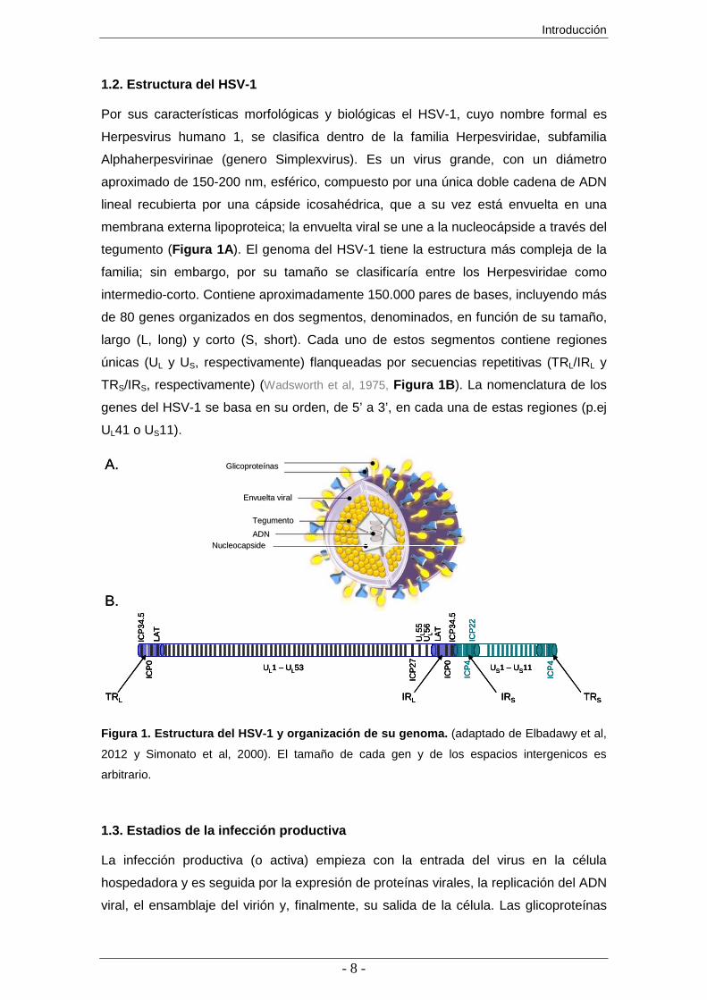
Por sus características morfológicas y biológicas el HSV-1, cuyo nombre formal es
Herpesvirus humano 1, se clasifica dentro de la familia Herpesviridae, subfamilia
Alphaherpesvirinae (genero Simplexvirus). Es un virus grande, con un diámetro
aproximado de 150-200 nm, esférico, compuesto por una única doble cadena de ADN
lineal recubierta por una cápside icosahédrica, que a su vez está envuelta en una
membrana externa lipoproteica; la envuelta viral se une a la nucleocápside a través del
tegumento (Figura 1A ). El genoma del HSV-1 tiene la estructura más compleja de la
familia; sin embargo, por su tamaño se clasificaría entre los Herpesviridae como
intermedio-corto. Contiene aproximadamente 150.000 pares de bases, incluyendo más
de 80 genes organizados en dos segmentos, denominados, en función de su tamaño,
largo (L, long) y corto (S, short). Cada uno de estos segmentos contiene regiones
únicas (UL y US, respectivamente) flanqueadas por secuencias repetitivas (TRL/IRL y
TRS/IRS, respectivamente) (Wadsworth et al, 1975, Figura 1B ). La nomenclatura de los
genes del HSV-1 se basa en su orden, de 5’ a 3’, en cada una de estas regiones (p.ej
UL41 o US11).
Figura 1. Estructura del HSV-1 y organización de su genoma. (adaptado de Elbadawy et al,
2012 y Simonato et al, 2000). El tamaño de cada gen y de los espacios intergenicos es
arbitrario.
1.3. Estadios de la infección productiva
La infección productiva (o activa) empieza con la entrada del virus en la célula
hospedadora y es seguida por la expresión de proteínas virales, la replicación del ADN
viral, el ensamblaje del virión y, finalmente, su salida de la célula. Las glicoproteínas
GlicoproteGlicoproteíínasnas
Envuelta viralEnvuelta viral
TegumentoTegumento
ADNADNNucleocapsideNucleocapside
A.
B.
UL1 – UL53 US1 – US11
ICP
27U
L55
ICP
0
ICP
4IC
P22
LAT
ICP
0IC
P34
.5
ICP
34.5
UL5
6LA
T
ICP
4
TRL IRL IRS TRS
GlicoproteGlicoproteíínasnas
Envuelta viralEnvuelta viral
TegumentoTegumento
ADNADNNucleocapsideNucleocapside
A.
B.
UL1 – UL53 US1 – US11
ICP
27U
L55
ICP
0
ICP
4IC
P22
LAT
ICP
0IC
P34
.5
ICP
34.5
UL5
6LA
T
ICP
4
TRL IRL IRS TRS
UL1 – UL53 US1 – US11
ICP
27U
L55
ICP
0
ICP
4IC
P22
LAT
ICP
0IC
P34
.5
ICP
34.5
UL5
6LA
T
ICP
4
TRL IRL IRS TRS
Introducción
- 9 -
virales B (gB), gC, gD, gH y gL están implicadas, de diferente manera, en la entrada
del virus en la célula hospedadora. Así, gC facilita la adhesión del virus a la membrana
plasmática, siendo este proceso estabilizado por gD y algunas proteínas de membrana
conocidas como receptores para la entrada de Herpesvirus (Hve) (Spear et al, 2000). El
siguiente paso es la fusión de la envuelta viral con la membrana celular, mecanismo
en el que participan gD y gB, así como el complejo gH/gL. Tras la fusión, la
nucleocápside y algunas proteínas del tegumento se liberan en el citoplasma y,
finalmente, la cápside es transportada al núcleo, donde se inicia la transcripción
génica.
La transcripción de los genes del HSV-1 sigue una secuencia temporal, dentro de la
que se distinguen tres grupos de genes: los muy tempranos (IE, immediate early o α),
los tempranos (E, early o β) y los tardíos (L, late o γ). Los primeros genes que se
transcriben (IE) no requieren síntesis previa de proteínas virales, ya que utilizan
mecanismos de transcripción de la propia célula infectada. Sin embargo, algunas
proteínas virales, como la VP16 (codificada por UL47), favorecen esta actividad de los
factores de transcripción celulares ya ensamblados en los promotores de los genes IE
(Taylor et al, 2002). A su vez, las proteínas virales codificadas por los genes IE inician la
transcripción de los genes tempranos y algunos tardíos, llamados tardíos iniciales o
γ1. Los genes tardíos verdaderos o γ2 se transcriben solo después del inicio de la
replicación viral.
El ADN viral linear se circulariza al pasar al núcleo. La duplicación del ADN viral se
inicia a partir de uno de los tres orígenes de replicación del genoma del virus,
generando muchos genomas virales nuevos en cada célula infectada. El ensamblaje
de la nucleocápside tiene lugar también en el núcleo de la célula infectada y requiere
la síntesis de varias proteínas virales tardías. Las cápsides vacías maduras se cargan
con ADN viral recién sintetizado y salen del núcleo hacía el espacio perinuclear,
proceso durante el cual adquieren envuelta viral y varias proteínas del tegumento. El
modelo de salida de los viriones más ampliamente aceptado implica su fusión con la
membrana nuclear externa y la liberación de la nucleocápside al citoplasma, seguida
por la re-adquisición de la envuelta viral en el Golgi y su secreción a través de una vía
de transporte vesicular. Alternativamente, los viriones saldrían del espacio perinuclear
en vesiculas o a través del retículo endoplasmico hacía el Golgi, y serían
transportados fuera de la célula por vías exocíticas o secretorias (Campadelli-Fiume,
2007; Taylor et al, 2002).

Introducción
- 10 -
1.4. La infección latente
Una característica de los Herpesvirus es su capacidad de establecer una infección
latente, que persiste a lo largo de toda la vida del individuo. Esta latencia se define
clásicamente como la retención del genoma completo del virus sin que haya
producción de viriones infectivos. Sin embargo, pese a la falta de una infección
productiva, durante el estado latente hay expresión, si bien limitada, de algunos
transcritos y proteínas virales (Derfuss et al, 2007; van Veltzen et al, 2013). Aunque
todavía se desconoce el mecanismo responsable del mantenimiento del estado latente
en las neuronas, se han realizado avances relevantes para su esclarecimiento.
En este sentido, se ha propuesto que ciertas diferencias entre el sistema
transcripcional de las neuronas y otros tipos celulares con capacidad proliferativa,
como la localización preferentemente citoplasmática del factor de transcripción HCF
(Host Cell Factor), limita su función sobre los genes IE (Preston y Efstathiou, 2007).
Otras teorías se refieren a que la proteína del tegumento VP16 puede que no llegue al
cuerpo neuronal junto con el genoma viral, o que no sea funcional, limitando
igualmente la transcripción de los genes IE (Kristie y Roizman, 1988; Preston y Efstathiou,
2007).
Los RNA virales más abundantes en las neuronas infectadas pertenecen a una familia
de transcritos denominados LAT (Latency Associated Transcripts), codificados en las
regiones repetitivas que flanquean la región UL (Figura 1B ). Aunque no se ha
demostrado expresión de proteínas codificadas por LAT y la presencia de estos
transcritos no es imprescindible para el estado de latencia, se les ha implicado en su
modulación. Así, las observaciones de que una parte de sus secuencias solapan con
la de los genes IE que codifican ICP0 e ICP4 y de que se transcriben en sentido
contrario, han llevado a la hipótesis de que la presencia de LAT bloquea la expresión
de estas proteínas, impidiendo así la infección productiva (Chen et al, 1997). Apoyando
esta hipótesis, varios estudios han demostrado presencia de RNA derivado de estos
genes IE (pero no de proteínas) en neuronas procedentes de ganglios trigeminales
humanos y murinos (Derfuss et al, 2007; Kramer et al, 1995; Theil et al, 2003). Una teoría
alternativa sobre el papel de los LAT en el mantenimiento de la latencia es su
capacidad de promover la supervivencia de las neuronas infectadas limitando su
apoptosis (Perng et al, 2000).
La hipótesis mas ampliamente aceptada sobre la reactivación del virus implica que los
promotores virales y, en particular, el de la ICP0, no están completamente bloqueados
sino inactivos. Estos promotores podrían activarse en respuesta a ciertos estímulos,
desencadenando así las reactivaciones (Preston y Efstathiou, 2007). Alternativamente,

Introducción
- 11 -
se ha propuesto un modelo dinámico de reactivación, en el que existe una producción
de viriones infectivos continua pero a bajos niveles, controlada estrechamente por el
sistema inmunitario del hospedador y acompañada solo esporádicamente de
sintomatología clínica (Wald et al, 1997). La liberación sorprendentemente frecuente de
viriones infectivos en las secreciones de sujetos asintomáticos apoya está ultima
hipótesis.
2. LA RESPUESTA INMUNITARIA FRENTE A HSV-1
El hecho de que la inmunosupresión, farmacológica o patológica, aumente el riesgo de
sufrir cuadros herpéticos de mayor trascendencia apoya el papel clave de la respuesta
inmunitaria en el control del HSV-1, tanto durante la primoinfección como en las
reactivaciones. Al resultado de la interacción entre el virus y su hospedador
contribuyen factores genéticos y ambientales, manifiestándose como susceptibilidad o
resistencia a la enfermedad. Tanto las respuestas inmunitarias innatas como las
adaptativas convergen para controlar la infección inicial y sus reactivaciones. Variantes
que modifiquen la intensidad de cualquiera de estos mecanismos podrían influir en la
probabilidad de sufrir una infección clínicamente manifiesta o asintomática.
2.1. Producción de citoquinas en respuesta a patrones moleculares virales
El desencadenante de cualquier respuesta inmunitaria frente a un microorganismo es
el reconocimiento de su presencia. Para ello, el hospedador dispone de varios tipos de
receptores expresados en diferentes estirpes celulares. Entre los sensores de la
inmunidad innata están varias familias de receptores de reconocimiento de patrones
(PRR), que se unen a motivos constitutivos y conservados de diferentes patógenos
(PAMPs) desencadenando respuestas tempranas en presencia de microorganismos.
La expresión de los diferentes PRRs en distintos compartimentos celulares y, por lo
general, en más de un subtipo celular, junto con la presencia de más de un PAMP en
la mayoría de los microorganismos, permiten una respuesta adecuada y coordinada
frente a virtualmente cualquier tipo de patógeno. Su activación da lugar a la producción
de citoquinas proinflamatorias, el reclutamiento de otras células implicadas en la
respuesta inmunitaria innata y, finalmente, la activación de una respuesta adaptativa y
el control de la infección.
Se han caracterizado interacciones entre varias estructuras del HSV-1 y diferentes
PRR. Así, el receptor de membrana TLR2 (Toll-like Receptor) reconoce la
glicoproteina viral gB y el complejo gH/gL (Cai et al, 2012; Leoni et al, 2012) y los
receptores endosomales TLR3 y TLR9 se activan en respuesta a dsRNA derivado de

Introducción
- 12 -
HSV-1 y ADN no metilado con islas CpG, respectivamente (Rasmussen et al, 2007;
Zhang et al, 2007). Además, varios sensores de ADN citosolicos, entre ellos RIG-1
(Retinoic acid-inducible gene 1) y MDA-5 (Melanoma Differentiation-Associated protein
5), reconocen también estructuras intracelulares derivadas de HSV-1 (Melchjorsen et al,
2010; Paludan et al, 2013; Rasmussen et al, 2009; Unterholzner et al, 2013).
Las vías de activación celular de los PRRs que reconocen estructuras del HSV-1
convergen en respuestas proinflamatorias, sobre todo la producción de interferones
(IFN). Los IFNs de tipo I y III (IFN-α/β e IFN-λ, respectivamente) son producidos en las
primeras horas después de la infección por una amplia gama de células, incluyendo
las células epiteliales infectadas y las células dendríticas. El IFN-γ, en cambio, se
secreta en un paso posterior de la respuesta inmunitaria, principalmente por los
linfocitos NK y T (Durbin et al, 2013). Los interferones, ampliamente aceptados como
primera línea de defensa frente a las infecciones virales, bloquean la replicación viral y
controlan la transcripción de varios genes en la célula hospedadora, generando un
estado de represión global de la síntesis de proteínas. Además, promueven la
producción de otras citoquinas y potencian la presentación antigénica, modulando así
la respuesta inmunitaria adaptativa frente al HSV-1 (Pollara et al, 2004). Especialmente
relevante es la potenciación de la producción de la IL-15, puesto que, a su vez, esta
citoquina estimula la proliferación y citotoxicidad de las células NK y de los linfocitos T
CD8+, efectores celulares clave en el control de las infecciones virales (Mattei et al,
2001; Musso et al, 1999; Ogasawara et al, 1998).
Asimismo, la producción de otras citoquinas (y quimioquinas) proinflamatorias tras la
activación de los PRRs parece ser igualmente importante en el control de la infección
herpética. Por ejemplo, el factor de necrosis tumoral (TNF)-α, producido principalmente
por monocitos, macrófagos, linfocitos T y células NK, actúa de manera sinérgica con el
IFN-γ y muestra efectos tanto protectores como inmunopatogénicos en diferentes
modelos de infección por HSV (Aravalli et al, 2005; Bryant-Hudson et al, 2014; Ghiasi et al,
1995; Keadle et al, 2000; Kodukula et al, 1999; Lundberg et al, 2007; Sergerie et al, 2007).
Mutaciones que predisponen al desarrollo de la encefalitis herpética (HSE) confirman
el papel clave de los IFNs y del reconocimiento de PAMPs en la defensa frente a HSV-
1. Varias mutaciones autosómicas dominantes y recesivas en genes que codifican
proteínas de la vía de activación TLR3 – IFN-I predisponen a sufrir cuadros aislados
de HSE. En concreto, se han descrito más de 10 casos de pacientes con mutaciones
en el propio TLR3, en UNC93B1 y en los genes que codifican TRAF3 (TNF receptor
associated factor 3), TRIF/TICAM1 (TIR-domain-containing adapter-inducing
interferon-β) y TBK1 (TANK-binding kinase 1, una proteína del complejo NF-κB),

Introducción
- 13 -
siendo todas ellas proteínas que participan en la señalización a través de TLR3, TLR7
y TLR9 (Casrouge et al, 2006; Guo et al, 2011; Herman et al, 2012; Perez de Diego et al,
2011; Sancho-Shimizu et al, 2011a; Zhang et al, 2007). Además, una minoría de pacientes
con otras inmunodeficiencias, como las causadas por mutaciones en NEMO (NF-κB
Essential Modulator) y STAT1 (Signal Transducer and Activator of Transcription 1),
que afectan a la señalización a través de NF-κB y a la respuesta a IFN-I, también
desarrolla HSE (Bustamante et al, 2008; Sancho-Shimizu et al, 2011b). Por tanto, la vía de
activación TLR3 – IFN-I en respuesta a HSV-1 parece fundamental para la protección
frente la primoinfección, al menos en el sistema nervioso central. Queda por demostrar
en qué medida el reconocimiento del HSV-1 a través de TLR3 y otros PRRs también
condicionaría las recurrencias.
2.2. Moléculas polimórficas en la interfaz entre la respuesta inmunitaria innata e
adaptativa
2.2.1. El Complejo Principal de Histocompatibilidad
Características estructurales de las moléculas HLA
El Complejo Principal de Histocompatibilidad (MHC) fue descubierto inicialmente en el
ratón, por su papel en la compatibilidad tisular entre individuos de la misma especie
(Snell, 1953). Las moléculas del MHC humanas (human leucocyte antigens, HLA) son
glicoproteínas de membrana especializadas en la presentación de péptidos
antigénicos a los linfocitos T. En función de sus características estructurales y
funcionales, se distinguen dos grandes clases, denominadas I y II. Las moléculas HLA
de clase I clásicas, que se expresan en prácticamente todas las células nucleadas,
presentan péptidos derivados de proteínas intracelulares a los linfocitos T CD8+,
mientras que las moléculas HLA de clase II se expresan casi exclusivamente en
células presentadoras de antígenos (APC) y son capaces de presentar péptidos
derivados de antígenos de origen extracelular a los linfocitos T CD4+ (Neefjes et al,
2011). Ademas de las moleculas HLA de clase I clásicas, o de clase Ia, se han
identificado varias otras no-clásicas, con características estructurales similares, pero
que difieren de las primeras en cuanto a su reducida variabilidad genética, su
distribución tisular y su función, en algunos casos todavía desconocida.
Las moléculas HLA de clase I clásicas están formadas por una glicoproteína integral
de membrana (cadena pesada o cadena α) unida de manera no covalente a la β2-
microglobulina (cadena ligera o cadena β). La cadena pesada presenta tres dominios
extracelulares globulares (α1, α2 y α3), un fragmento transmembrana y una región
intracitoplasmática corta. Una estructura similar se observa en las moléculas HLA de

Introducción
- 14 -
clase II, aunque estas están formadas por dos cadenas polipeptídicas transmembrana
de tamaño similar, α y β, cada una con dos dominios extracelulares globulares (α1, α2,
β1 y β2). Mientras que los dominios más próximos a la membrana de cada
heterodímero HLA son esencialmente invariantes y adoptan una conformación de tipo
dominio de inmunoglobulina, los dominios externos albergan gran parte del
polimorfismo de estas moléculas y tienen una conformación más compleja, que genera
el sitio de unión de péptidos antigénicos cuya base está formada por láminas β y los
bordes externos por helices α (Figura 2 ). Pequeñas diferencias estructurales entre las
moléculas HLA de clase I y II condicionan el tamaño de los fragmentos peptídicos que
pueden alojar; así, el sitio de unión de péptidos formado por los dominios α1 y α2 de la
cadena pesada de las moléculas HLA de clase I acomoda fragmentos peptídicos
cortos, de 8-11 aminoácidos, mientras que el de las moléculas HLA de clase II,
constituido por los dominios α1 y β1, permite la unión de péptidos de tamaño mayor y
algo más variable, habitualmente de 13-25 aminoácidos.
Figura 2. Estructura de las moléculas HLA de clase I y clase II.
A, B visión lateral del fragmento extracelular de moléculas HLA de clase I (A, HLA-A2, pdb
1B0R) y II (B, HLA-DR4, pdb 1D5Z). C, D visión superior del sitio de unión de péptidos
antigénicos de una molécula HLA de clase I (C) y II (D).

Introducción
- 15 -
Procesamiento y presentación antigénica
Los péptidos presentados por las moléculas HLA se generan en diferentes
compartimentos intracelulares. Las proteínas presentes en el citoplasma de una célula
(ya sean de origen propio o extraño) se degradan de forma controlada en el
proteasoma. Los péptidos generados son a continuación transportados activamente a
la luz del retículo endoplásmico a través del transportador de péptidos antigénicos
(TAP). Aquí, las moléculas HLA de clase I previamente ensambladas se cargan con
péptidos y maduran su estado conformacional, siendo transportadas vía Golgi hacía la
membrana plasmática. El correcto ensamblaje y la carga con péptidos de las
moléculas HLA de clase I dependen de un complejo multiproteico que incluye TAP,
tapasina y otras chaperonas. La tapasina facilita la co-localización de las moléculas
HLA vacías junto al TAP, estabiliza la conformación libre de péptidos de las moléculas
HLA y optimiza su repertorio de péptidos, favoreciendo a aquellos de alta afinidad
(Chen et al, 2007; Williams et al, 2002). Existe, sin embargo, un mecanismo alternativo de
presentación de péptidos antigénicos independiente de la tapasina, pero este proceso
es subóptimo y restringido a un número limitado de variantes alélicas de las moléculas
HLA de clase I (Rizvi et al, 2014; Thammavongsa et al, 2006; Williams et al, 2002).
La presentación antigénica de péptidos derivados de proteínas extracelulares
(procedentes de microorganismos extracelulares, partículas virales u otras proteínas
antigénicas solubles) por parte de moléculas HLA de clase II requiere su transporte
hacía el interior de la célula mediante fagocitosis o endocitosis. Estos procesos
generan vesiculas intracelulares en las que, tras fundirse con los lisosomas, las
proteínas internalizadas son degradadas mediante procesos enzimáticos. Las
moléculas HLA de clase II que han sido transportadas a través del reticulo
endoplasmico y el Golgi a estas vesiculas se cargan con los péptidos resultantes
mediante un proceso muy complejo tras en cual son finalmente transportadas hacia la
membrana plasmática.
Polimorfismo genético del MHC
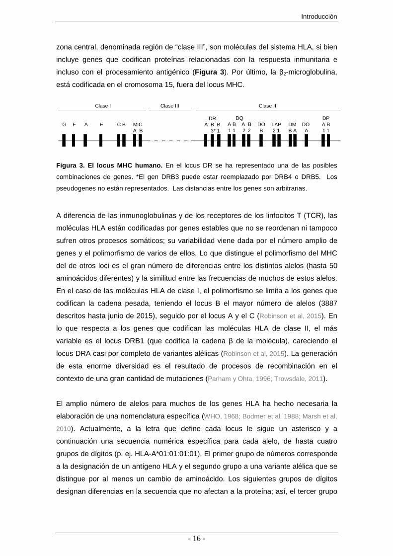
El MHC en humanos está localizado en el brazo corto del cromosoma 6 (6p21.3) y es
el locus más polimórfico y con mayor densidad de genes del genoma. El MHC abarca
~4 Mpb y está organizado en tres regiones. En la región telomérica se sitúan los genes
que codifican las distintas cadenas pesadas de las moléculas HLA de clase I, mientras
que en la zona más próxima al centrómero están los genes que codifican las cadenas
α y β de las moléculas HLA de clase II junto con varios otros genes implicados en el
procesamiento y presentación antigénica. Ninguno de los productos génicos de la
Introducción
- 16 -
zona central, denominada región de “clase III”, son moléculas del sistema HLA, si bien
incluye genes que codifican proteínas relacionadas con la respuesta inmunitaria e
incluso con el procesamiento antigénico (Figura 3 ). Por último, la β2-microglobulina,
está codificada en el cromosoma 15, fuera del locus MHC.
Figura 3. El locus MHC humano. En el locus DR se ha representado una de las posibles
combinaciones de genes. *El gen DRB3 puede estar reemplazado por DRB4 o DRB5. Los
pseudogenes no están representados. Las distancias entre los genes son arbitrarias.
A diferencia de las inmunoglobulinas y de los receptores de los linfocitos T (TCR), las
moléculas HLA están codificadas por genes estables que no se reordenan ni tampoco
sufren otros procesos somáticos; su variabilidad viene dada por el número amplio de
genes y el polimorfismo de varios de ellos. Lo que distingue el polimorfismo del MHC
del de otros loci es el gran número de diferencias entre los distintos alelos (hasta 50
aminoácidos diferentes) y la similitud entre las frecuencias de muchos de estos alelos.
En el caso de las moléculas HLA de clase I, el polimorfismo se limita a los genes que
codifican la cadena pesada, teniendo el locus B el mayor número de alelos (3887
descritos hasta junio de 2015), seguido por el locus A y el C (Robinson et al, 2015). En
lo que respecta a los genes que codifican las moléculas HLA de clase II, el más
variable es el locus DRB1 (que codifica la cadena β de la molécula), careciendo el
locus DRA casi por completo de variantes alélicas (Robinson et al, 2015). La generación
de esta enorme diversidad es el resultado de procesos de recombinación en el
contexto de una gran cantidad de mutaciones (Parham y Ohta, 1996; Trowsdale, 2011).
El amplio número de alelos para muchos de los genes HLA ha hecho necesaria la
elaboración de una nomenclatura específica (WHO, 1968; Bodmer et al, 1988; Marsh et al,
2010). Actualmente, a la letra que define cada locus le sigue un asterisco y a
continuación una secuencia numérica específica para cada alelo, de hasta cuatro
grupos de dígitos (p. ej. HLA-A*01:01:01:01). El primer grupo de números corresponde
a la designación de un antígeno HLA y el segundo grupo a una variante alélica que se
distingue por al menos un cambio de aminoácido. Los siguientes grupos de dígitos
designan diferencias en la secuencia que no afectan a la proteína; así, el tercer grupo
G
Clase I Clase III Clase II
F A E C B MICA B
DRA B B
3* 1
DQA B A B1 1 2 2
TAP2 1
DMB A
DOA
DPA B1 1
DOB
G
Clase I Clase III Clase II
F A E C B MICA B
DRA B B
3* 1
DQA B A B1 1 2 2
TAP2 1
DMB A
DOA
DPA B1 1
DOB
Introducción
- 17 -
designa sustituciones sinónimas, mientras que el cuarto abarca polimorfismos en la
secuencia de las regiones no codificantes.
Como se ha mencionado previamente, el polimorfismo HLA se concentra en los
dominios que interaccionan con los péptidos y el TCR, determinando las secuencias
de los péptidos que pueden alojar. Análisis exhaustivos de las secuencias peptídicas
que albergan moléculas HLA concretas han establecido que solamente algunos de sus
residuos son iguales (o muy similares). Así, en el caso de las moléculas HLA de clase
I, el segundo y el noveno aminoácidos de los péptidos que se unen a un determinado
alelo son muy parecidos y se acomodan en unos bolsillos generados en el sitio de
unión de antígenos (Falk et al, 1991). De esta manera, cada molécula HLA puede
presentar un gran abanico de péptidos, que, sin embargo, no es ilimitado. Además, el
procesamiento de las proteínas para la generación de péptidos antigénicos depende
de otros factores como la posibilidad de que se produzca un corte enzimático o la
probabilidad de paso a través del TAP en el caso de las moléculas HLA de clase I.
Todos estos factores contribuyen a la generación de un repertorio único, más o menos
amplio, de péptidos antigénicos para cada una de las moléculas HLA de un individuo,
ofreciendo una gran diversidad de antígenos para una respuesta T adecuada.
El papel del MHC en la salud humana
La enorme variabilidad del sistema HLA y sus particularidades ya mencionadas indican
que su evolución ha estado condicionada por fuertes presiones de selección. Desde
mediados del siglo XX, se ha postulado un papel clave de los patógenos en la
generación y mantenimiento de este gran polimorfismo genético (Haldane, 1949), y,
aunque apoyada por escasas evidencias experimentales, esta teoría sigue siendo la
más ampliamente aceptada. Así, diferentes procesos selectivos favorecerían la
supervivencia de individuos heterocigotos (sobredominancia), ya que estos tendrían
más posibilidades de presentar péptidos antigénicos derivados del agente patógeno
causante de una enfermedad. De manera similar, variantes alélicas raras, que
confieren protección a un cierto patógeno, aumentarían su frecuencia por un proceso
de selección positiva (Parham y Ohta, 1996).
Por tanto, no es sorprendente que la presencia de ciertas moléculas HLA se haya
relacionado con la susceptibilidad a varias enfermedades infecciosas. De ellas, una
asociación bien documentada es la infección por HIV, en la que se han establecido
papeles fundamentales en el progreso de la infección para ciertos aminoácidos del
sitio de unión de péptidos de las moléculas HLA-B, los niveles de expresión de HLA-C

Introducción
- 18 -
y la dependencia de la tapasina para la carga peptídica de las moléculas HLA de clase
I (Carrington, 2012; Rizvi et al, 2014).
La contribución de la diversidad genética del sistema MHC a la salud humana no se
limita al control de las enfermedades infecciosas. De hecho, el locus MHC es la región
del genoma humano más frecuentemente asociada a la susceptibilidad a enfermedad
(Tiwari y Terasaki, 1985). Más de cien enfermedades han sido asociadas al MHC, con
un rango de significación estadistica muy amplio, incluidas ciertas neoplasias,
enfermedades neurológicas como la narcolepsia o la esquizofrenia, pero sobre todo
muchas enfermedades autoinmunes (Trowsdale, 2011). La asociación del MHC con el
desarrollo de las enfermedades autoinmunes podría explicarse por la presentación
preferente de ciertos péptidos propios por moléculas HLA concretas. Esta pérdida de
la tolerancia frente a lo propio podría guardar relación con modificaciónes
postraduccionales de ciertos epítopos, o con el fenómeno de mimetismo molecular
(Caillat-Zucman, 2008).
2.2.2. Receptores de células NK para HLA de clase I
Las células NK son linfocitos con capacidad citolítica y secretora de citoquinas con un
papel clave en el control de las infecciones virales y los procesos neoplásicos.
Reconocen principalmente patrones de expresión molecular alterados por la presencia
de patógenos, de los que un ejemplo es la expresión anormalmente baja de moléculas
HLA de clase I (Lanier, 2005). Los receptores implicados en el reconocimiento de las
moléculas HLA pertenecen a las familias de receptores KIR (Killer-cell Ig-like
Receptors), LILR (Leukocyte Immunoglobulin-Like Receptor family, en concreto
LILRB1) y CD94/NKG2, e incluyen tanto receptores inhibidores como activadores.
Mientras que el papel de los receptores inhibidores para HLA se explicaría por la
hipótesis del “missing self”, según la cual la falta de su ligando desencadena una
respuesta citotóxica de las células NK que los expresa (Kärre, 1986), el papel de los
receptores activadores para HLA siguie siendo un tema de investigación abierto.
La expresión de estos receptores en las células NK es clonal, y esta expresión
variegada contribuye al proceso de maduración de las células NK conocido como
educación. Según los diferentes modelos propuestos para explicar la educación de las
células NK, solamente los clones de células NK que reconocen HLA propio a través de
receptores inhibidores serán funcionales, y su nivel de activación dependerá de la
señal neta recibida o, en otras palabras, del numero de receptores para MHC que
expresa y de la intensidad de su interacción con sus ligandos (Anfossi et al, 2006; Brodin
et al, 2009; Fauriat et al, 2010; Joncker et al, 2009; Kim et al, 2005). En analogía con el
Introducción
- 19 -
desarrollo de los linfocitos T, la educación a través de receptores para MHC
inhibidores sería comparable con la selección positiva, y la ejercida por los receptores
activadores, con la selección negativa, siendo una diferencia fundamental el hecho de
que en el caso de las células NK, esta selección no implica la muerte o la
supervivencia de los diferentes clones sino que afecta a su funcionalidad (es decir,
condiciona la activación o la anergia de estos clones en presencia de células que
expresan sus ligandos).
La expresión de estos receptores para HLA de clase I no está restringida a las células
NK. Una pequeña subpoblación de linfocitos T, sobre todo CD8+, expresa también
este tipo de receptores y, si bien su contribución al control de la activación de estas
células no se conoce en detalle, se les atribuye igualmente un papel modulador de su
actividad citolítica (TCR-dependiente) en presencia de moléculas propias (van Bergen y
Koning, 2010; Vivier y Anfossi, 2004).
2.2.2.1. Los receptores KIR (Killer-cell Ig-like Receptors)
Estructura de los KIR
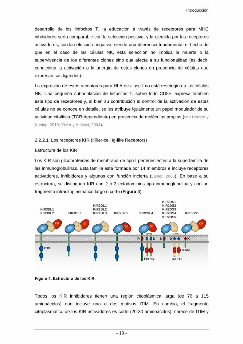
Los KIR son glicoproteínas de membrana de tipo I pertenecientes a la superfamilia de
las inmunoglobulinas. Esta familia está formada por 14 miembros e incluye receptores
activadores, inhibidores y algunos con función incierta (Lanier, 2005). En base a su
estructura, se distinguen KIR con 2 o 3 ectodominios tipo inmunoglobulina y con un
fragmento intracitoplasmático largo o corto (Figura 4 ).
Figura 4. Estructura de los KIR.
Todos los KIR inhibidores tienen una región citoplásmica larga (de 76 a 115
aminoácidos) que incluye uno o dos motivos ITIM. En cambio, el fragmento
citoplasmático de los KIR activadores es corto (20-30 aminoácidos), carece de ITIM y
D0 D0
D1
ITIM
KIR2DS1 KIR2DL1 KIR2DS2 KIR3DL1 KIR2DL2 KIR2DS3 KIR3DL2 KIR3DL3 KIR2DL3 KIR2DL5 KIR2DL4 KIR2DS4 KIR3DS1 KIR2DS5
D0 D1 D1
D2 D2 D0
D2
+
D2
D2
D1 D2
D0 D1
D2
+
ITAM
K – –
DAP12
+ D – – +
FcεRIγ
– D + K R + – – +

Introducción
- 20 -
su región transmembrana contiene un residuo básico de lisina que les permite unirse a
la molécula adaptadora DAP12. Hay además dos excepciones a esta regla; el receptor
KIR3DL3 tiene un fragmento citoplasmático de longitud intermedia y un solo ITIM
(Torkar et al, 1998), y KIR2DL4 tiene un fragmento intracelular con un ITIM y un residuo
basico de arginina en su región transmembrana, que le permitiría transmitir señales
intercelulares tanto activadoras como inhibidoras (Rajagopalan y Long, 1999).
La nomenclatura de los KIR se rige por las diferencias estructurales (Marsh et al, 2003);
así, el número que sigue al acrónimo KIR se corresponde con el de los dominios tipo
inmunoglobulina, y es seguido por la letra “D”, de dominio. La siguiente letra se refiere
a la longitud del fragmento intracelular (L, largo, o S, corto, de short) y el último
número se corresponde al número del gen que codifica un KIR con esta estructura.
Siguiendo estas reglas, por ejemplo, KIR2DS1 y KIR2DS5 son KIR activadores con
dos dominios tipo inmunoglobulina y cola citoplasmática corta, mientras que KIR3DL2
es un receptor inhibidor con tres dominios tipo inmunoglobulina y región citoplasmática
larga.
El complejo KIR
El complejo de genes KIR en humanos está localizado en el locus LRC (Leukocyte
Receptor Complex), en el cromosoma 19 (19q13.4, Wilson et al, 2000). El complejo KIR
incluye combinaciones de 15 genes y dos pseudogenes (KIR2DP1 y KIR3DP1)
dispuestos en tándem (Vilches y Parham, 2002). Además, fuera del complejo KIR, hacía
5’ en el LRC se encuentra otro pseudogén, KIR3DX1 (Sambrook et al, 2006). Una
característica importante de esta región del genoma es la gran variabilidad del número
de copias de cada gen KIR; así, con mucha frecuencia se encuentran individuos sanos
cuyo genoma incluye genes para solamente siete KIR y otros con genes KIR que
codifican hasta 15 receptores diferentes. Además, en un mismo cromosoma puede
haber cero, uno, dos o incluso más genes KIR que codifiquen el mismo receptor. Este
fenómeno se debe a una serie de recombinaciones, duplicaciones y deleciones que ha
sufrido este locus durante la evolución, procesos que han sido favorecidos por la alta
similitud entre las secuencias de los distintos genes KIR, su orientación idéntica y las
cortas regiones intergenicas (Pyo et al, 2010).
Los genes KIR, de manera similar a los genes del complejo MHC, se heredan en
bloques o haplotipos. Un atributo muy característico de los haplotipos KIR es la
variabilidad del contenido génico. Salvo cuatro genes conservados en casi todos los
individuos, la frecuencia del resto varia considerablemente en la población. De esta
manera, en cada haplotipo KIR hay genes que están casi siempre presentes,
Introducción
- 21 -
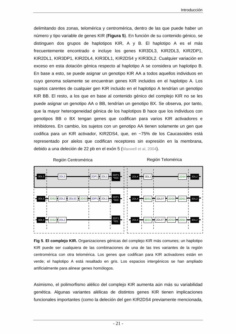
delimitando dos zonas, telomérica y centromérica, dentro de las que puede haber un
número y tipo variable de genes KIR (Figura 5 ). En función de su contenido génico, se
distinguen dos grupos de haplotipos KIR, A y B. El haplotipo A es el más
frecuentemente encontrado e incluye los genes KIR3DL3, KIR2DL3, KIR2DP1,
KIR2DL1, KIR3DP1, KIR2DL4, KIR3DL1, KIR2DS4 y KIR3DL2. Cualquier variación en
exceso en esta dotación génica respecto al haplotipo A se considera un haplotipo B.
En base a esto, se puede asignar un genotipo KIR AA a todos aquellos individuos en
cuyo genoma solamente se encuentran genes KIR incluidos en el haplotipo A. Los
sujetos carentes de cualquier gen KIR incluido en el haplotipo A tendrían un genotipo
KIR BB. El resto, a los que en base al contenido génico del complejo KIR no se les
puede asignar un genotipo AA o BB, tendrían un genotipo BX. Se observa, por tanto,
que la mayor heterogeneidad génica de los haplotipos B hace que los individuos con
genotipos BB o BX tengan genes que codifican para varios KIR activadores e
inhibidores. En cambio, los sujetos con un genotipo AA tienen solamente un gen que
codifica para un KIR activador, KIR2DS4, que, en ~75% de los Caucasoides está
representado por alelos que codifican receptores sin expresión en la membrana,
debido a una deleción de 22 pb en el exón 5 (Maxwell et al, 2004).
Fig 5 . El complejo KIR. Organizaciones génicas del complejo KIR más comunes; un haplotipo
KIR puede ser cualquiera de las combinaciones de una de las tres variantes de la región
centromérica con otra telomérica. Los genes que codifican para KIR activadores están en
verde; el haplotipo A está resaltado en gris. Los espacios intergénicos se han ampliado
artificialmente para alinear genes homólogos.
Asimismo, el polimorfismo alélico del complejo KIR aumenta aún más su variabilidad
genética. Algunas variantes alélicas de distintos genes KIR tienen implicaciones
funcionales importantes (como la deleción del gen KIR2DS4 previamente mencionada,
2DL3 2DP1 2DL1 3DL3
3DP1 *003
2DS2 2DL2 2DL5C 2DS3 2DP1 2DL1 3DL3 3DP1 *003
2DS2 2DL2 3DL3 3DP1 *001
3DL1 2DS4 2DL4 3DL2
3DS1 2DS5 2DS1 2DL5T 2DL4 3DL2
2DL4 3DL2 3DS1 2DS3 2DS1 2DL5T
Región Centromérica Región Telomérica

Introducción
- 22 -
mutaciones que impiden su expresión o variaciones que modifican la afinidad del
receptor para su ligando) pero el significado funcional de la mayoría de estos
polimorfismos es desconocido.
Ligandos y patrón de expresión de los KIR
El contenido génico variable del complejo KIR y el polimorfismo alélico condicionan el
repertorio KIR de cada individuo. Pero además, mecanismos sólo parcialmente
esclarecidos hacen que los diferentes clones de células NK expresen solo parte de los
KIR de su genoma (Valiante et al, 1997). Esta expresión variegada está controlada
epigenéticamente y determina la presencia de un amplio número de clones NK que
expresan KIR diferentes que, por tanto, tienen una capacidad diferente para responder
a diferentes estímulos y controlar individualmente la expresión de distintas moléculas
HLA (Uhrberg, 2005). En este contexto, infecciones o procesos tumorales que bajan la
expresión de moléculas HLA como mecanismo de protección frente a la respuesta
inmunitaria mediada por linfocitos T, activarían a las células NK con receptores KIR
inhibidores para estas moléculas HLA propias (según la hipótesis de missing self, o
falta de reconocimiento de lo propio). La menor afinidad de los receptores activadores
para sus ligandos HLA, comparada con los inhibidores que reconocen los mismos
ligandos contribuyen a que clones de células NK que expresen estos receptores
activadores no generen respuestas descontroladas frente a lo propio.
Durante las últimas décadas se han ido identificando ligandos de varios KIR, que, en
su mayoría, son moléculas HLA y este reconocimiento es, en general, alotípico. De
ellos, las mejor caracterizadas son las interacciones de KIR inhibidores con moléculas
HLA-C y HLA-B (Vilches y Parham, 2002). Los alelos HLA-C con lisina en la posición 80
de la cadena α (epítopo HLA-C2) son reconocidos por KIR2DL1 y, con menor afinidad,
por KIR2DS1. En cambio, KIR2DL2 y KIR2DL3 reconocen, con diferente afinidad,
alelos HLA-C con asparagina en la posición 80 (epítopo HLA-C1) aunque trabajos más
recientes demustran que esta preferencia no es absoluta, ya que estos receptores
reconocen también algunos alelos HLA-C2 (David et al, 2013; Moesta et al, 2008; Pende et
al, 2009; Schonberg et al, 2011; Winter et al, 1998). Los primeros estudios no encontraron
afinidad alguna de KIR2DS2 (el homólogo activador de KIR2DL2) para HLA-C (Moesta
et al, 2010; Vales-Gómez et al, 1998; Winter et al, 1998), pero otros observan que KIR2DS2
se une (con baja afinidad) a moléculas HLA-C1 y esta intertacción es capaz de activar
a las células NK KIR2DS2+KIR2DL2– (David et al, 2013; Stewart et al, 2005).
Inesperadamente, una publicación reciente encuentra que KIR2DS2 se une también a
HLA-A*11 (Liu et al, 2014).

Introducción
- 23 -
KIR3DL1 reconoce alotipos HLA-A y HLA-B con el epítopo Bw4 (LRI/TXLR en los
residuos 78-83 de la cadena pesada) (Gumperz et al, 1997); y KIR3DL2 se une a HLA-
A*03 y HLA-A*11 (Pende et al, 1996). Un estudio mas reciente demuestra que KIR2DS4
reconoce algunos alelos HLA-A y HLA-C (Graef et al, 2009). Si bien existen indicios de
que KIR3DS1 podría interaccionar con alotipos HLA-B con isoleucina en la posición 80
de la cadena α, estas interacciones no han sido todavía demostradas formalmente
(Alter et al, 2007; Carr et al, 2007; Martin et al, 2002). Por último, varios estudios apoyan
que KIR2DL4 reconoce moléculas HLA-G (Ponte et al, 1999; Rajagopalan et al, 1999).
Más recientemente se ha descrito el reconocimiento de proteínas diferentes de las
moléculas HLA por varios KIR. En concreto, KIR3DL2 (y en menor medida KIR3DL1,
KIR3DS1 y KIR2DL4) puede unirse a patrones moleculares derivados de patógenos
(PAMPs) como los oligonucleótidos CpG e internalizarlos para su reconocimiento por
TLR9 en los endosomas (Sivori et al, 2010). Además, KIR2DL4 puede unirse a
moléculas de heparán sulfato, interacción que podría tener un efecto modulador de la
disponibilidad del receptor para unirse a ligandos proteicos (Brusilovsky et al, 2013 y
2014). Ambas interacciones podrían guardar relación con la alta carga positiva del
dominio D0 de estos KIR, que ha sido directamente implicado en la unión de
oligonucleótidos CpG y de heparán sulfato (Brusilovsky et al, 2013; Sivori et al, 2010).
Los KIR en la salud humana
Los KIR controlan el nivel de activación de las células NK en diferentes escenarios. La
educación de las células NK, la variabilidad genética extrema de los KIR y de sus
ligandos y la expresión variagada de los KIR generan un sistema muy complejo de
regulación. El papel de este complejo sistema en la salud humana y sus posibles
implicaciones en el control de varios procesos patológicos representa todavía un tema
abierto de investigación.
Por ahora, la mayor parte de los datos disponibles proviene de estudios geneticos. Así,
la presencia o ausencia de algunos KIR y de sus ligandos en el genoma han sido
relacionadas con el control o el desarrollo de una serie de enfermedades infecciosas,
autoinmunes y neoplásicas, así como con complicaciones en la reproducción humana
(Khakoo y Carrington, 2006; Parham, 2005). En particular, el papel de los KIR en varias
infecciones virales ha sido ampliamente estudiado, sobre todo a nivel genético.
La presencia de KIR3DS1 y KIR3DL1 en el genoma, junto con alelos HLA con el
epitopo Bw4 con Ile en la posición 80 (Bw480I), se ha relacionado con un curso clínico
de la infeccion por HIV favorable (Boulet et al, 2000; Martin et al, 2002; Martin et al, 2007).
El efecto protector de KIR3DL1 se explicaría por su papel en la educación de las

Introducción
- 24 -
células NK: la presencia de alelos KIR3DL1 de alta expresión junto con su ligando
permitiría el desarrollo de una amplia serie de clones de células NK con capacidad
citolitica y altamente sensibles a pequeñas variaciones en la expresión de sus ligandos
HLA inducidas por el patógeno (Carrington y Alter, 2012). En cambio, el efecto protector
de KIR3DS1 junto con Bw480I frente a la infeccion por HIV implicaría un mejor
aclaramiento de las células infectadas mediado por las células NK KIR3DS1+ (Alter et
al, 2007). Adicionalmente, la identificación de variantes alélicas de HLA-C con mayor
expresión en la membrana que se correlacionan con la protección frente a la infección
por HIV ha generado hipótesis que implican a los KIR2D que reconocen HLA-C en el
control de la infección (Carrington y Alter, 2012). Por ultimo, recientemente se ha
relacionado a la presencia del alelo KIR2DS4 funcional en el genoma con la
patogenicidad de la infeccion crónica por HIV (Merino et al, 2014).
En la infeccion por el virus de la hepatitis C, algunos estudios han encontrado
asociaciones significativas entre la presencia de KIR2DS3 en el genoma y una mayor
probabilidad de desarrollar una infeccion crónica (Dring et al, 2011; de Vasconcelos et al,
2013), mientras que otro estudio más reciente relaciona la presencia en el genoma de
KIR2DS3 (en ausencia de KIR2DS5) con bajos niveles de viremia (Kuśnierczyk et al,
2015). La falta de un ligando conocido para KIR2DS3 dificulta la formulación de
hipótesis que expliquen estos resultados.
Los KIR activadores podrían estar implicados también en el control de la infección por
el citomegalovirus humano (hCMV). Así, la presencia en el genoma de haplotipos KIR
B ha sido asociada al control de la infección (Cook et al, 2006; Di Bona et al, 2014; Stern et
al, 2011) y en algunos individuos hCMV+ se ha observado una expansión de clones de
células NK que expresan KIR activadores (Beziat et al, 2013; Della Chiesa et al, 2014).
En la infección por HSV-1, la presencia de KIR2DL2 y KIR2DS2 en el genoma ha sido
relacionada con el desarrollo de recurrencias (Estefania et al, 2007), resultados que nos
hemos propuesto replicar en el trabajo actual.
2.2.2.2. Los heterodímeros CD94/NKG2
Estructura y función
La familia de receptores NKG2 incluye varias glicoproteínas integrales de membrana
de tipo II, pertenecientes a la superfamilia de las lectinas de tipo C: NKG2A/B, -C, -D, -
E/H y -F. Su expresión está restringida a las células NK y algunos linfocitos T y,
exceptuando NKG2D (el miembro de la familia más divergente filogenéticamente) y
NKG2F, forman heterodímeros con CD94 (Lanier 2005; Lazetic et al, 1996). El
procesamiento alternativo del ARNm de dos de los genes que codifican estos
Introducción
- 25 -
receptores da lugar a las isoformas NKG2B y NKG2H, que son variantes de NKG2A y
NKG2E, respectivamente, (Bellon et al, 1999; Plougastel et al, 1996). NKG2A/B, -C, y -E/H
tienen una alta homología en su región extracelular (~95%). Sin embargo, el fragmento
intracelular del receptor NKG2A/B es largo e incluye dos dominios ITIM transmitiendo,
por tanto, señales inhibidoras, mientras que NKG2C y -E/H tienen región
intracitoplasmática corta y una lisina en su fragmento transmembrana que les permite
asociarse a DAP12 para señalización intracelular (Houchins et al, 1997; Lanier, 1998;
Lopez-Botet et al, 1997). Si bien la capacidad de unirse a DAP12 les conferiría a priori
funciones activadores, solamente se ha establecido claramente un papel de receptor
activador para NKG2C (Cantoni et al, 1998; Muntasell et al, 2013).
Mientras que la expresión y la función de los heterodímeros CD94/NKG2A y
CD94/NKG2C han sido ampliamente estudiadas (Aramburu et al, 1991; Cantoni et al,
1998; Lee et al, 1998a; Perez-Villar et al, 1995; Muntasell et al, 2013), debido principalmente
a limitaciones técnicas se conoce mucho menos sobre el patrón de expresión y la
funcionalidad de NKG2B, NKG2E y NKG2H. Una observación reciente indica que los
complejos CD94/NKG2E/DAP12 están retenidos en el retículo endoplasmico,
sugiriendo que puedan tener un papel en la regulación de la disponibilidad de ligandos
para NKG2A o NKG2C (Orbelyan et al, 2014). Por el contrario, hay indicios de que
NKG2H (la isoforma de NKG2E) se expresa en la membrana celular de un pequeño
porcentaje de linfocitos, principalmente T CD8+ y de que es también funcional
(Dukovska, tesis doctoral UAM 2015).
Los heterodímeros CD94/NKG2 reconocen moléculas HLA-E cargadas con péptidos
derivados de la mayoría de las variantes alélicas de HLA-A y -C, HLA-G y varios
alotipos HLA-B, a través de un mecanismo TAP-dependiente (Braud et al, 1998a y b;
Kaiser et al, 2005; Lee et al, 1998a y b). El hecho de que los ligandos de los complejos
CD94/NKG2 sean moléculas MHC y la mayor afinidad para su ligando del receptor
inhibidor en comparación con los activadores, implica que estas proteínas tienen un
papel clave en el control de la expresión de moléculas propias y, por tanto, en la
vigilancia de los cambios inducidos por patógenos o procesos tumorales en la
expresión de estas moléculas HLA. Además, se ha demostrado que las moléculas
HLA-E pueden unirse también, con afinidad variable, a péptidos derivados de algunos
patógenos; en particular, un péptido de la secuencia señal de la proteína UL40 de
algunas cepas de hCMV se une a HLA-E y estabiliza su expresión en la membrana
celular (Nattermann et al, 2005; Tomasec et al, 2000; Ulbrecht et al, 2000). Este mecanismo
podría permitir la evasión del virus del control de la infección a través de CD94/NKG2A
y, a la vez, ofrecer una posible explicación para la presencia de receptores activadores
homólogos.
Introducción
- 26 -
Efectivamente, hay evidencias que sugieren una contribución importante del receptor
activador NKG2C en el control de la infección por el hCMV (López-Botet et al, 2014). En
concreto, en muchos de los individuos seropositivos para hCMV se ha observado un
aumento estable de la expresión de NKG2C en un porcentaje variable de sus células
NK, proceso que parece directamente relacionado con la infección activa (Gumá et al,
2004). Esta subpoblación NKG2CbrightNKG2A– se mantiene a lo largo de la vida del
individuo y se la considera un subtipo de células NK en un estado de diferenciación
más tardío que la mayoría de las células NK CD56dim, con características de memoria,
posiblemente generada por una expansión clonal inducida por el hCMV (Muntasell et al,
EJI 2013). Expansiones de subpoblaciónes NKG2Cbright se han observado también en
otras infecciones virales, pero en todas ellas hay una asociación con el hCMV (Beziat
et al, 2011; Björkström et al, 2011a; Gumá et al, 2006; Petitdemange et al, 2011). De manera
reminiscente a hCMV, la infección primaria por EBV cambia el repertorio de células NK
del individuo, aumentando el porcentaje de células que expresan NKG2A. A esta
subpoblación de células CD56dimNKG2A+NKG2C–, que también persiste a lo largo del
tiempo y tiene características de diferenciación terminal (Azzi et al, 2014; Hendricks et al,
2014), se la ha implicado en el control de la transformación maligna de las células
EBV+ (Lünemann et al, 2013).
El complejo NKC
La familia de receptores NKG2 está codificada en el locus NKC (NK complex) en el
cromosoma 12p12-13 humano (Figura 6 ). En contraposición con la gran variabilidad
genética del complejo KIR, el locus NKC está muy conservado (Shum et al, 2002). Sin
embargo, se han descrito tanto un limitado polimorfismo alélico, como la variación del
número de copias del gen que codifica NKG2C (KLRC2) debida a su deleción
completa. En dos poblaciones, japonesa y holandesa, se ha visto que más de un 30%
de los individuos llevan una sola copia de KLR2C y aproximadamente un 4% de la
población carece del gen por presentar la delecion en homocigosis (Hikami et al, 2003;
Miyashita et al, 2004).
Aunque los individuos KLRC2Del/Del, que no expresan NKG2C, no son más propensos a
desarrollar infección congénita sintomática por hCMV, los portadores de dos copias
KLRC2 tienen mayor expresión del receptor en la membrana y su activación es más
intensa que la de los sujetos KLRC2Wt/Del (Muntasell et al, 2013; Noyola et al, 2012). Si
bien la variación del número de copias del gen KLRC2 podría influir sobre el al control
de la infección por HSV-1, todavía no existen estudios al respecto.
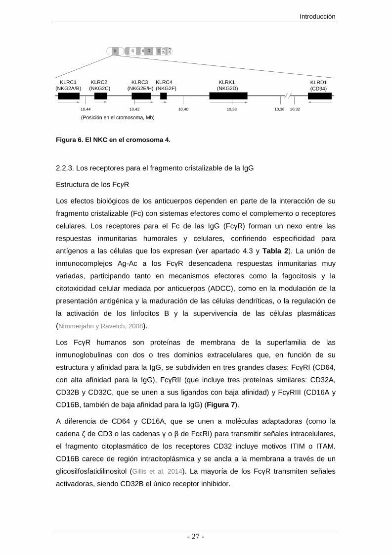
Introducción
- 27 -
Figura 6. El NKC en el cromosoma 4.
2.2.3. Los receptores para el fragmento cristalizable de la IgG
Estructura de los FcγR
Los efectos biológicos de los anticuerpos dependen en parte de la interacción de su
fragmento cristalizable (Fc) con sistemas efectores como el complemento o receptores
celulares. Los receptores para el Fc de las IgG (FcγR) forman un nexo entre las
respuestas inmunitarias humorales y celulares, confiriendo especificidad para
antígenos a las células que los expresan (ver apartado 4.3 y Tabla 2 ). La unión de
inmunocomplejos Ag-Ac a los FcγR desencadena respuestas inmunitarias muy
variadas, participando tanto en mecanismos efectores como la fagocitosis y la
citotoxicidad celular mediada por anticuerpos (ADCC), como en la modulación de la
presentación antigénica y la maduración de las células dendríticas, o la regulación de
la activación de los linfocitos B y la supervivencia de las células plasmáticas
(Nimmerjahn y Ravetch, 2008).
Los FcγR humanos son proteínas de membrana de la superfamilia de las
inmunoglobulinas con dos o tres dominios extracelulares que, en función de su
estructura y afinidad para la IgG, se subdividen en tres grandes clases: FcγRI (CD64,
con alta afinidad para la IgG), FcγRII (que incluye tres proteínas similares: CD32A,
CD32B y CD32C, que se unen a sus ligandos con baja afinidad) y FcγRIII (CD16A y
CD16B, también de baja afinidad para la IgG) (Figura 7 ).
A diferencia de CD64 y CD16A, que se unen a moléculas adaptadoras (como la
cadena ζ de CD3 o las cadenas γ o β de FcεRI) para transmitir señales intracelulares,
el fragmento citoplasmático de los receptores CD32 incluye motivos ITIM o ITAM.
CD16B carece de región intracitoplásmica y se ancla a la membrana a través de un
glicosilfosfatidilinositol (Gillis et al, 2014). La mayoría de los FcγR transmiten señales
activadoras, siendo CD32B el único receptor inhibidor.
KLRC1(NKG2A/B)
KLRC2 (NKG2C)
KLRC3 (NKG2E/H)
KLRC4(NKG2F)
KLRK1 (NKG2D)
(Posición en el cromosoma, Mb)
10,44 10,42 10,40 10,38 10,36 10,32
KLRD1(CD94)
12 13 21 23 24.3
24.1
15
KLRC1(NKG2A/B)
KLRC2 (NKG2C)
KLRC3 (NKG2E/H)
KLRC4(NKG2F)
KLRK1 (NKG2D)
(Posición en el cromosoma, Mb)
10,44 10,42 10,40 10,38 10,36 10,32
KLRD1(CD94)
12 13 21 23 24.3
24.1
15
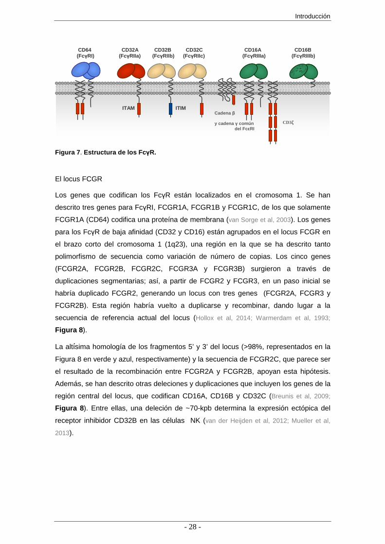
Introducción
- 28 -
Figura 7 . Estructura de los Fc γR.
El locus FCGR
Los genes que codifican los FcγR están localizados en el cromosoma 1. Se han
descrito tres genes para FcγRI, FCGR1A, FCGR1B y FCGR1C, de los que solamente
FCGR1A (CD64) codifica una proteína de membrana (van Sorge et al, 2003). Los genes
para los FcγR de baja afinidad (CD32 y CD16) están agrupados en el locus FCGR en
el brazo corto del cromosoma 1 (1q23), una región en la que se ha descrito tanto
polimorfismo de secuencia como variación de número de copias. Los cinco genes
(FCGR2A, FCGR2B, FCGR2C, FCGR3A y FCGR3B) surgieron a través de
duplicaciones segmentarias; así, a partir de FCGR2 y FCGR3, en un paso inicial se
habría duplicado FCGR2, generando un locus con tres genes (FCGR2A, FCGR3 y
FCGR2B). Esta región habría vuelto a duplicarse y recombinar, dando lugar a la
secuencia de referencia actual del locus (Hollox et al, 2014; Warmerdam et al, 1993;
Figura 8 ).
La altísima homología de los fragmentos 5’ y 3’ del locus (>98%, representados en la
Figura 8 en verde y azul, respectivamente) y la secuencia de FCGR2C, que parece ser
el resultado de la recombinación entre FCGR2A y FCGR2B, apoyan esta hipótesis.
Además, se han descrito otras deleciones y duplicaciones que incluyen los genes de la
región central del locus, que codifican CD16A, CD16B y CD32C (Breunis et al, 2009;
Figura 8 ). Entre ellas, una deleción de ~70-kpb determina la expresión ectópica del
receptor inhibidor CD32B en las células NK (van der Heijden et al, 2012; Mueller et al,
2013).
CD3ζ
Cadena β y cadena γ común del Fc εRI
ITAM ITIM
CD64 CD32A CD32B CD32C CD16A CD16B (FcγRI) (FcγRIIa) (FcγRIIb) (FcγRIIc) (FcγRIIIa) (FcγRIIIb)

Introducción
- 29 -
Figura 8. El complejo FCGR. Cada rectangulo corresponde a un gen del locus, cuyo nombre
aparece en la parte superior. Las flechas debajo de cada gen indican su orientación. En verde y
azul se han representado los dos fragmentos del locus FCGR que habrían surgido por
duplicación segmentaria. Las lineas grises indican que estas zonas del locus pueden estar
duplicadas o delecionadas. Se ha representado punteado un fragmento de 24,5 kb donde
estaría la zona de recombinación que condiciona la expresión de CD32B en las células NK
(Breunis et al, 2009; Hollox et al, 2014; Mueller et al, 2013).
Polimorfismos puntuales (SNP, single nucleotide polymorphism) en los genes del locus
FCGR tienen también implicaciones funcionales (Tabla 1 ). En este sentido, un SNP en
el intron 5 de FCGR2A conlleva la inclusión del exón críptico 6 en el marco de lectura
de una minoría de los individuos (generando un aumento de la activación celular tras
el reconocimiento de la IgG a traves de CD32A (van der Heijden et al, JACI 2013).
Tabla 1. Variaciones genéticas en el locus FCGR.
Gen (receptor) Variante Efecto SNP de
referencia R131H Aumento de la afinidad para IgG2 rs1801274 FCGR2A
(CD32A) c742+871A>G Aumento de la activación celular tras la unión de IgG
rs72717038
–386G>C1 –120T>A1
Aumento de la expresión rs3219018 rs34701572
FCGR2B (CD32B)
I232T Disminución de la expresión rs1050501 Q13X Expresión de proteína rs10917661 c798+1A>G Expresión de proteína rs76277413 FCGR2C
(CD32C) CNV
Correlación con los niveles de expresión Expresión ectópica de CD32B en células NK
F158V Aumento de la afinidad para IgG1, IgG2 e IgG3 rs396991 L48H Alteración funcional de las células NK rs10127939 FCGR3A
(CD16A) CNV Correlación con los niveles de expresión
HNA-1a/b/c Aumento de la fagocitosis (HNA-1a) Aumento de la expresión (HNA-1c)
n.a. FCGR3B (CD16B)
CNV Correlación con los niveles de expresión Expresión ectópica de CD32B en células NK
1 –386C–120A definen el haplotipo de mayor expresión. n.a., no aplicable.
FCGR2A FCGR3A FCGR2C FCGR3BHSPA7 FCGR2BHSPA6
Cromosoma 1
(Posición en el cromosoma, Mb)
161,50 161,55 161,60 161,65
2321 31 32 42132122313236.1
FCGR2A FCGR3A FCGR2C FCGR3BHSPA7 FCGR2BHSPA6
Cromosoma 1
(Posición en el cromosoma, Mb)
161,50 161,55 161,60 161,65
2321 31 32 42132122313236.1
2321 31 32 42132122313236.1

Introducción
- 30 -
La inhibición a través de CD32B está también modificada por variaciones genéticas
que afectan su expresión; así, un haplotipo determinado por varios SNPs en la región
promotora del FCGR2B aumenta su transcripción, mientras que el cambio de una
isoleucina por treonina en el fragmento transmembrana (I232T), reduce su expresión
en la membrana celular (Floto et al, 2005; Su et al, 2004a y 2004b).
Un SNP en el exón 3 de FCGR2C introduce un codón de parada prematuro (Q13X).
Su alta frecuencia en la población – aproximadamente un 80% de los individuos sanos
son homocigotos para este polimorfismo y, por tanto, carecen por completo de CD32C
– ha hecho que se le considerase un pseudogén (Breunis et al, 2008; Metes et al, 1998;
Su et al, 2002). Pero la expresión de CD32C no esta condicionada solamente por este
polimorfismo. Otro 4% de los individuos sin el cambio Q13X en al menos una de sus
copias FCGR2C, carecen también del receptor en la membrana. Esto es debido a otro
SNP en el sitio donador de splicing del intron 7 que cambia el marco de lectura
introduciendo otro codón de parada prematuro (van der Heijden et al, 2012).
Otros SNPs modifican la afinidad de los FcγR por su ligando; así, un cambio R131H de
la molécula CD32A aumenta su afinidad para IgG2 (Salmon et al, 1992). De manera
similar, el cambio F158V de CD16A aumenta su afinidad por IgG1 e IgG3 (Radaev at el,
2001; Sondermann et al, 2000; Wu et al, 1997) y el cambio L48H en CD16A (o L66H,
según se tome o no en cuenta la secuencia del péptido señal) ha sido implicado en la
alteración de la función de las células NK, pero su relevancia sigue siendo
controvertida (Forbes et al, 2009; Grier et al, 2012; Jawahar et al, 1993; de Vries et al, 1996).
Por último, varios SNPs en FCGR3B determinan los antígenos neutrofílicos HNA-1a,
HNA-1b y HNA-1c, que modifican la eficacia de la fagocitosis (Koene et al, 1998; Salmon
et al, 1992).
Todas estas variaciones genéticas del locus FCGR podrían tener implicaciones en el
desarrollo o en la severidad de diferentes enfermedades inmuno-mediadas.
Efectivamente, gran parte de los polimorfismos descritos han sido asociados con el
curso clínico de varias enfermedades autoinmunes e infecciosas y, además, con la
respuesta a terapias basadas en anticuerpos (Gillis et al, 2014; van Sorge et al, 2003).
Pero pese a su potencial relevancia, su papel en las infecciones virales ha sido
escasamente estudiado.
Patrón de expresión y afinidad para la IgG
Prácticamente todas las subpoblaciones de leucocitos, y también las plaquetas,
expresan al menos un subtipo de FcγR. Mientras que estirpes celulares implicadas en
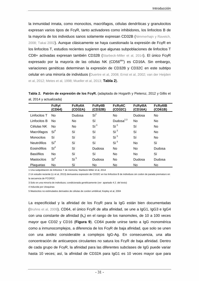
Introducción
- 31 -
la inmunidad innata, como monocitos, macrófagos, células dendríticas y granulocitos
expresan varios tipos de FcγR, tanto activadores como inhibidores, los linfocitos B de
la mayoría de los individuos sanos solamente expresan CD32B (Nimmerhajn y Ravetch,
2008; Takai 2002). Aunque clásicamente se haya cuestionado la expresión de FcγR en
los linfocitos T, estudios recientes sugieren que algunas subpoblaciones de linfocitos T
CD8+ activadas expresan también CD32B (Starbeck-Miller et al, 2014). El único FcγR
expresado por la mayoría de las células NK (CD56dim) es CD16A. Sin embargo,
variaciones genéticas determinan la expresión de CD32B y CD32C en este subtipo
celular en una minoría de individuos (Duertre et al, 2008; Ernst et al, 2002; van der Heijden
et al, 2012; Metes et al, 1998; Mueller et al, 2013; Tabla 2).
Tabla 2. Patrón de expresión de los Fc γR. (adaptada de Hogarth y Pietersz, 2012 y Gillis et
al, 2014 y actualizada)
FcRγI (CD64)
FcRγIIA (CD32A)
FcRγIIB (CD32B)
FcRγIIC (CD32C)
FcRγIIIA (CD16A)
FcRγIIIB (CD61B)
Linfocitos T No Dudosa Sí1 No Dudosa No
Linfocitos B No No Sí Dudosa2,3 No No
Células NK No No Sí 3 Sí 3 Sí No
Macrófagos Sí4 Sí Sí Sí 3 Sí No
Monocitos Sí Sí Sí Sí 3 Sí No
Neutrófilos Sí4 Sí Sí Sí 3 No Sí
Eosinófilos Sí4 Sí Dudosa No No Dudosa
Basófilos No Sí Sí No No Sí
Mastocitos Sí4 Sí 5 Dudosa No Dudosa Dudosa
Plaquetas No Sí No No No No 1 Una subpoblación de linfocitos T de memoria; Starbeck-Miller et al, 2014
2 Un estudio reciente (Li et al, 2013) demuestra expresión de CD32C en los linfocitos B de individuos sin codon de parada prematuro en
la secuencia de FCGR2C
3 Solo en una minoría de individuos; condicionada genéticamente (ver apartado 4.2. del texto)
4 Inducida por citoquinas
5 Mastocitos no estimulados derivados de células de cordon umbilical; Kepley et al, 2004
La especificidad y la afinidad de los FcγR para la IgG están bien documentadas
(Bruhns et al, 2009). CD64, el único FcγR de alta afinidad, se une a IgG1, IgG3 e IgG4
con una constante de afinidad (ka) en el rango de los nanomoles, de 10 a 100 veces
mayor que CD32 y CD16 (Figura 9 ). CD64 puede unirse tanto a IgG monomérica
como a inmunocomplejos, a diferencia de los FcγR de baja afinidad, que solo se unen
con una avidez considerable a complejos IgG-Ag. En consecuencia, una alta
concentración de anticuerpos circulantes no satura los FcγR de baja afinidad. Dentro
de cada grupo de FcγR, la afinidad para las diferentes subclases de IgG puede variar
hasta 10 veces; así, la afinidad de CD32A para IgG1 es 10 veces mayor que para
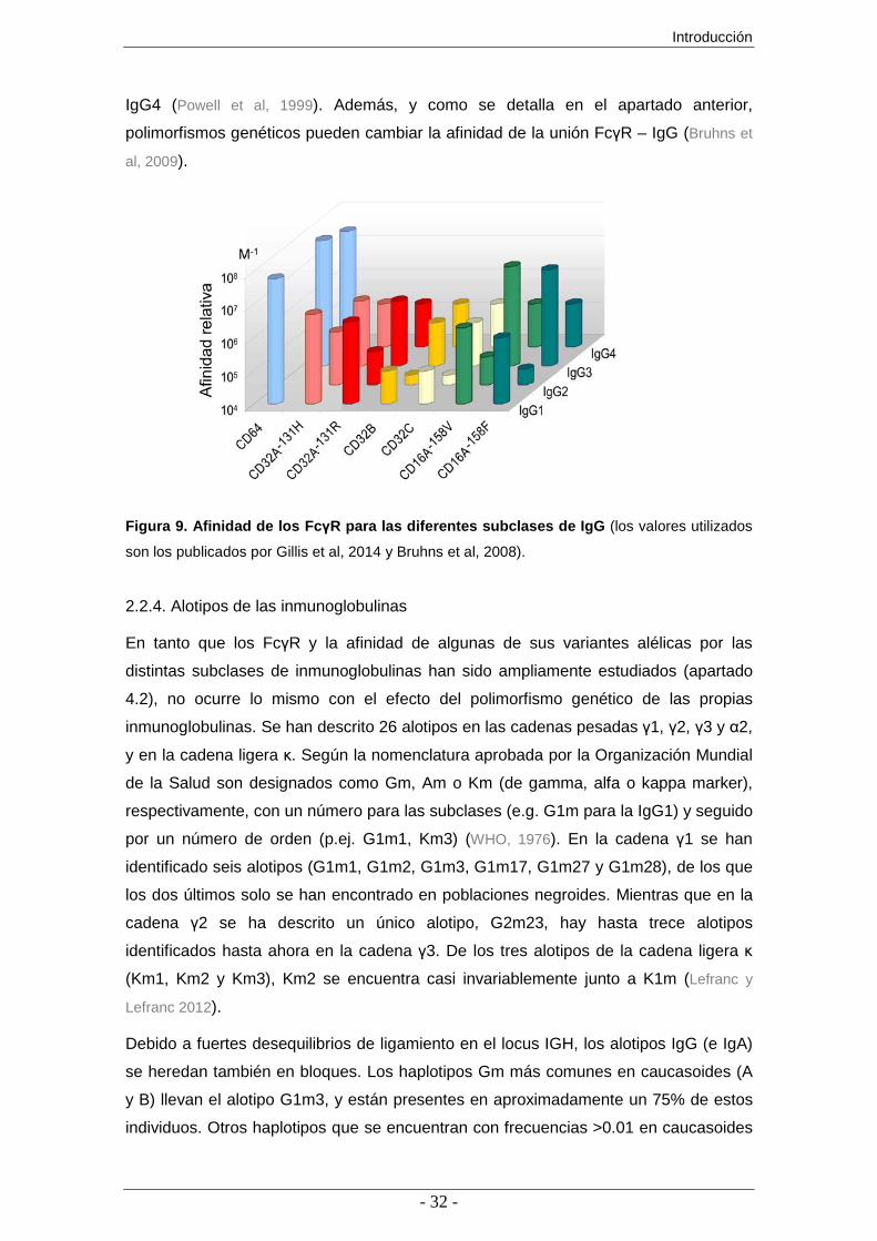
Introducción
- 32 -
IgG4 (Powell et al, 1999). Además, y como se detalla en el apartado anterior,
polimorfismos genéticos pueden cambiar la afinidad de la unión FcγR – IgG (Bruhns et
al, 2009).
Figura 9. Afinidad de los Fc γR para las diferentes subclases de IgG (los valores utilizados
son los publicados por Gillis et al, 2014 y Bruhns et al, 2008).
2.2.4. Alotipos de las inmunoglobulinas
En tanto que los FcγR y la afinidad de algunas de sus variantes alélicas por las
distintas subclases de inmunoglobulinas han sido ampliamente estudiados (apartado
4.2), no ocurre lo mismo con el efecto del polimorfismo genético de las propias
inmunoglobulinas. Se han descrito 26 alotipos en las cadenas pesadas γ1, γ2, γ3 y α2,
y en la cadena ligera κ. Según la nomenclatura aprobada por la Organización Mundial
de la Salud son designados como Gm, Am o Km (de gamma, alfa o kappa marker),
respectivamente, con un número para las subclases (e.g. G1m para la IgG1) y seguido
por un número de orden (p.ej. G1m1, Km3) (WHO, 1976). En la cadena γ1 se han
identificado seis alotipos (G1m1, G1m2, G1m3, G1m17, G1m27 y G1m28), de los que
los dos últimos solo se han encontrado en poblaciones negroides. Mientras que en la
cadena γ2 se ha descrito un único alotipo, G2m23, hay hasta trece alotipos
identificados hasta ahora en la cadena γ3. De los tres alotipos de la cadena ligera κ
(Km1, Km2 y Km3), Km2 se encuentra casi invariablemente junto a K1m (Lefranc y
Lefranc 2012).
Debido a fuertes desequilibrios de ligamiento en el locus IGH, los alotipos IgG (e IgA)
se heredan también en bloques. Los haplotipos Gm más comunes en caucasoides (A
y B) llevan el alotipo G1m3, y están presentes en aproximadamente un 75% de estos
individuos. Otros haplotipos que se encuentran con frecuencias >0.01 en caucasoides

Introducción
- 33 -
incluyen G1m17,1 (~15%) o G1m17,1,2 (~6%) (Mayr y Mickerts, 1970; Schanfield et al,
1975a y b). Por tanto, en poblaciones caucasoides el alotipo G1m17 se asocia
invariablemente a G1m1, encontrándose la combinación G1m3,1 solamente en un
15% de ciertas poblaciones mongoloides (van Loghem y Natvig, 1970).
En la infección por HSV-1, los únicos datos disponibles sobre la posible contribución
de los alotipos de las inmunoglobulinas al curso clínico de la enfermedad se limitan al
estudio de su unión al receptor para la IgG1 generado por las glicoproteínas virales gE
y gI (ver más adelante). En particular, este señuelo viral se une a inmunoglobulinas de
alotipo G1m3 con menor afinidad que las G1m17,1, cambio de afinidad directamente
relacionado con el residuo 214 de la cadena γ1 (Atherton et al, 2000). De acuerdo con
estos datos, se ha propuesto una hipótesis según la cual la susceptibilidad al HSV-1
pudiera estar determinada en parte por este polimorfismo (Pandey et al, 2012), lo que
trataremos de verificar en este trabajo.
Los alotipos G1m17 y G1m3 se distinguen por una sustitución Lys214Arg en el
dominio CH1 y son mutualmente excluyentes. G1m1 está situado en el dominio CH3,
fuera de la zona inferior bisagra del Fc, y se caracteriza por un aspártico en la posición
356 y un residuo de leucina en la posición 358. (Tabla 3).
Tabla 3. Los alotipos de la IgG1.
Dominio CH1 Dominio CH3 Alotipo 214* 356* 358* 431*
consenso Arg Glu Met Ala G1m1 Asp Leu G1m2 Gly G1m3 Arg G1m17 Lys * numeración de aminoácidos según la UE
Ninguno de estos aminoácidos ha sido directamente implicado en la interacción FcγR-
IgG (Shields et al, 2001; Sondermann et al, 2000), pero estos polimorfismos, al igual que
los que determinan los alotipos Km, podrían contribuir a interacciones alostéricas que
modifiquen la afinidad de esta unión. Esta hipótesis está apoyada por ensayos de
afinidad que han mostrado que CD16A SE une con mayor avidez A inmunoglobulinas
con alotipo G1m3 que G1m17,1 (Armour et al, 2010). Asimismo, ensayos de
competición, en los que se intenta bloquear la ADCC frente a dianas conocidas con
exceso de agregados de inmunoglobulinas de alotipo conocido, confirman que G1m3
compite eficazmente con anticuerpos monoclonales con alotipo G1m17 (Namboodiri y
Pandey, 2011).

Introducción
- 34 -
2.3. La respuesta inmunitaria celular y humoral
Las células citotóxicas, principalmente los linfocitos T CD8+ (CTL) y las células NK,
son los efectores celulares más importantes en las infecciones virales. En la infección
activa por HSV-1, los primeros linfocitos que infiltran la lesión son las células NK,
seguidas por los linfocitos T CD4+ y, por último, los CD8+, relacionándose
estrechamente su reclutamiento al sitio de la infección con el aclaramiento del virus
(Koelle et al, 2003). Apoyando que el papel de los anticuerpos específicos en la
respuesta inmunitaria al HSV-1 es igualmente relevante está la relación inversa entre
la gravedad de la infección neonatal y la presencia de anticuerpos anti-HSV en la
madre (Brown, 1991).
2.3.1. Las células NK
Las células NK son linfocitos que no expresan receptores clonotípicos para antígeno y
que pueden diferenciarse a partir de un progenitor común con los linfocitos T.
Constituyen aproximadamente un 10% de los linfocitos circulantes y se encuentran, en
proporciones variables, entre las células de origen linfoide del intestino, higado,
pulmón, decidua uterina y órganos linfoides secundarios (Trincheri, 1989; Yu et al, 2013).
Como se ha mencionado en un apartado anterior, las células NK expresan una serie
de receptores activadores e inhibidores, siendo dictada su función por el resultado de
una compleja relación entre las señales positivas y negativas desencadenadas tras la
unión de estos receptores a sus correspondientes ligandos. Aunque todavía quedan
varios receptores de células NK “huérfanos”, muchos de sus ligandos han sido
identificados y caracterizados en las últimas décadas, estando su gran mayoría
relacionados, de diferentes maneras, con la vigilancia de la presencia de células
infectadas o transformadas. Así, varias estructuras derivadas de patógenos son
reconocidas por TLRs y receptores citotóxicos naturales (NCR); algunas moléculas
inducidas por la infección o el estrés celular son ligandos para NKG2D, NCRs, DNAM-
1 (CD226) y 2B4 (CD244); las células opsonizadas pueden ser reconocidas a través
de FcγR; y varias subfamilias de receptores, tanto activadores como inhibidores,
controlan el nivel de expresión de las moléculas HLA de clase I (ver detalles en el
capítulo 3 de esta introducción) (Lanier, 2005; Li et al, 2014). La activación de las células
NK desencadena respuestas potentes e inmediatas tras el reconocimiento de una
célula diana, a través de mecanismos de citolisis (vía liberación de perforina y
granzimas), activación de la apoptosis y producción de citoquinas (como IFN-γ y TNF-
α) y quimioquinas, controlando así otros mecanismos inmunitarios innatos y
adaptativos implicados en la respuesta antiviral (Marras et al, 2014; Trincheri, 1989).
Introducción
- 35 -
Las células NK forman una población heterogénea en cuanto a sus características
fenotípicas, proliferativas y funcionales. La mayoría de células NK circulantes tiene una
expresión relativamente baja de CD56 (CD56dim), alta expresión de CD16 y
funcionalmente son citotóxicas y secretoras de citoquinas. En cambio,
aproximadamente un 5-10% de ellas tienen una mayor densidad de CD56 en su
superficie (CD56bright), no expresan CD16, pueden producir grandes cantidades de
citoquinas tras su estimulación y tienen una gran capacidad proliferativa, pero poca
citotóxica. A este subtipo de células NK, con un fenotipo mayoritariamente
CD56brightCD16–NKG2A+KIR–, algunos autores las consideran precursores de las
células NK CD56dimCD16+NKG2A–KIR+ en sangre, siendo la pérdida progresiva de
NKG2A y la adquisición de KIR un proceso activo de diferenciación (Beziat et al, 2010).
La generación de células NK con fenotipo “de memoria” inducido por patógenos como
el hCMV (detallado en el apartado 3.2.1.) apoya la existencia de características típicas
de la inmunidad adaptativa y diferenciación terminal para las células NK. En este
sentido, la expresión estable de NKG2C y CD57 y la pérdida de la cadena FcεRIγ se
perfilan como marcadores de diferenciación tardía de las células NK (Bjorkström et al,
2010; Muntasell et al, 2013; Zhang et al, 2013a).
La relevancia de las células NK en la infección por HSV-1 se ha confirmado en
pacientes con algunas inmunodeficiencias primarias graves, en los que la falta o la
disfunción de las células NK determinada por mutaciones en diferentes genes
implicados en su desarrollo, se acompaña, casi invariablemente, por infecciones
virales, sobre todo por herpesvirus (Jouanguy et al, 2013; Orange et al, 2013). Además,
varios estudios confirman la presencia de células NK en los infiltrados inflamatorios de
los sitios de infección activa y su papel en el aclaramiento del virus (Carr et al, 2008;
Cunningham et al, 1985; Frank et al, 2012; Sirianni et al, 1986; Vogel et al, 2014). Asimismo,
estudios en ratones deplecionados de células NK o a los que se les transfieren células
NK funcionales o no, apoyan su papel clave en el control de la infección (Kohl et al,
1986; Rager-Zisman et al, 1987). De manera similar, en un modelo murino de infección
por HSV-1 se ha demostrado la necesidad de una respuesta NK eficaz para el control
de la infección productiva, identificándose además un locus de resistencia a la
infección por HSV-1 cerca de la región NKC (Pereira et al, 2001).
Notablemente y a diferencia de otros herpesvirus, todavía no se han identificado
estructuras derivadas del HSV-1 que funcionen como ligandos virales para receptores
activadores expresados en las células NK. Sin embargo, el reconocimiento de
patrones moleculares alterados por la infección ha sido directamente implicado en el
control del HSV-1. Así, se ha demostrado que la expresión de la proteína viral ICP0 es

Introducción
- 36 -
necesaria y suficiente para aumentar la susceptibilidad de las células infectadas a la
lisis mediada por células NK, de manera dependiente de la activación de los NCR
(Chisholm et al, 2007). Además, DNAM1 (CD226) reconoce una proteína (nectina-2 o
CD112, también conocida como HveB o PRR2) que facilita la entrada de algunas
cepas de HSV-1 en las células diana, sugiriendo que, de manera similar a lo
observado en procesos tumorales, la presencia del virus aumentaría la expresión de
nectina-2, que funcionaría como estímulo para la activación de las células NK a través
de DNAM-1 (Bottino et al, 2003; Warner et al, 1998). Por último, se han descrito potentes
respuestas dependientes de anticuerpos mediadas por una subpoblación de células
NK frente a células infectadas por HSV-1. Dicha subpoblación tiene características de
memoria, un fenotipo CD56dimFcεRIγ–NCRlowNKG2AlowCD57hi y, predominantemente,
NKG2Cbright y una alta capacidad de producir IFN-γ en presencia de anticuerpos anti-
HSV-1 y fibroblastos infectados (Hwang et al, 2012; Zhang et al, 2013a).
2.3.2. Los linfocitos T
En la infección primaria por HSV, las APCs (principalmente células dendríticas, DC),
activadas directamente por patógenos o a través de IFN-I, se cargan con antígenos
virales que transportan a los órganos linfáticos secundarios (Allan et al, 2006; Pollara et
al, 2004). Los linfocitos T naïve específicos para el antígeno se activan y proliferan en
los ganglios linfáticos tras el reconocimiento de péptidos virales presentados por las
DCs. De esta manera, se generan linfocitos T efectores que migran a los sitios de la
infección activa. Una vez controlado el proceso patológico, hay una fase de
contracción, manteniéndose una pequeña población de linfocitos T de memoria. La
generación de linfocitos T efectores requiere varios días pero aunque las respuestas
adaptativas sean relativamente lentas, su intensidad y especificidad sobrepasan
ampliamente a las de la inmunidad innata.
Los linfocitos T CD8+ efectores específicos para antígenos virales, a través de
mecanismos citolíticos y producción de citoquinas, contribuyen al aclaramiento del
virus en el sitio de la infección. Además, estos linfocitos se acumulan alrededor de las
neuronas HSV-1+ en los ganglios sensitivos de ratones y humanos y se han implicado
en el control del mantenimiento del estado latente del virus (Feldman et al, 2002; Khana
et al, 2004; Knickelbein et al, 2008; Liu et al, 2000; Theil et al, 2003; Verjans et al, 2007).
Puesto que las neuronas infectadas no son destruidas por los linfocitos T CD8+ y
considerando que solo una fracción de los linfocitos T CD8+ presentes en los ganglios
sensitivos expresan granzima B (Held y Derfuss, 2011), se han propuesto mecanismos
no citolíticos implicados en este control. En este sentido, se ha demostrado en
modelos murinos que el IFN-γ producido por estos linfocitos impide la reactivación del

Introducción
- 37 -
virus en los ganglios sensitivos (Decman et al, 2005; Liu et al, 2001). Además, se ha
planteado la existencia de un efecto no citolítico de los productos de sus gránulos
sobre el control de la replicación viral. Evidencias que apoyan esta teoría incluyen (i) la
dificultad de mantener el estado latente del HSV en ratones deficientes de granzima A,
B y de perforina, (ii) el hecho de que la liberación de enzimas líticas por los linfocitos T
CD8+ en los ganglios sensitivos no induce la apoptosis de las neuronas infectadas, (iii)
la capacidad de los LAT de bloquear el efecto de la granzima B sobre las caspasas,
ofreciendo así una explicación para la supervivencia de las neuronas HSV-1+ y (iv) la
capacidad de la granzima B de degradar la proteína IE ICP4. Todo ello sugiere, por
tanto, que las granzimas liberadas por los linfocitos T CD8+ ejercen un efecto directo
(o indirecto, a través de la activación de otros enzimas) sobre proteínas virales en
fases tempranas del ciclo viral (Jiang et al, 2011; Knickelbein et al, 2008; Pereira et al,
2000).
Los linfocitos T CD4+ efectores están también presentes en las lesiones activas junto
con las células NK y los linfocitos T CD8+, siendo una importante fuente de IFN-γ.
Además, de manera similar a los linfocitos T CD8+, aunque en proporciones menores,
se ha demostrado que se acumulan en los ganglios sensitivos que albergan HSV-1 en
estado latente (Khana et al, 2004; Theil et al, 2003; van Velzen et al, 2013; Verjans et al,
2007). No obstante su papel clave en la respuesta adaptativa antiviral, colaborando en
la activación de los linfocitos B y T CD8+, se desconoce todavía su contribución al
mantenimiento del virus en estado latente (Frank et al, 2010; Shedlock et al, 2003; Sun et
al, 2003; van Velzen et al, 2013).
Mientras que la respuesta de los linfocitos T CD4+ puede ser desencadenada por una
amplia gama de antígenos virales, incluyendo proteínas de la envuelta, tegumento,
cápside y no estructurales, la respuesta mediada por los linfocitos T CD8+ parece ser
bastante restringida, sobre todo en los ganglios sensitivos. La mayoría de los epítopos
reconocidos por estos linfocitos, tanto en humanos como en ratones, proceden de
proteínas virales producidas antes del inicio de la replicación del ADN viral (Derfuss et
al, 2007; Koelle et al, 2003; St. Leger et al, 2011a; van Velzen et al, 2013). Aunque el
mecanismo responsable de esta selección de los epitopos T CD8+ no se conoce,
parece claro que debería ofrecer importantes ventajas para el control de las
reactivaciones. Alternativamente, esta restricción del repertorio de linfocitos T CD8+
podría derivar de la capacidad de ciertas moléculas HLA de clase I para presentar
péptidos procedentes de estas proteínas. Puesto que todavía no se han identificado
alelos HLA claramente relacionados con la resistencia o la susceptibilidad a la
infección por HSV-1, este campo sigue abierto a más estudios. En particular, la
capacidad de las moléculas HLA de clase I para presentar péptidos derivados de

Introducción
- 38 -
proteínas virales codificadas por genes IE requiere especial atención, sobre todo en
relación con la capacidad de los linfocitos T CD8+ para controlar el estado latente del
HSV-1.
2.3.3. La inmunidad humoral
En la primoinfección por HSV-1, los linfocitos B naïve específicos para estructuras
derivadas del HSV-1 que encuentran estos antígenos en los órganos linfáticos
secundarios se activan, proliferan y continuan diferenciándose. Algunos de ellos
maduran hacia células plasmáticas, otros migran a los centros germinales donde
sufren procesos adicionales de cambio de isotipo y maduración de la afinidad. Las
células que sobreviven a estos procesos selectivos proliferan y se diferencian a células
plasmáticas productoras de anticuerpos anti-HSV-1 de alta afinidad, o linfocitos B de
memoria. Puesto que todos estos mecanismos requieren más de una semana, durante
la primoinfección la respuesta de anticuerpos es tardía; sin embargo, su eficacia
mejora progresivamente, siendo retenidas las características adquiridas durante este
proceso por las células B de memoria para respuestas secundarias. Los anticuerpos
anti-HSV-1 de isotipo IgG ejercen efectos neutralizantes y de opsonización,
participando en la activación de la vía clásica del complemento y en la ADCC
(Mathiesen et al, 1998). Aunque su presencia no parece conferir protección en todos los
sujetos seropositivos, los distintos mecanismos de evasión desarrollados por el virus
para contrarrestar sus efectos (revisados en el apartado 3) apoyan su papel clave
durante la infección.
La gran mayoría de los individuos seropositivos tiene anticuerpos anti-HSV-1 de tipo
IgG1, mientras que solamente en la mitad de ellos se han detectado anticuerpos
específicos de tipo IgG3, siendo ocasional la presencia de anticuerpos anti-HSV-1-
IgG4 e -IgG2 (Mathiesen et al, 1988). Los anticuerpos específicos de tipo IgG1 e IgG3
tienen efectos neutralizantes, reclutan complemento y pueden mediar ADCC y son
reconocidos, con diferente afinidad, por los FcγR (Bruhns et al, 2009; Mathiesen et al,
1988). Pero, mientras que los pacientes deficientes de diferentes factores del
complemento no tienen mayor susceptibilidad a la infección que los individuos sanos,
se ha atribuido una mayor relevancia a la ADCC mediada por las células NK (Orange
2013; Walport 2001; Zhang et al, 2013b).
3. MECANISMOS DE INMUNOEVASIÓN DEL HSV-1
Para asegurar su supervivencia en la célula infectada, el HSV-1 ha desarrollado una
serie de mecanismos para controlar a la célula hospedadora y evadir la respuesta

Introducción
- 39 -
inmunitaria. Así, varias proteínas virales bloquean selectivamente la síntesis de
nuevas proteínas celulares, inhiben, degradan selectivamente o subvierten la función
de proteínas celulares ya sintetizadas, o restringen la señalización que alerta de que
una célula está infectada. Los mecanismos que afectan a las interacciones entre el
virus y el sistema inmunitario son especialmente relevantes para entender la
variabilidad clínica de la infección. Si bien el control de la respuesta innata es muy
importante en la primoinfección, puesto que la replicación viral se inicia antes de la
maduración de la respuesta inmunitaria adquirida, las estrategias de inmunoevasión
deberían ser más amplias para facilitar todos los estadios de la infección, afectando
tanto a mecanismos inmunitarios innatos como adaptativos.
Efectivamente, se ha descrito ya una larga serie de mecanismos de inmunoevasión
desarrollados por el HSV-1. De especial relevancia es la alteración de las vías de
activación mediadas por los interferones. La inducción de la producción de IFN-I
requiere la activación de factores de transcripción como IRF3 (interferon regulatory
factor) y el factor nuclear-κB (NF-κB). A su vez, la activación de sus receptores
desencadena una cascada de fosforilación de las quinasas de Jano (Jak) y las
moléculas Stat, que llevan a la transcripción de genes regulados por interferones y a
respuestas antivirales (Mossman y Ashkar, 2005). Tanto la producción de IFN-I como la
señalización a través de sus receptores son dianas para el HSV-1. Así, la proteína viral
ICP0 bloquea tanto la activación del NF-κB inducida por la activación de TLRs
(mediante deubiquitinilación específica de algunos de sus moduladores: TRAF6 y
NEMO), como la señalización a través de IRF, limitando la producción de interferón
(Daubeuf et al, 2009; Lin et al, 2004; van Lint et al, 2010; Melroe et al, 2004; Mossman y
Smiley, 2002; Orzalli et al, 2012). De manera similar, la proteínas del tegumento US3 y
VP16, codificadas por US3 y UL48, respectivamente, restringen la producción de IFN-I
deubiqutinilando TRAF6 y bloqueando el reclutamiento de un co-activador del IRF3
(CBP, CREB binding protein), respectivamente (Sen et al, 2013; Xing et al, 2013).
Además, la RNAasa UL41 y la proteína viral ICP27, codificada por el gen IE UL54,
reducen la disponibilidad de Jak1, Stat1 y Stat2, restringiendo así la señalización a
través de receptores para interferón (Chee y Roizman, 2004; Johnson y Knipe, 2009;
Suzutani et al, 2000). Adicionalmente, las proteínas virales US11 y RL1 interfieren con la
respuesta celular frente a ARN de doble cadena (dsRNA, un marcador de la infección
viral) impidiendo de esta manera tanto la activación del NF-κB como la muerte
programada de la célula infectada (Ishioka et al, 2013; Leib et al, 2000; Mossman y Smiley,
2002; Taddeo et al, 2003; Xing et al, 2012).
Adicionalmente, las proteínas virales ICP0, UL36, UL42 y VP16 bloquean la activación
del NF-κB inducida por TNF-α, deubiquitinilando TRAF3, o bloqueando su

Introducción
- 40 -
translocación (Wang et al, 2013; Xing et al, 2013; Zhang et al, 2013c y d). Además, uno de
los co-receptores celulares que facilitan la entrada del HSV-1 en las células diana es
un miembro de la familia de receptores para TNF (HVEM/HveA, herpes virus entry
mediator / protein A) que se une a la glicoproteína viral gD. La expresión prominente
de HVEM en los linfocitos T facilitaría su infección por HSV-1, permitiéndole
contrarrestar potencialmente los efectos antivirales de los linfocitos T (Benedict y Ware,
2001).
En consonancia con el papel clave de la presentación antigénica en la defensa frente
al HSV-1, son varios los mecanismos desarrollados por el virus para evadirla. En este
sentido, la proteína viral ICP47 se une al TAP en las células infectadas, interfiriendo
con el transporte de péptidos virales y su presentación por moléculas HLA de clase I a
linfocitos T citotóxicos (Hill et al, 1995). Se ha observado también que el HSV-1, que
infecta DCs inmaduras, bloquea su maduración e interfiere en su interacción con los
linfocitos T, disminuyendo además la expresión de la molécula de adhesión CD83 a
través de la proteína viral ICP0 (Kobelt et al, 2003; Kruse et al, 2000; Kummer et al, 2007).
Asimismo, por un mecanismo todavía no esclarecido independiente de la expresión de
moléculas HLA de clase II y coestimuladoras, la proteína viral ICP22 impide la
presentación antigénica a linfocitos T CD4+ (Barcy y Corey, 2001). Por último, aunque
formalmente no se haya implicado a las moléculas CD1 (una familia de proteínas MHC
de clase Ib que presentan derivados lipídicos) en la presentación de antígenos virales,
se ha observado una disminución de su expresión en células infectadas por HSV-1, lo
que sugiere que también podrían jugar un papel importante en este proceso (Raftery et
al, 2006).
Los múltiples efectos de los anticuerpos en la respuesta inmunitaria son también
dianas para los mecanismos de inmunoevasión del HSV-1. Las glicoproteínas virales
gE y gI generan un FcγR viral (FcγRv) señuelo que podría contrarrestar tanto la
ADCC, al competir con FcγR del hospedador, como la activación del complemento. En
efecto, se ha demostrado un mecanismo de antibody bipolar bridging por el que un
anticuerpo puede unirse por el paratopo a una proteína antigénica viral, y por el
fragmento Fc al FcγR señuelo, ambos en la superficie de la célula infectada. De esta
manera, los FcγRv, además de limitar la ADCC, impiden la unión de C1q a los
inmunocomplejos, y, consecuentemente, la activación de la vía clásica del
complemento (Dubin et al, 1991; Frank y Friedman, 1989; Johnson et al, 1988). Asimismo, la
glicoproteína viral C puede unirse al factor del complemento C3 y bloquear la
formación de las convertasas de C3 y C5 y, por tanto, la activación de todas las vías
del complemento (Friedman et al, 1984; Fries et al, 1986; Kostavasili et al, 1997).

OBJETIVOS


Objetivos
- 43 -
Objetivo general
Estudiar la contribución de la diversidad genética de moléculas potencialmente
implicadas en la respuesta inmunitaria frente a HSV-1, a la variabilidad cínica de la
infección.
Objetivos específicos
1. Analizar la influencia del polimorfismo genético del sistema HLA sobre el curso
clínico de la infección por HSV-1
2. Estudiar las implicaciones de la variabilidad de los KIR y sus ligandos en la
susceptibilidad a sufrir recurrencias de la infección por HSV-1
3. Diseñar un método eficiente para el genotipado del número de copias del gen que
codifica para el receptor NKG2C y estudiar su influencia en la infección por HSV-1
4. Explorar el papel del polimorfismo de los complejos genéticos FCGR e IGH en la
evolución clínica de la infección por HSV-1, y analizar in vitro su potencial
contribución a la modulación de la citotoxicidad celular dependiente de anticuerpos
(ADCC) mediada por células NK, en el contexto de la infección por HSV-1.


PUBLICACIONES


Artículo 1
J Immunol 2012: 188: 4412-20.
http://www.jimmunol.org/content/188/9/4412
Copyright © 2012 The American Association of Immunologists, Inc.


Publicaciones
- 49 -
Host genetic factors in susceptibility to Herpes simplex type 1 virus infection:
contribution of polymorphic genes at the interface of innate and adaptive
immunity
M Moraru,* E Cisneros,* N Gómez-Lozano,* R de Pablo,* F Portero,† M Cañizares,* M
Vaquero,‡ G Roustán,§ I Millán,¶ M López-Botet,|| and C Vilches2*
*Immunogenetics-HLA, †Microbiology, ‡Ophthalmology, §Dermatology, and ¶Statistics
Departments, Hospital Universitario Puerta de Hierro, Majadahonda; ||DCEX,
Universitat Pompeu Fabra, Barcelona, Spain
Abstract
HSV-1 establishes life-long latency which can result in clinical relapses or in
asymptomatic virus shedding. Although virtually all adults have been exposed to HSV-
1, the clinical course varies remarkably. Genetic host variability could be related to this
clinical diversity. In this study, we analyzed the contribution of gene families in
chromosomes 1, 6, 12, and 19, which encode key regulators of the innate and adaptive
immunity, in a cohort of 302 individuals. Class I and class II alleles of the HLA system,
the copy-number variation of NK-cell receptor genes (KIR and NKG2C), the
combinations of KIR and their HLA ligands, and CD16A and CD32A allotypes of
variable affinity for IgG subclasses were all studied. Although no major susceptibility
locus for HSV-1 was identified, our results show that the risk of suffering clinical HSV-1
infection is modified by MHC class I allotypes (B*18, C*15, and the group of alleles
encoding A19), the high affinity receptor/ligand pair KIR2DL2/HLA-C1 and the CD16A-
158V/F dimorphism. Conversely, HLA class II and CD32A polymorphisms, and NKG2C
deletion did not seem to influence the clinical course of herpetic infection. Collectively,
these findings support an important role in host defense against herpetic infection for
several polymorphic genes implicated in adaptive immunity and in surveillance of its
subversion. They confirm the crucial role of cytotoxic cells (CTL and NK) and the
contribution of genetic diversity to the clinical course of HSV-1 infection.
Keywords: cytotoxic T cells, Fc receptors, Herpes simplex, MHC, Natural Killer cell
receptors
Publicaciones
- 50 -
Introduction
Most adults have suffered HSV-1 infection. The virus migrates from mucosae or skin to
sensory ganglia, where it establishes life-long latent infection. Virus propagation is
controlled by innate and adaptive immunity, and the course of the infection depends
upon the host immune system capacity to control both the primary infection and the
reactivations. HSV-1 replication in keratinocytes and mucosal cells triggers a strong
cytokine response. IFN-alpha produced by dendritic cells (DC)3, and IFN-gamma
secreted by NK and, later, CD4+ T cells, represent a first line of direct antiviral defense
(1). IFNs also promote subsequent adaptive immune response by enhancing antigen
presentation (2).
CD8+ cytotoxic T cells (CTLs) eliminate infected cells presenting HSV-1 peptides on
HLA class I molecules, and they are responsible for immunosurveillance during viral
latency (3, 4). In addition, DC present viral antigens bound to HLA class II molecules to
CD4+ T lymphocytes, which enhance differentiation and establishment of functional
memory CTLs (1) and antibody-mediated control of HSV-1.
Key components of innate cellular immunity to viruses are NK cells, which kill infected
cells without presensitization, secrete antiviral cytokines, and favour DC maturation.
Their behaviour depends on the balance of signals transduced by activating and
inhibitory receptors that survey altered molecular patterns on infected cells (5, 6). Their
best-known trigger is HLA class I downregulation, monitored by several families of NK-
cell receptors (7). Furthermore, NK lymphocytes exert Ab-dependent cellular
cytotoxicity (ADCC) against Ab-coated infected cells by means of FcγR.
HSV-1, like other herpesviruses, has developed mechanisms to circumvent the host
immune response. Infected cell protein (ICP) ICP47 binds the transporter for antigenic
peptides (TAP), thus hampering HLA class I-mediated Ag presentation by infected cells
to T lymphocytes (8). Furthermore, HSV-1 glycoproteins gE and gI form an FcR on
both the viral envelope and the infected cell membrane, where it can compete, directly
and through Ab bipolar bridging, with host FcγR, protecting the infected cells from
complement- and cell-mediated lysis (9).
Although virtually all adults have been exposed to HSV-1, the clinical course of the
infection varies remarkably, ranging from asymptomatic virus shedding to frequent
inflammatory herpetic lesions and potentially life-threatening episodes of herpetic
encephalitis. Clinical variability is attributable to the virus itself (e.g. HSV-1 strains,
amount of inoculum), to the environment (e.g. exposure to UV light and other triggers)
and to variations in the host immune response. Herpetic encephalitis and isolated
cases of fatal HSV-1 infection have been associated with rare mutations in innate
Publicaciones
- 51 -
immunity-related genes (10-14). A recent linkage analysis detected in chromosome 21
a 2.5-megabase susceptibility region for herpes simplex labialis (15). However,
predictive genetic markers that explain the pathogenesis of the common outcomes of
HSV-1 infection are yet to be identified.
An attractive candidate to influence the course of HSV-1 infection is the enormous
diversity of the HLA complex in chromosome 6, credited with a large proportion of the
variability of immune responses, because it could modify the efficacy of HSV-1-antigen
presentation. Several groups have analyzed the contribution of HLA allotypes to the
clinical diversity of HSV-1 infection, but many results did not replicate in different
populations (16-27).
Killer-cell Ig-like Receptors (KIR), encoded by a highly polymorphic gene complex in
chromosome 19, represent another series of candidates to influence the HSV-1
infection outcome because they help NK cells fight viruses that tamper with HLA
expression (28). The most conspicuous aspect of KIR-gene diversity is copy-number
variation (CNV) – most humans are natural knockouts for multiple KIR genes, only the
genes at the centromeric and the telomeric ends of the KIR complex being conserved.
We have previously identified an association between symptomatic HSV-1 infection
and presence in the genome of the linked genes KIR2DL2 and KIR2DS2 (29).
Also used by NK cells to sense HLA expression are CD94/NKG2 heterodimers
encoded in the NK complex of chromosome 12, which comprise both activating and
inhibitory receptors for HLA-E (30, 31). In sharp contrast with KIR, the CD94 and most
NKG2 genes are well conserved (32), one remarkable exception being complete
deletion of NKG2C (KLRC2) in some individuals (33, 34). The consequence of NKG2C
deletion for immunity and health is unknown, but marked expansion of NKG2C-positive
NK- and T-lymphocytes in response to CMV infection (35, 36) suggests a role in
defense against herpesviruses.
Finally, the genes in chromosome 1 coding for the FcγR CD16A (FCGR3A) and
CD32A (FCGR2A) display allelic polymorphisms that modulate their affinity for IgG
(37). High-affinity FcγRs could enhance ADCC and compete with the decoy FcγR
formed by HSV-1 gE/gI, thus improving the efficacy of humoral response against HSV-
1.
Whether and how virus evasion mechanisms may be counteracted by host defense
and to which extent immunogenetic variability contributes to the clinical outcomes of
herpetic infection is yet to be established. In this study, we have analyzed the
contribution of the genetic diversity of the HLA system and the KIR, NKG2C, CD16A,
and CD32A receptors to the clinical variability of HSV-1 infection.

Publicaciones
- 52 -
Materials and Methods
Donor and patient samples.
Three hundred two individuals, in their vast majority of Spanish origin, were interviewed
about the presence and frequency of previous or actual clinical evidences of HSV-1
infection. Individuals reporting any diseases or therapies that might debilitate an
immune response or symptoms doubtfully attributable to HSV-1 infection were
excluded from this study; also excluded were donors seropositive for HSV-2. The total
group included 253 voluntary adults and 49 patients who sought medical assistance for
herpetic lesions. From the total population, 131 individuals had been included in a
previous study where we analyzed the influence of the KIR diversity on the clinical
course of the HSV-1 infection (29). In this study, we have replicated that study on KIR
diversity in a 171-individual population with similar characteristics. Moreover, we have
analyzed, in the whole sample of 302 individuals, the relationship between the outcome
of HSV-1 infection and HLA, NKG2C and FCGR polymorphisms.
Venous blood samples collected from all donors were used to isolate serum and
genomic DNA. Serum IgG/IgM antibodies against HSV-1+2 common antigens and
HSV-2 gG2 antigen were detected by ELISA and evaluated as recommended by the
manufacturer (Vircell, Granada, Spain).
Based on reported episodes of symptomatic herpes infection (most often, cold sores)
after a standardized questionnaire, the total sample was subdivided into two
categories: asymptomatic and symptomatic individuals. When considering the serologic
findings and the frequency of HSV-1 reactivations, each of the previous groups was
further separated, resulting in a total of four subcategories: (i) asymptomatic
seronegatives; (ii) asymptomatic seropositives; (iii) donors with sporadic clinical
reactivations (less than two reactivations yearly in the maximum activity period); and
(iv) patients with recurrent reactivations (two or more per year).
HLA, KIR, NKG2C, FCGR2A and FCGR3A genotyping.
HLA class I (HLA-A, -B and -C) and class II (HLA-DRB1, -DRB3, -DRB4 and -DRB5)
typing was carried out using a reverse line PCR sequence specific oligonucleotide
(PCR-SSO) test (Invitrogen-Dynal Reli-SSO, Wirral, UK). KIR genotyping was
performed using a PCR with sequence-specific primer method designed by our group
(38). On the basis of gene content, two categories of KIR haplotypes have been
established, designated A and B. These share some KIR genes and pseudogenes
(3DL2, 3DL3, and, in most cases, 2DL4 and 3DP1), whereas they diverge in the
remaining KIR genes. Besides the shared ‘framework’ genes, the A haplotype carries:

Publicaciones
- 53 -
genes coding for the inhibitory KIR 2DL1, 2DL3 and 3DL1; for KIR2DS4, as the only
activator; and one pseudogene, KIR2DP1. By contrast, the hallmark of B haplotypes is
the replacement of one or more of the latter six genes by variable combinations of KIR
not seen in the A haplotype: 2DL2, 2DL5, and genes encoding activating KIR different
from 2DS4. According to this, we assigned an AA genotype to individuals having all
and only KIR genes corresponding to an A-haplotype; a BX genotype, when all the
previous genes, as well as any of those characteristic of B-haplotypes, were present;
and a BB-genotype when any gene of the A-haplotype was missing and one or more of
those included in a B-haplotype were present. The assignment of a BX genotype was
based exclusively on gene content; such designation was used instead of AB so as not
to imply that the genes that form an A-haplotype were inherited in cis.
Joint KIR/ligand analysis considered the interactions of KIR 2DL1 and 2DS1 with HLA-
C alleles with lysine at position 80 of the alpha chain (HLA-C2 epitope); KIR 2DL2 and
2DL3 with HLA-C alleles bearing asparagine at the same position 80 (HLA-C1 epitope);
KIR3DL1 with HLA-B and HLA-A allotypes having the Bw4 epitope (excluding A*25);
KIR3DL2 with HLA-A*03 and A*11 alleles; the recently demonstrated interaction of full-
length KIR2DS4 with HLA-A*11 and subsets of HLA-C alleles (C*01, *02, *04, *05, *14,
and *1601) (39); and KIR3DS1 with Bw4, because of its apparent genetic interaction in
viral infection (40).
Homozygous and heterozygous deletion of the NKG2C gene were assessed by a PCR
method based on the approach used by Miyashita et al. (34), with modifications (M.
Moraru, M. Cañizares, A. Muntasell, R. de Pablo, M. López-Botet, C. Vilches,
submitted for publication, details available upon request).
Functional polymorphisms of FCGR2A and FCGR3A were determined using a PCR
with confronting two-pair primers, as described (41).
MHC Epitope prediction.
To predict HSV-1 peptide-binding to specific HLA molecules, we have used the
NetCTLpan tool (http://www.cbs.dtu.dk/services/NetCTLpan/), which integrates
information on proteasomal cleavage, TAP transport efficiency and MHC-binding
affinity (42).
Statistical analysis.
Gene frequencies were estimated by direct counting and they were compared using
the Fisher exact test. Influence of the activating KIR-gene number on the clinical
course of HSV-1 infection was analyzed using the Mann-Whitney U test. The Mantel-
Haenszel χ2 test for linear trends was used to analyze cumulative influence of high

Publicaciones
- 54 -
affinity KIR/ligand pairs 2DL2/C1 and 2DL1/C2; distribution of 2DL2/C1 in recurrent,
sporadic and asymptomatic patients; and allele-dose effect of CD16A valine-158. A
significance level of 5% was chosen.
Forward stepwise multiple logistic regression was performed, using the SPSS14
package (SPSS Inc., Chicago, IL), to measure the independent effect of various
genetic variables on the clinical course of herpetic infection, and to build a predictive
model of the probability of suffering symptomatic infection. A receiver operating
characteristic (ROC) curve was used to plot sensitivity and specificity of the predictive
model (43). For multiple logistic regression, we selected variables that showed
differences with P<0.1 in univariate analysis of their distribution in symptomatic and
asymptomatic donors. To reduce the number of variables, we grouped the alleles that
encode the following public HLA specificities: A9 (A*23–*24), A19 (A*29–*33), and A28
(A*68–*69). HLA-B*35, with P slightly over 0.1, was also included because of its
previous identification as a protective factor, as was the KIR2DL1/C2 combination, for
its apparently cumulative effect on the risk conferred by KIR2DL2/C1.
Results
Cohort demographic characteristics.
We studied 302 adult individuals, of whom 164 had symptomatic HSV-1 infection and
138 were asymptomatic. The mean age of the whole sample was 40.3 ± 12.7 y, the
symptomatic and the asymptomatic groups having similar ages (Supplemental Table I,
Table I). Sixty-five percent of the donors were women, who predominated in the group
developing clinical herpetic infection (p = 0.002), and, in particular, among individuals
with two or more reactivations per year (p = 0.0001, when compared with the rest of
the population). This finding is in line with previous observations on the gender
distribution of patients with recurrent HSV-1 infection (44).
Table I. Cohort demographic characteristics.
Category Women
n (%) Men n (%)
Mean age years (± s.d.)
Asymptomatics (N=138) 77 (55.8) 61(44.2) 38.9 (± 11.0)
Symptomatics (N=164) 120 (73.2) 44 (26.9) 41.6 (± 13.9)
TOTAL N=302 197 (65.2) 105 (34.8) 40.3 (± 12.7)

Publicaciones
- 55 -
Influence of HLA diversity on the outcome of HSV-1 infection.
To explore whether and how the extreme HLA polymorphism conditions the clinical
efficacy of adaptive responses against HSV-1, we analyzed the HLA alleles of all
individuals included in this study. HLA class II allotypes had remarkably similar
distributions in symptomatic and asymptomatic donors (Fig. 1, Supplemental Table II).
In contrast, several HLA-class I alleles displayed less even distributions in the two
subgroups (Fig. 1, Supplemental Table II), although only two of the apparent
differences between symptomatics and asymptomatics reached statistical significance.
In particular, HLA-B*18 was overrepresented in the asymptomatic group (p = 0.003;
odds ratio [OR], 0.34), and HLA-B*35, reported to associate negatively with herpes in
Italians (19), showed a trend in the same direction (one-tailed p = 0.075; OR, 0.60). On
the contrary, HLA-C*15 was more frequently encountered among symptomatic
individuals (p = 0.013; OR, 4.53).
FIGURE 1. Phenotypic frequencies of common HLA alleles in symptomatic and
asymptomatic individuals. Only the alleles with a frequency >5% in at least one subgroup are
represented here; the exact frequencies of all studied alleles are shown in Supplemental Table
II.
The most common HLA class I allotype, A*02, was slightly less represented in
asymptomatic than in symptomatic donors (Fig. 1). Stratification of asymptomatic
HLA-DRB1
0
10
20
30
40
50
60
*01 *03 *04 *07 *11 *13 *15
%
AsymptomaticsSymptomatics
HLA-A
0
10
20
30
40
50
60
*01 *02 *03 *11 *23 *24 *26 *29 *30 *31 *32 *33 *68
%
HLA-B
P=0.075**
0
10
20
30
40
50
60
*07 *08 *14 *15 *18 *27 *35 *40 *44 *49 *50 *51 *57
%
HLA-C
*
0
10
20
30
40
50
60
*01 *02 *03 *04 *05 *06 *07 *08 *12 *15 *16
%
*P<0.05**P<0.01

Publicaciones
- 56 -
donors for presence or absence of HSV-1-specific Abs showed that HLA-A*02 was
under-represented solely in seronegative individuals (37.3%), whereas seropositive
asymptomatic and symptomatic donors all had A*02 frequencies (52.1% and 49.4%,
respectively) similar to those reported in Spanish individuals (45, 46), but that
difference was only close to significance (p = 0.072 for the comparison between HSV-1
seronegatives and all other donors). Comparison of the HLA allele distribution in the
four subgroups of patients showed no other significant differences (Supplemental Table
II).
HSV-1 peptide binding profiles of HLA class I molecules
To identify functional differences between HLA-B*18, B*35 and C*15 that could explain
their divergent influence on risk of clinical HSV-1 infection, we used a tool for peptide-
binding prediction to MHC molecules (42). We focused on differences in presentation
of peptides derived from immediate early HSV-1 genes, because these are transcribed
upon viral entry without prior viral protein synthesis, and they encode antigens
expressed in latency (47). The products of all common B*18 and B*35 alleles of
Caucasoids (B*18:01, B*35:01–B*35:03, and B*35:08) were predicted to present a
unique 9-mer peptide (EPAPDVWVF) from ICP0; in contrast, no ICP0-derived peptides
were potential ligands for HLA-C*15 (alleles C*15:02 and C*15:05). The ICP27 gene
encodes several potential nonamer ligands for HLA-B*18 and C*15, but none for B*35.
Peptides from all other immediate early gene products were similarly presented by all
three MHC molecules.
Influence of KIR gene diversity on the clinical course of HSV-1 infection.
Previous analysis of KIR-gene CNV in 131 of the 302 donors studied in this article
showed that resistance to clinical HSV-1 infection associates with the absence of
KIR2DL2 and KIR2DS2 from the genome (29). In this study, we replicated that study in
another group of 171 individuals of similar characteristics and origins. We observed
small deviations in the same direction (e.g. 59.8% of symptomatics, versus only 55.7%
of asymptomatics, had a KIR2DL2 gene) (Fig. 2, Supplemental Table III), but they were
not statistically significant. Yet, in the total sample of 302 individuals, the increase of
KIR2DL2 and KIR2DS2 in clinical HSV-1 infection retained statistical significance (p =
0.011, OR = 1.83; and p = 0.027, OR = 1.69, respectively). As was the case in our first
study, all other KIR genes were distributed similarly in the symptomatic and
asymptomatic subgroups of the replication sample (Fig. 2A), as well as in patients with
recurrent and sporadic symptoms (data not shown).

Publicaciones
- 57 -
FIGURE 2. KIR-gene (A) and KIR-
genotype (B) frequencies, and number
of genes encoding activating KIR (C) in
symptomatic and asymptomatic
individuals. Each box plot represents the
median and the 25th and 75th percentiles.
Among KIR2DS4 alleles, only those with a
canonical structure were considered to
encode an activating receptor.
On the basis of gene content, two categories of KIR haplotypes have been established,
designated A and B, which combine into an array of diverse AA, BX, and BB
genotypes, for which distribution was compared in donors with or without HSV-1
symptoms. Consistent with the observed differences in the frequencies of KIR 2DL2
and 2DS2, genes present only in B-haplotypes, reciprocal deviations in the frequencies
of the AA and BB genotypes were seen (Fig. 2B, Supplemental Table IV), but those
deviations were only close to statistical significance (p<0.1).
No.
act
ivat
ing
KIR
6
54
3
2
1
n.s.
Asymptomatics Symptomatics
0
C.
KIR gene
0
20
40
60
80
100
2DL1
2DL2
2DL3
2DL5
All patients
3DL1
2DS1
2DS2
2DS3
2DS4
2DS4*
001
2DS5
n.s.
20
40
60
80
100
n.s.
Replication
2007 study
20
40
60
80
100
3DS1
%AsymptomaticsSymptomatics
A.
*P<0.05**P<0.01
** **
* *
B.
0
20
40
60
80
AA BBBX
AsymptomaticsSymptomatics
%

Publicaciones
- 58 -
The number of activating KIR encoded by different genotypes ranges from six to none
(when KIR2DS4 is represented in both chromosomes of an AA genotype by null
alleles, dominant in white populations), and this variation might modulate the activation
threshold of NK cells. However, search for possible association between the number of
genes for activating KIR and the clinical course of HSV-1 infection showed no
significant differences between asymptomatic donors and patients with either sporadic
or recurrent symptoms (Fig, 2C and data not shown).
KIR/HLA-ligand pairs of higher avidity increase the risk of clinical HSV-1 infection.
We refined the study of KIR genotypes by considering the simultaneous presence of a
receptor and its known or presumed ligand in the same individual. The receptor/ligand
pair 2DL2/HLA-C1 was more commonly encoded in symptomatic than in asymptomatic
donors (p = 0.011, OR = 1.87;) (Fig. 3, Supplemental Table IV). This had been
originally observed in our first study (29), and was reproduced with marginal
significance in the replication cohorts (58.7% vs. 45.6%; OR = 1.70; single-sided p =
0.059). Furthermore, when the HSV-1 reactivation rate was considered, a highly
significant gradient in the 2DL2/C1 distribution was observed: its frequency was highest
in patients with frequently recurrent HSV-1 reactivations and lowest in donors without
symptoms (p = 0.005; Table II).
Table II. Distribution of the receptor:ligand pair KIR2DL2:HLA-C1, according to HSV-1
serology and frequency of clinical reactivations.
2DL2:HLA-C1 frequency: % (n)
Asymptomatic seronegatives (n=67) 38.8 (26)
Asymptomatic seropositives (n=71) 43.7 (31)
Symptomatics, sporadic reactivations (n=63) 50.8 (32)
Symptomatics, recurrent reactivations (n=101) 59.4 (60)
P of linear trend: 0.005
The proportion of individuals encoding both high-affinity receptor/ligand pairs
2DL2/HLA-C1 and 2DL1/HLA-C2 is also increased among symptomatically infected
individuals (p = 0.005; OR = 2.13), in whom lack of both receptor/ligand pairs was rarer
than in asymptomatic individuals (p = 0.032, OR = 0.51, Fig. 3). As a consequence,
presence of both, either or none of the two pairs caused a linear increase in the risk of
having HSV-1 symptoms (Mantel-Haenszel chi-squared p = 0.002, supplemental table
S4). Coincident increase of both receptors and their respective ligands in the same
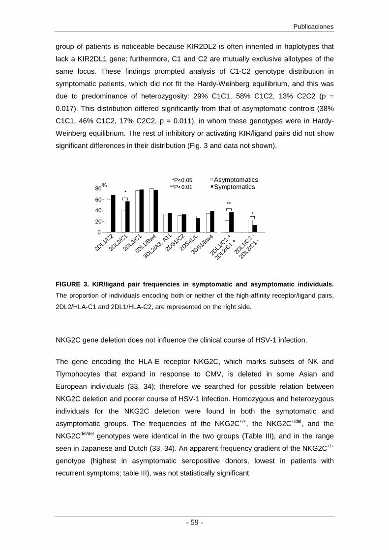
Publicaciones
- 59 -
group of patients is noticeable because KIR2DL2 is often inherited in haplotypes that
lack a KIR2DL1 gene; furthermore, C1 and C2 are mutually exclusive allotypes of the
same locus. These findings prompted analysis of C1-C2 genotype distribution in
symptomatic patients, which did not fit the Hardy-Weinberg equilibrium, and this was
due to predominance of heterozygosity: 29% C1C1, 58% C1C2, 13% C2C2 (p =
0.017). This distribution differed significantly from that of asymptomatic controls (38%
C1C1, 46% C1C2, 17% C2C2, p = 0.011), in whom these genotypes were in Hardy-
Weinberg equilibrium. The rest of inhibitory or activating KIR/ligand pairs did not show
significant differences in their distribution (Fig. 3 and data not shown).
FIGURE 3. KIR/ligand pair frequencies in symptomatic and asymptomatic individuals.
The proportion of individuals encoding both or neither of the high-affinity receptor/ligand pairs,
2DL2/HLA-C1 and 2DL1/HLA-C2, are represented on the right side.
NKG2C gene deletion does not influence the clinical course of HSV-1 infection.
The gene encoding the HLA-E receptor NKG2C, which marks subsets of NK and
Tlymphocytes that expand in response to CMV, is deleted in some Asian and
European individuals (33, 34); therefore we searched for possible relation between
NKG2C deletion and poorer course of HSV-1 infection. Homozygous and heterozygous
individuals for the NKG2C deletion were found in both the symptomatic and
asymptomatic groups. The frequencies of the NKG2C+/+, the NKG2C+/del, and the
NKG2Cdel/del genotypes were identical in the two groups (Table III), and in the range
seen in Japanese and Dutch (33, 34). An apparent frequency gradient of the NKG2C+/+
genotype (highest in asymptomatic seropositive donors, lowest in patients with
recurrent symptoms; table III), was not statistically significant.
**
*
2DL1
/C2
+
2DL2
/C1
+
2DL1
/C2
-
2DL2
/C1
-0
20
40
60
80
2DL1
/C2
2DL2/C
1
2DL3
/C1
3DL1
/Bw4
3DL2
/A3,
A11
2DS1/C
2
2DS4L
/L
3DS1/B
w4
AsymptomaticsSymptomatics
*
%*P<0.05
**P<0.01

Publicaciones
- 60 -
Table III. Distribution of NKG2C genotypes.
Asymptomatics Symptomatics
Seronegative
(N= 67) Seropositive
(N= 70) Total
(N=137) Sporadic
(N= 63) Recurrent (N= 100)
Total (N=163)
NKG2Cdel/del 4.5% 4.3% 4.4% 3.2% 5.0%
4.3%
NKG2Cwt/del 43.3% 27.1% 35.0% 31.7% 35.0% 33.7%
NKG2Cwt/wt 52.2% 68.6% 66.6% 65.1% 60.0%
62.0%
Homozygosity for the higher avidity allele of CD16A (FCGR3A) protects from clinical
HSV-1 infection.
Because FcγR mediate cellular responses (ADCC, phagocytosis) against infected cells
recognised by virus-specific IgG molecules, we investigated whether dimorphisms that
increase the avidity for IgG of two such receptors could improve the infection outcome.
The alleles encoding valine/phenylalanine-158 of CD16A (FCGR3A) and
arginine/histidine-131 of CD32A (FCGR2A) were genotyped, and their frequencies in
the whole sample were in the range reported for white populations (48, 49). After
excluding the individuals who lacked IgG antibodies for HSV-1, comparisons showed
that, as hypothesized, homozygosity for the higher affinity allele of CD16A (158V/V)
was less common in patients with clinical HSV-1 infection than in seropositive
individuals lacking HSV-1 symptoms (one tailed p = 0.024 , two tailed p = 0.037, OR =
0.43; Fig. 4). Furthermore, the dose of allele CD16A-158V had a significant effect on
protection from herpetic symptoms (Mantel-Haenszel test for linear trends p = 0.027;
table IV). In contrast with CD16A, the CD32A-131H/R dimorphism had no appreciable
influence on the clinical course of the infection (Fig. 4). No influence of either
dimorphism was seen on the frequency of clinical HSV-1 reactivations (data not
shown).
FIGURE 4. CD16A-158 (A) and
CD32A-131 (B) genotype
frequencies in symptomatic and
asymptomatic individuals.
B. CD32A-131A. CD16A-158
Asymptomatic seropositivesSymptomatics
*
0
20
40
60
80
100
V/V V/F + F/F
%
n.s.
0
20
40
60
80
100
H/H H/R + R/R
%
*P<0.05

Publicaciones
- 61 -
Table IV. CD16A-158 V allele dose and protection from symptomatic HSV-1 infection.
Genotype Asymptomatic seropositives
N=70, n (%) Symptomatics
N=163, n (%)
OR
CD16A-158 F/F 20 (28.6) 63 (38.6) 11
CD16A-158 V/F 35 (50.0) 83 (51.2) 0.75
CD16A-158 V/V 15 (21.4) 17 (10.4) 0.36
P of linear trend: 0.027
1In the Mantel-Haenszel test for linear trends, the reference genotype is assigned an arbitrary OR value of 1, to which the OR of the other genotypes are referred. The test measures the significance of a linear relationship between an ordinal variable, the dose of a risk or protective factor (the CD16A-158 V allele), and probability of an outcome (being symptomatic).
Multiple logistic regression confirms the association of several immunogenetic factors
with clinical HSV-1 infection.
Multiple logistic regression analysis was performed in order to determine the extent to
which each of the possibly associated variables identified in the univariate tests
contributes to clinical HSV-1 infection. This analysis revealed an independent and
cumulative effect of the presence or absence of several variables on the probability to
suffer symptomatic HSV-1 reactivations. These results confirmed that the presence of
HLA-B*18 and a CD16A-158V/V genotype grant protection from HSV-1 reactivation,
whereas HLA-C*15, the group of HLA-A alleles encoding the A19 allotype, and the
KIR2DL2/C1 receptor/ligand pair (but neither KIR2DL2 nor KIR2DS2 alone) confer
susceptibility to suffer herpetic reactivations (Table V). A predictive model on the
probability of suffering herpetic lesions was built with the parameters that, in multiple
regression, showed a significantly positive or negative association. Such model
classified correctly 70.6% of the patients with herpetic symptoms and 58.0% of the
asymptomatic donors. A ROC curve, which plots specificity and sensitivity of the model
(43), enclosed 68.7% of the maximum possible area (Fig. 5).
Table V. Variables that contribute independent and significantly to the risk of
symptomatic HSV-1 infection, according to multiple logistic regression analysis.
Variable p-value OR (C.I. 95%)
HLA-B*18 0.003 0.31 (0.14–0.66) HLA-C*15 0.007 5.93 (1.63–21.64) HLA-A19 0.035 1.71 (1.04–2.82) KIR2DL2:HLA-C1 0.002 2.15 (1.32–3.51) CD16A-158V/V 0.017 0.42 (0.21–0.85)
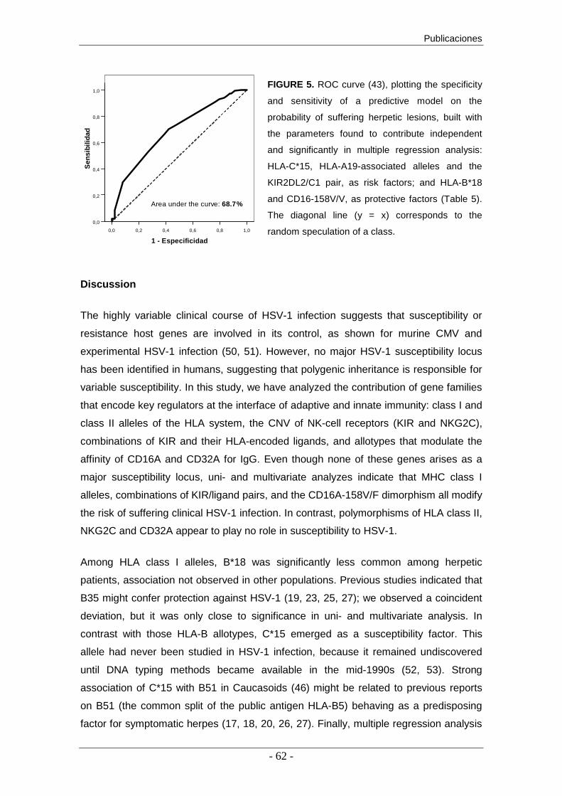
Publicaciones
- 62 -
1.0
0.8
0.6
0.4
0.2
0.00.0 0.2 0.4 0.6 0.8 1.0
Sen
sitiv
ity
Specificity
1,00,80,60,40,20,0
1 - Especificidad
1,0
0,8
0,6
0,4
0,2
0,0
Sen
sibi
lidad
1,00,80,60,40,20,0
1 - Especificidad
1,0
0,8
0,6
0,4
0,2
0,0
Sen
sibi
lidad
Area under the curve: 68.7%
FIGURE 5. ROC curve (43), plotting the specificity
and sensitivity of a predictive model on the
probability of suffering herpetic lesions, built with
the parameters found to contribute independent
and significantly in multiple regression analysis:
HLA-C*15, HLA-A19-associated alleles and the
KIR2DL2/C1 pair, as risk factors; and HLA-B*18
and CD16-158V/V, as protective factors (Table 5).
The diagonal line (y = x) corresponds to the
random speculation of a class.
Discussion
The highly variable clinical course of HSV-1 infection suggests that susceptibility or
resistance host genes are involved in its control, as shown for murine CMV and
experimental HSV-1 infection (50, 51). However, no major HSV-1 susceptibility locus
has been identified in humans, suggesting that polygenic inheritance is responsible for
variable susceptibility. In this study, we have analyzed the contribution of gene families
that encode key regulators at the interface of adaptive and innate immunity: class I and
class II alleles of the HLA system, the CNV of NK-cell receptors (KIR and NKG2C),
combinations of KIR and their HLA-encoded ligands, and allotypes that modulate the
affinity of CD16A and CD32A for IgG. Even though none of these genes arises as a
major susceptibility locus, uni- and multivariate analyzes indicate that MHC class I
alleles, combinations of KIR/ligand pairs, and the CD16A-158V/F dimorphism all modify
the risk of suffering clinical HSV-1 infection. In contrast, polymorphisms of HLA class II,
NKG2C and CD32A appear to play no role in susceptibility to HSV-1.
Among HLA class I alleles, B*18 was significantly less common among herpetic
patients, association not observed in other populations. Previous studies indicated that
B35 might confer protection against HSV-1 (19, 23, 25, 27); we observed a coincident
deviation, but it was only close to significance in uni- and multivariate analysis. In
contrast with those HLA-B allotypes, C*15 emerged as a susceptibility factor. This
allele had never been studied in HSV-1 infection, because it remained undiscovered
until DNA typing methods became available in the mid-1990s (52, 53). Strong
association of C*15 with B51 in Caucasoids (46) might be related to previous reports
on B51 (the common split of the public antigen HLA-B5) behaving as a predisposing
factor for symptomatic herpes (17, 18, 20, 26, 27). Finally, multiple regression analysis

Publicaciones
- 63 -
identified the group of alleles encoding an A19 antigen as possible risk factors; similar
results were obtained previously by some (16, 25, 27), but not all (19, 21), groups.
The picture that emerges from comparison of different studies is one in which MHC
class I polymorphism appears consistently associated with HSV-1 infection, the
predisposing or protective alleles varying in each study. Low reproducibility of results in
different populations might bear relation with geographical differences in distribution
and haplotypic combinations of HLA alleles. Confounding effects can also contribute to
conflicting results; among them, small cohorts of <50 patients in many previous
studies, control groups composed of general population instead of herpes-resistant
donors, and imprecision of serological HLA typing. We have addressed these problems
by studying >150 symptomatically infected individuals, and a similarly sized control
group comprising only asymptomatic individuals. Furthermore, this is the first study, to
our knowledge, performed entirely with DNA-based genotyping methods, of greater
precision and reproducibility, and the only techniques currently accepted for HLA-C and
class II alleles.
With the exception of C*15 and A*32 (part of the A19 allotype), the HLA class I alleles
that appear to modify the outcome of HSV-1 infection do not encode known KIR
ligands. Therefore, their divergent roles in the infection should derive from differential
functions as Ag-presenting molecules to CD8+ CTLs. A peptide prediction tool that
takes into account MHC binding, proteasomal C-terminal cleavage and TAP transport
efficacy, showed that the possibly protective (HLA-B*35 and B*18) and the risk alleles
(C*15 and most A19-related alleles) encode molecules that differ in their capacity to
present a unique ICP0 9-mer peptide. HSV-1 ICP0, a potential target for CTLs (54, 55),
is expressed during early infection and latency, and it triggers virus reactivation (47).
Therefore, efficacious presentation of ICP0-derived peptides by certain HLA class I
molecules could favour CTL control of infective virion production in sensory ganglia
(56).
Another aspect in which MHC molecules diverge functionally is in their capacity to
overcome the effect of viral immunoevasins. Independence of tapasin binding for
peptide loading enables some MHC molecules, including HLA-B35 (57), to migrate
more rapidly to the cell surface. Furthermore, although this characteristic is at the
expense of binding suboptimal peptides, it overcomes TAP blockade by protein ICP47
during HSV-1 infection (58). Tapasin-independent HLA molecules could therefore be
advantageous by presenting HSV-1 Ags early after infection/reactivation.
Unfortunately, tapasin-independence cannot be currently predicted reliably from the
primary structure of an HLA allotype, but B18 could share that feature with B35,
Publicaciones
- 64 -
because it carries a combination of two motifs (aspartate-114 and serine-116) that
associate with tapasin-independence (59-62).
Subversion of HLA class I expression in infected cells could facilitate HSV-1 evasion
from CTLs, but it is also a trigger for cytotoxicity and cytokine secretion by NK cells,
which sense it by means of KIR and other MHC receptors. We have previously
reported that KIR genes 2DL2 and 2DS2 contribute to the susceptibility to suffer
frequent, clinically relevant HSV-1 reactivations (29). Current results show that these
genes by themselves increase only modestly the risk of symptomatic herpes. It is
mainly the combination of KIR2DL2 with HLA-C alleles encoding its epitope ligand
[KIR2DS2 lacks any detectable affinity for HLA (63)] that increases noticeably
susceptibility to this disease.
KIR2DL2 is currently considered a 2DL3 allotype of higher affinity (64). Stronger
inhibition by KIR2DL2 in the presence of its cognate ligands could explain its
association with symptomatic herpes, because it might render NK cells less sensitive to
modest decreases in HLA-C-expression levels induced by HSV-1, and/or hamper
surveillance of viral reactivations by memory CD8 T cells expressing NK receptors.
Differences in KIR/ligand affinity are possibly relevant because IFN-γ could attenuate
HSV-1 blockade of HLA class I expression on infected epithelial cells (65). Consistent
with this model, another high-affinity receptor/ligand pair, 2DL1/C2, further increased
the risk conferred by 2DL2/C1. According to this reasoning, association of symptomatic
herpes of KIR2DS2 would only reflect its strong linkage disequilibrium with 2DL2. As an
anecdotal evidence, KIR genes 2DL2 and 2DS2 segregated separately in three donors
of this study – two 2DL2+2DS2– donors had HSV-1 symptoms, whereas the single
2DL2–2DS2+ individual was asymptomatic.
Our study on KIR diversity focused on CNV, but did not take into account possible
effects of allelic polymorphism. KIR2DL2 is represented in white populations by mostly
two alleles, KIR2DL2*001 and *003 (66). Their products share the three substitutions
that make KIR2DL2 a stronger receptor than KIR2DL3, and differ from each other by
two amino acid replacements: one in the second Ig-like domain (isoleucine/threonine-
200, respectively), and one in the cytoplasmic tail (alanine/threonine-313). The former
variation, located far from the ligand-binding region, seems not to modify the receptor
affinity (64), but more subtle effects of those polymorphisms on KIR2DL2 function and,
hence, on its association with HSV-1 infection, cannot be excluded. Similarly, we have
not considered quantitative differences in the affinity of KIR2DL2 for various HLA-C1,
and even -C2, ligands (64). Of note in the latter regard, C*15:02, the predominant C*15
allele in Spain (46, 52), encodes a C2 epitope for which KIR2DL2 lacks affinity, and it is

Publicaciones
- 65 -
recognised with average affinity by KIR2DL1 (64). Current data thus provide no clues
to understand an NK cell-mediated mechanism underlying the observed association of
C*15 with HSV-1 infection.
The KIR repertoire of different humans is enormously variable and variegated, not only
because of gene CNV and allelic polymorphism; both the receptor density on the cell
surface and the proportion of NK cells expressing each KIR vary in each individual.
Regulation of the KIR repertoire is incompletely understood, and it is genetically
controlled in part by KIR genes and by the HLA context (67). It is possible that certain
HLA-C ligands modulate differently the contribution of KIR2DL2 to the KIR repertoire,
or license to different extent the effector capacity of NK cells. Further investigation is
warranted to determine whether variable usage of KIR2DL2 modulates the influence
exerted on defense against HSV-1 by mere presence in the genome of the
genes/alleles encoding the receptor and its ligands.
Unidentified ligands for orphan KIR could have escaped our analysis, but no other KIR
and their known ligands associated with the clinical course of HSV-1 infection. And
neither did CNV of NKG2C, including its homozygous deletion. Confirmation of this
result would imply that this lectin-like NK receptor is redundant or plays no major role in
defense against HSV-1.
Finally, the higher affinity allotype of CD16A (158V) was identified as a protective factor
for symptomatic herpes. Valine-158 possibly enhances efficacy of humoral response
against HSV-1 by inducing a stronger interaction of CD16A with the lower hinge of IgG
(37). Importance of this interaction is illustrated by the decoy FcγR evolved by several
human viruses, including HSV-1, which, reciprocally, are possibly better counteracted
by competing host FcγR allotypes of higher affinity for IgG. Also supporting the
importance of CD16A for defense against HSV-1 would be the association of mutations
at aa 48 of CD16A with severe infections by herpesviruses, but that association is
controversial (68-71). Contrasting with the protective effect of CD16A-158V is the lack
of association between clinical herpes and CD32A-131 allotypes of variable affinity.
Lack of association might reflect greater importance of effective NK cell-mediated
cytotoxicity over phagocytosis in the elimination of HSV-1– infected cells and/or
predominance of the IgG subclasses bound preferentially by CD16A in the humoral
response to HSV-1.
This is, to our knowledge, the widest, hypothesis-based, immunogenetic study so far
performed on susceptibility to HSV-1 infection, and it includes several genes never
tested before. We have examined as many as 107 immunogenetic markers as potential
risk factors for symptomatic herpes (seventy-four HLA allotypes; sixteen KIR genes

Publicaciones
- 66 -
and genotypes; eight KIR:ligand pairs; and three NKG2C-, and six FCGR genotypes).
Paradoxically, this is also a pitfall of our approach, because the high number of
comparisons performed increases the probability of finding spurious associations.
Following Bonferroni’s correction for multiple tests would require setting a statistical
significance threshold at ~0.0005 (0.05 divided by 107 primary comparisons). For ORs
and allele frequencies in the range seen here, such confidence level could only have
been achieved by studying approximately 500-1000 donors. Consequently, the newly
identified associations (in particular those not suspected a priori, such as those of C*15
and B*18) should be considered preliminary until validated in other populations.
In summary, we have shown that susceptibility to clinical HSV-1 infection is modulated
by polymorphism in genes encoding molecules that control the effector functions of
cytotoxic T and NK lymphocytes (HLA class I, combinations of KIR and their ligands,
and CD16A); whereas the course of the infection is not modified by genetic
polymorphism in HLA class II and CD32A, molecules more implicated in cooperation
between CD4 T cells, B lymphocytes and phagocytic cells. Collectively, our results are
consistent with a crucial role of CTLs and NK cells in host defense against herpetic
infection, and they show that polymorphisms in genes controlling adaptive immune
responses, and surveillance of its sabotage, explain part of the variable susceptibility to
HSV-1 infection. Further research is warranted to determine the extent to which
diversity of innate immunity-related genes contributes to this disease.
Acknowledgements:
We are indebted to all individuals who generously donated blood samples; to the blood
bank staff and other colleagues who submitted and interviewed patients and healthy
voluntary donors; and to all technicians in the HLA and virology laboratories who
helped in sample collection and characterisation. This work was partially supported by
grants BFU2005–04622 and SAF2010-22153-C03-03, from the Spanish Ministerio de
Ciencia e Innovación.

Publicaciones
- 67 -
References
1. Cunningham, A. L., R. J. Diefenbach, M. Miranda-Saksena, L. Bosnjak, M. Kim, C. Jones, and M. W. Douglas. 2006. The cycle of human herpes simplex virus infection: virus transport and immune control. J Infect Dis. 194 Suppl 1: S11-18.
2. Pollara, G., M. Jones, M. E. Handley, M. Rajpopat, A. Kwan, R. S. Coffin, G. Foster, B. Chain, and D. R. Katz. 2004. Herpes simplex virus type-1-induced activation of myeloid dendritic cells: the roles of virus cell interaction and paracrine type I IFN secretion. J Immunol. 173: 4108-4119.
3. Cose, S. C., C. M. Jones, M. E. Wallace, W. R. Heath, and F. R. Carbone. 1997. Antigen-specific CD8+ T cell subset distribution in lymph nodes draining the site of herpes simplex virus infection. Eur J Immunol. 27: 2310-2316.
4. Frank, G. M., A. J. Lepisto, M. L. Freeman, B. S. Sheridan, T. L. Cherpes, and R. L. Hendricks. 2009. Early CD4(+) T cell help prevents partial CD8(+) T cell exhaustion and promotes maintenance of Herpes Simplex Virus 1 latency. J Immunol 184: 277-286.
5. Moretta, L., C. Bottino, D. Pende, M. Vitale, M. C. Mingari, and A. Moretta. 2004. Different checkpoints in human NK-cell activation. Trends Immunol 25: 670-676.
6. Lanier, L. L. 2005. NK cell recognition. Annu Rev Immunol 23: 225-274. 7. Karre, K. 1997. How to recognize a foreign submarine. Immunol Rev 155: 5-9. 8. Hill, A., P. Jugovic, I. York, G. Russ, J. Bennink, J. Yewdell, H. Ploegh, and D. Johnson.
1995. Herpes simplex virus turns off the TAP to evade host immunity. Nature. 375: 411-415.
9. Dubin, G., E. Socolof, I. Frank, and H. M. Friedman. 1991. Herpes simplex virus type 1 Fc receptor protects infected cells from antibody-dependent cellular cytotoxicity. J Virol. 65: 7046-7050.
10. Casrouge, A., S. Y. Zhang, C. Eidenschenk, E. Jouanguy, A. Puel, K. Yang, A. Alcais, C. Picard, N. Mahfoufi, N. Nicolas, L. Lorenzo, S. Plancoulaine, B. Senechal, F. Geissmann, K. Tabeta, K. Hoebe, X. Du, R. L. Miller, B. Heron, C. Mignot, T. B. de Villemeur, P. Lebon, O. Dulac, F. Rozenberg, B. Beutler, M. Tardieu, L. Abel, and J. L. Casanova. 2006. Herpes simplex virus encephalitis in human UNC-93B deficiency. Science. 314: 308-312.
11. Dupuis, S., E. Jouanguy, S. Al-Hajjar, C. Fieschi, I. Z. Al-Mohsen, S. Al-Jumaah, K. Yang, A. Chapgier, C. Eidenschenk, P. Eid, A. Al Ghonaium, H. Tufenkeji, H. Frayha, S. Al-Gazlan, H. Al-Rayes, R. D. Schreiber, I. Gresser, and J. L. Casanova. 2003. Impaired response to interferon-alpha/beta and lethal viral disease in human STAT1 deficiency. Nat Genet. 33: 388-391.
12. Niehues, T., J. Reichenbach, J. Neubert, S. Gudowius, A. Puel, G. Horneff, E. Lainka, U. Dirksen, H. Schroten, R. Doffinger, J. L. Casanova, and V. Wahn. 2004. Nuclear factor kappaB essential modulator-deficient child with immunodeficiency yet without anhidrotic ectodermal dysplasia. J Allergy Clin Immunol. 114: 1456-1462.
13. Perez de Diego, R., V. Sancho-Shimizu, L. Lorenzo, A. Puel, S. Plancoulaine, C. Picard, M. Herman, A. Cardon, A. Durandy, J. Bustamante, S. Vallabhapurapu, J. Bravo, K. Warnatz, Y. Chaix, F. Cascarrigny, P. Lebon, F. Rozenberg, M. Karin, M. Tardieu, S. Al-Muhsen, E. Jouanguy, S. Y. Zhang, L. Abel, and J. L. Casanova. 2010. Human TRAF3 adaptor molecule deficiency leads to impaired Toll-like receptor 3 response and susceptibility to herpes simplex encephalitis. Immunity. 33: 400-411.
14. Zhang, S. Y., E. Jouanguy, S. Ugolini, A. Smahi, G. Elain, P. Romero, D. Segal, V. Sancho-Shimizu, L. Lorenzo, A. Puel, C. Picard, A. Chapgier, S. Plancoulaine, M. Titeux, C. Cognet, H. von Bernuth, C. L. Ku, A. Casrouge, X. X. Zhang, L. Barreiro, J. Leonard, C. Hamilton, P. Lebon, B. Heron, L. Vallee, L. Quintana-Murci, A. Hovnanian, F. Rozenberg, E. Vivier, F. Geissmann, M. Tardieu, L. Abel, and J. L. Casanova. 2007. TLR3 deficiency in patients with herpes simplex encephalitis. Science. 317: 1522-1527.
15. Hobbs, M. R., B. B. Jones, B. E. Otterud, M. Leppert, and J. D. Kriesel. 2008. Identification of a herpes simplex labialis susceptibility region on human chromosome 21. J Infect Dis. 197: 340-346.
16. Blackwelder, W. C., R. Dolin, K. K. Mittal, P. M. McNamara, and F. J. Payne. 1982. A population study of herpesvirus infections and HLA antigens. Am J Epidemiol 115: 569-576.
17. Colin, J., C. Chastel, J. P. Saleun, J. F. Morin, and G. Renard. 1979. [Recurrent ocular herpes simplex infection and HLA antigens; preliminary results (author's transl)]. J Fr Ophtalmol 2: 263-266.

Publicaciones
- 68 -
18. Damgaard-Jensen, L., N. Ehlers, and F. Kissmeyer-Nielsen. 1979. HLA types in corneal diseases. Acta Ophthalmol (Copenh) 57: 982-985.
19. Gallina, G., V. Cumbo, P. Messina, M. A. Modica, and C. Caruso. 1987. MHC-linked genetic factors (HLA-B35) influencing recurrent circumoral herpetic lesions. Dis Markers 5: 191-197.
20. Jabbar, A. A., A. M. al-Samarai, and N. S. al-Amar. 1991. HLA antigens associated with susceptibility to herpes simplex virus infection. Dis Markers. 9: 281-287.
21. Legendre, C., A. S. Russell, and M. Jeannet. 1982. HLA antigens in patients with recrudescent herpes simplex infections. Tissue Antigens 19: 85-89.
22. Meyers-Elliott, R. H., J. H. Elliott, W. A. Maxwell, T. H. Pettit, D. M. O'Day, P. I. Terasaki, and D. Bernoco. 1980. HLA antigens in recurrent stromal herpes simplex virus keratitis. Am J Ophthalmol 89: 54-57.
23. Middleton, D., T. H. Hutchison, and J. Lynd. 1983. HLA antigen frequency in erythema multiforme and in recurrent herpes simplex. Tissue Antigens 21: 264-267.
24. Russell, A. S., and J. Schlaut. 1975. HL-A transplantation antigens in subjects susceptible to recrudescent herpes labialis. Tissue Antigens 6: 257-261.
25. Russell, A. S., and J. Schlaut. 1977. Association of HLA-A1 antigen and susceptibility to recurrent cold sores. Arch Dermatol. 113: 1721-1722.
26. Simon, M., Jr., D. Djawari, and A. Schonberger. 1984. [HLA typing of patients with herpes simplex recidivans and postherpetic erythema exsudativum multiforme]. Z Hautkr 59: 1087-1089.
27. Zimmerman, T. J., J. I. McNeill, A. Richman, H. E. Kaufman, and S. R. Waltman. 1977. HLA types and recurrent corneal herpes simplex infection. Invest Ophthalmol Vis Sci 16: 756-757.
28. Vilches, C., and P. Parham. 2002. KIR: Diverse, rapidly evolving receptors of innate and adaptive immunity. Annu Rev Immunol 20: 217-251.
29. Estefanía, E., N. Gómez-Lozano, F. Portero, R. de Pablo, R. Solís, S. Sepúlveda, M. Vaquero, M. A. González, E. Suárez, G. Roustán, and C. Vilches. 2007. Influence of KIR-gene diversity on the course of HSV-1 infection: resistance to the disease is associated with the absence of KIR2DL2 and KIR2DS2. Tissue Antigens 70: 34-41.
30. Lee, N., M. Llano, M. Carretero, A. Ishitani, F. Navarro, M. Lopez-Botet, and D. E. Geraghty. 1998. HLA-E is a major ligand for the natural killer inhibitory receptor CD94/NKG2A. Proc Natl Acad Sci U S A 95: 5199-5204.
31. Braud, V. M., D. S. Allan, C. A. O'Callaghan, K. Soderstrom, A. D'Andrea, G. S. Ogg, S. Lazetic, N. T. Young, J. I. Bell, J. H. Phillips, L. L. Lanier, and A. J. McMichael. 1998. HLA-E binds to natural killer cell receptors CD94/NKG2A, B and C. Nature 391: 795-799.
32. Shum, B. P., L. R. Flodin, D. G. Muir, R. Rajalingam, S. I. Khakoo, S. Cleland, L. A. Guethlein, M. Uhrberg, and P. Parham. 2002. Conservation and variation in human and common chimpanzee CD94 and NKG2 genes. J Immunol. 168: 240-252.
33. Hikami, K., N. Tsuchiya, T. Yabe, and K. Tokunaga. 2003. Variations of human killer cell lectin-like receptors: common occurrence of NKG2-C deletion in the general population. Genes Immun. 4: 160-167.
34. Miyashita, R., N. Tsuchiya, K. Hikami, K. Kuroki, T. Fukazawa, M. Bijl, C. G. Kallenberg, H. Hashimoto, T. Yabe, and K. Tokunaga. 2004. Molecular genetic analyses of human NKG2C (KLRC2) gene deletion. Int Immunol. 16: 163-168.
35. Gumá, M., A. Angulo, C. Vilches, N. Gómez-Lozano, N. Malats, and M. López-Botet. 2004. Imprint of human cytomegalovirus infection on the NK cell receptor repertoire. Blood 104: 3664-3671.
36. Gumá, M., M. Budt, A. Saez, T. Brckalo, H. Hengel, A. Angulo, and M. López-Botet. 2006. Expansion of CD94/NKG2C+ NK cells in response to human cytomegalovirus-infected fibroblasts. Blood. 107: 3624-3631.
37. van Sorge, N. M., W. L. van der Pol, and J. G. van de Winkel. 2003. FcgammaR polymorphisms: Implications for function, disease susceptibility and immunotherapy. Tissue Antigens 61: 189-202.
38. Vilches, C., J. Castaño, N. Gómez-Lozano, and E. Estefanía. 2007. Facilitation of KIR genotyping by a PCR-SSP method that amplifies short DNA fragments. Tissue Antigens 70: 415-422.
39. Graef, T., A. K. Moesta, P. J. Norman, L. Abi-Rached, L. Vago, A. M. Older Aguilar, M. Gleimer, J. A. Hammond, L. A. Guethlein, D. A. Bushnell, P. J. Robinson, and P. Parham. 2009. KIR2DS4 is a product of gene conversion with KIR3DL2 that introduced
Publicaciones
- 69 -
specificity for HLA-A*11 while diminishing avidity for HLA-C. J Exp Med. 206: 2557-2572.
40. Martin, M. P., X. Gao, J. H. Lee, G. W. Nelson, R. Detels, J. J. Goedert, S. Buchbinder, K. Hoots, D. Vlahov, J. Trowsdale, M. Wilson, S. J. O'Brien, and M. Carrington. 2002. Epistatic interaction between KIR3DS1 and HLA-B delays the progression to AIDS. Nat Genet 22: 22.
41. Vilches, C., J. Castano, P. Munoz, and J. Penalver. 2008. Simple genotyping of functional polymorphisms of the human immunoglobulin G receptors CD16A and CD32A: a reference cell panel. Tissue Antigens. 71: 242-246.
42. Stranzl, T., M. V. Larsen, C. Lundegaard, and M. Nielsen. 2010. NetCTLpan: pan-specific MHC class I pathway epitope predictions. Immunogenetics. 62: 357-368.
43. Fawcett, T. 2006. An introduction to ROC analysis. Pattern Recognition Letters 27: 861-874.
44. Bader, C., C. S. Crumpacker, L. E. Schnipper, B. Ransil, J. E. Clark, K. Arndt, and I. M. Freedberg. 1978. The natural history of recurrent facial-oral infection with herpes simplex virus. J Infect Dis. 138: 897-905.
45. de Pablo, R., C. Vilches, M. E. Moreno, M. Rementería, R. Solís, and M. Kreisler. 1992. Distribution of HLA antigens in Spanish Gypsies: a comparative study. Tissue Antigens 40: 187-196.
46. Balas, A., F. Garcia-Sanchez, and J. L. Vicario. 2011. Allelic and haplotypic HLA frequency distribution in Spanish hematopoietic patients. Implications for unrelated donor searching. Tissue Antigens 77: 45-53.
47. Khanna, K. M., A. J. Lepisto, V. Decman, and R. L. Hendricks. 2004. Immune control of herpes simplex virus during latency. Curr Opin Immunol. 16: 463-469.
48. Lehrnbecher, T., C. B. Foster, S. Zhu, S. F. Leitman, L. R. Goldin, K. Huppi, and S. J. Chanock. 1999. Variant genotypes of the low-affinity Fcgamma receptors in two control populations and a review of low-affinity Fcgamma receptor polymorphisms in control and disease populations. Blood. 94: 4220-4232.
49. Gonzalez-Escribano, M. F., F. Aguilar, J. Sanchez-Roman, and A. Nunez-Roldan. 2002. FcgammaRIIA, FcgammaRIIIA and FcgammaRIIIB polymorphisms in Spanish patients with systemic lupus erythematosus. Eur J Immunogenet. 29: 301-306.
50. Lee, S. H., S. Girard, D. Macina, M. Busa, A. Zafer, A. Belouchi, P. Gros, and S. M. Vidal. 2001. Susceptibility to mouse cytomegalovirus is associated with deletion of an activating natural killer cell receptor of the C-type lectin superfamily. Nat Genet 28: 42-45.
51. Pereira, R. A., A. Scalzo, and A. Simmons. 2001. Cutting edge: a NK complex-linked locus governs acute versus latent herpes simplex virus infection of neurons. J Immunol. 166: 5869-5873.
52. Vilches, C., R. de Pablo, M. J. Herrero, M. E. Moreno, and M. Kreisler. 1993. Molecular cloning and polymerase chain reaction-sequence-specific oligonucleotide detection of the allele encoding the novel allospecificity HLA-Cw6.2 (Cw*1502) in Spanish gypsies. Hum Immunol 37: 259-263.
53. Bunce, M., and K. I. Welsh. 1994. Rapid DNA typing for HLA-C using sequence-specific primers (PCR-SSP): identification of serological and non-serologically defined HLA-C alleles including several new alleles. Tissue Antigens 43: 7-17.
54. Mikloska, Z., A. M. Kesson, M. E. Penfold, and A. L. Cunningham. 1996. Herpes simplex virus protein targets for CD4 and CD8 lymphocyte cytotoxicity in cultured epidermal keratinocytes treated with interferon-gamma. J Infect Dis. 173: 7-17.
55. Mikloska, Z., and A. L. Cunningham. 1998. Herpes simplex virus type 1 glycoproteins gB, gC and gD are major targets for CD4 T-lymphocyte cytotoxicity in HLA-DR expressing human epidermal keratinocytes. J Gen Virol 79: 353-361.
56. Liu, T., K. M. Khanna, X. Chen, D. J. Fink, and R. L. Hendricks. 2000. CD8(+) T cells can block herpes simplex virus type 1 (HSV-1) reactivation from latency in sensory neurons. J Exp Med. 191: 1459-1466.
57. Thammavongsa, V., M. Schaefer, T. Filzen, K. L. Collins, M. Carrington, N. Bangia, and M. Raghavan. 2009. Assembly and intracellular trafficking of HLA-B*3501 and HLA-B*3503. Immunogenetics. 61: 703-716.
58. Zernich, D., A. W. Purcell, W. A. Macdonald, L. Kjer-Nielsen, L. K. Ely, N. Laham, T. Crockford, N. A. Mifsud, M. Bharadwaj, L. Chang, B. D. Tait, R. Holdsworth, A. G. Brooks, S. P. Bottomley, T. Beddoe, C. A. Peh, J. Rossjohn, and J. McCluskey. 2004.
Publicaciones
- 70 -
Natural HLA class I polymorphism controls the pathway of antigen presentation and susceptibility to viral evasion. J Exp Med 200: 13-24.
59. Neisig, A., R. Wubbolts, X. Zang, C. Melief, and J. Neefjes. 1996. Allele-specific differences in the interaction of MHC class I molecules with transporters associated with antigen processing. J Immunol. 156: 3196-3206.
60. Park, B., S. Lee, E. Kim, and K. Ahn. 2003. A single polymorphic residue within the peptide-binding cleft of MHC class I molecules determines spectrum of tapasin dependence. J Immunol 170: 961-968.
61. Turnquist, H. R., H. J. Thomas, K. R. Prilliman, C. T. Lutz, W. H. Hildebrand, and J. C. Solheim. 2000. HLA-B polymorphism affects interactions with multiple endoplasmic reticulum proteins. Eur J Immunol. 30: 3021-3028.
62. Williams, A. P., C. A. Peh, A. W. Purcell, J. McCluskey, and T. Elliott. 2002. Optimization of the MHC class I peptide cargo is dependent on tapasin. Immunity 16: 509-520.
63. Moesta, A. K., T. Graef, L. Abi-Rached, A. M. Older Aguilar, L. A. Guethlein, and P. Parham. 2010. Humans differ from other hominids in lacking an activating NK cell receptor that recognizes the C1 epitope of MHC class I. J Immunol 185: 4233-4237.
64. Moesta, A. K., P. J. Norman, M. Yawata, N. Yawata, M. Gleimer, and P. Parham. 2008. Synergistic polymorphism at two positions distal to the ligand-binding site makes KIR2DL2 a stronger receptor for HLA-C than KIR2DL3. J Immunol. 180: 3969-3979.
65. Koelle, D. M., and L. Corey. 2003. Recent progress in herpes simplex virus immunobiology and vaccine research. Clin Microbiol Rev. 16: 96-113.
66. Hou, L., M. Chen, J. Ng, and C. K. Hurley. 2012. Conserved KIR allele-level haplotypes are altered by microvariation in individuals with European ancestry. Genes Immun 13: 47-58.
67. Yawata, M., N. Yawata, M. Draghi, A. M. Little, F. Partheniou, and P. Parham. 2006. Roles for HLA and KIR polymorphisms in natural killer cell repertoire selection and modulation of effector function. J Exp Med. 203: 633-645.
68. Lenart, M., E. Trzyna, M. Rutkowska, K. Bukowska-Strakova, A. Szaflarska, A. Pituch-Noworolska, A. Szczepanik, M. Zembala, and M. Siedlar. 2010. The loss of the CD16 B73.1/Leu11c epitope occurring in some primary immunodeficiency diseases is not associated with the FcgammaRIIIa-48L/R/H polymorphism. Int J Mol Med 26: 435-442.
69. Jawahar, S., C. Moody, M. Chan, R. Finberg, R. Geha, and T. Chatila. 1996. Natural Killer (NK) cell deficiency associated with an epitope-deficient Fc receptor type IIIA (CD16-II). Clin Exp Immunol. 103: 408-413.
70. de Vries, E., H. R. Koene, J. M. Vossen, J. W. Gratama, A. E. von dem Borne, J. L. Waaijer, A. Haraldsson, M. de Haas, and M. J. van Tol. 1996. Identification of an unusual Fc gamma receptor IIIa (CD16) on natural killer cells in a patient with recurrent infections. Blood. 88: 3022-3027.
71. Forbes, L. R., M. Paessler, and J. S. Orange. 2009. CD16 Deficiency Discovered in a Cohort of Patients with Severe Herpesviral Infections Using a New Diagnostic Test. Journal of Allergy and Clinical Immunology 123: S67-S67.
Abbreviations used in this paper : ADCC, antibody-dependent cellular cytotoxicity;
CNV, copy number variation; DC, dendritic cells; HW, Hardy-Weinberg; ICP, infected
cell protein; KIR, killer-cell Ig-like receptor; OR, odds ratio; PCR-SSO, polymerase
chain reaction sequence specific oligonucleotide; ROC, receiver operating
characteristic.
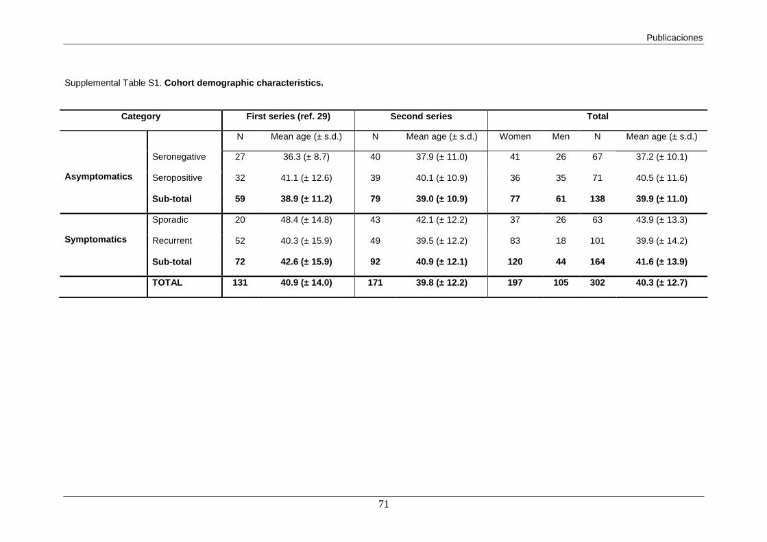
Publicaciones
71
Supplemental Table S1. Cohort demographic characteristics.
Category First series (ref. 29) Second series Total
N Mean age (± s.d.) N Mean age (± s.d.) Women Men N Mean age (± s.d.)
Seronegative 27 36.3 (± 8.7) 40 37.9 (± 11.0) 41 26 67 37.2 (± 10.1)
Seropositive 32 41.1 (± 12.6) 39 40.1 (± 10.9) 36 35 71 40.5 (± 11.6)
Asymptomatics
Sub-total 59 38.9 (± 11.2) 79 39.0 (± 10.9) 77 61 138 39.9 (± 11.0)
Sporadic 20 48.4 (± 14.8) 43 42.1 (± 12.2) 37 26 63 43.9 (± 13.3)
Recurrent 52 40.3 (± 15.9) 49 39.5 (± 12.2) 83 18 101 39.9 (± 14.2)
Symptomatics
Sub-total 72 42.6 (± 15.9) 92 40.9 (± 12.1) 120 44 164 41.6 (± 13.9)
TOTAL 131 40.9 (± 14.0) 171 39.8 (± 12.2) 197 105 302 40.3 (± 12.7)

Publicaciones
72
Supplemental table S2. HLA allele frequencies
Asymptomatics Symptomatics
Allele
Seronegatives
N= 67, n (%)
Seropositives
N= 71, n (%)
All
asymptomatics
N=138, n (%)
Sporadic
N= 63, n
(%)
Recurrent
N= 101,
n (%)
All
symptomatics
N=164, n (%)
A*01 10 (14.9) 10 (14.1) 20 (14.5) 7 (11.1) 18 (17.8) 25 (15.2)
A*02 25 (37.3) 37 (52.1) 62 (44.9) 32
(50.8) 49 (48.5) 81 (49.4)
A*03 16 (23.9) 12 (16.9) 28 (20.3) 20
(31.7) 20 (19.8) 40 (24.4)
A*11 11 (16.4) 9 (12.7) 20 (14.5) 6 (9.5) 14 (13.9) 20 (12.2)
A*23 8 (11.9) 7 (9.9) 15 (10.9) 4 (6.3) 7 (6.9) 11 (6.7)
A*24 12 (17.9) 16 (22.5) 28 (20.3) 10
(15.9) 16 (15.8) 26 (15.9)
A*25 2 (3.0) 2 (2.8) 4 (2.9) 1 (1.6) 5 (5.0) 6 (3.7)
A*26 9 (13.4) 6 (8.5) 15 (10.9) 5 (7.9) 4 (4.0) 9 (5.5)
A*29 11 (16.4) 12 (16.9) 23 (16.7) 12
(19.0) 23 (22.8) 35 (21.3)
A*30 7 (10.4) 3 (4.2) 10 (7.2) 6 (9.5) 8 (7.9) 14 (8.5)
A*31 4 (6.0) 3 (4.2) 7 (5.1) 3 (4.8) 2 (2.0) 5 (3.0)
A*32 2 (3.1) 5 (7.0) 7 (5.1) 7 (11.1) 10 (9.9) 17 (10.4)
A*33 3 (4.5) 6 (8.5) 9 (6.5) 5 (7.9) 8 (7.9) 13 (7.9)
A*66 1 (1.5) 0 (0) 1 (0.7) 0 (0) 2 (2.0) 2 (1.2)
A*68 2 (3.0) 1 (1.4) 3 (2.2) 3 (4.8) 9 (8.9) 12 (7.3)
A*69 1 (1.5) 0 (0) 1 (0.7) 0 (0) 0 (0) 0 (0)
A*80 0 (0) 1 (1.4) 1 (0.7) 2 (3.2) 0 (0) 2 (1.2)

Publicaciones
73
Supplemental table S2 (continued) Asymptomatics Symptomatics
Allele
Seronegatives
N= 67, n (%)
Seropositives
N= 71, n (%)
All
asymptomatics
N=138, n (%)
Sporadic
N= 63, n
(%)
Recurrent
N= 101,
n (%)
All
symptomatics
N=164, n (%)
B*07 14 (20.9) 9 (12.7) 23 (16.7) 12
(19.0)
17 (16.8) 29 (17.7)
B*08 4 (6.0) 10 (14.1) 14 (10.1) 8 (12.7) 8 (7.9) 16 (9.8)
B*13 3 (4.5) 3 (4.2) 6 (4.3) 4 (6.3) 3 (3.0) 7 (4.3)
B*14 8 (11.9) 13 (18.3) 21 (15.2) 9 (14.3) 17 (16.8) 26 (15.9)
B*15 3 (4.5) 7 (9.9) 10 (7.2) 5 (7.9) 9 (8.9) 14 (8.5)
B*18 13 (19.4) 13 (18.3) 26 (18.8) 6 (9.5) 6 (5.9) 12 (7.3)
B*27 6 (9.0) 4 (5.6) 10 (7.2) 6 (9.5) 3 (3.0) 9 (5.5)
B*35 11 (16.4) 15 (21.1) 26 (18.8) 8 (12.7) 13 (12.9) 20 (12.2)
B*37 2 (3.0) 0 (0) 2 (1.4) 2 (3.2) 1 (1.0) 3 (1.8)
B*38 2 (3.0) 3 (4.2) 5 (3.6) 4 (6.3) 2 (2.0) 6 (3.7)
B*39 2 (3.0) 2 (2.8) 4 (2.9) 1 (1.6) 3 (3.0) 4 (2.4)
B*40 3 (4.5) 5 (7.0) 8 (5.8) 3 (4.8) 8 (7.9) 11 (6.7)
B*41 1 (1.5) 2 (2.8) 3 (2.2) 3 (4.8) 2 (2.0) 5 (3.0)
B*44 26 (38.8) 20 (28.2) 46 (33.3) 19
(30.2)
36 (35.6) 55 (33.5)
B*45 4 (6.0) 1 (1.4) 5 (3.6) 4 (6.3) 0 (0) 4 (2.4)
B*47 2 (3.0) 0 (0) 2 (1.4) 0 (0) 1 (1.0) 1 (0.6)
B*48 1 (1.5) 0 (0) 1 (0.7) 0 (0) 0 (0) 0 (0)
B*49 6 (9.0) 4 (5.6) 10 (7.2) 6 (9.5) 12 (11.9) 18 (11.0)
B*50 2 (3.0) 6 (8.5) 8 (5.8) 3 (4.8) 12 (11.9) 15 (9.1)
B*51 10 (14.9) 11 (15.5) 21 (15.2) 13
(20.6)
17 (16.8) 30 (18.3)
B*52 2 (3.0) 1 (1.4) 3 (2.2) 1 (1.6) 3 (3.0) 4 (2.4)
B*53 0 (0) 1 (1.4) 1 (0.7) 3 (4.8) 1 (1.0) 4 (2.4)
B*55 0 (0) 3 (4.2) 3 (2.2) 0 (0) 3 (3.0) 3 (1.8)
B*56 0 (0) 0 (0) 0 (0) 1 (1.6) 2 (2.0) 3 (1.8)
B*57 6 (9.0) 3 (4.2) 9 (6.5) 0 (0) 13 (12.9) 13 (7.9)
B*58 1 (1.5) 1 (1.4) 2 (1.4) 0 (0) 3 (3.0) 3 (1.8)
B*67 0 (0) 0 (0) 0 (0) 1 (1.6) 0 (0) 1 (0.6)
B*78 0 (0) 1 (1.4) 1 (0.7) 0 (0) 0 (0) 0 (0)

Publicaciones
74
Supplemental table S2 (continued) Asymptomatics Symptomatics
Allele
Seronegatives
N= 67, n (%)
Seropositives
N= 71, n (%)
All
asymptomatics
N=138, n (%)
Sporadic
N= 63, n
(%)
Recurrent
N= 101,
n (%)
All
symptomatics
N=164, n (%)
C*01 6 (9.0) 4 (5.6) 10 (7.2) 4 (6.3) 8 (7.9) 12 (7.3)
C*02 5 (7.5) 5 (7.0) 10 (7.2) 9 (14.3) 14 (13.9) 23 (14.0)
C*03 6 (9.0) 10 (14.1) 16 (11.6) 6 (9.5) 14 (13.9) 20 (12.2)
C*04 16 (23.9) 20 (28.2) 36 (26.1) 12
(19.0)
19 (18.8) 31 (18.9)
C*05 15 (22.4) 12 (16.9) 27 (19.6) 10
(15.9)
17 (16.8) 27 (16.5)
C*06 10 (14.9) 11 (15.5) 21 (15.2) 11
(17.5)
22 (21.8) 33 (20.1)
C*07 30 (48.8) 35 (49.3) 65 (47.1) 24
(38.1)
42 (41.6) 66 (40.2)
C*08 7 (10.4) 12 (16.9) 19 (13.8) 9 (14.3) 17 (16.8) 26 (15.9)
C*12 8 (11.9) 10 (14.1) 18 (13.0) 7 (11.1) 9 (8.9) 16 (9.8)
C*14 2 (3.0) 3 (4.2) 5 (3.6) 3 (4.8) 3 (3.0) 6 (3.7)
C*15 2 (3.0) 1 (1.4) 3 (2.2) 8 (12.7) 7 (6.9) 15 (9.1)
C*16 19 (28.4) 11 (15.5) 30 (21.7) 15
(23.8)
18 (17.8) 33 (20.1)
C*17 1 (1.5) 2 (2.8) 3 (2.2) 1 (1.6) 1 (1.0) 2 (1.2)
DRB1*01 15 (22.4) 20 (28.2) 35 (25.4) 10
(15.9)
27 (26.7) 37 (22.6)
DRB1*03 13 (19.4) 14 (19.7) 27 (19.6) 14
(22.2) 20 (19.8) 34 (20.7)
DRB1*04 11 (16.4) 22 (31.0) 33 (23.9) 11
(17.5)
28 (27.7) 39 (23.8)
DRB1*07 28 (41.8) 24 (33.8) 52 (37.7) 21
(33.3)
35 (34.7) 56 (34.1)
DRB1*08 3 (4.5) 3 (4.2) 6 (4.3) 2 (3.2) 1 (1.0) 3 (1.8)
DRB1*09 3 (4.5) 1 (1.4) 4 (2.9) 1 (1.6) 3 (3.0) 4 (2.4)
DRB1*10 5 (7.5) 1 (1.4) 6 (4.3) 4 (6.3) 2 (2.0) 6 (3.7)
DRB1*11 15 (22.4) 18 (25.4) 33 (23.9) 24
(38.1)
21 (20.8) 45 (27.4)
DRB1*12 0 (0) 1 (1.4) 1 (0.7) 2 (3.2) 2 (2.0) 4 (2.4)
DRB1*13 15 (22.4) 16 (22.5) 31 (22.5) 16
(25.4)
25 (24.8) 41 (25.0)
DRB1*14 2 (3.0) 2 (2.8) 4 (2.9) 2 (3.2) 3 (3.0) 5 (3.0)
DRB1*15 15 (22.4) 7 (9.9) 22 (15.9) 11
(17.5)
18 (17.8) 29 (17.7)
DRB1*16 4 (6.0) 2 (2.8) 6 (4.3) 1 (1.6) 5 (5.0) 6 (3.7)
DRB3 40 (59.7) 45 (63.4) 85 (61.6) 45
(71.4)
64 (63.4) 109 (66.5)
DRB4 38 (56.7) 41 (57.7) 79 (57.2) 28
(44.4)
60 (59.4) 88 (53.7)
DRB5 18 (26.9) 9 (12.7) 27 (19.6) 12
(19.0) 23 (22.8) 35 (21.3)

Publicaciones
75
Supplemental table S3. KIR-gene frequencies.
First series (ref. 29) Replication series (this study) Total
Gene1
Asymptomatics
N=59, n (%)
Symptomatics
N=72, n (%) p
Asymptomatics
N=79, n (%)
Symptomatics
N=92, n (%) p
Asymptomatics
N=138, n (%)
Symptomatics
N=164, n (%) p
Total
N=302, n (%)
KIR2DL1 58 (98.3) 68 (94.4) n.s. 74 (93.7) 90 (97.8) n.s. 132 (95.7) 158 (96.3) n.s. 290 (96.0)
KIR2DL2 25 (42.4) 48 (66.7) 0.005 43 (54.4) 57 (62.0) n.s. 68 (49.3) 105 (64.0) 0.011 173 (57.3)
KIR2DL3 54 (91.5) 65 (90.3) n.s. 71 (89.9) 81 (88.0) n.s. 125 (90.6) 146 (89.0) n.s. 271 (89.7)
KIR2DL5 32 (54.2) 40 (55.6) n.s. 44 (55.7) 55 (59.8) n.s. 76 (55.1) 95 (57.9) n.s. 171 (56.6)
KIR3DL1 57 (96.6) 69 (95.8) n.s. 79 (100) 85 (92.4) n.s. 136 (98.6) 154 (93.9) n.s. 290 (96.0)
KIR2DS1 23 (39.0) 34 (47.2) n.s. 33 (41.8) 43 (46.7) n.s. 56 (40.6) 77 (47.0) n.s. 131 (43.4)
KIR2DS2 25 (42.4) 48 (66.7) 0.005 44 (55.7) 55 (59.8) n.s. 69 (50.0) 103 (62.8) 0.027 172 (57.0)
KIR2DS3 18 (30.5) 26 (36.1) n.s. 25 (31.6) 33 (35.9) n.s. 43 (31.2) 61 (37.2) n.s. 104 (34.4)
KIR2DS4 57 (96.6) 69 (95.8) n.s. 79 (100) 84 (91.3) n.s. 136 (98.6) 153 (93.3) n.s. 289 (95.7)
KIR2DS4*001 28 (47.4) 26 (36.1) n.s. 28 (35.4) 29 (31.5) n.s. 56 (40.6) 55 (33.5) n.s. 111 (36.7)
KIR2DS5 19 (32.2) 21 (29.2) n.s. 25 (31.6) 29 (31.5) n.s. 44 (31.9) 50 (30.5) n.s. 94 (31.1)
KIR3DS1 22 (37.3) 32 (44.4) n.s. 34 (43.0) 44 (47.8) n.s. 56 (40.6) 76 (46.3) n.s. 132 (43.7)
KIR2DP1 58 (98.3) 67 (93.1) n.s. 74 (93.7) 90 (97.8) n.s. 132 (95.7) 157 (95.7) n.s. 289 (95.7)
1The framework genes that mark the ends (KIR3DL3 and KIR3DL2) and the middle (KIR3DP1 and KIR2DL4) of the KIR complex were conserved in all donors, and they are not represented here.
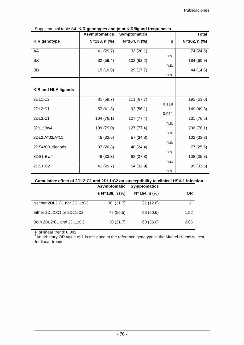
Publicaciones
- 76 -
Supplemental table S4. KIR genotypes and joint KIR/ligand frequencies.
KIR genotype
Asymptomatics
N=138, n (%)
Symptomatics
N=164, n (%) p
Total
N=302, n (%)
AA 41 (29.7) 33 (20.1) n.s.
74 (24.5)
BX 82 (59.4) 102 (62.2) n.s.
184 (60.9)
BB 15 (10.9) 29 (17.7) n.s.
44 (14.6)
KIR and HLA ligands
2DL1:C2 81 (58.7) 111 (67.7) 0.119
192 (63.6)
2DL2:C1 57 (41.3) 92 (56.1) 0.011
149 (49.3)
2DL3:C1 104 (76.1) 127 (77.4) n.s.
231 (76.5)
3DL1:Bw4 109 (79.0) 127 (77.4) n.s.
236 (78.1)
3DL2:A*03/A*11 45 (32.6) 57 (34.8) n.s.
102 (33.8)
2DS4*001:ligands 37 (26.8) 40 (24.4) n.s.
77 (25.5)
3DS1:Bw4 46 (33.3) 62 (37.8) n.s.
108 (35.8)
2DS1:C2 41 (29.7) 54 (32.9) n.s.
95 (31.5)
Cumulative effect of 2DL2:C1 and 2DL1:C2 on susceptibility to clinical HSV-1 infection
Asymptomatic
s N=138, n (%)
Symptomatics
N=164, n (%) OR
Neither 2DL2:C1 nor 2DL1:C2 30 (21.7) 21 (12.8) 11
Either 2DL2:C1 or 2DL1:C2 78 (56.5) 83 (50.6) 1.52
Both 2DL2:C1 and 2DL1:C2 30 (21.7) 60 (36.6) 2.86
P of linear trend: 0.002 1An arbitrary OR value of 1 is assigned to the reference genotype in the Mantel-Haenszel test for linear trends.


Artículo 2
Tissue Antigens 2012: 80: 184-7.


Publicaciones
- 80 -
Assessment of copy-number variation in the NKG2C receptor gene in a single-
tube and characterization of a reference cell panel, using standard polymerase
chain reaction
M Moraru,1 M Cañizares,1 A Muntasell,2 R de Pablo,1 M López-Botet,2,3 and C Vilches1. 1Laboratorio de Inmunogenética-HLA, Servicio de Inmunología, Hospital Universitario
Puerta de Hierro, Majadahonda, Madrid; 2IMIM-Hospital del Mar Research Institute,
Barcelona; and 3DCEX, Universitat Pompeu Fabra, Barcelona, Spain
Abstract
Natural Killer and T-lymphocytes monitor HLA-E expression through CD94:NKG2
heterodimers. Structural polymorphism is not a hallmark for NK-complex genes on
chromosome 12, except for complete NKG2C deletion in some humans. We present a
method for fast, simple and accurate assessment of NKG2C copy-number variation –
presence or absence in the genome of an NKG2C gene, in homo- or heterozygosis, is
detected by a single conventional polymerase chain reaction that yields amplicons of
different lengths in each genotype. We have also determined the NKG2C genotypes of
a reference cell panel comprising 13 NK- and tumor-cell lines and 39 Epstein-Barr virus
transformed cells from the International Histocompatibility Workshop. Our results
should facilitate research on the importance of NKG2C and its deletion for immunity.
Key words : copy number variation, gene deletion, genotyping, human, NK cell
receptors, polymerase-chain reaction

Publicaciones
- 81 -
Natural Killer (NK) cells and CD8+ cytotoxic T-lymphocytes (CTL) are key components
of the cellular immune response to infections and tumours. While CTLs recognize
target cells in an antigen-dependent manner, NK cells detect and kill, without previous
sensitization, cells displaying altered molecular patterns. Mammal NK cells sense
major histocompatibility complex (MHC) expression through at least four families of
receptors, all of which include both activating and inhibitory members. Paired
inhibitory/activating receptors are believed to balance NK-cell behaviour in the
presence of a potential target cell, and an important set of these receptors are also
expressed on CTLs (1). Inhibitory MHC receptors are fundamental to the missing self
model of NK-cell regulation (2). In contrast, the role of their activating counterparts in
immunity is obscure – whilst some may recognise pathogen-encoded (3) or -modified
molecules, most activating homologues of inhibitory MHC receptors have self or
unknown ligands.
One of the human families of MHC receptors comprises CD94:NKG2 heterodimers of
the C-type lectin-like superfamily encoded in the NK complex (NKC) on chromosome
12p12-13 (4). Three homologous genes of this complex, NKG2A (KLRC1), NKG2C
(KLRC2), and NKG2E (KLRC3), code for NKG2 molecules that complex with CD94;
the first two heterodimers have been formally shown to recognize human leukocyte
antigen (HLA)-E molecules presenting the leader peptides of other HLA class I
products (5, 6). Whereas CD94/NKG2A transmits an inhibitory signal, the
CD94/NKG2C and -E heterodimers are activating receptors.
NKG2C is expressed on subsets of NK- and T-lymphocytes, which expand to variable
extent in response to cytomegalovirus (CMV) (7-10). Expansions of NKG2C+ cells have
also been observed concomitantly with other viral infections (8, 11-13), but they appear
to be also associated to CMV. The manner in which NKG2C+ lymphocytes contribute to
cellular immunity to CMV is so far unknown.
Contrasting with the enormous variability of the Killer-cell Immunoglobulin-like Receptor
complex on chromosome 19, the NKC on chromosome 12 is mostly conserved in terms
of gene content and allelic polymorphism. An exception is NKG2C – although it shows
only modest polymorphism (14, 15), complete deletion of NKG2C (KLR2C) has been
described in two different populations of Asian and Caucasoid origins (15, 16). Up to
32.4% of the studied cohorts carried this deletion in heterozygosis and about 4%
completely lacked NKG2C. Thus far, this gene deletion has not been related to any

Publicaciones
- 82 -
functional feature and only a very limited number of studies have analysed its possible
influence on human health (16-18). Enabling large population studies could help to
shed light into this yet largely unexplored area of knowledge.
Polymerase-chain reaction with sequence-specific primers (PCR-SSP) is a fast, simple
and reliable method for genotype assignment (19). Alleles or highly homologous genes
are discriminated in this technique by means of appropriate sets of oligonucleotide
primers that target distinctive single-nucleotide polymorphisms under stringent PCR
conditions. The integrity of each individual PCR is usually warranted by an internal
positive control band, amplified from a conserved gene by an additional primer pair.
Previous studies have used PCR-SSP methods to study either the presence in the
genome of an NKG2C gene, or the gene arrangement resulting from its deletion (7, 14-
16, 20).
We aimed at improving the assessment of NKG2C copy-number variation by designing
a simpler and more comprehensive method that showed, in a single reaction, either
presence or deletion of NKG2C. To that end, we adapted the PCR-SSP technique and
designed a single-tube PCR with two pairs of oligonucleotides per test. Two primers
(KLRdelF and KLRdelR, Table 1) are specific for the intergenic regions flanking
NKG2C and generate a 411-bp amplicon only in carriers of an NKG2C deletion, in
whom the two regions come to close proximity (16); whereas a second primer pair
(KLRFg669 and KLRR+135, Table 1) amplifies, in DNAs having a NKG2C gene, a 201-
bp fragment from exon 6 and its 3’untranslated region (Figure 1). The target sequences
of these primers are, according to the entries currently deposited in the
GenBank/EMBL/DDBJ databases, non-polymorphic to ensure that all carriers of a
NKG2C gene, irrespective of the allele they bear, generate the expected 201-bp PCR
product. To enhance specificity, all four primers recognise unique NKG2C sequences
seen in none of its highly similar paralogues.
From every individual, the multiplex reaction always amplifies one or both of the
possible amplicons, therefore it does not require an additional primer pair serving as an
internal positive control. The amplicons thus generated can then be separated and
distinguished, without further manipulation, by their different electrophoretic mobilities
in agarose gels (Figure 1). This way, small DNA amounts and only few hours are
required to analyse up to 95 samples in a single assay, using conventional
thermocyclers and appropriate electrophoresis chambers.
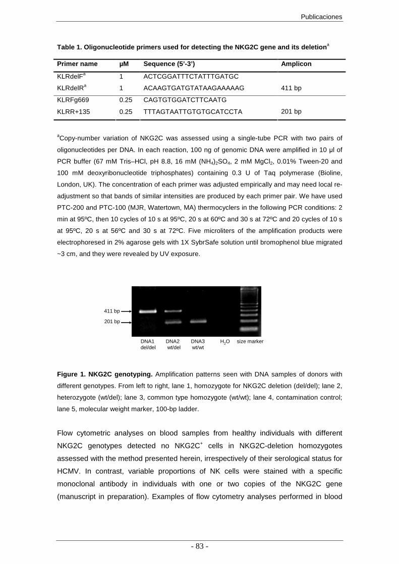
Publicaciones
- 83 -
Table 1. Oligonucleotide primers used for detecting the NKG2C gene and its deletion a
Primer name µM Sequence (5’-3’) Amplicon
KLRdelFa 1 ACTCGGATTTCTATTTGATGC
KLRdelRa 1 ACAAGTGATGTATAAGAAAAAG
411 bp
KLRFg669 0.25 CAGTGTGGATCTTCAATG
KLRR+135 0.25 TTTAGTAATTGTGTGCATCCTA
201 bp
aCopy-number variation of NKG2C was assessed using a single-tube PCR with two pairs of
oligonucleotides per DNA. In each reaction, 100 ng of genomic DNA were amplified in 10 µl of
PCR buffer (67 mM Tris–HCl, pH 8.8, 16 mM (NH4)2SO4, 2 mM MgCl2, 0.01% Tween-20 and
100 mM deoxyribonucleotide triphosphates) containing 0.3 U of Taq polymerase (Bioline,
London, UK). The concentration of each primer was adjusted empirically and may need local re-
adjustment so that bands of similar intensities are produced by each primer pair. We have used
PTC-200 and PTC-100 (MJR, Watertown, MA) thermocyclers in the following PCR conditions: 2
min at 95ºC, then 10 cycles of 10 s at 95ºC, 20 s at 60ºC and 30 s at 72ºC and 20 cycles of 10 s
at 95ºC, 20 s at 56ºC and 30 s at 72ºC. Five microliters of the amplification products were
electrophoresed in 2% agarose gels with 1X SybrSafe solution until bromophenol blue migrated
~3 cm, and they were revealed by UV exposure.
Figure 1. NKG2C genotyping. Amplification patterns seen with DNA samples of donors with
different genotypes. From left to right, lane 1, homozygote for NKG2C deletion (del/del); lane 2,
heterozygote (wt/del); lane 3, common type homozygote (wt/wt); lane 4, contamination control;
lane 5, molecular weight marker, 100-bp ladder.
Flow cytometric analyses on blood samples from healthy individuals with different
NKG2C genotypes detected no NKG2C+ cells in NKG2C-deletion homozygotes
assessed with the method presented herein, irrespectively of their serological status for
HCMV. In contrast, variable proportions of NK cells were stained with a specific
monoclonal antibody in individuals with one or two copies of the NKG2C gene
(manuscript in preparation). Examples of flow cytometry analyses performed in blood
DNA1 DNA2 DNA3 H2O size markerdel/del wt/del wt/wt
411 bp
201 bp

Publicaciones
- 84 -
samples from three healthy individuals with different NKG2C genotypes are shown in
Figure 2.
Figure 2. Expression of NKG2C in peripheral blood NK cells of three CMV-seronegative
individuals harbouring different NKG2C genotypes, assessed by multicolour flow cytometry.
NKG2C was analyzed in CD56+CD3– cells from the lymphocyte gate upon staining with anti-
NKG2C-Phycoerythrin (clone 134591, R&D, Minneapolis, MN), anti-CD3-peridin-chlorophyll-
protein, and anti-CD56-allophycocyanin (BD, Biosciences, San Diego, CA). Samples were
acquired on a FACSCalibur Cytometer, and CellQuest software was used for data analysis (BD
Biosciences). Surface NKG2C was undetectable in NK cells from NKG2Cdel/del CMV-
seropositive donors (not shown).
The consequence of NKG2C deletion has scarcely been investigated. Research on
NKG2C copy-number variation is hampered by a lack of appropriate control DNAs. To
solve this difficulty, we used our method to characterise worldwide available cell lines
that should serve as an NKG2C-genotyped reference panel. This panel comprises 52
cells, including: (i) 39 Epstein-Barr virus transformed B-lymphoblastoid cell lines (B-
LCLs) from the International Histocompatibility Workshop, a well-characterized
reference cell panel for the HLA and the Killer-cell Immunoglobulin-like Receptor
genes; (ii) four NK cell lines; and (iii), nine common tumour cells of haemopoietic and
non-haemopoietic lineages (Table 2). All three possible genotypes (NKG2Cwt/wt,
NKG2Cwt/del and NKG2Cdel/del) are represented by at least four cells of this panel, which
should allow access to appropriate control DNAs to laboratories willing to study the
copy number variation of NKG2C. The amplicons derived from six cell lines (two for
each genotype) were tested by direct sequencing to rule out possible cross-
amplification of undesired NKG2 genes. We further validated the method by excluding
the possibility that presence of a single amplicon could be a false-negative result due to
preferential amplification in an NKG2Cwt/del individual. To that end, ten DNAs with
apparent NKG2Cwt/wt and another ten genotyped as NKG2Cdel/del, were assayed with
both the primer pair recognising NKG2C and that specific for its deletion in two

Publicaciones
- 85 -
separate regular PCR-SSP assays. In all twenty samples, both primer pairs yielded
separately results identical to those obtained when they were combined in the same
tube (not shown).
Table 2. An NKG2C-genotyped reference cell panel.
IHW B-LCLs (no.) Other cell lines
NKG2C genotype: wt/wt NKG2C genotype: wt/del NKG2C genotype: wt/wt
AMALA 9064 CB6B 9060 NK3.3 NK cell line
BH 9046 DUCAF 9019 NK92 NK cell line
BM16 9038 HOM-2 9005 NKL NK cell line
BM21 9043 HOR 9053 YT NK cell line
BOLETH 9031 KT12 9073 JURKAT T cell leukemia
COX 9022 LE023 9267 RPMI 8866 B-lymphoblastoid
DBB 9052 MZ070782 9002 HeLa Cervix carcinoma
DEU 9025 PLH 9047 DU145 Prostate carcinoma
E4181324 9011 RSH 9021
IBW9 9049 SPOO10 9036
JBUSH 9035 TEM 9057 NKG2C genotype: wt/del
JESTHOM 9004 WDV 9062 HEK-293T Embryonic kidney
KAS116 9003 WT24 9015 LICR-LON-HMy-2 leukemia-derived B-cell line
LUY 9070 WT51 9029
OLGA 9071
PAR 9266 NKG2C genotype: del/del
PE117 9028 NKG2C genotype: del/del K562 Erythroleukemia
PITOUT 9051 BRI-P 9044 RAJI Burkitt Lymphoma
RML 9016 721.221 derived from B-LCL
SWEIG 9037 IHW 9249
T7527 9077
TAB089 9066
WT8 9017
YAR 9026
PCR-SSP methods combine simplicity and efficacy, as they require minimum amounts
of reagents and time for accurate allele assignation. We present here a variant of such
method for the determination of NKG2C copy-number variation. With the proposed
approach, similar to one used previously to study copy-number variation of the LILRA3
gene in multiple sclerosis (21), DNA fragments corresponding to both the complete and
the deleted genotypes are detected simultaneously in a single PCR reaction, further
facilitating the analysis of large DNA collections. Such fast and robust method should
enable research on the role of NKG2C in innate and adaptive immunity, and help to
establish the importance of its copy-number variation for human health.

Publicaciones
- 86 -
References
1. Lanier LL. NK cell recognition. Annu Rev Immunol 2005: 23: 225-74. 2. Karre K. How to recognize a foreign submarine. Immunol Rev 1997: 155: 5-9. 3. Arase H, Mocarski ES, Campbell AE, Hill AB, Lanier LL. Direct recognition of
cytomegalovirus by activating and inhibitory NK cell receptors. Science 2002: 296: 1323-6.
4. Trowsdale J, Barten R, Haude A, Stewart CA, Beck S, Wilson MJ. The genomic context of natural killer receptor extended gene families. Immunol Rev 2001: 181: 20-38.
5. Lee N, Llano M, Carretero M, et al. HLA-E is a major ligand for the natural killer inhibitory receptor CD94/NKG2A. Proc Natl Acad Sci U S A 1998: 95: 5199-204.
6. Braud VM, Allan DS, O'Callaghan CA, et al. HLA-E binds to natural killer cell receptors CD94/NKG2A, B and C. Nature 1998: 391: 795-9.
7. Gumá M, Angulo A, Vilches C, Gómez-Lozano N, Malats N, López-Botet M. Imprint of human cytomegalovirus infection on the NK cell receptor repertoire. Blood 2004: 104: 3664-71.
8. Gumá M, Cabrera C, Erkizia I, et al. Human cytomegalovirus infection is associated with increased proportions of NK cells that express the CD94/NKG2C receptor in aviremic HIV-1-positive patients. J Infect Dis 2006: 194: 38-41.
9. Gumá M, Budt M, Saez A, et al. Expansion of CD94/NKG2C+ NK cells in response to human cytomegalovirus-infected fibroblasts. Blood 2006: 107: 3624-31.
10. Monsivais-Urenda A, Noyola-Cherpitel D, Hernandez-Salinas A, et al. Influence of human cytomegalovirus infection on the NK cell receptor repertoire in children. Eur J Immunol 2010: 40: 1418-27.
11. Beziat V, Dalgard O, Asselah T, et al. CMV drives clonal expansion of NKG2C(+) NK cells expressing self-specific KIRs in chronic hepatitis patients. Eur J Immunol 2011: 42: 447-57.
12. Bjorkstrom NK, Lindgren T, Stoltz M, et al. Rapid expansion and long-term persistence of elevated NK cell numbers in humans infected with hantavirus. J Exp Med 2011: 208: 13-21.
13. Petitdemange C, Becquart P, Wauquier N, et al. Unconventional repertoire profile is imprinted during acute chikungunya infection for natural killer cells polarization toward cytotoxicity. PLoS Pathog 2011: 7: e1002268.
14. Shum BP, Flodin LR, Muir DG, et al. Conservation and variation in human and common chimpanzee CD94 and NKG2 genes. J Immunol 2002: 168: 240-52.
15. Hikami K, Tsuchiya N, Yabe T, Tokunaga K. Variations of human killer cell lectin-like receptors: common occurrence of NKG2-C deletion in the general population. Genes Immun 2003: 4: 160-7.
16. Miyashita R, Tsuchiya N, Hikami K, et al. Molecular genetic analyses of human NKG2C (KLRC2) gene deletion. Int Immunol 2004: 16: 163-8.
17. Thomas R, Low HZ, Kniesch K, Jacobs R, Schmidt RE, Witte T. Nkg2c Deletion Is a Risk Factor of Hiv Infection. AIDS Res Hum Retroviruses 2011: doi:10.1089/AID.2011.0253.
18. Moraru M, Cisneros E, de Pablo R, et al. Host genetic factors in susceptibility to Herpes Simplex type 1 virus infection: contribution of polymorphic genes at the interface of innate and adaptive immunity. J Immunol 2012: 188: 4412–20.
19. Newton CR, Graham A, Heptinstall LE, et al. Analysis of any point mutation in DNA. The amplification refractory mutation system (ARMS). Nucleic Acids Res 1989: 17: 2503-16.
20. Uhrberg M, Valiante NM, Shum BP, et al. Human diversity in killer cell inhibitory receptor genes. Immunity 1997: 7: 753-63.
21. Ordóñez D, Sánchez AJ, Martínez-Rodríguez JE, et al. Multiple sclerosis associates with LILRA3 deletion in Spanish patients. Genes Immun 2009: 10: 579-85.
Acknowledgments
This work was funded by grant SAF2010-22153-C03, from the Spanish Ministerio de
Ciencia e Innovación. A.M. is supported by the AECC Foundation. The authors declare
no conflict of interest.


Artículo 3
Aceptado para publicación, J Immunol 2015
Copyright © 2015 The American Association of Immunologists, Inc.


Publicaciones
- 90 -
NK cells and immunoglobulins interplay in defense against Herpes simplex virus
type 1: epistatic interaction of CD16A and IgG1 allotypes of variable affinity
modulates antibody-dependent cellular cytotoxicity and susceptibility to clinical
reactivation.
Manuela Moraru,* Laurel E. Black,† Aura Muntasell,‡ Francisca Portero,§ Miguel López-
Botet,‡ Hugh T. Reyburn,¶ Janardan P. Pandey,† and Carlos Vilches*
Departments of *Immunogenetics-HLA and §Microbiology, Instituto de Investigación Sanitaria
Puerta de Hierro, Majadahonda, Madrid, Spain; †Department of Microbiology and Immunology,
Medical University of South Carolina, Charleston, South Carolina, United States of America; ‡IMIM-Hospital del Mar Medical Research Institute, Barcelona, Spain; ¶Centro Nacional de
Biotecnología, Consejo Superior de Investigaciones Científicas, Madrid, Spain.
Abstract
Herpes simplex virus type 1 (HSV-1) latently infects most humans, causing a variable
clinical picture, which depends in part on host genetic factors. Both IgG and its cellular
Fc receptors CD16A and CD32A–C (encoded by genes FCGR3A and FCGR2A–C,
respectively, on chromosome 1) display polymorphisms that could affect their
defensive function. Of potential relevance are: a FCGR3A dimorphism resulting in
CD16A-valine/phenylalanine-158 allotypes with different IgG affinity; variations
conditioning NK-cell expression of CD32B or CD32C; and IgG1 heavy-chain (IGHG1)
and kappa-chain (IGKC) polymorphisms determining allotypes designated G1m and
Km. In this study we assessed the contribution of immunoglobulin genetic variations
and their interaction with FcR polymorphism, to HSV-1 susceptibility, and their impact
on NK-cell mediated antibody-dependent cellular cytotoxicity (ADCC). Our results show
an epistatic interaction between IGHG1 and FCGR3A, genotype G1m3/3 associating
with an asymptomatic course of HSV-1 infection only in homozygotes for higher-affinity
CD16A-158V, and vice versa. Furthermore, CD16A-158V and G1m3 allotypes
enhanced ADCC against opsonized, HSV-1-infected, fibroblasts. Conversely, Km
allotypes and CD32B or CD32C expression on NK cells did not significantly influence
HSV-1 susceptibility or ADCC. NK cells degranulating against immune serum-
opsonized, HSV-1-infected fibroblasts had heterogeneous phenotypes. Yet, enhanced
ADCC was observed among NK cells showing a differentiated, memory-like phenotype
(NKG2CbrightNKG2A–CD57+FcRγ–), which expand in response to human
cytomegalovirus. These results extend knowledge on the importance of immunogenetic
polymorphisms and NK-cell-antibody interplay in host response against HSV-1, and
point to the relevance of interactions between immune responses elicited during
chronic co-infection by multiple herpesviruses.

Publicaciones
- 91 -
Introduction
Herpes simplex virus-1 (HSV-1) infects most individuals, establishing life-long latency
with periodical reactivations that result in clinical relapses or asymptomatic virus
shedding. Although exposure to HSV-1 is virtually universal in adults, the clinical
course of the infection varies largely, which depends in part on host genetic factors,
including polymorphisms at the interface of innate and adaptive immunity, like those of
KIR and their HLA ligands and the IgG receptor CD16A (1-3). Viral replication is
controlled by both branches of the immune response, of which key components are
human NK cells. This lymphocyte subset exerts both cytokine production and cellular
toxicity and its behavior in the presence of a target cell depends on the balance of
activating and inhibitory signals transduced by multiple NK-cell receptors (4). HSV-1-
specific antibodies are detectable approximately one month after primary infection and
are life-lasting. The great majority of seropositive individuals carry antibodies of IgG1
subclass, while in only about half of them HSV-specific IgG3 is detected, HSV-IgG4
and HSV-IgG2 being rare findings (5). These specific antibodies have a demonstrated
neutralizing activity and readily mediate complement-dependent and antibody-
dependent cellular cytotoxicity (ADCC) (5). Yet, their presence in virtually all infected
individuals does not prevent sporadic or recurrent episodes of symptomatic reactivation
in up to half of them.
Several mechanisms help HSV-1 to evade the host immune system (6). Among these,
HSV-1 glycoproteins E and I (gE/gI) form a viral receptor for the crystallizable fraction
of immunoglobulins (vFcγR), which binds human IgG1, 2 and 4 with different affinities
and is expressed on both the viral envelope and the infected cell membrane (7-11).
The gE/gI decoy vFcγR does not transmit any activating signal but it can hamper host
cell FcγR and C1q binding to immune complexes, thus protecting the infected cells
from ADCC, complement-dependent neutralization and phagocytosis.
ADCC, a major pathway in the clearance of pathogens, depends on simultaneous
binding of IgG molecules to infected cells and to FcγR expressed on a variety of
effector cells, most importantly NK cells (12). Essential for cellular immune response to
antigen-bound IgG are low-intermediate affinity receptors for the Fc of IgG. Among
these, CD16A, CD16B, CD32A, CD32B and CD32C are encoded by five genes
(named FCGR3A–B and FCGR2A–C, respectively) clustered in ~150.000 bp on the
long arm of chromosome 1 – the FCGR locus. Several of these genes display
functional polymorphism, including both sequence and copy-number variation (13, 14).
Whereas CD32A (FCGR2A) is expressed mainly on myeloid leukocytes and CD16B
(FCGR3B) on polymorphonuclear cells, potentially relevant for NK cell-mediated ADCC
are genetic variations involving three of the FCGR genes (FCGR2B, FCGR2C and
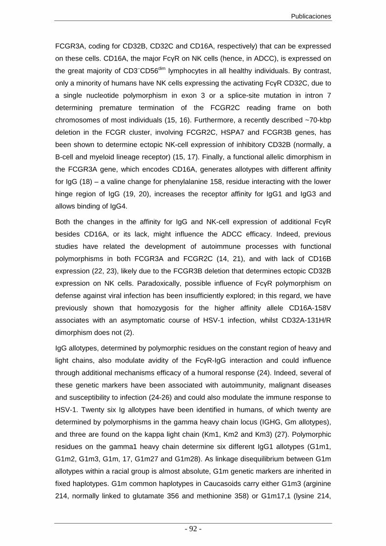
Publicaciones
- 92 -
FCGR3A, coding for CD32B, CD32C and CD16A, respectively) that can be expressed
on these cells. CD16A, the major FcγR on NK cells (hence, in ADCC), is expressed on
the great majority of CD3–CD56dim lymphocytes in all healthy individuals. By contrast,
only a minority of humans have NK cells expressing the activating FcγR CD32C, due to
a single nucleotide polymorphism in exon 3 or a splice-site mutation in intron 7
determining premature termination of the FCGR2C reading frame on both
chromosomes of most individuals (15, 16). Furthermore, a recently described ~70-kbp
deletion in the FCGR cluster, involving FCGR2C, HSPA7 and FCGR3B genes, has
been shown to determine ectopic NK-cell expression of inhibitory CD32B (normally, a
B-cell and myeloid lineage receptor) (15, 17). Finally, a functional allelic dimorphism in
the FCGR3A gene, which encodes CD16A, generates allotypes with different affinity
for IgG (18) – a valine change for phenylalanine 158, residue interacting with the lower
hinge region of IgG (19, 20), increases the receptor affinity for IgG1 and IgG3 and
allows binding of IgG4.
Both the changes in the affinity for IgG and NK-cell expression of additional FcγR
besides CD16A, or its lack, might influence the ADCC efficacy. Indeed, previous
studies have related the development of autoimmune processes with functional
polymorphisms in both FCGR3A and FCGR2C (14, 21), and with lack of CD16B
expression (22, 23), likely due to the FCGR3B deletion that determines ectopic CD32B
expression on NK cells. Paradoxically, possible influence of FcγR polymorphism on
defense against viral infection has been insufficiently explored; in this regard, we have
previously shown that homozygosis for the higher affinity allele CD16A-158V
associates with an asymptomatic course of HSV-1 infection, whilst CD32A-131H/R
dimorphism does not (2).
IgG allotypes, determined by polymorphic residues on the constant region of heavy and
light chains, also modulate avidity of the FcγR-IgG interaction and could influence
through additional mechanisms efficacy of a humoral response (24). Indeed, several of
these genetic markers have been associated with autoimmunity, malignant diseases
and susceptibility to infection (24-26) and could also modulate the immune response to
HSV-1. Twenty six Ig allotypes have been identified in humans, of which twenty are
determined by polymorphisms in the gamma heavy chain locus (IGHG, Gm allotypes),
and three are found on the kappa light chain (Km1, Km2 and Km3) (27). Polymorphic
residues on the gamma1 heavy chain determine six different IgG1 allotypes (G1m1,
G1m2, G1m3, G1m, 17, G1m27 and G1m28). As linkage disequilibrium between G1m
allotypes within a racial group is almost absolute, G1m genetic markers are inherited in
fixed haplotypes. G1m common haplotypes in Caucasoids carry either G1m3 (arginine
214, normally linked to glutamate 356 and methionine 358) or G1m17,1 (lysine 214,

Publicaciones
- 93 -
aspartate 356, leucine 358) (27). Of note, G1m3 binds to the HSV-1-encoded FcγR
with lower affinity than G1m1,17 (7), leading to hypothesize a differential risk to
develop HSV-1-driven disease determined by these allotypes (28). Furthermore,
affinity-binding assays found slightly but consistently higher affinity of both CD16A-
158V and -158F for G1m3, compared to G1m17,1 allotype (29).
In the current study, we aim at extending knowledge on the influence of the FCGR
cluster genetic variation and other immunogenetic factors on immunity to HSV-1. To
that end, we analyze the contribution of the major IgG1 and kappa chain allotypes, and
their interaction with FcγR allelic variants, to the clinical course of HSV-1 infection.
Furthermore, using an in-vitro setting of HSV-1 infection, we study the functional
influence of IgG1 and CD16A allotypes, and of CD32B and CD32C NK-cell expression,
on NK cell-mediated ADCC.
Materials and methods
Subjects and samples
Two hundred and twenty six healthy individuals were selected from a previously
studied cohort (2), based on the presence of specific IgG antibodies against HSV-1 and
clinical HSV-1 manifestation, or the lack of them. A group of sixty-five seronegative
subjects were also studied as controls for several genetic markers and their genotypes
distributions did not differ from those of seropositive individuals (Suppl. Table). Frozen
DNA, serum and PBMC samples previously extracted from venous blood after
informed consent were used for genotyping and functional studies, respectively. When
required, new blood samples were obtained and PBMCs were isolated by density
gradient separation using Lymphoprep (Axis-Shield PoC AS, Oslo, Norway).
For ADCC experiments, we selected PBMCs from ten donors carrying a CD16A-
158V/V genotype and another ten with a CD16A-158F/F genotype. Additionally, sera
from ten donors were selected – two seronegative and eight seropositive for HSV-1; of
these, four with G1m3/3 and four with G1m17/17 genotypes. Pairs of G1m3/3 and
17/17 sera were made ensuring that both sera of each pair had similar specific anti-
HSV-1 IgG titers on ELISA tests (Vircell, Granada, Spain). Serum isolated from these
subjects was heat inactivated and frozen in individual aliquots for each ADCC
experiment. For the assessment of CD32B and CD32C influence on NK-cell mediated
ADCC against HSV-1 infected targets we selected twelve individuals with different
CD32B/C genotypes and expression patterns (four donors with each of the following
genotypes: CD32B–/CD32C–, CD32B+/CD32C– and CD32B–/CD32C+, Suppl. figure).

Publicaciones
- 94 -
For phenotype analysis of degranulating NK cells, we selected six individuals
displaying neatly identifiable expansions of NKG2Cbright NK cells.
Genotyping
For the determination of IgG1 markers G1m3 and 17 (a G to A substitution determining
an arginine to lysine change at residue 214 of the CH1 region), we used a TaqMan®
genotyping assay from Applied Biosystems Inc. (Foster City, CA). In brief, the assay
includes a PCR with primers 5’-cccagacctacatctgcaacgtga-3’ (forward) and 5’-
ctgccctggactgggactgcat-3’ (reverse), which specifically amplify a 161 bp fragment of
the IGHG1 gene; and probes that discriminate the single nucleotide polymorphism:
VIC-ctctcaccaactttcttgt-NFQ (G1m17-specific) and FAM-ctctcaccaactctcttgt-NFQ
(G1m3-specific). A PCR-restriction fragment-length polymorphism-based approach
(30) was used to identify Km1 and Km3 allotypes. Three alleles – Km1, Km1,2, and
Km3 – segregate at the Km locus in IGKC. Km1 allele, without Km2, is extremely rare;
over 98% of the individuals positive for Km1 are also positive for Km2. Here, and in
most other publications, positivity for Km1 includes both Km1 and Km1,2 alleles.
FCGR cluster polymorphism was analyzed with a multiplex ligation-dependent probe
amplification (MLPA) method (MRC-Holland, Amsterdam, The Netherlands). The
FCGR-specific MLPA assay includes gene-specific probes designed to determine the
copy-number variation in FCGR2A, FCGR2B, FCGR2C, FCGR3A, FCGR3B, as well
as HSPA6 and HSPA7 which lay in the same gene cluster, thus allowing for a highly
accurate prediction of ectopic CD32B expression on NK cells. Furthermore, it also
contains probes for multiple single nucleotide polymorphisms in FCGR2A, FCGR2B,
FCGR2C, FCGR3A and FCGR3B genes, including those encoding CD16A-158V/F and
CD32A-131H/R dimorphisms, and determining a premature stop codon in FCGR2C.
Therefore, this method further validated our original results on CD16A-158V/F and
CD32A-131H/R genotyping using a PCR method with confronting two-pair primers, as
previously published (2, 31).
Cell lines, virus and antibodies
Human telomerase reverse transcriptase-immortalized (hTERT) HCA2 dermal
fibroblasts (HCA2 hTERT, HLA-A*01, B*08,*41, C*07,*17, kindly provided by Dr. Gavin
Wilkinson, University of Wales College of Medicine, Cardiff, UK) and BHK21 cells (a
gift from Dr S. Efstathiou, Department of Pathology University of Cambridge) were
grown in DMEM (Lonza, Verviers, Belgium) supplemented with 10% FBS, penicillin and
streptomycin. The 49-44 cell line was grown in DMEM supplemented with 10% FBS,
0.6 mg/mL G418 and 4 mg/mL puromycin.
Publicaciones
- 95 -
To ensure high viability of infected fibroblasts as stimulators of ADCC we used, unless
stated otherwise, a gH-null HSV-1 mutant, dh1a, derived from HSV-1 strain 17 (32).
Dh1a stocks were prepared and titered in 49-44 cells, a complementing cell line
expressing gH. In selected experiments we used s∆US8, gE– mutant derived from
SC16 HSV-1 strain (33) (kindly provided by Dr Helena Browne, Department of
Pathology, University of Cambridge); this virus was titred on both HCA2 hTERT and
BHK21 cell lines. Viral infections were performed with a multiplicity of infection of 10,
conditions under which >95% of fibroblasts were infected, as confirmed by flow
cytometry assays using anti-gE antibody (clone H600, Santa Cruz Biotechnology,
Santa Cruz, CA) and anti-ICP4 (hybridoma 58S (34), purchased from ATCC) (data not
shown).
Degranulation of NK cells was assessed with anti-CD107a-PE-Cy5 (clone H4A3), anti-
CD56-ECD and anti-CD3-FITC (clone UCHT1) and (clone N901/HLDA6) form
Beckman Coulter (Fullerton, CA, USA). Surface expression of FcγRs and other NK cell
receptors was analyzed using anti-CD16-FITC (clone NKP15, BD Biosciences, San
Diego, CA), anti-CD32B/C clone 2B6 (purchased from LCG Standards, Middlesex, UK,
and used as culture supernatant), anti-CD57-PE (clone hNK-1, BD Biosciences) or
anti-CD57-VioBlue (clone hNK-1, Miltenyi Biotec, Bergisch Gladbach, DE), anti-
NKG2C-PE (clone 134591, R&D Systems, Minneapolis, MN, USA), anti-NKG2A-FITC
(clone REA110, Miltenyi Biotec), anti-CD3-PE-Cy7 (clone SK7, Biolegend, San Diego,
CA, USA), anti-CD56-APC (clone CMSSB, EBioscience, San Diego, CA, USA), anti-
CD107a-APC-Cy7 (clone 1D4B, Biolegend), anti-FcεRIγ subunit (Millipore, Temecula,
CA, USA), PE-conjugated F(ab’)2 goat anti-mouse Ig, PE-conjugated F(ab’)2 goat anti-
rabbit Ig and PerCP-conjugated F(ab’)2 goat anti-rabbit Ig (Jackson, ImmunoResearch,
West Grove, PA, USA). For vFcR-blocking experiments we tried two different
monoclonal anti-gE antibodies (clones H600, Santa Cruz Biotechnology, Santa Cruz,
CA, USA and M612452, Abcam, Cambridge, U.K.) and purified rabbit IgG (Jackson,
ImmunoResearch, West Grove, PA, USA) aggregated at 63ºC for 20 minutes; blocking
was assessed by comparing binding of human serum obtained from two different HSV-
1 seronegative individuals to HSV-1 infected fibroblasts in the presence or absence of
anti-gE antibodies.
Functional assays
Cryopreserved PBMCs were thawed and rested overnight in RPMI 1640 supplemented
with 10% FBS, glutamine, penicillin and streptomycin. HCA2 hTERT cells were infected
with dh1a HSV-1 strain for 2 hours, then washed with PBS to remove unadsorbed virus
and cultured overnight in DMEM supplemented with 10% FBS, glutamine, Penicillin
and Streptomycin. The next day, fibroblasts were harvested using a 5 mM EDTA

Publicaciones
- 96 -
solution in PBS. Infected or uninfected HCA2 hTERT cells were cultured with PBMCs
and 0.5% of heat-inactivated human serum in 96-well round bottom plates for 2 hours
at 37°C. PBMC numbers were adjusted to achieve an effector:target (i.e. NK
cells:fibroblasts) ratio of 1:1. When indicated, 30 minutes incubation with blocking
antibodies preceded co-culture of HCA2 hTERT infected fibroblasts with PBMCs. Each
experiment was performed with PMBCs from two age-matched individuals with
opposed CD16A-158V/F genotypes in parallel, and serum from at least one
seronegative and two seropositive individuals with G1m3/3 and G1m17/17 allotypes,
respectively.
As the presence of CD32B or CD32C on NK cells from some individuals might
influence the ADCC potency, we selected the paired samples for the functional assays
ensuring that both individuals in each pair either lacked or expressed at similar levels
CD32C on their NK cells, and that no individual carrying a CD16A-158F/F allotype
expressed the inhibitory Fc receptor CD32B. In line with this, all selected donors had
two copies of FCGR3A, and comparable CD16A baseline expression levels.
NK cell degranulation was assessed by four-color flow cytometry using an Epics XL-
MCL Flow Cytometer (Coulter Electronics, Hialeah, FL) and analyzed with Expo 32
ADC software (Beckman Coulter). The memory-like phenotype of degranulating NK
cells was assessed by eight-color flow cytometry using a MACSQuant Analyzer and
MACSQuantify software (Miltenyi Biotec GmbH, Bergisch Gladbach, DE). Percentages
of degranulating NK cells were obtained by selecting CD107a+ from CD56dimCD3–
lymphocytes, or from specific subsets of these cells (gating details in Suppl. figure).
Background degranulation levels, attributable to natural cytotoxicity against infected
fibroblasts, were assessed in all ADCC experiments in presence of serum lacking HSV-
1-specific IgG (mean value 8.7%, range 2.5%–19.6%); all reported percentages of
degranulating NK cells are the result of subtracting this background from degranulation
levels obtained in presence of HSV-1 immune sera.
A possible confounding effect derived from interference of KIR2DL2 and its HLA-C1-
ligands (associated with HSV-1 disease) in ADCC assays was doubly controlled. First,
through normalization of degranulation levels by subtracting natural cytotoxicity, as
described above; in addition, we used multiple pairs of NK-cell donors with divergent
CD16A-158 genotypes such that both, neither or one of the donors could have NK cells
educated by KIR2DL2 and C1 (as ascertained by presence of both factors in their
genomes). In no experiment did these KIR/ligand phenotype combinations modify the
predominance of the ADCC response exerted by CD16A-158V/V over CD16A-158F/F
NK cells (see Results section).

Publicaciones
- 97 -
Statistical analysis
Gene frequencies were estimated by direct counting, and they were compared using
the Fisher exact test. The non-parametric Wilcoxon matched-pairs signed-rank test
was used to compare NK cell degranulation between different subgroups. A
significance level of 5% was chosen. A two-sided p value was always chosen, unless
otherwise stated.
Forward stepwise multiple logistic regression was used to measure the independent
effect on HSV-1 disease of the joint presence of a CD16A-158V/V and G1m3/3
allotypes, and of another five previously identified susceptibility and protection genetic
markers (SPSS14 package, SPSS, Chicago, IL).
Results
IgG1 heavy-chain and Kappa light-chain allotypes (G1m3/17 and Km1/3) do not
independently associate with symptomatic HSV-1 reactivation
Immunoglobulins allelic polymorphisms could, either by themselves or through
interaction with other genes, modify the defense against infection. To assess their
possible influence on HSV-1-driven host response, and extend our previous analysis
on immunogenetic factors affecting susceptibility to the pathogen, we genotyped the
two main heavy-chain allotypes of IgG1, a major component of the humoral response
against HSV-1, in symptomatic and asymptomatic seropositive individuals. The
frequencies of G1m3 and G1m17 allotypes in our study group (Suppl. table) were
similar to those previously reported in Caucasoids (35, 36) and G1m polymorphism
alone did not associate significantly with the clinical course of HSV-1 infection (G1m3/3
frequency in asymptomatic and symptomatic, seropositive individuals was 55.88% and
51.27%, respectively, OR=0.83, n.s.; Figure 1A and Suppl. Table).
We also explored association of the major light-chain allotypes with HSV-1 disease,
and possible interactions with G1m allotypes. Assessment of the distribution of Km
allotypes showed that 17.25% of individuals with HSV-1 antibodies carried Km1 (Suppl.
Table), in line with the published frequencies for this allotype in Caucasoids (35, 36).
Again, neither Km allotypes alone, nor their combination with G1m, were found to
influence significantly susceptibility to HSV-1 (Suppl. Table).

Publicaciones
- 98 -
FIGURE 1. Combined
association of CD16A-158V and
G1m3 allotypes with clinical
HSV-1 recurrence. A. CD16A-
158V/V, G1m3/3 and the combined
G1m3/3+CD16A-158V/V genotype
frequencies in symptomatic and
asymptomatic individuals. B and C.
Stratification of CD16A-158V/V
frequency for G1m genotypes, and
vice versa, respectively. Fisher
exact test was used to compare
frequencies between subgroups.
Epistatic interaction between the genes encoding IgG1 and its CD16A receptor: a
higher-affinity CD16A-158V/V genotype protects from clinical HSV-1 reactivations only
in G1m3/3 individuals.
Previous analysis of CD16A-158V/F dimorphism in a cohort that included the 226
donors studied here showed that homozygosity for the higher-affinity CD16A-158V
allele was more common in subjects without clinically manifest HSV-1 reactivation than
in symptomatic patients (2). To evaluate whether the apparently protective effect of
CD16A-158V (higher affinity for IgG) might be modified by the G1m3 allotype (lower
affinity for vFcγR and higher affinity for CD16A), we analyzed the joint effect of both
polymorphisms in our study population. Indeed, homozygosis for both CD16A-158V
and G1m3 associated negatively with a symptomatic course of HSV-1 infection (OR vs.
all other genotypes: 0.19; p=0.003) more strongly than CD16A-158V/V alone (OR:
0.38, p=0.017) (Figure 1A). Furthermore, stratification for IgG1 allotypes revealed that
the CD16A-158V/V genotype was protective against clinical HSV-1 reactivations only in

Publicaciones
- 99 -
presence of a G1m3/3 genotype (OR=0.19; p=0.003, Figure 1B), but not in its absence
(OR=0.97, n.s.). Reciprocally, the G1m3/3 genotype was also underrepresented
among symptomatically infected individuals when its distribution was analyzed in the
subgroup of 28 individuals carrying a CD16A-158V/V genotype, but the small sample
size of CD16A-158V/V donors prevented reaching a statistically valid conclusion
(OR=0.22, n.s., Figure 1C); in contrast, G1m3/3 was equally frequent among
symptomatic and asymptomatic donors with other CD16A-158 genotypes (OR=1.04,
n.s.).
Our previously published study showed that, by contrast with CD16A-158V/F, CD32A-
131H/R dimorphism did not modify HSV-1 susceptibility (2). In line with that result, the
combined presence of genotypes G1m3/3 (lower vFcγR affinity) and CD32A-131H/H
(higher IgG2 and IgG3 affinity) had no appreciable influence on the probability to suffer
clinical HSV-1 recurrences (Suppl. Table). Similarly, stratification for Km allotypes
revealed no new, unsuspected interactions of these with the genes encoding CD16A
and CD32A (Suppl. Table).
Genetic polymorphisms determining CD32B or CD32C expression on NK cells do not
modify the risk to suffer HSV-1 reactivations
By contrast with CD16A, expressed on most NK cells of all healthy humans, only a
minority of individuals express the low affinity IgG receptors CD32B and/or CD32C on
NK cells, which is controlled by genetic polymorphisms. These are, therefore, potential
candidates to influence immunity to HSV-1. To test this hypothesis, we used multiplex
ligation-dependent probe amplification (MLPA) to genotype the FCGR cluster. The
frequencies of CD32CORF (determined by the absence of two point mutations in the
FCGR2C gene – c37C>T and G>A at the donor splice-site in intron 7) and the FCGR
~70-kbp deletion conditioning CD32B ectopic expression on NK cells (CD32BNK
henceforth) are shown in Suppl. Table. FCGR2C-genotype frequencies approached
those previously observed in Caucasoids (21, 37), whereas the distribution of
CD32BNK-related genotypes had, to our knowledge, never been reported. Neither
CD32CORF nor CD32BNK showed any appreciable association with herpetic
susceptibility (OR=1.11 and OR=0.79, respectively, n.s.) (Figure 2). In agreement with
previously published data (37), we found strong linkage disequilibrium between
CD32CORF and CD16A-158V (D=0.025, D’=0.61, p<0.001). To rule out that association
of the latter dimorphism with HSV-1 protection might reflect one of a CD16A-CD32C
haplotype, we analyzed CD32CORF frequencies in the presence and absence of
CD16A-158V/V and vice versa but, again, we found no new significant associations
with the risk to suffer HSV-1 recurrences (not shown).
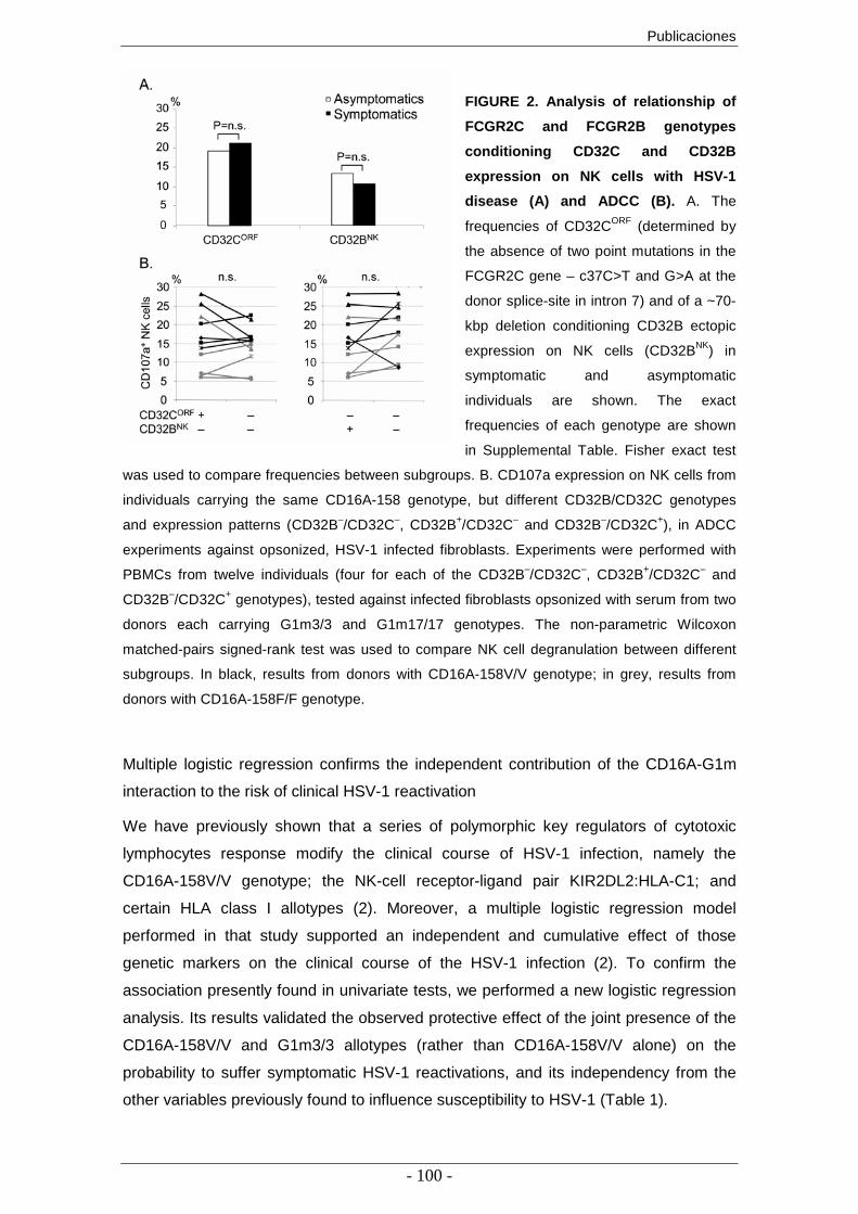
Publicaciones
- 100 -
FIGURE 2. Analysis of relationship of
FCGR2C and FCGR2B genotypes
conditioning CD32C and CD32B
expression on NK cells with HSV-1
disease (A) and ADCC (B). A. The
frequencies of CD32CORF (determined by
the absence of two point mutations in the
FCGR2C gene – c37C>T and G>A at the
donor splice-site in intron 7) and of a ~70-
kbp deletion conditioning CD32B ectopic
expression on NK cells (CD32BNK) in
symptomatic and asymptomatic
individuals are shown. The exact
frequencies of each genotype are shown
in Supplemental Table. Fisher exact test
was used to compare frequencies between subgroups. B. CD107a expression on NK cells from
individuals carrying the same CD16A-158 genotype, but different CD32B/CD32C genotypes
and expression patterns (CD32B–/CD32C–, CD32B+/CD32C– and CD32B–/CD32C+), in ADCC
experiments against opsonized, HSV-1 infected fibroblasts. Experiments were performed with
PBMCs from twelve individuals (four for each of the CD32B–/CD32C–, CD32B+/CD32C– and
CD32B–/CD32C+ genotypes), tested against infected fibroblasts opsonized with serum from two
donors each carrying G1m3/3 and G1m17/17 genotypes. The non-parametric Wilcoxon
matched-pairs signed-rank test was used to compare NK cell degranulation between different
subgroups. In black, results from donors with CD16A-158V/V genotype; in grey, results from
donors with CD16A-158F/F genotype.
Multiple logistic regression confirms the independent contribution of the CD16A-G1m
interaction to the risk of clinical HSV-1 reactivation
We have previously shown that a series of polymorphic key regulators of cytotoxic
lymphocytes response modify the clinical course of HSV-1 infection, namely the
CD16A-158V/V genotype; the NK-cell receptor-ligand pair KIR2DL2:HLA-C1; and
certain HLA class I allotypes (2). Moreover, a multiple logistic regression model
performed in that study supported an independent and cumulative effect of those
genetic markers on the clinical course of the HSV-1 infection (2). To confirm the
association presently found in univariate tests, we performed a new logistic regression
analysis. Its results validated the observed protective effect of the joint presence of the
CD16A-158V/V and G1m3/3 allotypes (rather than CD16A-158V/V alone) on the
probability to suffer symptomatic HSV-1 reactivations, and its independency from the
other variables previously found to influence susceptibility to HSV-1 (Table 1).
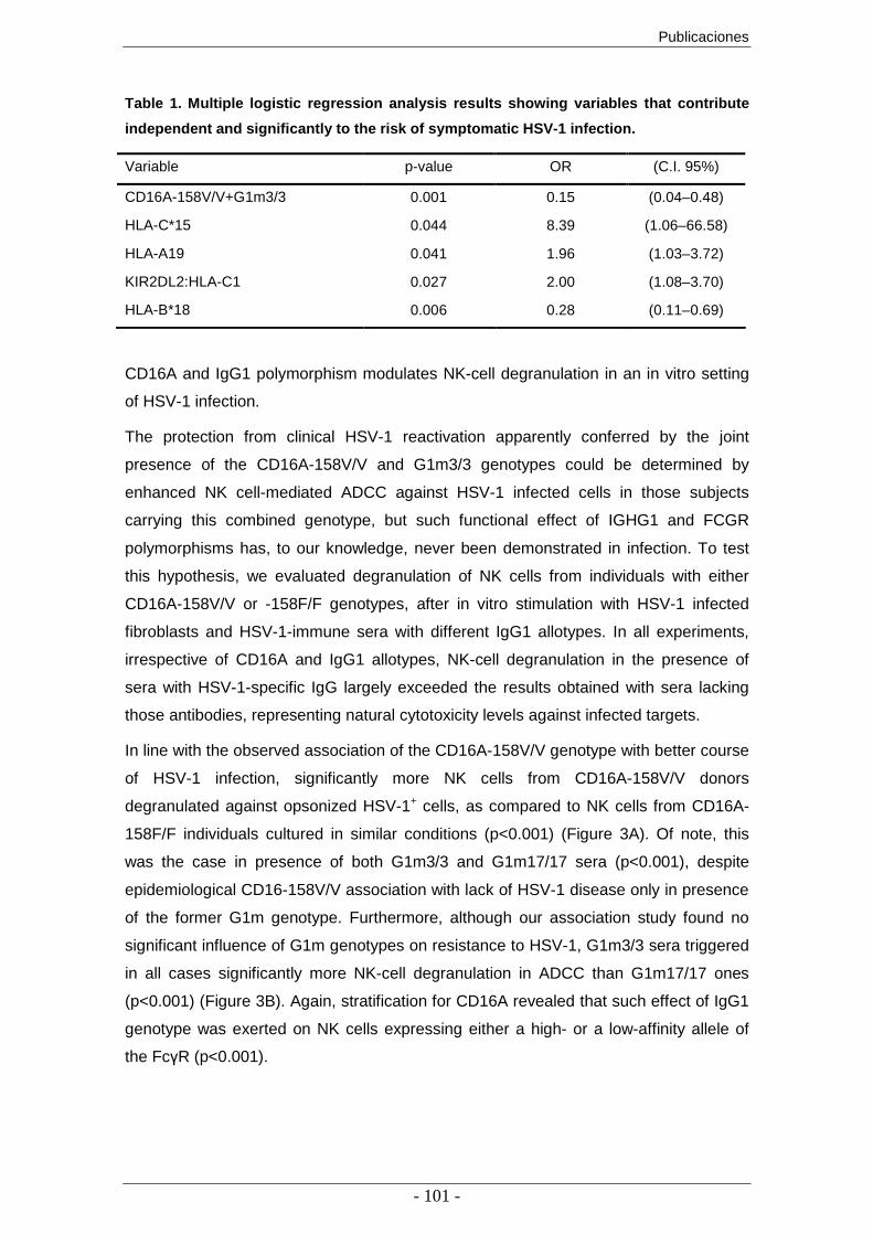
Publicaciones
- 101 -
Table 1. Multiple logistic regression analysis results showing variables that contribute
independent and significantly to the risk of symptomatic HSV-1 infection.
Variable p-value OR (C.I. 95%)
CD16A-158V/V+G1m3/3 0.001 0.15 (0.04–0.48)
HLA-C*15 0.044 8.39 (1.06–66.58)
HLA-A19 0.041 1.96 (1.03–3.72)
KIR2DL2:HLA-C1 0.027 2.00 (1.08–3.70)
HLA-B*18 0.006 0.28 (0.11–0.69)
CD16A and IgG1 polymorphism modulates NK-cell degranulation in an in vitro setting
of HSV-1 infection.
The protection from clinical HSV-1 reactivation apparently conferred by the joint
presence of the CD16A-158V/V and G1m3/3 genotypes could be determined by
enhanced NK cell-mediated ADCC against HSV-1 infected cells in those subjects
carrying this combined genotype, but such functional effect of IGHG1 and FCGR
polymorphisms has, to our knowledge, never been demonstrated in infection. To test
this hypothesis, we evaluated degranulation of NK cells from individuals with either
CD16A-158V/V or -158F/F genotypes, after in vitro stimulation with HSV-1 infected
fibroblasts and HSV-1-immune sera with different IgG1 allotypes. In all experiments,
irrespective of CD16A and IgG1 allotypes, NK-cell degranulation in the presence of
sera with HSV-1-specific IgG largely exceeded the results obtained with sera lacking
those antibodies, representing natural cytotoxicity levels against infected targets.
In line with the observed association of the CD16A-158V/V genotype with better course
of HSV-1 infection, significantly more NK cells from CD16A-158V/V donors
degranulated against opsonized HSV-1+ cells, as compared to NK cells from CD16A-
158F/F individuals cultured in similar conditions (p<0.001) (Figure 3A). Of note, this
was the case in presence of both G1m3/3 and G1m17/17 sera (p<0.001), despite
epidemiological CD16-158V/V association with lack of HSV-1 disease only in presence
of the former G1m genotype. Furthermore, although our association study found no
significant influence of G1m genotypes on resistance to HSV-1, G1m3/3 sera triggered
in all cases significantly more NK-cell degranulation in ADCC than G1m17/17 ones
(p<0.001) (Figure 3B). Again, stratification for CD16A revealed that such effect of IgG1
genotype was exerted on NK cells expressing either a high- or a low-affinity allele of
the FcγR (p<0.001).

Publicaciones
- 102 -
Figure 3. Influence of CD16A-
158 and G1m genotypes on
NK-cell degranulation in an in
vitro setting of ADCC against
opsonized HSV-1+ targets. A.
Percentages of degranulating
NK cells in paired experiments
using PMBCs from CD16A-
158V/V and CD16A-158F/F
donors. In black, results
obtained with G1m3/3 sera, in
grey, results obtained with
G1m17/17 sera. B. Percentages
of degranulating NK cells in
paired experiments using sera
from G1m3/3 and G1m17/17
individuals with similar HSV-1-
specific IgG titers. In black,
results from donors with CD16A-
158V/V genotype; in grey,
results from donors with CD16A-
158F/F genotype. C.
Percentages of degranulating
CD16A-158V/V NK cells in the
presence of G1m3/3 sera
(‘protective’ genotype),
compared with CD16A-158F/F NK cells in the presence of G1m17/17 sera (‘susceptible’
genotype). In all cases, natural cytotoxicity (estimated by degranulation seen in presence of
serum lacking HSV-1-specific IgG) has been subtracted in order to show degranulation
specifically induced by ADCC. Experiments were performed with PBMCs from ten different
individuals carrying each CD16A-158 allotype in homozygosis, tested against infected
fibroblasts opsonized with serum from two (6 experiments) or four (6 experiments) paired
donors, based on their IgG1 genotype. The non-parametric Wilcoxon matched-pairs signed-
rank test was used to compare NK cell degranulation between different subgroups.
The previous experiments defined CD16A and IgG1 genotypes associated with more
potent ADCC and/or a milder course of HSV-1 infection. The interaction of both
polymorphisms allows defining combined genotypes of theoretical maximal protection
(CD16A-158V/V+G1m3/3) and maximal susceptibility (CD16A-158F/F+G1m17/17).

Publicaciones
- 103 -
Comparison of such combined extreme genotypes in ADCC assays revealed in every
experiment higher NK-cell response when PBMCs from individuals carrying CD16A-
158V/V genotype were cultured with G1m3/3 sera, as compared to NK cells expressing
lower-affinity CD16A-158F/F cultured with serum containing G1m17/17 IgG1 (p<0.001)
(Figure 3C). On average, the presence of a combined susceptibility genotype reduced
the percentage of CD107a+ NK cells by 65.3% (range: 31.2%–100%) relative to the
combined protective genotype. Of note, differences between degranulation levels of NK
cells with each CD16A-158 genotype were consistently seen in all donor combinations,
regardless of whether cells derived from donors having or lacking a KIR2DL2/HLA-C1
receptor-ligand pair, associated with HSV1 disease (not shown).
Modulation of ADCC efficacy by CD16A/IgG1 polymorphism is partially independent of
viral FcγR and CD32B/CD32C expression on NK cells
The observed differences in ADCC intensity determined by IgG1 major allotypes might
be due to G1m3/3 IgG having lower binding affinity for vFcγR, higher affinity for CD16A
or both. To assess the contribution of the decoy vFcγR, we aimed to analyze in its
absence the effect of IgG1 allotypes on NK-cell degranulation. Viral FcγR-blocking
experiments were unsuccessful since the capacity of infected cells to bind human IgG
was efficaciously abolished by neither aggregated rabbit IgG (capable of binding to the
HSV-1 FcγR (10) ) nor two commercially available monoclonal antibodies against HSV-
1 gE, (data not shown). As an alternative approach, we used HSV-1 gE– mutant strain
s∆US8 to infect target fibroblasts. These were then exposed to NK cells and immune
sera of different genotypes, and the results were compared with those obtained with
gE+ HSV-1. As it can be appreciated in Figure 4, comparison between degranulation
induced by G1m3/3 and 17/17 sera showed greater variability with the gE– than with
the gE+ virus. Still, sera of the former genotype triggered significantly greater ADCC
than the latter (Figure 4), suggesting that the effect of G1m polymorphism on ADCC is,
at least, partially independent of the vFcγR. In the same line, comparison of the fold
ratios of CD107a+ NK cells induced by G1m3/3 or 17/17 sera in each experiment
revealed no significant difference between the results obtained with the gE– (0.9–3.5)
and the gE+ (1.2–2.9) HSV-1 strains (Figure 4).
Although our association studies showed that polymorphisms linked to CD32B or
CD32C expression on NK-cell surface had no significant influence on susceptibility to
HSV-1, those results do not exclude a possible influence on ADCC potency. Hence, we
performed ADCC assays to compare individuals carrying the same CD16A-158
genotype, but different CD32B/CD32C genotypes and expression patterns (CD32B–
/CD32C–, CD32B+/CD32C– and CD32B–/CD32C+), as assessed by both DNA typing
and flow cytometry. These assays revealed that NK-cell expression of CD32B or

Publicaciones
- 104 -
CD32C did not significantly or consistently influence ADCC potency in our in vitro
setting of HSV-1 infection (Figure 2).
FIGURE 4. HSV-1 vFcγR contribution to NK-cell mediated ADCC, studied using mutant
(gE–) or wild type (gE +) strains for the gE subunit of the decoy IgG receptor. Left and
middle panels show percentages of CD107a+ cells induced by G1m3/3 serum, in comparison
with G1m17/17 serum in paired ADCC experiments; right panel, fold reduction of CD107a+ cells
in G1m17/17 serum is compared between paired experiments performed using gE– and gE+
HSV-1 strains. Experiments were performed with PBMCs from four different individuals carrying
a CD16A-158V/V genotype and three with a CD16A-158F/F one, tested against infected
fibroblasts opsonized with sera from six paired donors, based on their IgG1 genotypes. The
non-parametric Wilcoxon matched-pairs signed-rank test was used to compare NK-cell
degranulation between subgroups. *A one-sided p-value was calculated to compare IgG1-
allotype effect on NK-cell mediated ADCC against HSV-1 gE‒infected targets. In black, results
from donors with CD16A-158V/V genotype, in grey, results from donors with CD16A-158F/F
genotype.
Phenotype of NK cells exerting ADCC against HSV-1-infected targets
Mature CD56dim NK cells can undergo clonal expansion and further differentiation, a
process characterized by phenotypic and functional changes, which eventually
generates memory-like, long-lived NK cells of unique phenotypes (38-42). Best known
example of this is an NKG2Cbright memory-like NK-cell subset with a defined phenotype
(NKG2A–KIR+, low Natural Cytotoxicity Receptor (NCR)-expression) that expands in
many, but not all, healthy individuals infected with human cytomegalovirus (HCMV) (40,
42). Moreover, an overlapping memory-like, long-lived, mature CD57+NKG2A–NCRlow
NK-cell subset, observed in HCMV-exposed individuals, frequently downregulates the
common signaling adaptor gamma-chain of Fc receptors (FcRγ, also known as
FcεRIγ); those cells have been proposed to be highly responsive to HCMV and HSV-1
infected cells in the presence of specific antibodies (43). To address whether and to
which extent NK cells with memory-like phenotypes exert ADCC against HSV-1
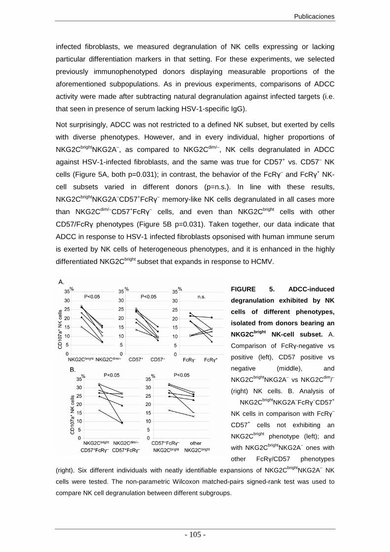
Publicaciones
- 105 -
infected fibroblasts, we measured degranulation of NK cells expressing or lacking
particular differentiation markers in that setting. For these experiments, we selected
previously immunophenotyped donors displaying measurable proportions of the
aforementioned subpopulations. As in previous experiments, comparisons of ADCC
activity were made after subtracting natural degranulation against infected targets (i.e.
that seen in presence of serum lacking HSV-1-specific IgG).
Not surprisingly, ADCC was not restricted to a defined NK subset, but exerted by cells
with diverse phenotypes. However, and in every individual, higher proportions of
NKG2CbrightNKG2A–, as compared to NKG2Cdim/–, NK cells degranulated in ADCC
against HSV-1-infected fibroblasts, and the same was true for CD57+ vs. CD57– NK
cells (Figure 5A, both p=0.031); in contrast, the behavior of the FcRγ– and FcRγ+ NK-
cell subsets varied in different donors (p=n.s.). In line with these results,
NKG2CbrightNKG2A–CD57+FcRγ– memory-like NK cells degranulated in all cases more
than NKG2Cdim/–CD57+FcRγ– cells, and even than NKG2Cbright cells with other
CD57/FcRγ phenotypes (Figure 5B p=0.031). Taken together, our data indicate that
ADCC in response to HSV-1 infected fibroblasts opsonised with human immune serum
is exerted by NK cells of heterogeneous phenotypes, and it is enhanced in the highly
differentiated NKG2Cbright subset that expands in response to HCMV.
FIGURE 5. ADCC-induced
degranulation exhibited by NK
cells of different phenotypes,
isolated from donors bearing an
NKG2Cbright NK-cell subset. A.
Comparison of FcRγ-negative vs
positive (left), CD57 positive vs
negative (middle), and
NKG2CbrightNKG2A– vs NKG2Cdim/–
(right) NK cells. B. Analysis of
NKG2CbrightNKG2A–FcRγ–CD57+
NK cells in comparison with FcRγ–
CD57+ cells not exhibiting an
NKG2Cbright phenotype (left); and
with NKG2CbrightNKG2A– ones with
other FcRγ/CD57 phenotypes
(right). Six different individuals with neatly identifiable expansions of NKG2CbrightNKG2A– NK
cells were tested. The non-parametric Wilcoxon matched-pairs signed-rank test was used to
compare NK cell degranulation between different subgroups.
Publicaciones
- 106 -
Discussion
Previous studies support the hypothesis that polygenic inheritance controls common
variations in the clinical course of the HSV-1 infection (6). We reported that a series of
polymorphic key regulators at the interface of innate and adaptive immunity (i.e. HLA
class I molecules, KIR and CD16A) associate with this clinical variability, strengthening
the essential role of cytotoxic lymphocytes in the immune control of HSV-1 (2).
Polymorphisms of other genes, including APOE and CSSG-1, have also been related
to susceptibility to recurrences, but their exact implication in HSV-1 control is as yet
unknown (1, 44, 45). In turn, inborn mutations of TLR3-IFN signaling pathway genes
predisposing to herpetic encephalitis support a crucial role for TLR3 in the control of
HSV-1 primary infection in the central nervous system (3), but those mutations are
apparently not connected with common forms of HSV-1 infection. In this study, we
extend our previous findings, assessing the contribution of interaction between
immunoglobulin and FcγR genetic polymorphisms to HSV-1 susceptibility.
Genetic variations in both FcγR and IgG heavy chain are attractive candidates to
influence the immune responses against HSV-1, because they potentially modulate
ADCC intensity, an important NK-cell effector mechanism. We previously showed that
a CD16A-158V/V genotype confers protection from symptomatic herpes (2).
Furthermore, major G1m allotypes in Caucasoids were recently pointed out as potential
candidates to influence susceptibility to HSV-1, based on their different affinity-binding
to HSV-1 gE/gI (7, 29), hence variable likeliness to be scavenged by the vFcγR decoy
(28). The G1m1 allotype has actually been reported as a possible susceptibility factor
for HSV-2 infection (46). According to our present results, G1m3/17 allotypes do not
associate by themselves with the clinical course of HSV-1 infection, but they interact
epistatically with a FCGR3A (CD16A) functional dimorphism, thus conditioning
susceptibility to symptomatic HSV-1 recurrences. Specifically, the previously reported
protective effect conferred by a CD16A-158V/V genotype (2) was enhanced in, or
limited to, G1m3/3 individuals. Conversely, herpetic disease did not associate with
Km1/3 allotypes (with or without stratification for CD16A genotype), and we detected
interaction neither between G1m or Km allotypes, nor between them and CD32A-
131H/R dimorphism, revealing a dominant effect of IgG1-CD16A interaction among the
analyzed polymorphisms of immunoglobulins and their cellular receptors.
Notwithstanding the demonstrated effect of CD16A-158V/F polymorphism on IgG
affinity, and vice versa, few studies have addressed functionally the influence of those
polymorphisms on ADCC potency (47-49). In the current study, we observed a
consistently enhanced NK-cell degranulation against opsonized, HSV-1-infected
targets in CD16A-158V/V carriers compared to CD16A-158F/F ones. Similarly, G1m3/3

Publicaciones
- 107 -
antibodies induced stronger NK-cell degranulation in this context, as compared to
G1m17/17 ones, despite our observation that G1m allotypes appear not to associate
on their own with HSV-1 immunity. The combined genotype CD16A-158V/V+G1m3/3
was hence associated with the most potent NK-cell mediated ADCC, as compared to
any other combination of CD16A+IgG1 genotypes. We thus expand knowledge on
FcγR-IgG interaction and provide further evidence on the implication of IGHG
polymorphism in NK-cell response by demonstrating that CD16A-158V/F and G1m
allotypes condition ADCC intensity against HSV-1 infected cells.
The strength of Fc receptor-antibody interaction has previously been shown to
condition NK-cell activation and ADCC potency (47, 50). In an infection setting, HSV-1
vFcγR has been proposed to hamper ADCC efficacy (8, 51), therefore a higher affinity
host FcγR could better compete with the vFcγR decoy and compensate this subversion
mechanism. Indeed, the estimated Kd for vFcγR-G1m17,1 binding is similar to the Kd
of the CD16A-158F-IgG1 interaction (7, 29, 47, 52). Thus, lower-affinity G1m3-vFcγR,
together with a higher-affinity G1m3-CD16A-158V/V, interactions could suffice to
counteract the viral immune evasion mechanism, explaining all our functional and
clinical observations. However, we observed that antibodies of the G1m3 allotype
induced stronger NK-cell degranulation even against targets infected with a HSV-1
strain lacking vFcγR. This reveals a direct effect of G1m polymorphism on ADCC
intensity, independent of the virus subversion mechanism, and possibly mediated by
the affinity binding of CD16A to different IgG1 allotypes.
Even though our in vitro setting might not accurately mirror the overall response to
HSV-1 in vivo, murine models showed that human anti-HSV-1 antibodies confer
significant protection to subsequent HSV-1 infection, irrespective of the vFcγR+ (53-56).
Likewise, our findings point to a limited capacity of vFcγR to subvert FcγR-driven NK-
cell activation and they are consistent with previous reports indicating that G1m
polymorphism also affects IgG1-CD16A binding itself. IgG1 allotype G1m17 differs
from G1m3 at position 214 (CH1, arginine for lysine), and it associates almost
invariably in Caucasoids with another two substitutions (Glu356Asp and Met358Leu,
CH3), that determine the G1m1 allotype (35, 36, 57). Site-directed mutagenesis assays
demonstrated the importance of IgG1 heavy-chain residue 214 (and to a lesser extent
356 and 358) for HSV-1 FcγR binding (7, 29), but none of these interacts directly with
human FcγR (19, 50). Yet, these substitutions might allosterically influence IgG1 avidity
for cellular FcγR. Supporting this hypothesis are affinity-binding assays showing higher
affinity of G1m3, as compared to G1m17,1, for both CD16A-158V and -158F (29).
Likewise, ADCC competition assays showed that IgG aggregates carrying the
G1m17,1 allotype (nb – but not the G1m3 one) bind with lower affinity to CD16A-
Publicaciones
- 108 -
158F/F than to 158V/V NK cells (58). All those results, like ours, point collectively to a
G1m3 allotype directly enhancing ADCC regardless of presence or absence of a
pathogen FcγR.
The inhibitory receptor CD32B, classically considered to be expressed only on B
lymphocytes or myeloid cells, and the activating CD32C, generally not expressed, can
both be detected on the NK cells of some individuals, and their expression on this
lymphocyte subset has been reported to modulate ADCC potency in some, but not all,
assays (15, 16, 59, 60). In our in-vitro setting of HSV-1 infection, significant CD32B and
CD32C contributions to NK-cell mediated ADCC were undetectable. Among FcγRs,
CD32B and CD32C bind to IgG1 with lowest affinity (52, 61), and the CD32C staining
pattern in flow cytometry is consistent with low expression on NK cells (Suppl. figure),
therefore, inefficient competition of CD32B/C with CD16A could explain the latter FcγR
dominating the overall NK cell mediated ADCC. In addition, lower-affinity CD32B/C
binding to IgG could be more efficiently competed by vFcγR than that of CD16A. More
precise assessment of the CD32C+/CD32B+ NK-cell role in immunity falls outside the
purpose of this work, therefore we can not exclude a marginal contribution of these
receptors to antibody-dependent, cytotoxic or non-cytotoxic NK-cell responses to HSV-
1. Nevertheless, against a major role for NK-cell expression of CD32B and CD32C in
immune response to HSV-1 also stands our association study showing that genetic
variations controlling such expression do not significantly influence the clinical course
of the infection.
A recent study showed increased antibody-driven IFN-γ production for FcRγ– NK cells,
in response to both HSV-1 and HCMV infected cells (43). Likewise, enhanced
degranulation and IFN-γ production in response to HIV+ ADCC targets was observed
for differentiated, CD57+/KIR3DL1+ NK cells (62, 63). Furthermore, a major role in
cytokine production and specific degranulation triggered by opsonized HCMV-infected
targets has been established for NKG2Cbright NK cells that expand in response to the
latter virus, many of which express CD57 and downregulate FcRγ as memory-like
differentiation markers (64, 65). Of note, NKG2Cbright-cell expansions are specifically
triggered, through unknown mechanisms, by HCMV, but not by HSV-1 or other human
viruses (40, 41). In our in vitro setting of NK-cell response to opsonized, HSV-1
infected targets, phenotype analysis showed that HSV-1 can readily trigger ADCC in
NK cells of different phenotypes and maturation stages. Nonetheless, an enhanced
capacity to degranulate in antibody-mediated cytotoxic response was seen among
NKG2CbrightNKG2A– and CD57+ NK cells, and, in particular, among those with the
combined NKG2CbrightNKG2A–FcRγ–CD57+ phenotype. These data, in line with those
obtained with HCMV, underline a phenotypic variable that seems to modify efficacy of

Publicaciones
- 109 -
the NK-cell response. Since these phenotypic traits are in part determined by
environment (i.e. HCMV infection), and current genetic tests cannot predict them,
further specific studies are warranted to determine whether, how, and to what extent an
expanded NKG2CbrightNKG2A–FcRγ–CD57+ NK subset might modify immunity to HSV-
1.
In summary, our results show that both CD16A-158V and G1m3 allotypes enhance NK
cell-mediated cytotoxicity against HSV-1 infected, opsonized targets, and that those
polymorphisms interact epistatically, protecting from clinical HSV-1 recurrence. This
underlines NK-cell mediated ADCC as an important anti-viral mechanism controlling
HSV-1 reactivation, and combined FCGR3A (CD16A) and IGHG1 (IgG1) genetic
variability as a key contributor to susceptibility to infection.
Acknowledgments
We thank Drs. S. Efstathiou and H. Browne (Department of Pathology, University of
Cambridge) for the gifts of viruses and cell lines, Dr. Elvira Ramil and Dr. Aránzazu
García Grande, from the DNA sequencing and Flow cytometry facilities of Instituto de
Investigación Sanitaria Puerta de Hierro, for expert support, and all of the individuals
who kindly donated blood samples. This work was supported by Ministerio de
Economía y Competitividad grants BFU2005-04622 (to C.V.), SAF2010–22153-C03 (to
M.L.-B. and C.V.), Fundació La Marató TV3 (grant 121531 to M.L.-B. and C.V.) and
Fondo de Investigación Sanitaria PI11/00298 (to HTR). M.M. was supported,
successively, by SAF2010–22153-C03 and Fundació La Marató TV3 grant 121531,
and AM was supported by AECC Foundation.

Publicaciones
- 110 -
References
1. Hobbs, M. R., B. B. Jones, B. E. Otterud, M. Leppert, and J. D. Kriesel. 2008. Identification of a herpes simplex labialis susceptibility region on human chromosome 21. J Infect Dis. 197: 340-346.
2. Moraru, M., E. Cisneros, N. Gomez-Lozano, R. de Pablo, F. Portero, M. Canizares, M. Vaquero, G. Roustan, I. Millan, M. Lopez-Botet, and C. Vilches. 2012. Host genetic factors in susceptibility to herpes simplex type 1 virus infection: contribution of polymorphic genes at the interface of innate and adaptive immunity. J Immunol 188: 4412-4420.
3. Zhang, S. Y., L. Abel, and J. L. Casanova. 2013. Mendelian predisposition to herpes simplex encephalitis. Handb Clin Neurol 112: 1091-1097.
4. Lanier, L. L. 2005. NK cell recognition. Annu Rev Immunol. 23: 225-274. 5. Mathiesen, T., M. A. Persson, V. A. Sundqvist, and B. Wahren. 1988. Neutralization
capacity and antibody dependent cell-mediated cytotoxicity of separated IgG subclasses 1, 3 and 4 against herpes simplex virus. Clin Exp Immunol. 72: 211-215.
6. Moraru, M., and C. Vilches. 2015. Host genetics and response to Herpes simplex type I infection. In Genomics, personalized medicine and oral disease. S. Sonis, ed. Springer Publisher, p. in press.
7. Atherton, A., K. L. Armour, S. Bell, A. C. Minson, and M. R. Clark. 2000. The herpes simplex virus type 1 Fc receptor discriminates between IgG1 allotypes. Eur J Immunol. 30: 2540-2547.
8. Dubin, G., E. Socolof, I. Frank, and H. M. Friedman. 1991. Herpes simplex virus type 1 Fc receptor protects infected cells from antibody-dependent cellular cytotoxicity. J Virol. 65: 7046-7050.
9. Frank, I., and H. M. Friedman. 1989. A novel function of the herpes simplex virus type 1 Fc receptor: participation in bipolar bridging of antiviral immunoglobulin G. J Virol. 63: 4479-4488.
10. Johansson, P. J., T. Hallberg, V. A. Oxelius, A. Grubb, and J. Blomberg. 1984. Human immunoglobulin class and subclass specificity of Fc receptors induced by herpes simplex virus type 1. J Virol. 50: 796-804.
11. Johnson, D. C., M. C. Frame, M. W. Ligas, A. M. Cross, and N. D. Stow. 1988. Herpes simplex virus immunoglobulin G Fc receptor activity depends on a complex of two viral glycoproteins, gE and gI. J Virol 62: 1347-1354.
12. Nimmerjahn, F., and J. V. Ravetch. 2008. Fcgamma receptors as regulators of immune responses. Nat Rev Immunol 8: 34-47.
13. Breunis, W. B., E. van Mirre, J. Geissler, N. Laddach, G. Wolbink, E. van der Schoot, M. de Haas, M. de Boer, D. Roos, and T. W. Kuijpers. 2009. Copy number variation at the FCGR locus includes FCGR3A, FCGR2C and FCGR3B but not FCGR2A and FCGR2B. Hum Mutat. 30: E640-650.
14. van Sorge, N. M., W. L. van der Pol, and J. G. van de Winkel. 2003. FcgammaR polymorphisms: Implications for function, disease susceptibility and immunotherapy. Tissue Antigens. 61: 189-202.
15. van der Heijden, J., W. B. Breunis, J. Geissler, M. de Boer, T. K. van den Berg, and T. W. Kuijpers. 2012. Phenotypic variation in IgG receptors by nonclassical FCGR2C alleles. J Immunol 188: 1318-1324.
16. Metes, D., L. K. Ernst, W. H. Chambers, A. Sulica, R. B. Herberman, and P. A. Morel. 1998. Expression of functional CD32 molecules on human NK cells is determined by an allelic polymorphism of the FcgammaRIIC gene. Blood. 91: 2369-2380.
17. Mueller, M., P. Barros, A. S. Witherden, A. L. Roberts, Z. Zhang, H. Schaschl, C. Y. Yu, M. E. Hurles, C. Schaffner, R. A. Floto, L. Game, K. M. Steinberg, R. K. Wilson, T. A. Graves, E. E. Eichler, H. T. Cook, T. J. Vyse, and T. J. Aitman. 2013. Genomic pathology of SLE-associated copy-number variation at the FCGR2C/FCGR3B/FCGR2B locus. Am J Hum Genet 92: 28-40.
18. Wu, J., J. C. Edberg, P. B. Redecha, V. Bansal, P. M. Guyre, K. Coleman, J. E. Salmon, and R. P. Kimberly. 1997. A novel polymorphism of FcgammaRIIIa (CD16) alters receptor function and predisposes to autoimmune disease. J Clin Invest. 100: 1059-1070.
19. Sondermann, P., R. Huber, V. Oosthuizen, and U. Jacob. 2000. The 3.2-A crystal structure of the human IgG1 Fc fragment-Fc gammaRIII complex. Nature. 406: 267-273.
Publicaciones
- 111 -
20. Radaev, S., S. Motyka, W. H. Fridman, C. Sautes-Fridman, and P. D. Sun. 2001. The structure of a human type III Fcgamma receptor in complex with Fc. J Biol Chem. 276: 16469-16477.
21. Breunis, W. B., E. van Mirre, M. Bruin, J. Geissler, M. de Boer, M. Peters, D. Roos, M. de Haas, H. R. Koene, and T. W. Kuijpers. 2008. Copy number variation of the activating FCGR2C gene predisposes to idiopathic thrombocytopenic purpura. Blood. 111: 1029-1038.
22. Aitman, T. J., R. Dong, T. J. Vyse, P. J. Norsworthy, M. D. Johnson, J. Smith, J. Mangion, C. Roberton-Lowe, A. J. Marshall, E. Petretto, M. D. Hodges, G. Bhangal, S. G. Patel, K. Sheehan-Rooney, M. Duda, P. R. Cook, D. J. Evans, J. Domin, J. Flint, J. J. Boyle, C. D. Pusey, and H. T. Cook. 2006. Copy number polymorphism in Fcgr3 predisposes to glomerulonephritis in rats and humans. Nature. 439: 851-855.
23. Fanciulli, M., P. J. Norsworthy, E. Petretto, R. Dong, L. Harper, L. Kamesh, J. M. Heward, S. C. Gough, A. de Smith, A. I. Blakemore, P. Froguel, C. J. Owen, S. H. Pearce, L. Teixeira, L. Guillevin, D. S. Graham, C. D. Pusey, H. T. Cook, T. J. Vyse, and T. J. Aitman. 2007. FCGR3B copy number variation is associated with susceptibility to systemic, but not organ-specific, autoimmunity. Nat Genet. 39: 721-723.
24. Pandey, J. P., and Z. Li. 2013. The forgotten tale of immunoglobulin allotypes in cancer risk and treatment. Exp Hematol Oncol 2: 2162-3619.
25. Oxelius, V. A., and J. P. Pandey. 2013. Human immunoglobulin constant heavy G chain (IGHG) (Fcgamma) (GM) genes, defining innate variants of IgG molecules and B cells, have impact on disease and therapy. Clin Immunol 149: 475-486.
26. Pandey, J. P. 2000. Immunoglobulin GM and KM allotypes and vaccine immunity. Vaccine. 19: 613-617.
27. Lefranc, M. P., and G. Lefranc. 2012. Human Gm, Km, and Am allotypes and their molecular characterization: a remarkable demonstration of polymorphism. Methods Mol Biol 882: 635-680.
28. Pandey, J. P. 2012. Immunoglobulin genes and immunity to herpes simplex virus type 1. J Infect Dis 206: 143-144.
29. Armour, K. L., C. S. Smith, and M. R. Clark. 2010. Expression of human FcgammaRIIIa as a GPI-linked molecule on CHO cells to enable measurement of human IgG binding. J Immunol Methods 354: 20-33.
30. Moxley, G., and R. S. Gibbs. 1992. Polymerase chain reaction-based genotyping for allotypic markers of immunoglobulin kappa shows allelic association of Km with kappa variable segment. Genomics. 13: 104-108.
31. Vilches, C., J. Castano, P. Munoz, and J. Penalver. 2008. Simple genotyping of functional polymorphisms of the human immunoglobulin G receptors CD16A and CD32A: a reference cell panel. Tissue Antigens. 71: 242-246.
32. Chisholm, S. E., K. Howard, M. V. Gomez, and H. T. Reyburn. 2007. Expression of ICP0 is sufficient to trigger natural killer cell recognition of herpes simplex virus-infected cells by natural cytotoxicity receptors. J Infect Dis. 195: 1160-1168.
33. Balan, P., N. Davis-Poynter, S. Bell, H. Atkinson, H. Browne, and T. Minson. 1994. An analysis of the in vitro and in vivo phenotypes of mutants of herpes simplex virus type 1 lacking glycoproteins gG, gE, gI or the putative gJ. J Gen Virol. 75: 1245-1258.
34. Showalter, S. D., M. Zweig, and B. Hampar. 1981. Monoclonal antibodies to herpes simplex virus type 1 proteins, including the immediate-early protein ICP 4. Infect Immun. 34: 684-692.
35. Schanfield, M. S., P. Herzog, and H. H. Fudenberg. 1975. Immunoglobulin allotypes of European populations. II.Gm, Am and Km(Inv) allotypic markers in Czechoslovakians. Hum Hered. 25: 382-392.
36. Schanfield, M. S., J. Gergely, and H. H. Fudenberg. 1975. Immunoglobulin Allotypes of European Populations. I. Gm and Km(Inv) allotypic markers in Hungarians. Hum Hered. 25: 370-377.
37. Lejeune, J., B. Piegu, V. Gouilleux-Gruart, M. Ohresser, H. Watier, and G. Thibault. 2012. FCGR2C genotyping by pyrosequencing reveals linkage disequilibrium with FCGR3A V158F and FCGR2A H131R polymorphisms in a Caucasian population. MAbs. 4: 784-787.
38. Beziat, V., B. Descours, C. Parizot, P. Debre, and V. Vieillard. 2010. NK cell terminal differentiation: correlated stepwise decrease of NKG2A and acquisition of KIRs. PLoS One 5: 0011966.

Publicaciones
- 112 -
39. Bjorkstrom, N. K., P. Riese, F. Heuts, S. Andersson, C. Fauriat, M. A. Ivarsson, A. T. Bjorklund, M. Flodstrom-Tullberg, J. Michaelsson, M. E. Rottenberg, C. A. Guzman, H. G. Ljunggren, and K. J. Malmberg. 2010. Expression patterns of NKG2A, KIR, and CD57 define a process of CD56dim NK-cell differentiation uncoupled from NK-cell education. Blood 116: 3853-3864.
40. Guma, M., A. Angulo, C. Vilches, N. Gomez-Lozano, N. Malats, and M. Lopez-Botet. 2004. Imprint of human cytomegalovirus infection on the NK cell receptor repertoire. Blood. 104: 3664-3671.
41. Lopez-Botet, M., A. Muntasell, and C. Vilches. 2014. The CD94/NKG2C+ NK-cell subset on the edge of innate and adaptive immunity to human cytomegalovirus infection. Semin Immunol 26: 145-151.
42. Muntasell, A., M. Lopez-Montanes, A. Vera, G. Heredia, N. Romo, J. Penafiel, M. Moraru, J. Vila, C. Vilches, and M. Lopez-Botet. 2013. NKG2C zygosity influences CD94/NKG2C receptor function and the NK-cell compartment redistribution in response to human cytomegalovirus. Eur J Immunol 43: 3268-3278.
43. Zhang, T., J. M. Scott, I. Hwang, and S. Kim. 2013. Cutting edge: antibody-dependent memory-like NK cells distinguished by FcRgamma deficiency. J Immunol 190: 1402-1406.
44. Kriesel, J. D., B. B. Jones, N. Matsunami, M. K. Patel, C. A. St Pierre, E. A. Kurt-Jones, R. W. Finberg, M. Leppert, and M. R. Hobbs. 2011. C21orf91 genotypes correlate with herpes simplex labialis (cold sore) frequency: description of a cold sore susceptibility gene. J Infect Dis 204: 1654-1662.
45. Itzhaki, R., and M. Wozniak. 2008. Susceptibility to herpes simplex labialis conferred by the gene encoding apolipoprotein E. J Infect Dis. 198: 624-625; author reply 625-626.
46. Seppanen, M., S. Meri, I. L. Notkola, I. J. Seppala, E. Hiltunen-Back, H. Sarvas, M. Lappalainen, H. Valimaa, A. Palikhe, V. V. Valtonen, and M. L. Lokki. 2006. Subtly impaired humoral immunity predisposes to frequently recurring genital herpes simplex virus type 2 infection and herpetic neuralgia. J Infect Dis. 194: 571-578.
47. Vance, B. A., T. W. Huizinga, K. Wardwell, and P. M. Guyre. 1993. Binding of monomeric human IgG defines an expression polymorphism of Fc gamma RIII on large granular lymphocyte/natural killer cells. J Immunol. 151: 6429-6439.
48. Varchetta, S., N. Gibelli, B. Oliviero, E. Nardini, R. Gennari, G. Gatti, L. S. Silva, L. Villani, E. Tagliabue, S. Menard, A. Costa, and F. F. Fagnoni. 2007. Elements related to heterogeneity of antibody-dependent cell cytotoxicity in patients under trastuzumab therapy for primary operable breast cancer overexpressing Her2. Cancer Res. 67: 11991-11999.
49. Hatjiharissi, E., L. Xu, D. D. Santos, Z. R. Hunter, B. T. Ciccarelli, S. Verselis, M. Modica, Y. Cao, R. J. Manning, X. Leleu, E. A. Dimmock, A. Kortsaris, C. Mitsiades, K. C. Anderson, E. A. Fox, and S. P. Treon. 2007. Increased natural killer cell expression of CD16, augmented binding and ADCC activity to rituximab among individuals expressing the Fc{gamma}RIIIa-158 V/V and V/F polymorphism. Blood. 110: 2561-2564.
50. Shields, R. L., A. K. Namenuk, K. Hong, Y. G. Meng, J. Rae, J. Briggs, D. Xie, J. Lai, A. Stadlen, B. Li, J. A. Fox, and L. G. Presta. 2001. High resolution mapping of the binding site on human IgG1 for Fc gamma RI, Fc gamma RII, Fc gamma RIII, and FcRn and design of IgG1 variants with improved binding to the Fc gamma R. J Biol Chem. 276: 6591-6604.
51. Corrales-Aguilar, E., M. Trilling, K. Hunold, M. Fiedler, V. T. Le, H. Reinhard, K. Ehrhardt, E. Merce-Maldonado, E. Aliyev, A. Zimmermann, D. C. Johnson, and H. Hengel. 2014. Human cytomegalovirus Fcgamma binding proteins gp34 and gp68 antagonize Fcgamma receptors I, II and III. PLoS Pathog 10: e1004131. doi:1004110.1001371/journal.ppat.1004131.
52. Bruhns, P., B. Iannascoli, P. England, D. A. Mancardi, N. Fernandez, S. Jorieux, and M. Daeron. 2009. Specificity and affinity of human Fcgamma receptors and their polymorphic variants for human IgG subclasses. Blood. 113: 3716-3725.
53. Erlich, K. S., and J. Mills. 1986. Passive immunotherapy for encephalitis caused by herpes simplex virus. Rev Infect Dis. 8 Suppl 4 : S439-445.
54. LeBlanc, R. A., L. Pesnicak, M. Godleski, and S. E. Straus. 1999. Treatment of HSV-1 infection with immunoglobulin or acyclovir: comparison of their effects on viral spread, latency, and reactivation. Virology. 262: 230-236.

Publicaciones
- 113 -
55. Dalai, S. K., L. Pesnicak, G. F. Miller, and S. E. Straus. 2002. Prophylactic and therapeutic effects of human immunoglobulin on the pathobiology of HSV-1 infection, latency, and reactivation in mice. J Neurovirol. 8: 35-44.
56. Sanna, P. P., A. De Logu, R. A. Williamson, Y. L. Hom, S. E. Straus, F. E. Bloom, and D. R. Burton. 1996. Protection of nude mice by passive immunization with a type-common human recombinant monoclonal antibody against HSV. Virology. 215: 101-106.
57. Mayr, W. R., and D. Mickerts. 1970. [Human gamma globulin polymorphism. Calculation of its distribution in Vienna and its suitability in paternity cases]. Acta Biol Med Ger. 25: 473-482.
58. Namboodiri, A. M., and J. P. Pandey. 2011. Differential inhibition of trastuzumab- and cetuximab-induced cytotoxicity of cancer cells by immunoglobulin G1 expressing different GM allotypes. Clin Exp Immunol 166: 361-365.
59. Dutertre, C. A., E. Bonnin-Gelize, K. Pulford, D. Bourel, W. H. Fridman, and J. L. Teillaud. 2008. A novel subset of NK cells expressing high levels of inhibitory FcgammaRIIB modulating antibody-dependent function. J Leukoc Biol. 84: 1511-1520.
60. Ernst, L. K., D. Metes, R. B. Herberman, and P. A. Morel. 2002. Allelic polymorphisms in the FcgammaRIIC gene can influence its function on normal human natural killer cells. J Mol Med (Berl). 80: 248-257.
61. Maenaka, K., P. A. van der Merwe, D. I. Stuart, E. Y. Jones, and P. Sondermann. 2001. The human low affinity Fcgamma receptors IIa, IIb, and III bind IgG with fast kinetics and distinct thermodynamic properties. J Biol Chem. 276: 44898-44904.
62. Parsons, M. S., L. Wren, G. Isitman, M. Navis, I. Stratov, N. F. Bernard, and S. J. Kent. 2012. HIV infection abrogates the functional advantage of natural killer cells educated through KIR3DL1/HLA-Bw4 interactions to mediate anti-HIV antibody-dependent cellular cytotoxicity. J Virol 86: 4488-4495.
63. Parsons, M. S., L. Loh, S. Gooneratne, R. J. Center, and S. J. Kent. 2014. Role of education and differentiation in determining the potential of natural killer cells to respond to antibody-dependent stimulation. AIDS 28: 2781-2786.
64. Wu, Z., C. Sinzger, G. Frascaroli, J. Reichel, C. Bayer, L. Wang, R. Schirmbeck, and T. Mertens. 2013. Human cytomegalovirus-induced NKG2C(hi) CD57(hi) natural killer cells are effectors dependent on humoral antiviral immunity. J Virol 87: 7717-7725.
65. Costa-Garcia, M., A. Vera, M. Moraru, C. Vilches, M. Lopez-Botet, and A. Muntasell. 2015. Antibody-Mediated Response of NKG2Cbright NK Cells against Human Cytomegalovirus. J Immunol 194: 2715-2724.
Abbreviations used in this article : ADCC, antibody dependent cellular cytotoxicity;
FcRγ, FcεRI gamma-chain; FCGR, FcγR locus; gE, glycoprotein E; gI, glycoprotein I;
gH, glycoprotein I; Gm, gamma heavy chain allotypes; HCMV, human cytomegalovirus;
hTERT, Human telomerase reverse transcriptase-immortalization; IGHG, gamma
heavy chain locus; Km, kappa light chain allotypes; MLPA, multiplex ligation-dependent
probe amplification; NCR, Natural Cytotoxicity Receptors; vFcγR, viral (HSV-1)
receptor for the crystallizable fraction of immunoglobulins.

Publicaciones
- 114 -
Supplemental table I. G1m, Km, CD32B NK, CD32C and combined genotype frequencies.
Genotype
Seronegatives
N=65, n (%)
Seropositives
(all)
N=226, n (%)
Seropositves
(asymptomatics)
N=68, n (%)
Seropositves
(symptomatics)
N=158, n (%)
P-value
(asymptomatics
vs. symptomatics)
G1m3/3 34 (52.3) 119 (52.7) 38 (55.9) 81 (51.3) n.s.
G1m3/17 26 (40.0) 91 (40.3) 26(38.2) 65 (41.1) n.s.
G1m17/17 5 (7.7) 16 (7.1) 4 (5.9) 12 (7.6) n.s.
Km3/3 48 (73.9) 187 (82.7) 58 (85.3) 129 (81.7) n.s.
Km1/3 16 (24.6) 36 (15.9) 9 (13.2) 27 (17.1) n.s.
Km1/1 1 (1.5) 3 (1.3) 1 (1.5) 2 (1.3) n.s.
G1m3/3 + Km3/3 26 (40.0) 98 (43.4) 32 (47.1) 66 (41.8) n.s.
CD16A-158V/V
+ G1m3/3 5 (7.7) 15 (6.6) 10 (14.7) 5 (3.2) 0.005
CD16A-158V/V
+ Km3/3 7 (10.8) 24 (10.6) 11 (16.2) 13 (8.2) n.s.
CD32A-131H/H
+ G1m3/3 7 (10.8) 22 (9.7) 4 (5.9) 18 (11.4) n.s.
CD32A-131H/H
+ Km3/3 8 (12.3) 38 (16.8) 9 (13.2) 29 (18.3) n.s.
CD32BNK/NK n.d. 1(0.4) 0 (0) 1 (0.6) n.s.
CD32BNK/WT n.d. 25 (11.1) 9 (13.2) 16 (10.1) n.s.
CD32BWT/WT n.d. 200 (88.5) 59 (86.8) 141 (89.2) n.s.
CD32CORF/ORF n.d. 3 (1.3) 0 (0) 3 (1.9) n.s.
CD32CORF/– n.d. 43 (19.0) 13 (19.1) 30 (19.0) n.s.
CD32C–/– n.d. 180 (72.1) 55 (80.9) 125 (79.1) n.s.
CD32C-13Gln/Gln n.d. 7 (3.1) 0 (0) 7 (4.4) n.s.
CD32C-13Gln/TAG n.d. 56 (24.8) 18 (26.5) 38 (24.1) n.s.
CD32C-13TAG/TAG n.d. 163 (72.1) 50 (73.5) 113 (71.5) n.s.
N.d., not determined; n.s. not significant. Seronegative and seropositive individuals did not differ significantly at any of the studied markers.

Publicaciones
- 115 -
Supplemental figure. (A) FcγR expression pattern (CD16A and CD32B/C) on NK cells.
Representative plots from selected donors carrying different CD32B/C genotypes are shown.
(B) Gating Strategy used for NK cell subpopulations.

DISCUSIÓN


Discusión
- 118 -
El curso clínico de muchas infecciones virales en humanos es altamente variable y a
esta variabilidad contribuye, entre otros factores, la diversidad genética del
hospedador. En un contexto amplio, el resultado de la interacción patógeno-
hospedador se traduce en susceptibilidad o resistencia a la infección. Clásicamente,
se consideraba que la susceptibilidad genética a la infección puede agruparse en dos
categorías: (i) la predisposición a infecciones múltiples determinada por mutaciones de
herencia mendeliana en genes únicos, que corresponde a la definición tradicional de
una inmunodeficiencia primaria, o (ii) la susceptibilidad a la infección por un único
patógeno común, que suele ser el resultado de la presencia de una serie de
polimorfismos en genes múltiples. Sin embargo, con la caracterización de nuevas
inmunodeficiencias primarias no convencionales a lo largo de los últimos años, ha
surgido una tercera categoría (iii) en la que defectos genéticos en genes únicos
confieren susceptibilidad a infecciones por un solo patógeno (Casanova y Abel, 2007).
Notablemente, la susceptibilidad a la infección por HSV-1, en sus diferentes formas,
puede incluirse en las tres categorías; y es que esta tercera categoría de
predisposición genética a la infección ha surgido precisamente a partir de la
caracterización de defectos genéticos que predisponen al desarrollo de la encefalitis
herpética (Casanova y Abel, 2007). En cuanto a la primera categoría, varios tipos de
inmunodeficiencias primarias, sobre todo las que afectan al número o a la
funcionalidad de las células NK, o a la activación de la respuesta de interferones,
cursan con infecciones herpéticas recurrentes, algunas de ellas de gran
transcendencia clínica (Jouanguy et al, 2013; Orange 2013; Sancho-Shimizu et al, 2011b;
Bustamente et al, 2008). Mientras que los diferentes tipos de susceptibilidad a la
infección determinados por mutaciones patogénicas (i y iii) están restringidos a un
número muy limitado de pacientes inmunodeficientes, hay evidencias que apoyan una
herencia poligénica como responsable de la resistencia a sufrir infecciones recurrentes
de herpes labial en la población inmunocompetente (Moraru y Vilches, 2015). Esta
segunda categoría (ii) refleja el objetivo global de esta tesis. Para alcanzarlo,
analizamos la contribución de la diversidad inmunogenética del hospedador a la
susceptibilidad de sufrir infecciones recurrentes por el HSV-1, e identificamos varios
genes polimórficos en la interfaz de las respuestas inmunitarias innata y adaptativa,
relacionados principalmente con la activación de linfocitos citotóxicos, que condicionan
la variabilidad de la respuesta clínica a la infección.

Discusión
- 119 -
1. La influencia del polimorfismo genético del sistema HLA
El polimorfismo extremo del complejo MHC humano y su probable condicionamiento
por presiones selectivas, sobre todo en relación con la presencia de diferentes
patógenos, lo sitúa entre los candidatos punteros para influir en el curso clínico de la
infección por HSV-1. Además, las moléculas HLA tienen un papel clave en la
presentación antigénica y en la modulación de la activación de las células NK. Eso
implica su control directo sobre la actividad de los linfocitos citotóxicos, que, como se
ha reseñado previamente, son los efectores celulares de mayor relevancia para la
defensa frente a las infecciones virales. Por tanto, no es sorprendente que durante los
años 70 y 80 se llevaran a cabo varios estudios que trataron de establecer
asociaciones significativas entre los distintos alelos HLA y el curso clínico de la
infección herpética (revisado en Moraru et al, 2012a). En años posteriores se realizaron
además importantes avances en la metodología para el estudio del polimorfismo HLA,
ampliando enormemente el número de alelos HLA conocidos.
Por todo ello y considerando la escasa reproducibilidad de los resultados previos,
eran necesarios nuevos estudios de asociación que aclarasen el papel del
polimorfismo genético del locus HLA en la susceptibilidad a HSV-1. En nuestro estudio
HLA-B*18 se perfila como factor protector y HLA-C*15 como factor de riesgo de sufrir
enfermedad herpética recurrente (Moraru et al, 2012a). Encontramos también una
tendencia a que HLA-B*35 confiera protección frente a las recurrencias de la infección,
en concordancia con los resultados publicados previamente en una serie de individuos
de origen siciliano (Gallina et al, 1987). De manera similar, algunos de los alelos HLA-A
que codifican el alotipo A19, al que nuestro análisis de regresión múltiple identifica
como factor riesgo para una infección recurrente, han sido también relacionados
previamente con el curso clínico de la infección (Russell y Schlaut, 1977: Zimmerman et al,
1977).
En conjunto, los datos publicados hasta ahora indican que la presencia de diferentes
alelos HLA de clase I se correlaciona con el curso clínico de la infección por HSV-1,
mientras que el polimorfismo en los genes que codifican moléculas HLA de clase II no
parece estar relacionado con ello. Este efecto predominante de las moléculas HLA de
clase I podría explicarse por su papel en la presentación antigénica a los linfocitos T
CD8+ o en la modulación de la activación de las células NK, siendo ambos subtipos
linfocitarios clave en el control de las infecciones virales. De los alelos HLA de clase I
que contribuyen al curso clínico de la infección herpética identificados en nuestro
estudio, solo HLA-C*15 es ligando conocido para receptores de células NK. En el

Discusión
- 120 -
apartado siguiente se discuten las implicaciones de la expresión conjunta de
receptores KIR y sus ligandos HLA-C.
Para activarse, los linfocitos T CD8+ requieren la presentación de péptidos antigénicos
en complejo con moléculas HLA de clase I, aunque solo una pequeña fracción de los
péptidos que podrían generarse a partir de las diferentes proteínas puede activar a
estos linfocitos. Esta inmunodominancia de ciertos péptidos guarda relación con una
serie de factores como los niveles de expresión génica, la capacidad del proteasoma
de degradar estas proteínas, la eficiencia del TAP para transportar los péptidos
resultantes al retículo endoplásmico, la probabilidad de unión de estos péptidos a las
moléculas HLA de clase I de cada individuo, la afinidad del TCR para estos complejos
HLA-péptido, la frecuencia de los precursores T específicos y su capacidad para
expandirse y sobrevivir (St Leger y Hendricks, 2011b). Efectivamente, en la infección por
HSV-1 se ha observado que la respuesta de los linfocitos T CD8+ está restringida a un
repertorio de péptidos bastante limitado y, al igual que en el caso del hCMV, la
mayoría de ellos proceden de proteínas virales producidas antes del inicio de la
replicación del ADN viral (Koelle et al, 2003; Reddehase et al, 1987).
En la infección experimental del ratón, más de un 50% de los linfocitos T CD8+ que
infiltran los ganglios trigeminales reconocen un solo péptido derivado de la gB, una
proteína viral codificada por un gen γ1, limitandose la especificidad del resto a otros
pocos epítopos subdominantes derivados de solo 10 proteínas virales (St. Leger et al,
2011a; Wallace et al, 1999; Khana et al, 2003). Se desconoce si esta situación es
extrapolable a la infección en humanos. Sin embargo, las vacunas anti-HSV
generadas a partir de péptidos o proteínas truncadas derivadas de gB (y gD) no
confieren protección a largo plazo (Corey et al, 1999; Straus et al, 1997). Los datos
disponibles de estudios de predicción de epítopos inmunogénicos en humanos indican
que, si bien el repertorio de péptidos derivados de HSV-1 que son reconocidos por los
linfocitos T CD8+ está muy restringido en cada individuo, hay una importante
heterogeneidad interindividual (Mikloska et al, 2000; Dervillez et al, 2013; Srivastava et al,
2015; van Veltzen et al, 2013).
Nuestros estudios de predicción de la presentación de péptidos de origen viral por las
moléculas HLA de clase I que confieren riesgo o susceptibilidad al HSV-1 indican que
el producto de los alelos (de protección) HLA-B*18:01 y HLA-B*35 más comunes
(HLA-B*35:01, *35:02, *35:03 y *35:08) presentan un único péptido derivado del
dominio RING finger de la ICP0 (requerido para todas sus actividades biológicas
(Everett 2000)), mientras que las moléculas codificadas por HLA-C*15 (de riesgo para
la infección) no se estima que puedan presentar péptidos derivados de este fragmento

Discusión
- 121 -
proteico (Moraru et al, 2012a). La ICP0 es una proteína viral multifuncional codificada
por un gen IE que regula la estabilidad de varias proteínas intracelulares, las
respuestas al daño del ADN o la producción de IFN (Boutell y Everett 2013). Tiene un
papel clave en el mantenimiento del equilibrio entre el estado latente y las
reactivaciones del HSV-1 y existen indicios de que su ausencia permite el
silenciamiento de la transcripción viral por mecanismos celulares todavía
desconocidos (Preston, 2000; Everett, 2000). Además, su unión a USP7, una proteína de
la familia de las proteasas específicas de la ubiquitina, impediría la degradación de
proteínas en el proteasoma, por lo que podría limitar también la presentación
antigénica de péptidos virales (Everett et al, 1997).
La detección de transcritos de ICP0 en los ganglios sensitivos infectados, que se
correlaciona con la expansión clonal y la acumulación de linfocitos T CD8+ activados
en estos ganglios, apoya un modelo en el que ICP0 se expresa durante el estado
latente (Derfuss et al, 2007). Su degradación temprana favorecería el mantenimiento del
estado latente. Nuestros resultados indican que la susceptibilidad a sufrir recurrencias
podría estar relacionada, en parte, con la capacidad de las moléculas HLA de clase I
para presentar péptidos derivados de esta proteína viral (Moraru et al, 2012a). Si bien
otro estudio más reciente ha identificado también un péptido inmunodominante
derivado de ICP0 (van Veltzen et al, 2013) y pese a las ventajas que ofrecería el
reconocimiento de péptidos derivados de proteínas que se expresan antes o incluso
en ausencia de la replicación viral, se requieren más estudios para confirmar esta
relación causal.
Varios estudios indican que la generación del repertorio de péptidos antigénicos no es
fácilmente predecible en el contexto de una infección viral. Clásicamente se
consideraba que los péptidos presentados por moléculas HLA de clase I provienen de
la degradación de proteínas maduras, pero datos más recientes indican que una
fracción de estos péptidos podría derivar de productos génicos defectuosos conocidos
como DRiPs (defective ribosomal products) (Bourdetsky et al; 2014; Yewdell y Nicchitta,
2006). La generación de los DRiPs podría estar aumentada en células infectadas,
siendo el origen de estas proteínas defectuosas el propio genoma viral o la alteración
de la síntesis de proteínas endógenas debida a la infección. A favor de la presentación
preferente de péptidos derivados de DRiPs está la rapidez del procesamiento
antigénico de las proteínas virales (que puede llegar a requerir una sola hora, aunque
su vida media es de varios días) y la observación de que los epítopos virales para
linfocitos T CD8+ se generan principalmente a partir de proteínas recién sintetizadas
(Khan et al, 2001; Yewdell y Nicchitta, 2006). Sin embargo, la degradación de proteínas
virales no alteradas inmediatamente después de su síntesis sería una explicación

Discusión
- 122 -
alternativa para ambas observaciones (Rock et al, 2014). Un escenario en el que los
DRiPs sean una fuente importante de péptidos de origen viral complicaría aun más la
predicción de los péptidos antigénicos que reconocerían los linfocitos T CD8+ en el
contexto de la infección por HSV-1.
Además de la selección del repertorio de péptidos antigénicos que presenta a los
linfocitos T, el polimorfismo HLA modifica también otros aspectos de la presentación
antigénica. Varios ejemplos de ello son su influencia sobre sus niveles de expresión, la
velocidad de su ensamblaje, la estabilidad de sus formas carentes de péptidos o la
dependencia para su expresión de un complejo de carga plenamente funcional. En la
infección por HSV-1, cuya proteína ICP47 puede bloquear el TAP, la capacidad de
ciertas moléculas HLA para cargarse de péptidos de manera independiente de la
tapasina les conferiría una importante ventaja sobre las demás. Si bien esta
independencia de la tapasina implica una carga subóptima con péptidos antigénicos,
su paso hacia la membrana celular es más rápido en comparación con las variantes
dependientes de la tapasina. Efectivamente, un estudio reciente demuestra que tanto
HLA-B*18 como HLA-B*35 funcionan de manera independiente de la tapasina (Rizvi et
al, 2014), lo que apoya nuestros resultados que clasifican a estos alelos como
protectores frente a la infección clínica por HSV-1.
Puesto que la eficacia de la presentación antigénica en cada individuo depende del
conjunto de moléculas HLA que expresa, estudios dirigidos al análisis de genotipos
HLA completos (en lugar de genes aislados) deberían facilitar el entendimiento del
papel del polimorfismo HLA en el control del curso clínico de la infección por HSV-1.
2. Las implicaciones de la variabilidad de los KIR y sus ligandos
Las células NK son un componente clave en la respuesta frente a infecciones virales
activándose, mediante la integración de señales recibidas de receptores inhibidores e
activadores, en respuesta a una serie de alteraciones de lo propio inducidas por
patógenos. Los KIR, considerando sus características funcionales y su gran
variabilidad genética, podrían ser altamente relevantes en el control de la infección por
HSV-1. En efecto, un estudio previo de nuestro grupo ha encontrado que la presencia
de KIR2DL2 y KIR2DS2 en el genoma confiere susceptibilidad a sufrir recurrencias de
la infección. Confirmamos este resultado en la serie actual y, además, encontramos
que la asociación con la enfermedad es aún más fuerte al analizar la presencia
conjunta de los genes KIR2DL2 y KIR2DS2 y su ligando HLA-C-1.
La alta complejidad genética, estructural y funcional de los KIR no permite formular
una hipótesis única que explique estos resultados. En primer lugar, la asociación casi

Discusión
- 123 -
completa de KIR2DL2 y KIR2DS2 en un haplotipo muy conservado impide identificar el
principal responsable de la asociación encontrada, basándose exclusivamente en los
datos epidemiológicos. En segundo lugar, la alta homología entre los dos receptores
en su fragmento extracelular apunta a un ligando común, aunque todavía es
controvertido si KIR2DS2 reconoce o no HLA-C1 (más detalles sobre este aspecto
están recogidos en la introducción). Esa alta homología entre KIR2DL2 y KIR2DS2 ha
complicado también la generación de anticuerpos monoclonales monoespecíficos. Sin
embargo, el grupo de Cristelle Retière ha desarrollado recientemente un anticuerpo
anti-KIR2DS2/2DL3 (David et al, 2009) que, en combinación con anticuerpos anti-
KIR2DL3 y anti-KIR2DL2/2DL3/2DS2 podría permitir distinguir las células NK que
expresen KIR2DS2 en presencia o ausencia de KIR2DL2 y elucidar, por tanto, el papel
de cada uno en la infección por HSV-1. En tercer lugar, el polimorfismo alélico y el
repertorio de péptidos que presentan las diferentes moléculas HLA, que modifican
también las interacciones KIR-ligando, dificultan aun más este escenario.
Se han descrito alelos de KIR2DL2 y KIR2DS2 con baja frecuencia en la población
que codifican proteínas con expresión o función alteradas. Así, KIR2DL2*004 codifica
una proteína que está retenida intracelularmente, mientras que KIR2DL2*006,
KIR2DL2*009 y KIR2DL2*011 son muy similares a KIR2DL3*001 en la región
responsable del reconocimiento de su ligando y, por tanto, podrían codificar receptores
de menor afinidad para HLA-C (VandenBussche et al, 2006; Falco et al, 2013).
KIR2DS2*003 codifica una proteína que carece de lisina en el fragmento
transmembrana, por lo que no se asocia a DAP12 y, por tanto, no transmite señales
activadoras (Bottino et al, 2000). En el estudio actual no hemos tenido en cuenta el
polimorfismo alélico de estos KIR, pero la baja frecuencia en la población de estas
variantes alélicas frente a los alelos comunes (Hou et al, 2012) hace que sea poco
probable que nuestros resultados se vean afectados significativamente por la inclusión
de algún individuo con un alelo KIR2DL2 o KIR2DS2 con función alterada.
De manera similar al reconocimiento de HLA-E por CD94/NKG2, la naturaleza de los
péptidos unidos a las moléculas HLA modifica en ocasiones la afinidad de los KIR por
sus ligandos, lo que ha sido confirmado también para KIR2DL2 (Boyington et al, 2000;
Fadda et al, 2010). Pero mientras que la unión de KIR2DL2 a su ligando HLA es muy
sensible a mutaciones en estos péptidos, no parece que ocurra lo mismo con
KIR2DS2 (Stewart et al, 2005). En el contexto de la infección por HSV-1, las células NK
podrían explotar la selectividad para su ligando generada por péptidos virales,
activándose en presencia de células infectadas independientemente de la disminución
de la expresión de moléculas HLA.
Discusión
- 124 -
La importancia de KIR2DL2 en la susceptibilidad a HSV-1 podría estar relacionada con
su mayor afinidad para HLA-C en comparación con KIR2DL3 y KIR2DS2 (Moesta et al,
2008; David et al, 2013). Esta alta afinidad implicaría una menor sensibilidad de las
células NK KIR2DL2+ a la disminución de los niveles de HLA en las células infectadas.
O bien, si no hubiera disminución significativa de la expresión de HLA-C, KIR2DL2
establecería un umbral de inhibición demasiado alto para ser contrarrestado por otros
receptores activadores estimulados por la infección. Esta inhibición excesiva de las
células NK ejercida por KIR2DL2, en cualquiera de los dos escenarios, está apoyada
por el hecho de que la presencia conjunta de dos parejas KIR–ligando de alta afinidad,
KIR2DL2–HLA-C1 y KIR2DL1–HLA-C2, aumenta aún mas el riesgo de sufrir
reactivaciones de la infección (Moraru et al, 2012a). En contraposición a esta hipótesis,
ensayos in vitro con clones de células NK KIR2DL2+ o KIR2DS2+ enfrentadas a
fibroblastos HSV-1+ sugieren un papel importante de KIR2DL2 en la lisis de estas
dianas, en las que la presencia del virus disminuye la expresión de sus ligandos (Huard
y Früh, 2000). Variaciones significativas de la afinidad de KIR2DL2 para su ligando
introducidas por los repertorios de péptidos de las células HSV-1+ podrían también
explicar estos resultados. En ambos casos, una activación excesiva de los clones de
células NK KIR2DL2+ en presencia del virus en la piel o mucosas, podría ser
responsable de que las reactivaciones sean clínicamente manifiestas. Análisis
exhaustivos del nivel de activación de los clones de células NK KIR2DL2+ presentes
en las lesiones de pacientes con recurrencias de la infección podrían esclarecer su
papel en la susceptibilidad a HSV-1, pero este tipo de ensayos resultan a priori
complicados. Igualmente, diferencias significativas en la proporción de células
KIR2DL2+ entre los individuos sintomáticos y asintomáticos podrían justificar el
aumento de la susceptibilidad al HSV-1 determinada por este receptor, pero estos
estudios fenotípicos no se han realizado todavía.
Aunque en las células NK la inhibición vía KIR2DL2 sobrepasa el efecto de KIR2DS2,
la distribución variegada de los receptores de las células NK permite que haya clones
KIR2DS2+KIR2DL2– que, además, podrían carecer de otros receptores inhibidores
para moléculas HLA. Los diferentes modelos de educación de las células NK indican
que en individuos que expresan HLA-C1 estos clones estarían anérgicos. Sin
embargo, modelos murinos sugieren que este proceso podría ser reversible, siendo
ciertos estímulos capaces de generar unas potentes señales moduladoras de la
respuesta inmunitaria mediada por receptores para MHC activadores (Bolanos y
Tripathy, 2011; Jonker et al, 2010). Puesto que un modelo similar sería también válido
para clones KIR2DS1+KIR2DL1– en sujetos que expresan HLA-C2, este modelo no

Discusión
- 125 -
explica, por sí solo, por qué la presencia de KIR2DS2 podría aumenta el riesgo de
sufrir enfermedad herpetica recurrente.
En el contexto de la infección por HSV-1, un estudio previo encontró una alta actividad
citolítica de los clones de células NK KIR2DS2+ en presencia de fibroblastos autólogos
infectados con HSV-1 (Pietra et al, 2000). La presencia de estos clones en la piel o
mucosas de individuos infectados con HSV-1 podría ser responsable de la lisis
extensa de los queratinocitos HSV-1+, contribuyendo así a que las reactivaciones del
virus sean clínicamente manifiestas. O, por el contrario, un efecto subversivo
moderado del HSV-1 sobre la expresión de HLA-C, que es generalmente baja, podría
ser suficiente para impedir la activación de las células KIR2DS2+KIR2DL2– en
presencia de dianas infectadas, disminuyendo así el control dependiente de las células
NK sobre la infección. Una confirmación de está segunda hipótesis requeriría más
estudios, ya que los datos disponibles hasta ahora relativos al efecto del HSV-1 sobre
la expresión de HLA-C son contradictorios. Mientras que en los experimentos de Pietra
et al, los fibroblastos HSV-1+ mantienen la expresión de HLA-C (a diferencia de HLA-A
y –B), Huard y Früh encuentran que la infección con HSV-1 o la transfección con
ICP47 disminuyen la expresión de HLA-C de células HeLa (Huard y Früh, 2000; Pietra et
al, 2000). La controversia respecto a si KIR2DS2 reconoce o no HLA-C1 complica aun
más este escenario.
Si KIR2DS2 reconoce (también), directa- o indirectamente, ligandos virales se
desconoce todavía. Igualmente inexplorado está el papel de KIR2DS2 en los linfocitos
T en el contexto de la infección por HSV-1. Sin embargo, cabe destacar que la
presencia de KIR2DS2 y de su ligando en el genoma, acompañada de una alta
frecuencia de clones de linfocitos T citotóxicos CD4+CD28nullKIR2DS2+KIR2DL2– ha
sido propuesta como factor de riesgo para la vasculitis reumatoide (Yen et al, 2001). La
contribución de KIR2DS2 a la patogénesis de esta enfermedad se explicaría por su
función co-estimuladora en presencia de estímulos subóptimos recibidos a través del
TCR. Una hipótesis similar ha sido formulada para explicar la susceptibilidad a
diabetes tipo I determinada por KIR2DS2 y su ligando (van der Silk et al, 2003) y podría
justificar también su efecto deletéreo en la infección por HSV-1.
En conjunto, aunque sean en gran medida contradictorios, los datos disponibles hasta
ahora parecen apoyar más un modelo en el que KIR2DL2 y no KIR2DS2 predispone a
sufrir recurrencias de la infección. Pero a falta de más ensayos fenotípicos y
funcionales, queda por demostrar de qué manera la expresión de estos KIR
modificaría la respuesta inmunitaria frente a dianas HSV-1+.
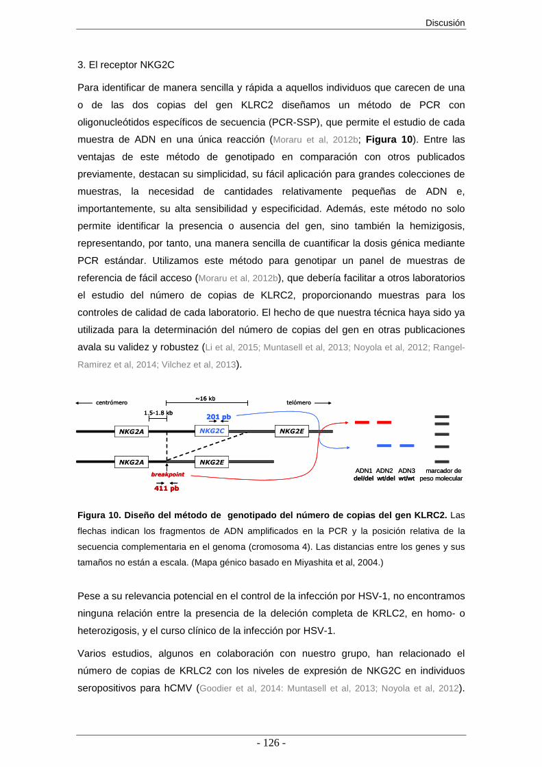
Discusión
- 126 -
3. El receptor NKG2C
Para identificar de manera sencilla y rápida a aquellos individuos que carecen de una
o de las dos copias del gen KLRC2 diseñamos un método de PCR con
oligonucleótidos específicos de secuencia (PCR-SSP), que permite el estudio de cada
muestra de ADN en una única reacción (Moraru et al, 2012b; Figura 10 ). Entre las
ventajas de este método de genotipado en comparación con otros publicados
previamente, destacan su simplicidad, su fácil aplicación para grandes colecciones de
muestras, la necesidad de cantidades relativamente pequeñas de ADN e,
importantemente, su alta sensibilidad y especificidad. Además, este método no solo
permite identificar la presencia o ausencia del gen, sino también la hemizigosis,
representando, por tanto, una manera sencilla de cuantificar la dosis génica mediante
PCR estándar. Utilizamos este método para genotipar un panel de muestras de
referencia de fácil acceso (Moraru et al, 2012b), que debería facilitar a otros laboratorios
el estudio del número de copias de KLRC2, proporcionando muestras para los
controles de calidad de cada laboratorio. El hecho de que nuestra técnica haya sido ya
utilizada para la determinación del número de copias del gen en otras publicaciones
avala su validez y robustez (Li et al, 2015; Muntasell et al, 2013; Noyola et al, 2012; Rangel-
Ramirez et al, 2014; Vilchez et al, 2013).
Figura 10. Diseño del método de genotipado del número de copias del gen KLRC2 . Las
flechas indican los fragmentos de ADN amplificados en la PCR y la posición relativa de la
secuencia complementaria en el genoma (cromosoma 4). Las distancias entre los genes y sus
tamaños no están a escala. (Mapa génico basado en Miyashita et al, 2004.)
Pese a su relevancia potencial en el control de la infección por HSV-1, no encontramos
ninguna relación entre la presencia de la deleción completa de KRLC2, en homo- o
heterozigosis, y el curso clínico de la infección por HSV-1.
Varios estudios, algunos en colaboración con nuestro grupo, han relacionado el
número de copias de KRLC2 con los niveles de expresión de NKG2C en individuos
seropositivos para hCMV (Goodier et al, 2014: Muntasell et al, 2013; Noyola et al, 2012).
NKG2CNKG2A NKG2E
1.5-1.8 kb
breakpoint
NKG2ENKG2A
411 pb
201 pb
ADN1 ADN2 ADN3 marcador dedel/del wt/del wt/wt peso molecular
centrómero telómero~16 kb
NKG2CNKG2A NKG2ENKG2CNKG2A NKG2E
1.5-1.8 kb1.5-1.8 kb
breakpoint
NKG2ENKG2A NKG2ENKG2A
411 pb411 pb
201 pb201 pb
ADN1 ADN2 ADN3 marcador dedel/del wt/del wt/wt peso molecular
centrómero telómero~16 kb~16 kb

Discusión
- 127 -
Además, se ha observado una expansión preferente de células NKG2Cbright en
infecciones agudas por otros virus (p. ej. hantavirus y chikungunya virus) en individuos
coinfectados con hCMV (Björkström et al, 2011a; Petidemange et al, 2011). Aunque se
desconoce si HSV-1 induce también alguna reconfiguración del repertorio de células
NK, no se ha encontrado una correlación entre la expansión de NKG2Cbright y la
seropositividad HSV-1 (Gumá et al, 2004). Apoyando este último dato, en otro estudio
tampoco se ha observado una expansión significativa de células NK altamente
diferenciadas en respuesta a HSV-2 (Björkström et al, 2011b).
Los resultados anteriores apuntan a un papel redundante del receptor NKG2C en la
infección por HSV-1. Sin embargo, la expansión de una población de células NK
NKG2Cbright en respuesta a hCMV se acompaña de importantes cambios, no solo
fenotípicos sino también funcionales (Muntasell et al, 2013; Muntasell et al, Costa-Garcia et
al, 2015) que sugieren que esta subpoblación linfocitaria, presente en proporciones
muy variables en los individuos expuestos a hCMV, podría modular la respuesta de las
células NK en otras infecciones. En este sentido, sería de interés estudiar el posible
papel de esta subpoblación altamente diferenciada de células NK en el control de la
infección por HSV-1. Notablemente, un estudio reciente ha encontrado que una
subpoblación de células NK FcεRIγ– que, en su mayoría, tienen un fenotipo
NKG2CbrightCD57+, producen cantidades significativamente mayores de IFN-γ en
comparación con el resto de las células NK en presencia de células infectadas por
HSV-1 y hCMV y opsonizadas con anticuerpos específicos (Zhang et al, 2013a).
Si bien la respuesta citotóxica directa de este subtipo de células NK con
caracterísiticas de memoria frente a dianas HSV-1+ no se ha estudiado todavía,
analizamos su papel en la ADCC in vitro. Los estudios fenotípicos de las células NK
que degranulan en respuesta a fibroblastos HSV-1+ opsonizados señalan también una
contribución preferente a la ADCC de las células NK NKG2Cbright y las CD57+, siendo la
subpoblación NKG2CbrightNKG2A–FcεRIγ–CD57+ la más citotóxica en el contexto de la
infección por HSV-1 (Moraru et al, 2015b). Si la presencia de este subtipo de células NK
altamente diferenciadas se correlaciona con el curso clínico de la infección por HSV-1,
queda por ser demostrado.
La presencia de al menos una copia de KRLC2 parece ser prescindible para el control
de la infeccion por HSV-1 y hCMV, resultado coincidente con su redundancia para el
control de otra infeccion viral, por el papiloma virus humano (Vilchez et al, 2013).
Estudios en pacientes seropositivos para hCMV sugieren que la deleción completa de
KRLC2 podría estar compensada por la expresión de KIR activadores (Della Chiessa et
al, 2014). Análisis fenotípicos de las células NK, incluyendo los patrones de expresión

Discusión
- 128 -
de KIR y NKG2C, podrían, por tanto, contribuir a explicar las diferencias
interindividuales en la eficacia del control de la infeccion por HSV-1.
4. El polimorfismo genético de los complejos FCGR e IGH
La respuesta inmunitaria mediada por anticuerpos abarca mecanismos complejos,
estableciendo un puente entre la inmunidad innata y la adaptativa. De particular
relevancia en las infecciones virales es su contribución a la citotoxicidad celular
mediada por anticuerpos. La ADCC, un mecanismo de gran importancia para el
aclaramiento de muchos agentes patógenos, depende de la unión de la IgG a
antígenos virales y a FcγR (Nimmerjahn and Ravetch, 2008). Mientras que la alta afinidad
de la unión del parátopo a su epitopo viral es el resultado de una serie de procesos
somáticos en los genes que codifican las regiones variables de las IgG, la fuerza de
unión Fc – FcγR depende en parte de polimorfismos genéticos en los loci FCGR e
IGHG. Igualmente, la variación del número de copias de varios de los genes del
complejo FCGR, que condicionaría los niveles de expresión de los receptores que
codifican, podría influir sobre la intensidad de las señales transmitidas a través de
estos FcγR. Puesto que la afinidad del FcγR por la IgG podría determinar el resultado
global de la respuesta inmunitaria frente al HSV-1, estudiamos la posible contribución
de varios polimorfismos funcionales en FCGR e IGHG al curso clínico de la infección.
Notablemente, al analizar una amplia serie de marcadores genéticos del locus FCGR,
descartamos posibles asociaciones debidas a desequilibrios de ligamiento entre ellos.
En concreto, el estudio se centró en los alotipos de la IgG1 y de la cadena ligera
kappa, las variaciones genéticas en FCGR2A, FCGR2B, FCGR2C y FCGR3A, y las
posibles interacciones entre los mismos.
4.1. Los alotipos CD16A-158V y G1m3
El dimorfismo funcional de FCGR3A, que cambia la afinidad de CD16A por la IgG,
podría modificar el aclaramiento del HSV-1 porque este FcγR se expresa a altos
niveles en la mayoría de las células NK, generando potentes respuestas citotóxicas
tras su activación (Lanier, 1998). Efectivamente, encontramos que la presencia de un
alotipo CD16A de alta afinidad (158-V) en homocigosis confiere protección frente a las
reactivaciones de la infección por HSV-1 (Moraru et al, 2012a). Pero un interés similar
tienen también los polimorfismos en los genes que codifican la región constante de la
cadena pesada de las IgG, sobre todo los que determinan los alotipos de la IgG1 más
frecuentes en caucasoides (Pandey et al, 2012). Si bien la presencia de un alotipo IgG1
de mayor afinidad para CD16 (G1m3) no contribuye por si sola significativamente a la
resistencia a HSV-1, análisis exhaustivos encontraron que el efecto protector del

Discusión
- 129 -
genotipo CD16A-158V/V está restringido a aquellos individuos homocigotos para
G1m3 (Moraru et al, 2015b). Por el contrario, no encontramos asociaciones significativas
entre el curso clínico de la infección por HSV-1 y los alotipos de la cadena ligera κ.
Puesto que este efecto protector de los alotipos CD16A e IgG1 podría estar
determinado, al menos en parte, por su contribución a la ADCC mediada por las
células NK, analizamos la capacidad de degranulación de estas células en un modelo
in vitro de ADCC frente a fibroblastos infectados por HSV-1. CD107a (LAMP-1) es un
marcador de los gránulos citotóxicos que puede ser detectado en la membrana celular
durante su exocitosis. La detección de la expresión de CD107a en las células NK es
un método comúnmente utilizado para la evaluación de la respuesta citotóxica.
Aunque no permite una cuantificación de la muerte de las dianas, se convierte en una
técnica de primera elección en modelos in vitro de infección, donde la muerte puede
ser causada tanto por el propio microorganismo como por las células efectoras a
evaluar. Además, la seguridad de su uso y el hecho de que permite la caracterización
fenotípica de las células que están degranulando justifican nuestra elección.
De acuerdo con nuestra hipótesis de partida, confirmamos que la presencia conjunta
de los genotipos CD16A-158V/V y G1m3/3 se asocia a mayor frecuencia de células
NK que degranulan en presencia de dianas HSV-1+ opsonizadas, comparado con las
demás combinaciones de alotipos CD16A e IgG1 (Moraru et al, 2015b). Además y pese
a que no hayamos encontrado una influencia significativa de los alotipos IgG1 aislados
sobre el curso clínico de la infección, encontramos que un porcentaje mayor de células
NK degranulan en presencia de sueros de donantes G1m3/3 comparado con
G1m17/17, independientemente del genotipo CD16A. De manera similar, aunque el
alotipo CD16A-158V no tiene efecto protector frente a las reactivaciones de la
infección en los sujetos G1m17/17, el efecto del dimorfismo CD16A-158V/F sobre la
intensidad de la ADCC es significativo tanto en presencia de sueros G1m3/3 como
G1m17/17.
En el contexto de la infección por HSV-1, otro factor que podría modificar la intensidad
de la ADCC frente a células infectadas es el señuelo viral que se une a IgG (FcγRv).
Estudios previos han demostrado que el FcγRv se une con mayor afinidad a IgG con
alotipo G1m17,1 comparado con G1m3 (Armour et al, 2002; Atherton et al, 2000). La
menor afinidad de la unión FcγRv – G1m3 podría favorecer la unión a CD16A de los
anticuerpos anti-HSV-1 con alotipo IgG1, contribuyendo, por tanto, al aumento de la
intensidad de la ADCC en los individuos G1m3/3 observado en nuestros experimentos
funcionales. Sin embargo, el efecto de los alotipos IgG sobre la intensidad de la ADCC
mediada por células NK en nuestro modelo de infección in vitro no se explica

Discusión
- 130 -
exclusivamente por la presencia del FcγRv. Aunque no se pueda descartar el papel del
FcγRv in vivo, nuestros resultados sugieren que el efecto del genotipo G1m3/3 sobre
la intensidad del ADCC está relacionado en parte con su modulación directa de la
afinidad CD16A-IgG1.
4.2. El dimorfismo funcional CD32A-131H/R
Se ha descrito que el alotipo CD32A-131H se une con mayor afinidad a IgG2 que
CD32A-131R y este aumento de afinidad modula la intensidad de la fagocitosis
mediada por CD32A (Salmon et al, 1992). Nuestros resultados demuestran que el curso
clínico de la infección herpética no se correlaciona con los alotipos CD32A (Moraru et
al, 2012a). Como se ha mencionado en la introducción, los anticuerpos anti-HSV-1 de
subclase IgG2 se encuentran solo en un pequeño porcentaje de los individuos
seropositivos, mientras que casi todos tienen anticuerpos específicos de tipo IgG1
(Mathiesen et al, 1998). No obstante, no hemos determinado las subclases de los
anticuerpos anti-HSV-1 en nuestra cohorte. Dado que el dimorfismo CD32A-131H/R
solo reduce marginalmente la afinidad de CD32A por los complejos inmunes con IgG1
(ambos alotipos reconocen eficazmente estos complejos) (Bruhns et al, 2009), el sesgo
poblacional entre las dos subclases (la escasez de individuos con anticuerpos de tipo
IgG2) podría explicar nuestros resultados. Otra posible explicación residiría en la
expresión preferente de CD32A en monocitos/macrófagos y el papel marginal que
juega la fagocitosis en el control de la infección por HSV-1 en comparación con la
ADCC.
4.3. Los polimorfismos genéticos que condicionan la expresión de CD32B y CD32C en
las células NK
La alta homología entre varios de los genes del locus FCGR, la variación del número
de copias de algunos de ellos y, particularmente, la casi completa identidad en las
secuencias de nucleótidos que existe entre las regiones 5’ (hasta el exón 4) de
FCGR2B y FCGR2C, y 3’ (a partir del exón 5) de FCGR2A y FCGR2C, ha dificultado
el estudio del polimorfismo genético de esta región mediante técnicas de PCR
estándar. Con el desarrollo del método de amplificación múltiple de sondas
dependiente de la ligación (MLPA), que permite cuantificar simultáneamente el número
de copias de hasta 50 genes y la presencia de diferentes SNPs en varios de ellos en
una única reacción, estos problemas han sido, en parte, superados. Utilizamos esta
técnica para identificar a la minoría de individuos con una secuencia de FCGR2C que
predice su expresión en la membrana plasmática (CD32CORF) y estudiar su posible
contribución a la susceptibilidad al HSV-1. Aunque, hipotéticamente, la expresión de

Discusión
- 131 -
CD32C en las células NK de una minoría de individuos podría proporcionarles una
ventaja, aumentando los niveles de activación de estas células en presencia de dianas
infectadas y opsonizadas, no encontramos asociaciones significativas entre la
presencia de CD32CORF y el desarrollo de reactivaciones de la infección por HSV-1
(Moraru et al, 2015b).
El desequilibrio de ligamiento entre los diferentes polimorfismos del locus FCGR
podría introducir sesgos en los estudios de asociación. Efectivamente, un trabajo
previo había encontrado que CD32CORF y CD16A-158V se heredan juntos en un
porcentaje significativo de individuos (Lejeune et al, 2012). Confirmamos el desequilibrio
de ligamiento entre CD32CORF y CD16A-158V en nuestra serie y analizamos la
contribución de cada uno de estos marcadores genéticos en ausencia del otro al curso
clínico de la infección por HSV-1. La falta de asociación entre CD32CORF y la
predisposición a sufrir recurrencias en los individuos con y sin un genotipo CD16AV/V
(que confiere protección frente a la infección) confirman que la expresión de CD32C en
las células NK no contribuye significativamente a la susceptibilidad al HSV-1 (Moraru et
al, 2015b).
Estudios de lisis redirigida (rADCC) indican que la expresión de CD32C en las células
NK condiciona la intensidad del rADCC (Metes et al, 1998; Ernst et al, 2002). Sin
embargo, el único ensayo publicado hasta ahora que analiza la contribución de CD32C
a la ADCC mediada por células NK en presencia de CD16 como competidor para la
IgG no ha podido demostrar que CD32C tenga un papel significativo en ello (Heijden et
al, 2012). En nuestro modelo de infección por HSV-1 tampoco observamos una
contribución significativa de la presencia de CD32C a la intensidad global de la ADCC
(degranulación de células NK) frente a dianas HSV-1+ opsonizadas (Moraru et al,
2015b). La menor afinidad de CD32C para la IgG, su expresión en una minoría de las
células NK y los niveles de expresión relativamente bajos en comparación con CD16
podrían explicar nuestros resultados. Por otro lado, una competición eficaz entre
FcγRv y CD32C podría impedir que este último se una a IgG anti-HSV-1. Para
descartar completamente su posible contribución a la ADCC en el contexto de la
infección por HSV-1 serían necesarios estudios con cantidades no limitantes de IgG
específica y un análisis de la contribución a la ADCC de células NK CD32C+. Aún así,
nuestros resultados (epidemiológicos y funcionales) descartan que, en condiciones
fisiológicas, la expresión de CD32C contribuya significativamente al control de la
infección.
Discusión
- 132 -
Si bien clásicamente se consideraba que el receptor inhibidor CD32B se expresa
solamente en linfocitos B y en células de estirpe mieloide, durante la última década
varios estudios han encontrado también expresión de CD32B en las células NK de una
minoría de individuos (Ernst et al, 2002; Stewart-Akers et al, 2004; Dutertre et al, 2008).
Estudios posteriores han demostrado que la expresión de CD32B en las células NK
(CD32BNK) se asocia con una deleción de ~70 Kpb en el locus FCGR. Esta deleción
incluye FCGR2C, FCGR3B y parte de la región intergénica FCGR3B – FCGR2B, lo
que sugiere que los elementos reguladores que determinan la expresión de CD32C en
las células NK controlan la expresión de CD32B en estos individuos (Heijden et al, 2012;
Muller et al, 2013, ver figura 8 de la introducción). Su expresión en este subtipo
linfocitario sería potencialmente relevante, ya que podría competir con CD16A por la
unión de la IgG, bloqueando, por tanto, la activación de las células NK en presencia de
inmunocomplejos.
Dos estudios independientes indican que la expresión de CD32B en las células NK
modifica la intensidad de la ADCC in vitro (Dutertre et al, 2018; Heijden et al, 2012). En
nuestro modelo de ADCC frente a dianas HSV-1+ opsonizadas no encontramos una
inhibición general significativa de la degranulación de las células NK por CD32B ni
asociación con la enfermedad (Moraru et al, 2015b). En el contexto de la infección por
HSV-1, el FcγRv, que se une a IgG humana con mayor afinidad que CD32B (Atherton
et al, 2000; Bruhns et al, 2009), podría contrarrestar el efecto de CD32B sobre la ADCC
mediada por células NK. Los resultados de los ensayos funcionales y de asociación
apoyan un modelo en el que la expresión de CD32B no modifica significativamente la
respuesta inmunitaria frente a HSV-1 (Moraru et al, 2015b). Sin embargo, una limitación
de nuestro estudio es que no analizamos selectivamente la degranulación de las
células NK CD32B+ y, por tanto, no podemos descartar un efecto menor de CD32B en
esta subpoblación.
En resumen, encontramos que interacciones epistáticas entre los alotipos CD16A-
158V y G1m3 modifican la intensidad de la ADCC frente a dianas HSV-1+ y el curso
clínico de la infección. Por el contrario, esta respuesta es independiente de variaciones
genéticas que afectan a la afinidad de CD32A por su ligando o a la expresión de
CD32B y CD32C en las células NK. En conjunto, estos datos apoyan el papel clave de
la ADCC mediada por células NK y del polimorfismo genético que condiciona su
intensidad en la respuesta inmunitaria frente a HSV-1.
Discusión
- 133 -
5. Otros polimorfismos genéticos del hospedador que modifican el curso clínico de la
infección
Los avances significativos en la genómica de los últimos años y su disponibilidad para
la investigación clínica han contribuido a esclarecer el papel de la variabilidad genética
del hospedador en la evolución de varias infecciones virales (Cooke y Hill, 2001; Tuite y
Gross, 2006). Sin embargo, aunque este tipo de estudios podrían mejorar nuestro
entendimiento sobre el control de la infección por HSV-1, solamente se ha realizado
por ahora un rastreo del genoma humano en búsqueda de marcadores genéticos que
condicionen las reactivaciones de la infección. Este estudio identificó una región en el
cromosoma 21 (Hobbs et al, 2008) y, posteriormente un gen en esta región como
responsable de la susceptibilidad a sufrir reactivaciones, que denominaron CSSG-1
(Cold Sore Susceptibility Gene 1) (Kriesel et al, 2011). CSSG-1 codifica una proteína
intracelular con función todavía desconocida, por lo que todavía se requieren más
estudios para identificar mecanismos potenciales a través de los que CSSG-1
modificaría el curso clínico de la infección.
Notablemente, los rastreos del genoma han sido diseñados para identificar
polimorfismos puntuales, pero no son adecuados para analizar otro tipo de
alteraciones como las deleciones o inserciones, la variación de número de copias, las
variantes alélicas raras y el polimorfismo de familias multigénicas altamente
homólogas, como las estudiadas en este trabajo. Para ello, los ensayos basados en
una hipótesis previa, construida sobre la relevancia de ciertos genes en el control de la
infección y métodos específicos, siguen siendo clave en la búsqueda marcadores
genéticos predictivos de resistencia a la infección.
En este sentido, otros estudios basados en hipótesis previas han identificado, por
ejemplo, una serie de mutaciones en genes implicados en la vía de activación TLR3-
IFN como responsables del desarrollo de la HSE (detallado en el apartado 5.1. de la
introducción), confirmando la importancia de esta ruta de activación celular en el
control de la infección por HSV-1 en el sistema nervioso central (Zhang et al, 2013b). Su
papel en el desarrollo de las recurrencias de la infección es, sin embargo, cuestionado.
Así, solo una pequeña minoría de los niños portadores de estos defectos genéticos
han sufrido más de un episodio de HSE y muy pocos de estos pacientes (y de sus
familiares) han tenido herpes labial (Abel et al, 2010; Guo et al, 2011; Spiegel et al, 2008).
Si bien un estudio posterior indica que un cambio de una leucina por fenilalanina
(F412L) en la secuencia de TLR3 aumenta el riesgo de sufrir recurrencias de la
infección, estos resultados requieren confirmación en series más amplias (Yang et al,
2012).
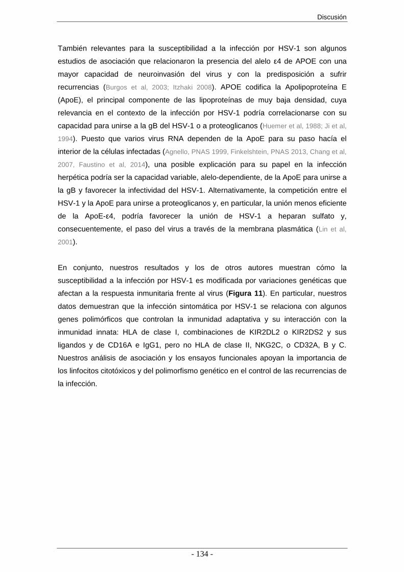
Discusión
- 134 -
También relevantes para la susceptibilidad a la infección por HSV-1 son algunos
estudios de asociación que relacionaron la presencia del alelo ε4 de APOE con una
mayor capacidad de neuroinvasión del virus y con la predisposición a sufrir
recurrencias (Burgos et al, 2003; Itzhaki 2008). APOE codifica la Apolipoproteína E
(ApoE), el principal componente de las lipoproteínas de muy baja densidad, cuya
relevancia en el contexto de la infección por HSV-1 podría correlacionarse con su
capacidad para unirse a la gB del HSV-1 o a proteoglicanos (Huemer et al, 1988; Ji et al,
1994). Puesto que varios virus RNA dependen de la ApoE para su paso hacía el
interior de la células infectadas (Agnello, PNAS 1999, Finkelshtein, PNAS 2013, Chang et al,
2007, Faustino et al, 2014), una posible explicación para su papel en la infección
herpética podría ser la capacidad variable, alelo-dependiente, de la ApoE para unirse a
la gB y favorecer la infectividad del HSV-1. Alternativamente, la competición entre el
HSV-1 y la ApoE para unirse a proteoglicanos y, en particular, la unión menos eficiente
de la ApoE-ε4, podría favorecer la unión de HSV-1 a heparan sulfato y,
consecuentemente, el paso del virus a través de la membrana plasmática (Lin et al,
2001).
En conjunto, nuestros resultados y los de otros autores muestran cómo la
susceptibilidad a la infección por HSV-1 es modificada por variaciones genéticas que
afectan a la respuesta inmunitaria frente al virus (Figura 11 ). En particular, nuestros
datos demuestran que la infección sintomática por HSV-1 se relaciona con algunos
genes polimórficos que controlan la inmunidad adaptativa y su interacción con la
inmunidad innata: HLA de clase I, combinaciones de KIR2DL2 o KIR2DS2 y sus
ligandos y de CD16A e IgG1, pero no HLA de clase II, NKG2C, o CD32A, B y C.
Nuestros análisis de asociación y los ensayos funcionales apoyan la importancia de
los linfocitos citotóxicos y del polimorfismo genético en el control de las recurrencias de
la infección.
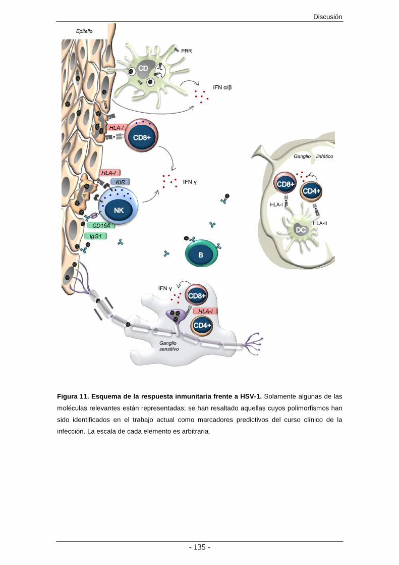
Discusión
- 135 -
Figura 11. Esquema de la respuesta inmunitaria frente a HSV-1. Solamente algunas de las
moléculas relevantes están representadas; se han resaltado aquellas cuyos polimorfismos han
sido identificados en el trabajo actual como marcadores predictivos del curso clínico de la
infección. La escala de cada elemento es arbitraria.

CONCLUSIONES


Conclusiones
- 138 -
1. HLA-B*18 disminuye la susceptibilidad a la enfermedad recurrente por HSV-1,
mientras que HLA-C*15 y el grupo de alelos que codifican A19 aumentan el riesgo
de sufrirla.
2. El polimorfismo genético del locus HLA de clase II DRB1 no modifica el curso
clínico de la infección por HSV-1.
3. La presencia en el genoma de KIR2DL2 y KIR2DS2 junto con alelos HLA con el
epítopo C1, predispone a sufrir infección recurrente por HSV-1.
4. El número de copias del gen KLRC2, que codifica el receptor activador NKG2C, no
modifica la susceptibilidad a HSV-1.
5. Los alotipos 158V de CD16A y G1m3 de la IgG1 aumentan la intensidad de la
ADCC mediada por células NK frente a dianas infectadas con HSV-1 incluso en
ausencia de un señuelo FcγRv, y su presencia conjunta en homozigosis confiere
protección frente a las recurrencias de la infección.
6. El dimorfismo funcional CD32A-131H/R y los alotipos de la cadena ligera κ de la Ig
no modifican el riesgo de sufrir enfermedad por HSV-1.
7. La expresión de los receptores para el Fc de la IgG CD32B y CD32C en las células
NK no afecta a la susceptibilidad al HSV-1 y a la ADCC.
8. En conclusión, la susceptibilidad al HSV-1 es modificada por variaciones genéticas
que afectan a la respuesta inmunitaria frente al virus, particularmente la función de
los linfocitos citotóxicos.


BIBLIOGRAFÍA

Bibliografía
- 142 -
Abel L, Plancoulaine S, Jouanguy E, et al. Age-dependent Mendelian predisposition to herpes simplex virus type 1 encephalitis in childhood. J Pediatr 2010: 157: 623-9, 629.e1.
Agnello V, Abel G, Elfahal M, Knight GB, Zhang QX. Hepatitis C virus and other flaviviridae viruses enter cells via low density lipoprotein receptor. Proc Natl Acad Sci U S A 1999: 96: 12766-71.
Allan RS, Waithman J, Bedoui S, et al. Migratory dendritic cells transfer antigen to a lymph node-resident dendritic cell population for efficient CTL priming. Immunity 2006: 25: 153-62.
Alter G, Martin MP, Teigen N, et al. Differential natural killer cell-mediated inhibition of HIV-1 replication based on distinct KIR/HLA subtypes. J Exp Med 2007: 204: 3027-36.
Anfossi N, André P, Guia S, et al. Human NK cell education by inhibitory receptors for MHC class I. Immunity 2006: 25: 331-42.
Aramburu J, Balboa MA, Izquierdo M, López-Botet M. A novel functional cell surface dimer (Kp43) expressed by natural killer cells and gamma/delta TCR+ T lymphocytes. II. Modulation of natural killer cytotoxicity by anti-Kp43 monoclonal antibody. J Immunol 1991: 147: 714-21.
Aravalli RN, Hu S, Rowen TN, Palmquist JM, Lokensgard JR. TLR2-mediated proinflammatory cytokine and chemokine production by microglial cells in response to herpes simplex virus. J Immunol 2005: 175: 4189-93.
Arduino PG, Porter SR. Herpes Simplex Virus Type 1 infection: overview on relevant clinico-pathological features. J Oral Pathol Med 2008: 37: 107-21.
Armour KL, Smith CS, Clark MR. Expression of human FcgammaRIIIa as a GPI-linked molecule on CHO cells to enable measurement of human IgG binding. J Immunol Methods 2010: 354: 20-33.
Atherton A, Armour KL, Bell S, Minson AC, Clark MR. The herpes simplex virus type 1 Fc receptor discriminates between IgG1 allotypes. Eur J Immunol 2000: 30: 2540-7.
Azzi T, Lünemann A, Murer A, et al. Role for early-differentiated natural killer cells in infectious mononucleosis. Blood 2014: 124: 2533-43.
Barcy S, Corey L. Herpes simplex inhibits the capacity of lymphoblastoid B cell lines to stimulate CD4+ T cells. J Immunol 2001: 166: 6242-9.
Bellón T, Heredia AB, Llano M, et al. Triggering of effector functions on a CD8+ T cell clone upon the aggregation of an activatory CD94/kp39 heterodimer. J Immunol 1999: 162: 3996-4002.
Benedict CA, Ware CF. Virus targeting of the tumor necrosis factor superfamily.Virology 2001: 289: 1-5.
van Bergen J, Koning F. The tortoise and the hare: slowly evolving T-cell responses take hastily evolving KIR. Immunology 2010: 131: 301-9.
Béziat V, Descours B, Parizot C, Debré P, Vieillard V. NK cell terminal differentiation: correlated stepwise decrease of NKG2A and acquisition of KIRs. PLoS One 2010: 5: e11966.
Béziat V, Dalgard O, Asselah T, et al. CMV drives clonal expansion of NKG2C+ NK cells expressing self-specific KIRs in chronic hepatitis patients. Eur J Immunol 2012: 42: 447-57.
Béziat V, Liu LL, Malmberg JA, et al. NK cell responses to cytomegalovirus infection lead to stable imprints in the human KIR repertoire and involve activating KIRs. Blood 2013: 121: 2678-88.
Björkström NK, Riese P, Heuts F, et al. Expression patterns of NKG2A, KIR, and CD57 define a process of CD56dim NK-cell differentiation uncoupled from NK-cell education. Blood 2010: 116: 3853-64.
Björkström NK, Lindgren T, Stoltz M, et al. Rapid expansion and long-term persistence of elevated NK cell numbers in humans infected with hantavirus. J Exp Med 2011a: 208: 13-21.
Bodmer WF, Albert E, Bodmer JG, et al. Nomenclature for factors of the HLA system, 1987. Tissue Antigens 1988: 32: 177-87.

Bibliografía
- 143 -
Bolanos FD, Tripathy SK. Activation receptor-induced tolerance of mature NK cells in vivo requires signaling through the receptor and is reversible. J Immunol 2011: 186: 2765-71.
di Bona D, Scafidi V, Plaia A, et al. HLA and killer cell immunoglobulin-like receptors influence the natural course of CMV infection. J Infect Dis 2014: 210: 1083-9.
Bottino C, Falco M, Sivori S, Moretta L, Moretta A, Biassoni R. Identification and molecular characterization of a natural mutant of the p50.2/KIR2DS2 activating NK receptor that fails to mediate NK cell triggering. Eur J Immunol 2000: 30: 3569-74.
Bottino C, Castriconi R, Pende D, et al. Identification of PVR (CD155) and Nectin-2 (CD112) as cell surface ligands for the human DNAM-1 (CD226) activating molecule. J Exp Med 2003: 198: 557-67.
Boulet S, Sharafi S, Simic N, et al. Increased proportion of KIR3DS1 homozygotes in HIV-exposed uninfected individuals. AIDS 2008: 22: 595-9.
Bourdetsky D, Schmelzer CE, Admon A. The nature and extent of contributions by defective ribosome products to the HLA peptidome. Proc Natl Acad Sci U S A 2014: 111: E1591-9.
Boutell C, Everett RD. Regulation of alphaherpesvirus infections by the ICP0 family of proteins. J Gen Virol 2013: 94: 465-81.
Boyington JC, Motyka SA, Schuck P, Brooks AG, Sun PD. Crystal structure of an NK cell immunoglobulin-like receptor in complex with its class I MHC ligand. Nature 2000 : 405: 537-43.
Braud VM, Allan DS, O'Callaghan CA, et al. HLA-E binds to natural killer cell receptors CD94/NKG2A, B and C. Nature 1998a: 391: 795-9.
Braud VM, Allan DS, Wilson D, McMichael AJ. TAP- and tapasin-dependent HLA-E surface expression correlates with the binding of an MHC class I leader peptide. Curr Biol 1998b: 8: 1-10.
Breunis WB, van Mirre E, Bruin M, et al. Copy number variation of the activating FCGR2C gene predisposes to idiopathic thrombocytopenic purpura. Blood 2008: 111: 1029-38.
Breunis WB, van Mirre E, Geissler J, et al. Copy number variation at the FCGR locus includes FCGR3A, FCGR2C and FCGR3B but not FCGR2A and FCGR2B. Hum Mutat 2009: 30: E640-50.
Brodin P, Kärre K, Höglund P. NK cell education: not an on-off switch but a tunable rheostat. Trends Immunol 2009: 30: 143-9.
Brown ZA, Benedetti J, Ashley R, et al. Neonatal herpes simplex virus infection in relation to asymptomatic maternal infection at the time of labor. N Engl J Med 1991: 324: 1247-52.
Bruhns P, Iannascoli B, England P, et al. Specificity and affinity of human Fcgamma receptors and their polymorphic variants for human IgG subclasses. Blood 2009: 113: 3716-3725.
Brusilovsky M, Cordoba M, Rosental B, et al. Genome-wide siRNA screen reveals a new cellular partner of NK cell receptor KIR2DL4: heparan sulfate directly modulates KIR2DL4-mediated responses. J Immunol 2013: 191: 5256-67.
Brusilovsky M, Radinsky O, Yossef R, Campbell KS, Porgador A. Carbohydrate-mediated modulation of NK cell receptor function: structural and functional influences of heparan sulfate moieties expressed on NK cell surface. Front Oncol 2014: 4: 185.
Bryant-Hudson KM, Gurung HR, Zheng M, Carr DJ. Tumor necrosis factor alpha and interleukin-6 facilitate corneal lymphangiogenesis in response to herpes simplex virus 1 infection. J Virol 2014. 88: 14451-7.
Burgos JS, Ramirez C, Sastre I, Bullido MJ, Valdivieso F. ApoE4 is more efficient than E3 in brain access by herpes simplex virus type 1. Neuroreport 2003: 14: 1825-7.
Bustamante J, Boisson-Dupuis S, Jouanguy E, et al. Novel primary immunodeficiencies revealed by the investigation of paediatric infectious diseases. Curr Opin Immunol 2008: 20: 39-48.
Bibliografía
- 144 -
Cai MS, Li ML, Zheng CF. Herpesviral infection and Toll-like receptor 2. Protein Cell 2012: 3: 590-601.
Caillat-Zucman S. Molecular mechanisms of HLA association with autoimmune diseases. Tissue Antigens 2009: 73: 1-8.
Campadelli-Fiume G. The egress of alphaherpesviruses from the cell. Human Herpesviruses: Biology, Therapy, and Immunoprophylaxis. Cambridge: Cambridge University Press 2007: 12: 151-62.
Cantoni C, Biassoni R, Pende D, et al. The activating form of CD94 receptor complex: CD94 covalently associates with the Kp39 protein that represents the product of the NKG2-C gene. Eur J Immunol 1998: 28: 327-38.
Carr DJ, Wuest T, Ash J. An increase in herpes simplex virus type 1 in the anterior segment of the eye is linked to a deficiency in NK cell infiltration in mice deficient in CXCR3. J Interferon Cytokine Res 2008: 28: 245-51.
Carr WH, Rosen DB, Arase H, Nixon DF, Michaelsson J, Lanier LL. Cutting Edge: KIR3DS1, a gene implicated in resistance to progression to AIDS, encodes a DAP12-associated receptor expressed on NK cells that triggers NK cell activation. J Immunol 2007: 178: 647-51.
Carrington M, Alter G. Innate immune control of HIV. Cold Spring Harb Perspect Med 2012: 2: a007070.
Carrington M, Walker BD. Immunogenetics of spontaneous control of HIV. Annu Rev Med 2012: 63: 131-45.
Casanova JL, Abel L. Primary immunodeficiencies: a field in its infancy. Science 2007: 317: 617-9.
Casrouge A, Zhang SY, Eidenschenk C, et al. Herpes simplex virus encephalitis in human UNC-93B deficiency. Science 2006: 314: 308-12. Epub 2006 Sep 14.
Chang KS, Jiang J, Cai Z, Luo G. Human apolipoprotein e is required for infectivity and production of hepatitis C virus in cell culture. J Virol 2007: 81: 13783-93.
Chase RA, Pottage JC, Haber MH, Kistler G, Jensen D, Levin S. Herpes simplex viral hepatitis in adults: two case reports and review of the literature. Rev Infect Dis 1987: 9: 329-33.
Chee AV, Roizman B. Herpes simplex virus 1 gene products occlude the interferon signalling pathway at multiple sites. J Virol 2004: 78: 4185-96.
Chen M, Bouvier M. Analysis of interactions in a tapasin/class I complex provides a mechanism for peptide selection. EMBO J 2007: 26: 1681-90.
Chen SH, Kramer MF, Schaffer PA, Coen DM. A viral function represses accumulation of transcripts from productive-cycle genes in mouse ganglia latently infected with herpes simplex virus. J Virol 1997: 71: 5878-84.
della Chiesa M, Falco M, Bertaina A, et al. Human cytomegalovirus infection promotes rapid maturation of NK cells expressing activating killer Ig-like receptor in patients transplanted with NKG2C-/- umbilical cord blood. J Immunol 2014: 192: 1471-9.
Chisholm SE, Howard K, Gómez MV, Reyburn HT. Expression of ICP0 is sufficient to trigger natural killer cell recognition of herpes simplex virus-infected cells by natural cytotoxicity receptors. J Infect Dis 2007: 195: 1160-8.
Cook M, Briggs D, Craddock C, et al. Donor KIR genotype has a major influence on the rate of cytomegalovirus reactivation following T-cell replete stem cell transplantation. Blood 2006: 107: 1230-2.
Cooke GS, Hill AV. Genetics of susceptibility to human infectious disease. Nat Rev Genet 2001: 2: 967-77.
Corey L, Langenberg AGM, Ashley R, et al. Two double-blind, placebo-controlled trials of a vaccine containing recombinant gD2 and gB2 antigens in MF59 adjuvant for the prevention of genital HSV-2 acquisition. JAMA 1999: 282: 331–340.

Bibliografía
- 145 -
Costa-Garcia M, Vera A, Moraru M, Vilches C, López-Botet M, Muntasell A. Antibody-mediated response of NKG2Cbright NK cells against human cytomegalovirus. J Immunol. 2015: 194: 2715-24.
Cunningham AL, Turner RR, Miller AC, Para MF, Merigan TC. Evolution of recurrent herpes simplex lesions. An immunohistologic study. J Clin Invest 1985: 75: 226-33.
Daubeuf S, Singh D, Tan Y, et al. HSV ICP0 recruits USP7 to modulate TLR-mediated innate response. Blood 2009: 113: 3264-75.
David G, Morvan M, Gagne K, et al. Discrimination between the main activating and inhibitory killer cell immunoglobulin-like receptor positive natural killer cell subsets using newly characterized monoclonal antibodies. Immunology 2009. 128: 172-84.
David G, Djaoud Z, Willem C, et al. Large spectrum of HLA-C recognition by killer Ig-like receptor (KIR)2DL2 and KIR2DL3 and restricted C1 specificity of KIR2DS2: dominant impact of KIR2DL2/KIR2DS2 on KIR2D NK cell repertoire formation. J Immunol 2013: 191: 4778-88.
Decman V, Kinchington PR, Harvey SA, Hendricks RL. Gamma interferon can block herpes simplex virus type 1 reactivation from latency, even in the presence of late gene expression. J Virol. 2005 Aug;79(16):10339-47.
Derfuss T, Segerer S, Herberger S, et al. Presence of HSV-1 immediate early genes and clonally expanded T-cells with a memory effector phenotype in human trigeminal ganglia. Brain Pathol 2007: 17: 389-98.
Dervillez X, Qureshi H, Chentoufi AA, et al. Asymptomatic HLA-A*02:01-restricted epitopes from herpes simplex virus glycoprotein B preferentially recall polyfunctional CD8+ T cells from seropositive asymptomatic individuals and protect HLA transgenic mice against ocular herpes. J Immunol 2013 : 191: 5124-38.
Dring MM, Morrison MH, McSharry BP, et al. Innate immune genes synergize to predict increased risk of chronic disease in hepatitis C virus infection. Proc Natl Acad Sci U S A 2011: 108: 5736-41.
Dubin G, Socolof E, Frank I, Friedman HM. Herpes simplex virus type 1 Fc receptor protects infected cells from antibody-dependent cellular cytotoxicity. J Virol 1991: 65: 7046-50.
Durbin RK, Kotenko SV, Durbin JE. Interferon induction and function at the mucosal surface. Immunol Rev 2013: 255: 25-39.
Dutertre CA, Bonnin-Gélizé E, Pulford K, Bourel D, Fridman WH, Teillaud JL. A novel subset of NK cells expressing high levels of inhibitory FcgammaRIIB modulating antibody-dependent function. J Leukoc Biol 2008: 84: 1511-20.
Elbadawy HM, Gailledrat M, Desseaux C, Ponzin D, Ferrari S. Targeting herpetic keratitis by gene therapy. J Ophthalmol. 2012: 2012: 594869.
Ernst LK, Metes D, Herberman RB, Morel PA. Allelic polymorphisms in the FcgammaRIIC gene can influence its function on normal human natural killer cells. J Mol Med. 2002: 80: 248-57.
Estefanía E, Gómez-Lozano N, Portero F, et al. Influence of KIR gene diversity on the course of HSV-1 infection: resistance to the disease is associated with the absence of KIR2DL2 and KIR2DS2. Tissue Antigens 2007: 70: 34-41.
Everett RD, Meredith M, Orr A, Cross A, Kathoria M, Parkinson J. A novel ubiquitin-specific protease is dynamically associated with the PML nuclear domain and binds to a herpesvirus regulatory protein. EMBO J 1997: 16: 1519-30.
Everett RD. ICP0, a regulator of herpes simplex virus during lytic and latent infection. Bioessays 2000: 22: 761-70.
Fadda L, Borhis G, Ahmed P, et al. Peptide antagonism as a mechanism for NK cell activation. Proc Natl Acad Sci U S A 2010: 107: 10160-5.
Bibliografía
- 146 -
Falk K, Rötzschke O, Stevanović S, Jung G, Rammensee HG. Allele-specific motifs revealed by sequencing of self-peptides eluted from MHC molecules. Nature 1991: 351: 290-6.
Falco M, Moretta L, Moretta A, Bottino C. KIR and KIR ligand polymorphism: a new area for clinical applications? Tissue Antigens 2013 : 82: 363-73.
Fauriat C, Ivarsson MA, Ljunggren HG, Malmberg KJ, Michaëlsson J. Education of human natural killer cells by activating killer cell immunoglobulin-like receptors. Blood 2010: 115: 1166-74.
Faustino AF, Carvalho FA, Martins IC, et al. Dengue virus capsid protein interacts specifically with very low-density lipoproteins. Nanomedicine 2014: 10: 247-55.
Feldman LT, Ellison AR, Voytek CC, Yang L, Krause P, Margolis TP. Spontaneous molecular reactivation of herpes simplex virus type 1 latency in mice. Proc Natl Acad Sci U S A. 2002: 99: 978-83.
Finkelshtein D, Werman A, Novick D, Barak S, Rubinstein M. LDL receptor and its family members serve as the cellular receptors for vesicular stomatitis virus. Proc Natl Acad Sci U S A 2013: 110: 7306-11.
Floto RA, Clatworthy MR, Heilbronn KR, et al. Loss of function of a lupus-associated FcgammaRIIb polymorphism through exclusion from lipid rafts. Nat Med 2005: 11: 1056-8.
Forbes LR, Paessler M, Orange JS. CD16 deficiency discovered in a cohort of patients with severe herpesviral infections using a new diagnostic test. J Allergy Clin Immunol 2009: 123: S67.
Frank I, Friedman HM. A novel function of the herpes simplex virus type 1 Fc receptor: participation in bipolar bridging of antiviral immunoglobulin G. J Virol 1989: 63: 4479-88.
Frank GM, Lepisto AJ, Freeman ML, Sheridan BS, Cherpes TL, Hendricks RL. Early CD4(+) T cell help prevents partial CD8(+) T cell exhaustion and promotes maintenance of Herpes Simplex Virus 1 latency. J Immunol 2010: 184: 277-86.
Frank GM, Buela KA, Maker DM, Harvey SA, Hendricks RL. Early responding dendritic cells direct the local NK response to control herpes simplex virus 1 infection within the cornea. J Immunol 2012: 188: 1350-9.
Frederick DM, Bland D, Gollin Y. Fatal disseminated herpes simplex virus infection in a previously healthy pregnant woman. A case report. J Reprod Med 2002: 47: 591-6.
Friedman HM, Cohen GH, Eisenberg RJ, Seidel CA, Cines DB. Glycoprotein C of herpes simplex virus 1 acts as a receptor for the C3b complement component on infected cells. Nature 1984: 309: 633-5.
Fries LF, Friedman HM, Cohen GH, Eisenberg RJ, Hammer CH, Frank MM. Glycoprotein C of herpes simplex virus 1 is an inhibitor of the complement cascade. J Immunol 1986: 137: 1636-41.
Gallina G, Cumbo V, Messina P, Modica MA, Caruso C. MHC-linked genetic factors (HLA-B35) influencing recurrent circumoral herpetic lesions. Dis Markers 1987: 5: 191-7.
Ghiasi H, Wechsler SL, Kaiwar R, Nesburn AB, Hofman FM. Local expression of tumor necrosis factor alpha and interleukin-2 correlates with protection against corneal scarring after ocular challenge of vaccinated mice with herpes simplex virus type 1. J Virol 1995: 69: 334-40.
Gillis C, Gouel-Chéron A, Jönsson F, Bruhns P. Contribution of Human FcγRs to Disease with Evidence from Human Polymorphisms and Transgenic Animal Studies. Front Immunol 2014: 5: 254.
Goodier MR, White MJ, Darboe A, et al. Rapid NK cell differentiation in a population with near-universal human cytomegalovirus infection is attenuated by NKG2C deletions. Blood 2014: 124: 2213-22.
Graef T, Moesta AK, Norman PJ, et al. KIR2DS4 is a product of gene conversion with KIR3DL2 that introduced specificity for HLA-A*11 while diminishing avidity for HLA-C. J Exp Med 2009: 206: 2557-72.

Bibliografía
- 147 -
Grier JT, Forbes LR, Monaco-Shawver L, et al. Human immunodeficiency-causing mutation defines CD16 in spontaneous NK cell cytotoxicity. J Clin Invest 2012: 122: 3769-80.
Gumá M, Angulo A, Vilches C, Gómez-Lozano N, Malats N, López-Botet M. Imprint of human cytomegalovirus infection on the NK cell receptor repertoire. Blood 2004: 104: 3664-71.
Gumá M, Cabrera C, Erkizia I, et al. Human cytomegalovirus infection is associated with increased proportions of NK cells that express the CD94/NKG2C receptor in aviremic HIV-1-positive patients. J Infect Dis 2006 : 194: 38-41.
Gumperz JE, Barber LD, Valiante NM, et al. Conserved and variable residues within the Bw4 motif of HLA-B make separable contributions to recognition by the NKB1 killer cell-inhibitory receptor. J Immunol 1997: 158: 5237-41.
Guo Y, Audry M, Ciancanelli M, et al. Herpes simplex virus encephalitis in a patient with complete TLR3 deficiency: TLR3 is otherwise redundant in protective immunity. J Exp Med 2011: 208: 2083-98.
Haldane J. Disease and evolution. Ric Sci Suppl 1949 A19: 68–76.
van der Heijden J, Breunis WB, Geissler J, de Boer M, van den Berg TK, Kuijpers TW. Phenotypic variation in IgG receptors by nonclassical FCGR2C alleles. J Immunol 2012: 188: 1318-24.
van der Heijden J, Geissler J, van Mirre E, et al. A novel splice variant of FcγRIIa: a risk factor for anaphylaxis in patients with hypogammaglobulinemia. J Allergy Clin Immunol 2013: 131: 1408-16.e5.
Held K, Derfuss T. Control of HSV-1 latency in human trigeminal ganglia--current overview. J Neurovirol 2011: 17: 518-27.
Hendricks DW, Balfour HH Jr, Dunmire SK, Schmeling DO, Hogquist KA, Lanier LL. Cutting edge: NKG2C(hi)CD57+ NK cells respond specifically to acute infection with cytomegalovirus and not Epstein-Barr virus. J Immunol 2014: 192: 4492-6.
Herman M, Ciancanelli M, Ou YH, et al. Heterozygous TBK1 mutations impair TLR3 immunity and underlie herpes simplex encephalitis of childhood. J Exp Med 2012: 209: 1567-82.
Hikami K, Tsuchiya N, Yabe T, Tokunaga K. Variations of human killer cell lectin-like receptors: common occurrence of NKG2-C deletion in the general population. Genes Immun 2003: 4: 160-7.
Hill A, Jugovic P, York I, et al. Herpes simplex virus turns off the TAP to evade host immunity. Nature 1995: 375: 411-5.
Hobbs MR, Jones BB, Otterud BE, Leppert M, Kriesel JD. Identification of a herpes simplex labialis susceptibility region on human chromosome 21. J Infect Dis 2008: 197: 340-6.
Hogarth PM, Pietersz GA. Fc receptor-targeted therapies for the treatment of inflammation, cancer and beyond. Nat Rev Drug Discov 2012: 11: 311-31.
Hollox EJ, Hoh BP. Human gene copy number variation and infectious disease. Hum Genet 2014: 133: 1217-33.
Hou L, Chen M, Ng J, Hurley CK. Conserved KIR allele-level haplotypes are altered by microvariation in individuals with European ancestry. Genes Immun 2012: 13: 47-58.
Houchins JP, Lanier LL, Niemi EC, Phillips JH, Ryan JC. Natural killer cell cytolytic activity is inhibited by NKG2-A and activated by NKG2-C. J Immunol 1997: 158: 3603-9.
Huard B, Früh K. A role for MHC class I down-regulation in NK cell lysis of herpes virus-infected cells. Eur J Immunol 2000: 30: 509-15.
Huemer HP, Menzel HJ, Potratz D, et al. Herpes simplex virus binds to human serum lipoprotein. Intervirology 1988: 29: 68-76.
Hwang I, Zhang T, Scott JM, et al. Identification of human NK cells that are deficient for signaling adaptor FcRγ and specialized for antibody-dependent immune functions. Int Immunol 2012: 24: 793-802.
Bibliografía
- 148 -
Ishioka K, Ikuta K, Sato Y, et al. Herpes simplex virus type 1 virion-derived US11 inhibits type 1 interferon-induced protein kinase R phosphorylation. Microbiol Immunol 2013: 57: 426-36.
Itzhaki R, Wozniak M. Susceptibility to herpes simplex labialis conferred by the gene encoding apolipoprotein E. J Infect Dis 2008: 198: 624-5.
Jawahar S, Moody C, Chan M, Finberg R, Geha R, Chatila T. Natural Killer (NK) cell deficiency associated with an epitope-deficient Fc receptor type IIIA (CD16-II). Clin Exp Immunol 1996: 103: 408-13.
Ji ZS, Fazio S, Lee YL, Mahley RW. Secretion-capture role for apolipoprotein E in remnant lipoprotein metabolism involving cell surface heparan sulfate proteoglycans. J Biol Chem 1994: 269: 2764-72.
Jiang X, Chentoufi AA, Hsiang C, et al. The herpes simplex virus type 1 latency-associated transcript can protect neuron-derived C1300 and Neuro2A cells from granzyme B-induced apoptosis and CD8 T-cell killing. J Virol 2011: 85: 2325-32.
Jouanguy E, Gineau L, Cottineau J, Beziat V, Vivier E, Casanova JL. Inborn errors of the development of human natural killer cells. Curr Opin Allergy Clin Immunol 2013: 13: 589-95.
Johnson DC, Frame MC, Ligas MW, Cross AM, Stow ND. Herpes simplex virus immunoglobulin G Fc receptor activity depends on a complex of two viral glycoproteins, gE and gI. J Virol 1988: 62: 1347-54.
Johnson KE, Knipe DM. Herpes simplex virus-1 infection causes the secretion of a type I interferon-antagonizing protein and inhibits signaling at or before Jak-1 activation. Virology 2010: 396: 21-9.
Joncker NT, Fernandez NC, Treiner E, Vivier E, Raulet DH. NK cell responsiveness is tuned commensurate with the number of inhibitory receptors for self-MHC class I: the rheostat model. J Immunol 2009: 182: 4572-80.
Joncker NT, Shifrin N, Delebecque F, Raulet DH. Mature natural killer cells reset their responsiveness when exposed to an altered MHC environment. J Exp Med 2010: 207: 2065-72.
Kaiser BK, Barahmand-Pour F, Paulsene W, Medley S, Geraghty DE, Strong RK. Interactions between NKG2x immunoreceptors and HLA-E ligands display overlapping affinities and thermodynamics. J Immunol 2005: 174: 2878-84.
Kärre K, Ljunggren HG, Piontek G, Kiessling R. Selective rejection of H-2-deficient lymphoma variants suggests alternative immune defence strategy. Nature 1986: 319: 675-8.
Kärre K. Natural killer cells and the MHC class I pathway of peptide presentation. Semin Immunol 1993: 5: 127-45.
Keadle TL, Usui N, Laycock KA, Miller JK, Pepose JS, Stuart PM. IL-1 and TNF-alpha are important factors in the pathogenesis of murine recurrent herpetic stromal keratitis. Invest Ophthalmol Vis Sci 2000: 41: 96-102.
Khan S, de Giuli R, Schmidtke G, et al. Neosynthesis is required for the presentation of a T cell epitope from a long-lived viral protein. J Immunol 2001: 167: 4801-4.
Khanna KM, Lepisto AJ, Decman V, Hendricks RL. Immune control of herpes simplex virus during latency. Curr Opin Immunol. 2004: 16: 463-9.
Khakoo SI, Carrington M. KIR and disease: a model system or system of models? Immunol Rev 2006: 214: 186-201.
Kim S, Poursine-Laurent J, Truscott SM, et al. Licensing of natural killer cells by host major histocompatibility complex class I molecules. Nature 2005: 436: 709-13.
Knickelbein JE, Khanna KM, Yee MB, Baty CJ, Kinchington PR, Hendricks RL. Noncytotoxic lytic granule-mediated CD8+ T cell inhibition of HSV-1 reactivation from neuronal latency. Science 2008: 322: 268-71.
Bibliografía
- 149 -
Kobelt D, Lechmann M, Steinkasserer A. The interaction between dendritic cells and herpes simplex virus-1. Curr Top Microbiol Immunol 2003: 276: 145-61.
Kodukula P, Liu T, Rooijen NV, Jager MJ, Hendricks RL. Macrophage control of herpes simplex virus type 1 replication in the peripheral nervous system. J Immunol 1999: 162: 2895-905.
Koelle DM, Corey L. Recent progress in herpes simplex virus immunobiology and vaccine research. Clin Microbiol Rev 2003: 16: 96-113.
Koene HR, Kleijer M, Roos D, de Haas M, von dem Borne AE. Fc gamma RIIIB gene duplication: evidence for presence and expression of three distinct Fc gamma RIIIB genes in NA(1+,2+)SH(+) individuals. Blood 1998: 91: 673-9.
Kohl S, Loo LS, Schmalstieg FS, Anderson DC. The genetic deficiency of leukocyte surface glycoprotein Mac-1, LFA-1, p150,95 in humans is associated with defective antibody-dependent cellular cytotoxicity in vitro and defective protection against herpes simplex virus infection in vivo. J Immunol 1986: 137: 1688-94.
Kostavasili I, Sahu A, Friedman HM, Eisenberg RJ, Cohen GH, Lambris JD. Mechanism of complement inactivation by glycoprotein C of herpes simplex virus. J Immunol 1997: 158: 1763-71.
Kramer MF, Coen DM. Quantification of transcripts from the ICP4 and thymidine kinase genes in mouse ganglia latently infected with herpes simplex virus. J Virol 1995: 69: 1389-99.
Kriesel JD, Jones BB, Matsunami N, et al. C21orf91 genotypes correlate with herpes simplex labialis (cold sore) frequency: description of a cold sore susceptibility gene. J Infect Dis 2011: 204: 1654-62.
Kristie TM, Roizman B. Differentiation and DNA contact points of host proteins binding at the cis site for virion-mediated induction of alpha genes of herpes simplex virus 1. J Virol 1988: 62: 1145-57.
Kruse M, Rosorius O, Kratzer F, et al. Mature dendritic cells infected with herpes simplex virus type 1 exhibit inhibited T-cell stimulatory capacity. J Virol 2000: 74: 7127-36.
Kummer M, Turza NM, Muhl-Zurbes P, et al. Herpes simplex virus type 1 induces CD83 degradation in mature dendritic cells with immediate-early kinetics via the cellular proteasome. J Virol 2007: 81: 6326-38.
Kuśnierczyk P, Mozer-Lisewska I, Zwolińska K, et al. Contribution of genes for killer cell immunoglobulin-like receptors (KIR) to the susceptibility to chronic hepatitis C virus infection and to viremia. Hum Immunol 76: 102-8.
Lanier LL. NK cell receptors. Annu Rev Immunol 1998: 16: 359-93.
Lanier LL. NK cell recognition. Annu Rev Immunol 2005: 23: 225-74.
Lanier LL, Corliss B, Wu J, Phillips JH. Association of DAP12 with activating CD94/NKG2C NK cell receptors. Immunity. 1998: 8: 693-701.
Lazetic S, Chang C, Houchins JP, Lanier LL, Phillips JH. Human natural killer cell receptors involved in MHC class I recognition are disulfide-linked heterodimers of CD94 and NKG2 subunits. J Immunol 1996: 157: 4741-5.
Lee N, Llano M, Carretero M, et al. HLA-E is a major ligand for the natural killer inhibitory receptor CD94/NKG2A. Proc Natl Acad Sci U S A 1998a 95: 5199-204.
Lee N, Goodlett DR, Ishitani A, Marquardt H, Geraghty DE. HLA-E surface expression depends on binding of TAP-dependent peptides derived from certain HLA class I signal sequences. J Immunol 1998b: 160: 4951-60.
Lefranc MP, Lefranc G. Human Gm, Km, and Am allotypes and their molecular characterization: a remarkable demonstration of polymorphism. Methods Mol Biol 2012: 882: 635-80.
Leib DA, Machalek MA, Williams BR, Silverman RH, Virgin HW. Specific phenotypic restoration of an attenuated virus by knockout of a host resistance gene. Proc Natl Acad Sci U S A 2000: 97: 6097-101.

Bibliografía
- 150 -
Lejeune J, Piègu B, Gouilleux-Gruart V, Ohresser M, Watier H, Thibault G. FCGR2C genotyping by pyrosequencing reveals linkage disequilibrium with FCGR3A V158F and FCGR2A H131R polymorphisms in a Caucasian population. MAbs 2012: 4: 784-7.
Leoni V, Gianni T, Salvioli S, Campadelli-Fiume G. Herpes simplex virus glycoproteins gH/gL and gB bind Toll-like receptor 2, and soluble gH/gL is sufficient to activate NF-kappaB. J Virol 2012: 86: 6555-62.
Leung DY. Why is eczema herpeticum unexpectedly rare? Antiviral Res 2013: 98: 153-7.
Li Y, Mariuzza RA. Structural basis for recognition of cellular and viral ligands by NK cell receptors. Front Immunol 2014: 5: 123.
Liesegang TJ. Herpes simplex virus epidemiology and ocular importance. Cornea 2001: 20: 1-13.
Lin R, Noyce RS, Collins SE, Everett RD, Mossman KL. The herpes simplex virus ICP0 RING finger domain inhibits IRF3- and IRF7-mediated activation of interferon-stimulated genes. J Virol 2004: 78: 1675-84.
Lin WR, Wozniak MA, Esiri MM, Klenerman P, Itzhaki RF. Herpes simplex encephalitis: involvement of apolipoprotein E genotype. J Neurol Neurosurg Psychiatry 2001: 70: 117-9.
van Lint AL, Murawski MR, Goodbody RE, et al. Herpes simplex virus immediate-early ICP0 protein inhibits Toll-like receptor 2-dependent inflammatory responses and NF-kappaB signaling. J Virol 2010: 84: 10802-11.
Liu J, Xiao Z, Ko HL, Shen M, Ren EC. Activating killer cell immunoglobulin-like receptor 2DS2 binds to HLA-A*11. Proc Natl Acad Sci U S A 2014: 111: 2662-7.
Liu T, Khanna KM, Chen X, Fink DJ, Hendricks RL. CD8(+) T cells can block herpes simplex virus type 1 (HSV-1) reactivation from latency in sensory neurons. J Exp Med 2000: 191: 1459-66.
Liu T, Khanna KM, Carriere BN, Hendricks RL. Gamma interferon can prevent herpes simplex virus type 1 reactivation from latency in sensory neurons. J Virol 2001: 75: 11178-84.
van Loghem E, Natvig JB. Uncommon Gm gene complexes. Vox Sang 1970: 18: 421-34.
López-Botet M, Pérez-Villar JJ, Carretero M, et al. Structure and function of the CD94 C-type lectin receptor complex involved in recognition of HLA class I molecules. Immunol Rev 1997: 155: 165-74.
López-Botet M, Muntasell A, Vilches C. The CD94/NKG2C+ NK-cell subset on the edge of innate and adaptive immunity to human cytomegalovirus infection. Semin Immunol 2014: 26: 145-51.
Lundberg P, Welander PV, Edwards CK 3rd, van Rooijen N, Cantin E. Tumor necrosis factor (TNF) protects resistant C57BL/6 mice against herpes simplex virus-induced encephalitis independently of signaling via TNF receptor 1 or 2. J Virol 2007: 81: 1451-60.
Lünemann A, Vanoaica LD, Azzi T, Nadal D, Münz C. A distinct subpopulation of human NK cells restricts B cell transformation by EBV. J Immunol 2013: 191: 4989-95.
Marras F, Bozzano F, Ascierto ML, De Maria A. Baseline and Dynamic Expression of Activating NK Cell Receptors in the Control of Chronic Viral Infections: The Paradigm of HIV-1 and HCV. Front Immunol 2014: 5: 305.
Marsh SG, Parham P, Dupont B, et al. Killer-cell immunoglobulin-like receptor (KIR) nomenclature report, 2002. Immunogenetics. 2003: 55: 220-6.
Marsh SG, Albert ED, Bodmer WF, et al. An update to HLA nomenclature, 2010. Bone Marrow Transplant 2010: 45: 846-8.
Martin MP, Gao X, Lee JH, et al. Epistatic interaction between KIR3DS1 and HLA-B delays the progression to AIDS. Nat Genet 2002: 31: 429–34.
Martin MP, Qi Y, Gao X, et al. Innate partnership of HLA-B and KIR3DL1 subtypes against HIV-1. Nat Genet 2007: 39: 733–740.
Bibliografía
- 151 -
Mattei F, Schiavoni G, Belardelli F, Tough DF. IL-15 is expressed by dendritic cells in response to type I IFN, double-stranded RNA, or lipopolysaccharide and promotes dendritic cell activation. J Immunol 2001: 167: 1179-87.
Mathiesen T, Persson MA, Sundqvist VA, Wahren B. Neutralization capacity and antibody dependent cell-mediated cytotoxicity of separated IgG subclasses 1, 3 and 4 against herpes simplex virus. Clin Exp Immunol 1988: 72: 211-215.
Maxwell LD, Williams F, Gilmore P, Meenagh A, Middleton D. Investigation of killer cell immunoglobulin-like receptor gene diversity: II. KIR2DS4. Hum Immunol 2004: 65: 613-21.
Mayr WR, Mickerts D. [Human gamma globulin polymorphism. Calculation of its distribution in Vienna and its suitability in paternity cases]. Acta Biol Med Ger 1970: 25: 473-82.
Melchjorsen J, Rintahaka J, Soby S, et al. Early innate recognition of herpes simplex virus in human primary macrophages is mediated via the MDA5/MAVS-dependent and MDA5/MAVS/RNA polymerase III-independent pathways. J Virol 2010: 84: 11350-8.
Melroe GT, DeLuca NA, Knipe DM. Herpes simplex virus 1 has multiple mechanisms for blocking virus-induced interferon production. J Virol 2004: 78: 8411-20.
Merino AM, Dugast AS, Wilson CM, et al. KIR2DS4 promotes HIV-1 pathogenesis: new evidence from analyses of immunogenetic data and natural killer cell function. PLoS One 2014: 9: e99353.
Metes D, Ernst LK, Chambers WH, Sulica A, Herberman RB, Morel PA. Expression of functional CD32 molecules on human NK cells is determined by an allelic polymorphism of the FcgammaRIIC gene. Blood 1998: 91: 2369-80.
Mikloska Z, Rückholdt M, Ghadiminejad I, et al. Monophosphoryl lipid A and QS21 increase CD8 T lymphocyte cytotoxicity to herpes simplex virus-2 infected cell proteins 4 and 27 through IFN-gamma and IL-12 production. J Immunol. 2000 May 15;164(10):5167-76.
Miyashita R, Tsuchiya N, Hikami K, et al. Molecular genetic analyses of human NKG2C (KLRC2) gene deletion. Int Immunol 2004: 16:163-8.
Moesta AK, Norman PJ, Yawata M, Yawata N, Gleimer M, Parham P. Synergistic polymorphism at two positions distal to the ligand-binding site makes KIR2DL2 a stronger receptor for HLA-C than KIR2DL3. J Immunol 2008: 180: 3969-79.
Moesta AK, Graef T, Abi-Rached L, Older Aguilar AM, Guethlein LA, Parham P. Humans differ from other hominids in lacking an activating NK cell receptor that recognizes the C1 epitope of MHC class I. J Immunol. 2010: 185: 4233-7.
Moraru M, Cisneros E, Gómez-Lozano N, et al. Host genetic factors in susceptibility to herpes simplex type 1 virus infection: contribution of polymorphic genes at the interface of innate and adaptive immunity. J Immunol 2012a: 188: 4412-20.
Moraru M, Cañizares M, Muntasell A, de Pablo R, López-Botet M, Vilches C. Assessment of copy-number variation in the NKG2C receptor gene in a single-tube and characterization of a reference cell panel, using standard polymerase chain reaction. Tissue Antigens 2012b: 80: 184-7.
Moraru M, Vilches C. Host genetics and response to Herpes simplex type I infection. Genomics, personalized medicine and oral disease. Springer Publisher 2015a. In press.
Moraru M, Black LE, Muntasell A, et al. NK cells and immunoglobulins interplay in defense against Herpes simplex virus type 1: epistatic interaction of CD16A and IgG1 allotypes of variable affinity modulates antibody-dependent cellular cytotoxicity and susceptibility to clinical reactivation. J Immunol 2015b. aceptado.
Mossman KL, Smiley JR. Herpes simplex virus ICP0 and ICP34.5 counteract distinct interferon-induced barriers to virus replication. J Virol 2002: 76: 1995-8.
Mossman KL, Ashkar AA. Herpesviruses and the innate immune response. Viral Immunol 2005: 18: 267-81.
Mueller M, Barros P, Witherden AS, et al. Genomic pathology of SLE-associated copy-number variation at the FCGR2C/FCGR3B/FCGR2B locus. Am J Hum Genet 2013: 92: 28-40.
Bibliografía
- 152 -
Muntasell A, Lopez-Montanes M, Vera A, et al. NKG2C zygosity influences CD94/NKG2C receptor function and the NK-cell compartment redistribution in response to human cytomegalovirus. Eur J Immunol 2013: 43: 3268-78.
Musso T, Calosso L, Zucca M, et al. Human monocytes constitutively express membrane-bound, biologically active, and interferon-gamma-upregulated interleukin-15. Blood 1999: 93: 3531-9.
Namboodiri AM, Pandey JP. Differential inhibition of trastuzumab- and cetuximab-induced cytotoxicity of cancer cells by immunoglobulin G1 expressing different GM allotypes. Clin Exp Immunol 2011: 166: 361-5.
Nattermann J, Nischalke HD, Hofmeister V, et al. The HLA-A2 restricted T cell epitope HCV core 35-44 stabilizes HLA-E expression and inhibits cytolysis mediated by natural killer cells. Am J Pathol 2005: 166: 443-53.
Neefjes J, Jongsma MLM, Paul P, Oddmund Bakke O. Towards a systems understanding of MHC class I and MHC class II antigen presentation. Nat Rev Immunol 2011: 11: 823-36.
Nimmerjahn F, Ravetch JV. Fcgamma receptors as regulators of immune responses. Nat Rev Immunol 2008: 8: 34-47.
Noyola DE, Fortuny C, Muntasell A, et al. Influence of congenital human cytomegalovirus infection and the NKG2C genotype on NK-cell subset distribution in children. Eur J Immunol 2012: 42: 3256-66.
Ogasawara K, Hida S, Azimi N, et al. Requirement for IRF-1 in the microenvironment supporting development of natural killer cells. Nature 1998: 391: 700-3.
Orange JS. Natural killer cell deficiency. J Allergy Clin Immunol 2013: 132: 515-25.
Orbelyan GA, Tang F, Sally B et al. Human NKG2E is expressed and forms an intracytoplasmic complex with CD94 and DAP12. J Immunol 2014 : 193: 610-6.
Orzalli MH, DeLuca NA, Knipe DM. Nuclear IFI16 induction of IRF-3 signaling during herpesviral infection and degradation of IFI16 by the viral ICP0 protein. Proc Natl Acad Sci U S A 2012: 109: E3008-17.
Paludan SR, Bowie AG. Immune sensing of DNA. Immunity 2013: 38: 870-80.
Pandey JP. Immunoglobulin genes and immunity to herpes simplex virus type 1. J Infect Dis 2012: 206: 143-4.
Parham P. MHC class I molecules and KIRs in human history, health and survival. Nat Rev Immunol 2005: 5: 201-14.
Pende D, Biassoni R, Cantoni C, et al. The natural killer cell receptor specific for HLA-A allotypes: a novel member of the p58/p70 family of inhibitory receptors that is characterized by three immunoglobulin-like domains and is expressed as a 140-kD disulphide-linked dimer. J Exp Med 1996: 184: 505-18.
Pende D, Marcenaro S, Falco M, et al. Anti-leukemia activity of alloreactive NK cells in KIR ligand-mismatched haploidentical HSCT for pediatric patients: evaluation of the functional role of activating KIR and redefinition of inhibitory KIR specificity. Blood 2009: 113: 3119-29.
Pepose JS, Keadle TL, Morrison LA. Ocular Herpes Simplex: Changing Epidemiology, Emerging Disease Patterns, and the Potential of Vaccine Prevention and Therapy Am J Ophthalmol 2006: 141: 547-57.
Pereira RA, Simon MM, Simmons A. Granzyme A, a noncytolytic component of CD8(+) cell granules, restricts the spread of herpes simplex virus in the peripheral nervous systems of experimentally infected mice. J Virol 2000: 74: 1029-32.
Pereira RA, Scalzo A, Simmons A. A NK complex-linked locus governs acute versus latent herpes simplex virus infection of neurons. J Immunol 2001: 166: 5869-73.
Bibliografía
- 153 -
Pérez de Diego R, Sancho-Shimizu V, Lorenzo L, et al. Human TRAF3 adaptor molecule deficiency leads to impaired Toll-like receptor 3 response and susceptibility to herpes simplex encephalitis. Immunity 2010: 33: 400-11.
Pérez-Villar JJ, Melero I, Rodríguez A, et al. Functional ambivalence of the Kp43 (CD94) NK cell-associated surface antigen. J Immunol 1995: 154: 5779-88.
Perng GC, Jones C, Ciacci-Zanella J, et al. Virus-induced neuronal apoptosis blocked by the herpes simplex virus latency-associated transcript. Science 2000: 287: 1500-3.
Petitdemange C, Becquart P, Wauquier N, et al. Unconventional repertoire profile is imprinted during acute chikungunya infection for natural killer cells polarization toward cytotoxicity. PLoS Pathog 2011: 7: e1002268.
Pietra G, Semino C, Cagnoni F, et al. Natural killer cells lyse autologous herpes simplex virus infected targets using cytolytic mechanisms distributed clonotypically. J Med Virol 2000: 62: 354-63.
Plougastel B, Jones T, Trowsdale J. Genomic structure, chromosome location, and alternative splicing of the human NKG2A gene. Immunogenetics 1996: 44: 286-91.
Pollara G, Jones M, Handley ME, et al. Herpes simplex virus type-1-induced activation of myeloid dendritic cells: the roles of virus cell interaction and paracrine type I IFN secretion. J Immunol 2004: 173: 4108-19.
Ponte M, Cantoni C, Biassoni R, et al. Inhibitory receptors sensing HLA-G1 molecules in pregnancy: decidua-associated natural killer cells express LIR-1 and CD94/NKG2A and acquire p49, an HLA-G1-specific receptor. Proc Natl Acad Sci U S A 1999: 96: 5674-9.
Powell MS, Barton PA, Emmanouilidis D, et al. Biochemical analysis and crystallisation of Fc gamma RIIa, the low affinity receptor for IgG. Immunol Lett 1999: 68: 17-23.
Preston CM. Repression of viral transcription during herpes simplex virus latency. J Gen Virol 2000: 81: 1-19.
Preston CM, Efstathiou S. Molecular basis of HSV latency and reactivation.Human Herpesviruses: Biology, Therapy, and Immunoprophylaxis. Cambridge: Cambridge University Press 2007: 33: 602-15.
Pyo CW, Guethlein LA, Vu Q, et al. Different patterns of evolution in the centromeric and telomeric regions of group A and B haplotypes of the human killer cell Ig-like receptor locus. PLoS One. 2010: 5: e15115.
Radaev S, Motyka S, Fridman WH, Sautes-Fridman C, Sun PD. The structure of a human type III Fcgamma receptor in complex with Fc. J Biol Chem 2001: 276: 16469-77.
Raftery MJ, Winau F, Kaufmann SH, Schaible UE, Schonrich G. CD1 antigen presentation by human dendritic cells as a target for herpes simplex virus immune evasion. J Immunol 2006: 177: 6207-14.
Rager-Zisman B, Quan PC, Rosner M, Moller JR, Bloom BR. Role of NK cells in protection of mice against herpes simplex virus-1 infection. J Immunol 1987: 138: 884-8.
Rajagopalan S, Long EO. A human histocompatibility leukocyte antigen (HLA)-G-specific receptor expressed on all natural killer cells. J Exp Med 1999: 189: 1093-100.
Rangel-Ramírez VV, Garcia-Sepulveda CA, Escalante-Padrón F, et al. NKG2C gene deletion in the Mexican population and lack of association to respiratory viral infections. Int J Immunogenet 2014: 41: 126-30.
Rasmussen SB, Sorensen LN, Malmgaard L, et al. Type I interferon production during herpes simplex virus infection is controlled by cell-type-specific viral recognition through Toll-like receptor 9, the mitochondrial antiviral signaling protein pathway, and novel recognition systems. J Virol 2007: 81: 13315-24.
Rasmussen SB, Jensen SB, Nielsen C, et al. Herpes simplex virus infection is sensed by both Toll-like receptors and retinoic acid-inducible gene- like receptors, which synergize to induce type I interferon production. J Gen Virol 2009: 90: 74-8.
Bibliografía
- 154 -
Reddehase MJ, Mutter W, Münch K, Bühring HJ, Koszinowski UH. CD8-positive T lymphocytes specific for murine cytomegalovirus immediate-early antigens mediate protective immunity. J Virol 1987: 61: 3102-8.
Rizvi SM, Salam N, Geng J et al. Distinct assembly profiles of HLA-B molecules. J Immunol 2014: 192: 4967-76.
Robinson J, Halliwell JA, Hayhurst JH, Flicek P, Parham P, Marsh SGE. The IPD and IMGT/HLA database: allele variant databases. Nucleic Acids Research 2015 : 43: D423-431.
Rock KL, Farfán-Arribas DJ, Colbert JD, Goldberg AL. Re-examining class-I presentation and the DRiP hypothesis. Trends Immunol 2014: 35: 144-52.
Russell AS, Schlaut J. Association of HLA-A1 antigen and susceptibility to recurrent cold sores. Arch Dermatol. 1977: 113: 1721-2.
Salmon JE, Edberg JC, Brogle NL, Kimberly RP. Allelic polymorphisms of human Fc gamma receptor IIA and Fc gamma receptor IIIB. Independent mechanisms for differences in human phagocyte function. J Clin Invest 1992: 89: 1274-81.
Sambrook JG, Bashirova A, Andersen H, et al. Identification of the ancestral killer immunoglobulin-like receptor gene in primates. BMC Genomics 2006: 7: 209.
Sancho-Shimizu V, Perez de Diego R, Lorenzo L, et al. Herpes simplex encephalitis in children with autosomal recessive and dominant TRIF deficiency. J Clin Invest 2011a: 121: 4889-902.
Sancho-Shimizu V, Perez de Diego R, Jouanguy E, Zhang SY, Casanova JL. Inborn errors of anti-viral interferon immunity in humans. Curr Opin Virol 2011b: 1: 487-96.
Schanfield MS, Gergely J, Fudenberg HH. Immunoglobulin Allotypes of European Populations. I. Gm and Km(Inv) allotypic markers in Hungarians. Hum Hered 1975a: 25: 370-7.
Schanfield MS, Herzog P, Fudenberg HH. Immunoglobulin allotypes of European populations. II.Gm, Am and Km (Inv) allotypic markers in Czechoslovakians. Hum Hered 1975b: 25: 382-92.
Schönberg K, Sribar M, Enczmann J, Fischer JC, Uhrberg M. Analyses of HLA-C-specific KIR repertoires in donors with group A and B haplotypes suggest a ligand-instructed model of NK cell receptor acquisition. Blood 2011: 117: 98-107.
Sen J, Liu X, Roller R, Knipe DM. Herpes simplex virus US3 tegument protein inhibits Toll-like receptor 2 signaling at or before TRAF6 ubiquitination. Virology 2013: 439: 65-73.
Sergerie Y, Rivest S, Boivin G. Tumor necrosis factor-alpha and interleukin-1 beta play a critical role in the resistance against lethal herpes simplex virus encephalitis. J Infect Dis 2007: 196: 853-60.
Shedlock DJ, Shen H. Requirement for CD4 T cell help in generating functional CD8 T cell memory. Science 2003: 300: 337-9.
Shields RL, Namenuk AK, Hong K, et al. High resolution mapping of the binding site on human IgG1 for Fc gamma RI, Fc gamma RII, Fc gamma RIII, and FcRn and design of IgG1 variants with improved binding to the Fc gamma R. J Biol Chem 2001: 276: 6591-604.
Shum BP, Flodin LR, Muir DG, et al. Conservation and variation in human and common chimpanzee CD94 and NKG2 genes. J Immunol 2002: 168: 240-52.
van der Slik AR, Koeleman BP, Verduijn W, Bruining GJ, Roep BO, Giphart MJ. KIR in type 1 diabetes: disparate distribution of activating and inhibitory natural killer cell receptors in patients versus HLA-matched control subjects. Diabetes 2003: 52: 2639-42.
Simonato M, Manservigi R, Marconi P, et al. Gene transfer into neurones for the molecular analysis of behaviour: focus on herpes simplex vectors. Trends Neurosci. 2000: 23: 183-90.
Sirianni MC, Bonomo R, Scarpati B, et al. Immunological responses of patients with recurrent herpes genitalis. Diagn Immunol 1986: 4: 294-8.
Sivori S, Falco M, Carlomagno S, et al. A novel KIR-associated function: evidence that CpG DNA uptake and shuttling to early endosomes is mediated by KIR3DL2. Blood 2010: 116: 1637-47.

Bibliografía
- 155 -
Snell GD. The genetics of transplantation. J Natl Cancer Inst 1953: 14: 691-704.
Sondermann P, Huber R, Oosthuizen V, Jacob U. The 3.2-A crystal structure of the human IgG1 Fc fragment-Fc gammaRIII complex. Nature 2000: 406: 267-73.
van Sorge NM, van der Pol WL, van de Winkel JG. FcgammaR polymorphisms: Implications for function, disease susceptibility and immunotherapy. Tissue Antigens 2003: 61: 189-202.
Spear PG, Eisenberg RJ, Cohen GH. Three classes of cell surface receptors for alphaherpesvirus entry. Virology. 2000: 275: 1-8.
Spiegel R, Miron D, Yodko H, Lumelsky D, Habib A, Horovitz Y. Late relapse of herpes simplex virus encephalitis in a child due to reactivation of latent virus: clinicopathological report and review. J Child Neurol 2008: 23: 344-8.
Srivastava R, Khan AA, Spencer D, et al. HLA-A02:01-restricted epitopes identified from the herpes simplex virus tegument protein VP11/12 preferentially recall polyfunctional effector memory CD8+ T cells from seropositive asymptomatic individuals and protect humanized HLA-A*02:01 transgenic mice against ocular herpes. J Immunol 2015: 194: 2232-48.
St Leger AJ, Peters B, Sidney J, Sette A, Hendricks RL. Defining the herpes simplex virus-specific CD8+ T cell repertoire in C57BL/6 mice. J Immunol 2011a: 186: 3927-33.
St Leger AJ, Hendricks RL. CD8+ T cells patrol HSV-1-infected trigeminal ganglia and prevent viral reactivation. J Neurovirol 2011b: 17: 528-34.
Starbeck-Miller GR, Badovinac VP, Barber DL, Harty JT. Expression of FcγRIIB tempers memory CD8 T cell function in vivo. J Immunol 2014: 192: 35-9.
Stern M, Hadaya K, Hönger G, et al. Telomeric rather than centromeric activating KIR genes protect from cytomegalovirus infection after kidney transplantation. Am J Transplant 2011: 11: 1302-7.
Stewart CA, Laugier-Anfossi F, Vély F, et al. Recognition of peptide-MHC class I complexes by activating killer immunoglobulin-like receptors. Proc Natl Acad Sci U S A 2005: 102: 13224-9.
Stewart-Akers AM, Cunningham A, Wasko MC, Morel PA. Fc gamma R expression on NK cells influences disease severity in rheumatoid arthritis. Genes Immun 2004: 5: 521-9.
Straus SE, Wald A, Kost RG, et al. Immunotherapy of recurrent genital herpes with recombinant herpes simplex virus type 2 glycoproteins D and B: results of a placebo-controlled vaccine trial. J Infect Dis 1997: 176: 1129-34.
Su K, Wu J, Edberg JC, McKenzie SE, Kimberly RP. Genomic organization of classical human low-affinity Fcgamma receptor genes. Genes Immun 2002: Suppl 1: S51-6.
Su K, Wu J, Edberg JC, et al. A promoter haplotype of the immunoreceptor tyrosine-based inhibitory motif-bearing FcgammaRIIb alters receptor expression and associates with autoimmunity. I. Regulatory FCGR2B polymorphisms and their association with systemic lupus erythematosus. J Immunol 2004a: 172: 7186-91.
Su K, Li X, Edberg JC, Wu J, Ferguson P, Kimberly RP. A promoter haplotype of the immunoreceptor tyrosine-based inhibitory motif-bearing FcgammaRIIb alters receptor expression and associates with autoimmunity. II. Differential binding of GATA4 and Yin-Yang1 transcription factors and correlated receptor expression and function. J Immunol. 2004b: 172: 7192-9.
Sun JC, Bevan MJ. Defective CD8 T cell memory following acute infection without CD4 T cell help. Science 2003: 300: 339-42.
Suzutani T, Nagamine M, Shibaki T, et al. The role of the UL41 gene of herpes simplex virus type 1 in evasion of non-specific host defence mechanisms during primary infection. J Gen Virol 2000: 81: 1763-71.
Taddeo B, Luo TR, Zhang W, Roizman B. Activation of NF-kappaB in cells productively infected with HSV-1 depends on activated protein kinase R and plays no apparent role in blocking apoptosis. Proc Natl Acad Sci U S A 2003: 100: 12408-13.
Takai T. Roles of Fc receptors in autoimmunity. Nat Rev Immunol 2002: 2: 580-92.
Bibliografía
- 156 -
Taylor TJ, Brockman MA, McNamee EE, Knipe DM. Herpes simplex virus. Front Biosci 2002: 7: 752-64.
Thammavongsa V, Raghuraman G, Filzen TM, Collins KL, Raghavan M. HLA-B44 polymorphisms at position 116 of the heavy chain influence TAP complex binding via an effect on peptide occupancy. J Immunol 2006: 177: 3150-61.
Theil D, Derfuss T, Paripovic I et al. Latent herpesvirus infection in human trigeminal ganglia causes chronic immune response. Am J Pathol 2003:163: 2179-84.
Tiwari JL, Terasaki PI. HLA and disease associations. Springer-Verlag, 1985.
Tomasec P, Braud VM, Rickards C, et al. Surface expression of HLA-E, an inhibitor of natural killer cells, enhanced by human cytomegalovirus gpUL40. Science 2000: 287: 1031.
Torkar M, Norgate Z, Colonna M, Trowsdale J, Wilson MJ. Isotypic variation of novel immunoglobulin-like transcript/killer cell inhibitory receptor loci in the leukocyte receptor complex. Eur J Immunol 1998: 28: 3959-67.
Trinchieri G. Biology of natural killer cells. Adv Immunol 1989: 47: 187-376.
Trowsdale J. The MHC, disease and selection. Immunol Lett 2011: 137: 1-8.
Tuite A, Gros P. The impact of genomics on the analysis of host resistance to infectious disease. Microbes Infect 2006: 8: 1647-53.
Uhrberg M. Shaping the human NK cell repertoire: an epigenetic glance at KIR gene regulation. Mol Immunol 2005: 42: 471-5.
Ulbrecht M, Martinozzi S, Grzeschik M, et al. Cutting edge: the human cytomegalovirus UL40 gene product contains a ligand for HLA-E and prevents NK cell-mediated lysis. J Immunol 2000: 164: 5019-22.
Unterholzner L. The interferon response to intracellular DNA: why so many receptors? Immunobiology 2013: 218: 1312-21.
Valés-Gómez M, Reyburn HT, Erskine RA, Strominger J. Differential binding to HLA-C of p50-activating and p58-inhibitory natural killer cell receptors. Proc Natl Acad Sci U S A 1998: 95: 14326-31.
Valiante NM, Uhrberg M, Shilling HG, et al. Functionally and structurally distinct NK cell receptor repertoires in the peripheral blood of two human donors. Immunity 1997: 7: 739-51.
VandenBussche CJ, Dakshanamurthy S, Posch PE, Hurley CK. A single polymorphism disrupts the killer Ig-like receptor 2DL2/2DL3 D1 domain. J Immunol 2006: 177: 5347-57.
de Vasconcelos JM, de Jesus Maués Pereira Móia L, Amaral Ido S, et al. Association of killer cell immunoglobulin-like receptor polymorphisms with chronic hepatitis C and responses to therapy in Brazil. Genet Mol Biol 2013: 36: 22-7.
van Velzen M, Jing L, Osterhaus AD, Sette A, Koelle DM, Verjans GM. Local CD4 and CD8 T-cell reactivity to HSV-1 antigens documents broad viral protein expression and immune competence in latently infected human trigeminal ganglia. PLoS Pathog. 2013: 9:e1003547.
Verjans GM, Hintzen RQ, van Dun JM, et al. Selective retention of herpes simplex virus-specific T cells in latently infected human trigeminal ganglia. Proc Natl Acad Sci U S A. 2007: 104: 3496-501.
Vilches C, Parham P. KIR: diverse, rapidly evolving receptors of innate and adaptive immunity. Annu Rev Immunol 2002: 20: 217-51.
Vilchez JR, Torres-Moreno D, Martínez-Senac MM, Trujillo-Santos J, Conesa-Zamora P. Evaluation of the association of NKG2C copy number variations with susceptibility to human papillomavirus-induced cervical lesions. Hum Immunol 2013: 74: 1352-6.
Vogel K, Thomann S, Vogel B, Schuster P, Schmidt B. Both plasmacytoid dendritic cells and monocytes stimulate natural killer cells early during human herpes simplex virus type 1 infections. Immunology 2014: 143: 588-600.
Bibliografía
- 157 -
de Vries E, Koene HR, Vossen JM, et al. Identification of an unusual Fc gamma receptor IIIa (CD16) on natural killer cells in a patient with recurrent infections. Blood 1996: 88: 3022-7.
Vivier E, Anfossi N. Inhibitory NK-cell receptors on T cells: witness of the past, actors of the future. Nat Rev Immunol 2004: 4: 190-8.
Wadsworth S, Jacob RJ, Roizman B. Anatomy of herpes simplex virus DNA. II. Size, composition, and arrangement of inverted terminal repetitions. J Virol. 1975:15: 1487-97.
Wallace ME, Keating R, Heath WR, Carbone FR. The cytotoxic T-cell response to herpes simplex virus type 1 infection of C57BL/6 mice is almost entirely directed against a single immunodominant determinant. J Virol 1999: 73: 7619-26.
Wald A, Corey L, Cone R, Hobson A, Davis G, Zeh J. Frequent genital herpes simplex virus 2 shedding in immunocompetent women. Effect of acyclovir treatment. J Clin Invest 1997: 99: 1092-7.
Walport MJ. Complement. First of two parts. N Engl J Med 2001: 344: 1058-66.
Wang S, Wang K, Li J, Zheng C. Herpes simplex virus 1 ubiquitin-specific protease UL36 inhibits beta interferon production by deubiquitinating TRAF3. J Virol 2013: 87: 11851-60.
Warmerdam PA, Nabben NM, van de Graaf SA, van de Winkel JG, Capel PJ. The human low affinity immunoglobulin G Fc receptor IIC gene is a result of an unequal crossover event. J Biol Chem 1993: 268: 7346-9.
Warner MS, Geraghty RJ, Martinez WM, et al. A cell surface protein with herpesvirus entry activity (HveB) confers susceptibility to infection by mutants of herpes simplex virus type 1, herpes simplex virus type 2, and pseudorabies virus. Virology 1998: 246: 179-89.
Whitley R, Arvin A, Prober C, et al. Predictors of morbidity and mortality in neonates with herpes simplex virus infections. The National Institute of Allergy and Infectious Diseases Collaborative Antiviral Study Group. N Engl J Med 1991: 324: 450-4.
Whitley RJ, Kimberlin DW, Roizman B. Herpes simplex viruses. Clin Infect Dis 1998: 26: 541-53.
WHO Nomenclature Committee. Nomenclature for Factors of the HL-A System, 1968. Bull World Health Organ 1968: 39: 483-6.
WHO. Review of the notation for the allotypic and related markers of human immunoglobulins. WHO meeting on human immunoglobulin allotypic markers, held 16-19 July 1974, Rouen, France. Report amended June 1976. J Immunogenet 1976: 3: 357-62.
Williams AP, Peh CA, Purcell AW, McCluskey J, Elliott T. Optimization of the MHC class I peptide cargo is dependent on tapasin. Immunity 2002: 16: 509-20.
Wilson MJ, Torkar M, Haude A, et al. Plasticity in the organization and sequences of human KIR/ILT gene families. Proc Natl Acad Sci U S A 2000: 97: 4778-83.
Winter CC, Gumperz JE, Parham P, Long EO, Wagtmann N. Direct binding and functional transfer of NK cell inhibitory receptors reveal novel patterns of HLA-C allotype recognition. J Immunol. 1998: 161: 571-7.
Wu J, Edberg JC, Redecha PB, et al. A novel polymorphism of FcgammaRIIIa (CD16) alters receptor function and predisposes to autoimmune disease. J Clin Invest. 1997: 100: 1059-70.
Xing J, Wang S, Lin R, Mossman KL, Zheng C. Herpes simplex virus 1 tegument protein US11 downmodulates the RLR signaling pathway via direct interaction with RIG-I and MDA-5. J Virol 2012: 86: 3528-40.
Xing J, Ni L, Wang S, Wang K, Lin R, Zheng C. Herpes simplex virus 1-encoded tegument protein VP16 abrogates the production of beta interferon (IFN) by inhibiting NF-κB activation and blocking IFN regulatory factor 3 to recruit its coactivator CBP. J Virol 2013: 87: 9788-801.
Bibliografía
- 158 -
Yang CA, Raftery MJ, Hamann L, et al. Association of TLR3-hyporesponsiveness and functional TLR3 L412F polymorphism with recurrent herpes labialis. Hum Immunol 2012: 73: 844-51.
Yen JH, Moore BE, Nakajima T, et al. Major histocompatibility complex class I-recognizing receptors are disease risk genes in rheumatoid arthritis. J Exp Med 2001: 193: 1159-67.
Yewdell JW, Nicchitta CV. The DRiP hypothesis decennial: support, controversy, refinement and extension. Trends Immunol 2006: 27: 368-73.
Yu J, Freud AG, Caligiuri MA. Location and cellular stages of natural killer cell development. Trends Immunol. 2013 : 34: 573-82.
Zhang SY, Jouanguy E, Ugolini S, et al. TLR3 deficiency in patients with herpes simplex encephalitis. Science 2007: 317: 1522-7.
Zhang T, Scott JM, Hwang I, Kim S. Antibody-dependent memory-like NK cells distinguished by FcRγ deficiency. J Immunol. 2013a: 190: 1402-6.
Zhang SY, Abel L., Casanova JL. Mendelian predisposition to herpes simplex encephalitis. Handb Clin Neurol 2013b: 112, 1091-7.
Zhang J, Wang K, Wang S, Zheng C. Herpes simplex virus 1 E3 ubiquitin ligase ICP0 protein inhibits tumor necrosis factor alpha-induced NF-κB activation by interacting with p65/RelA and p50/NF-κB1. J Virol 2013c: 87: 12935-48.
Zhang J, Wang S, Wang K, Zheng C. Herpes simplex virus 1 DNA polymerase processivity factor UL42 inhibits TNF-α-induced NF-κB activation by interacting with p65/RelA and p50/NF-κB1. Med Microbiol Immunol 2013d: 202: 313-25.
Zimmerman TJ, McNeill JI, Richman A, Kaufman HE, Waltman SR. HLA types and recurrent corneal herpes simplex infection. Invest Ophthalmol Vis Sci 1977: 16: 756-7.


ANEXO


Anexo
- 162 -
El trabajo realizado durante este periodo de formación postgrado se ha materializado
en una serie de publicaciones que se recogen en este siguiente listado:
1. Moraru M. , Vilches C. Host genetics and response to Herpes simplex type I
infection. Genomics, Personalized Medicine and Oral Disease, Springer 2015 in press.
2. Moraru M , Black LE, Muntasell A, Portero F, Millán I, Miguel López-Botet M,
Reyburn HT, Pandey JP, Vilches C. NK cells and immunoglobulins interplay in defense
against Herpes simplex virus type 1: epistatic interaction of CD16A and IgG1 allotypes
of variable affinity modulates antibody-dependent cellular cytotoxicity and susceptibility
to clinical infection. J Immunol 2015. aceptado. (artículo 3 de esta tesis )
3. Costa-Garcia M, Vera A, Moraru M , Vilches C, López-Botet M, Muntasell A.
Antibody-mediated response of NKG2Cbright NK cells against human cytomegalovirus.
J Immunol 2015: 194: 2715-24.
4. Cisneros E, Martínez-Pomar N, Vilches M, Martín P, de Pablo R, Nuñez Del Prado
N, Nieto A, Matamoros N, Moraru M , Vilches C. Advancing allele group-specific
amplification of the complete HLA-C gene--isolation of novel alleles from three allele
groups (C*04, C*07 and C*08). Tissue Antigens 2013: 82: 280-5.
5. Muntasell A, López-Montañés M, Vera A, Heredia G, Romo N, Peñafiel J, Moraru
M, Vila J, Vilches C, López-Botet M. NKG2C zygosity influences CD94/NKG2C
receptor function and the NK-cell compartment redistribution in response to human
cytomegalovirus. Eur J Immunol 2013: 43: 3268-78.
6. Cisneros E*, Moraru M *, Gómez-Lozano N, López-Botet M, Vilches C. KIR2DL5: An
Orphan Inhibitory Receptor Displaying Complex Patterns of Polymorphism and
Expression. Front Immunol 2012: 3: 289. *ambos primeros autores
7. Noyola DE, Fortuny C, Muntasell A, Noguera-Julian A, Muñoz-Almagro C, Alarcón
A, Juncosa T, Moraru M , Vilches C, López-Botet M. Influence of congenital human
cytomegalovirus infection and the NKG2C genotype on NK-cell subset distribution in
children. Eur J Immunol 2012: 42: 3256-66.
8. Moraru M , Cañizares M, Muntasell A, de Pablo R, López-Botet M, Vilches C.
Assessment of copy-number variation in the NKG2C receptor gene in a single-tube and
characterization of a reference cell panel, using standard polymerase chain reaction.
Tissue Antigens 2012: 80:184-7. (artículo 2 de esta tesis )
Anexo
- 163 -
9. Ordóñez D, Moraru M , Gómez-Lozano N, Cisneros E, Vilches C. KIR typing by non-
sequencing methods: polymerase-chain reaction with sequence-specific primers.
Methods Mol Biol. Springer 2012: 882: 415-30.
10. Moraru M , Cisneros E, Gómez-Lozano N, de Pablo R, Portero F, Cañizares M,
Vaquero M, Roustán G, Millán I, López-Botet M, Vilches C. Host genetic factors in
susceptibility to herpes simplex type 1 virus infection: contribution of polymorphic
genes at the interface of innate and adaptive immunity. J Immunol 2012: 188: 4412-20.
(artículo 1 de esta tesis )
11. Moraru M , Balas A, de Pablo R, Vicario JL, Vilches C. Allele-specific amplification
of the complete HLA-C gene from genomic DNA - a novel Cw4 allele (C*04:71) with a
Cw1 motif in the peptide-binding site. Tissue Antigens 2012: 79: 291-4.
12. Cisneros E, Moraru M , de Pablo R, Vilches C. A method for simple and accurate
identification of the multiple sclerosis associated allele HLA-DRB1*1501 in
neuroscience research laboratories. J Neuroimmunol. 2010: 225: 143-8.