

VASCULAR ANOMALIES OF THE FACE
48
VASCULAR ANOMALIES OF THE FACE www.craniofacialinstitute.org Prof. Dr. Dr. Srinivas Gosla Reddy MBBS, MDS, FRCS (Edin.), FDSRCS (Edin), FDSRCS (Eng.), FDSRCPS (Glasg.), PhD Dr. Rajgopal R. Reddy MBBS, BDS, FDSRCPS (Glasg.), PhD Dr. Ashish Fanan M.D.S Dr. Avni Pandey M.D.S GSR Institute of Craniofacial Surgery, Hyderabad India
Transcript of VASCULAR ANOMALIES OF THE FACE
VASCULAR ANOMALIES OF THE FACE
www.craniofacialinstitute.org
Prof. Dr. Dr. Srinivas Gosla Reddy MBBS, MDS, FRCS (Edin.), FDSRCS (Edin), FDSRCS (Eng.), FDSRCPS (Glasg.), PhD
Dr. Rajgopal R. Reddy MBBS, BDS, FDSRCPS (Glasg.), PhD
Dr. Ashish Fanan M.D.S
Dr. Avni Pandey M.D.S
GSR Institute of Craniofacial Surgery,
Hyderabad India
Vascular Anomalies
are the
abnormal formation or development
of blood vessels
affecting capillaries, arteries, veins and lymphatic channels
Vascular anomalies are localized defects of vascular development
www.craniofacialinstitute.org

Vascular anomalies are histopathologically characterized by a
focal increase in the number of vessels that are abnormally
tortuous and enlarged1.
Vascular malformation
localized defect in vascular
morphogenesis
Vascular Anomalies2
Vascular tumor
grows by cellular
hyperplasia
slow-flow lesions fast-flow lesions
1. Boon LM, Ballieux F, Vikkula M. Pathogenesis of Vascular Anomalies. Clin Plast Surg. 2011 Jan 1; 38(1): 7–19
2. Richter GT, Friedman AB. Hemangiomas and Vascular Malformations: Current Theory and Management. Int J Ped. 2012, Article
ID 645678, 10 pages
www.craniofacialinstitute.org

ISSVA classification for vascular anomalies
www.craniofacialinstitute.org
ISSVA classification for vascular anomalies
(Approved at the 20th ISSVA Workshop, Melbourne, April 2014)
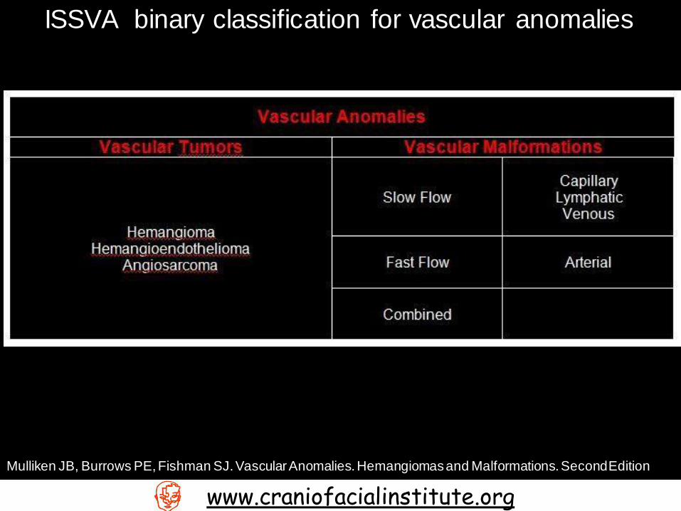
ISSVA binary classification for vascular anomalies
Mulliken JB, Burrows PE, Fishman SJ. Vascular Anomalies. Hemangiomas and Malformations. Second Edition
www.craniofacialinstitute.org
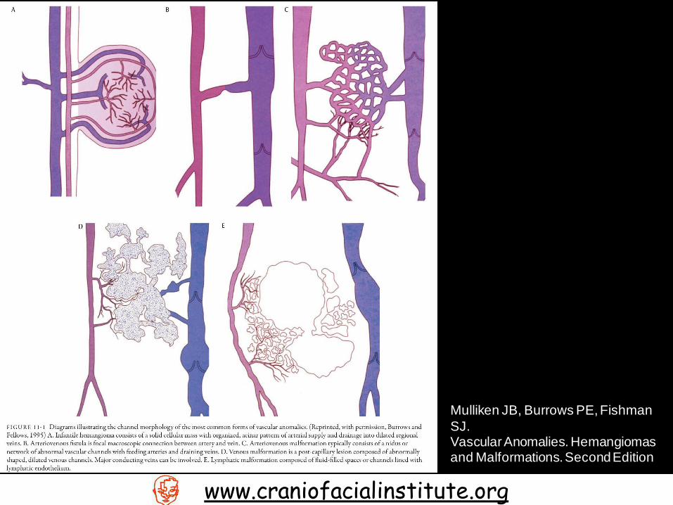
Mulliken JB, Burrows PE, Fishman
SJ. Vascular Anomalies. Hemangiomas and Malformations. Second Edition
www.craniofacialinstitute.org
Clinical Manifestation
Vascular Tumors
www.craniofacialinstitute.org

Infantile Hemangioma
Benign vascular neoplasms
Have a characteristic clinical course marked by
early proliferation and
followed by spontaneous involution.
Third
Trimester
PROLIFERATION PHASE
Neonate
Proliferation of primitive cells
INVOLUTING PHASE
1-5 years
Due to Apoptosis
Mast cells interact with macrophages and
fibroblasts = Transgranulation
INVOLUTED PHASE
>7 years
Revascularization
Deposition of fat cells
www.craniofacialinstitute.org
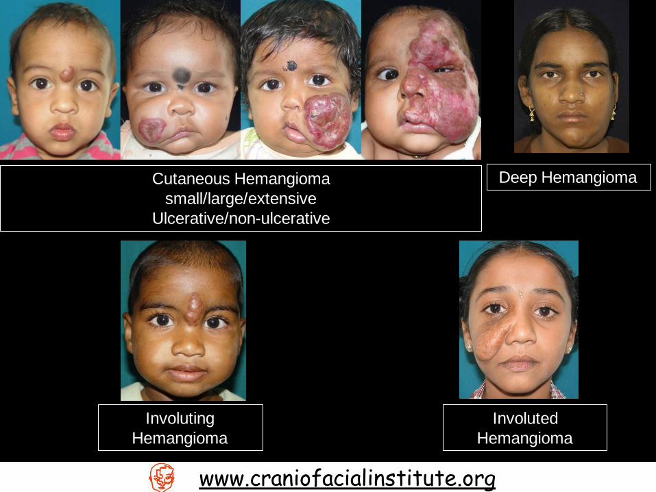
Cutaneous Hemangioma
small/large/extensive
Ulcerative/non-ulcerative
Deep Hemangioma
Involuting
Hemangioma
Involuted
Hemangioma
www.craniofacialinstitute.org
Clinical Manifestation
Vascular Malformations
www.craniofacialinstitute.org
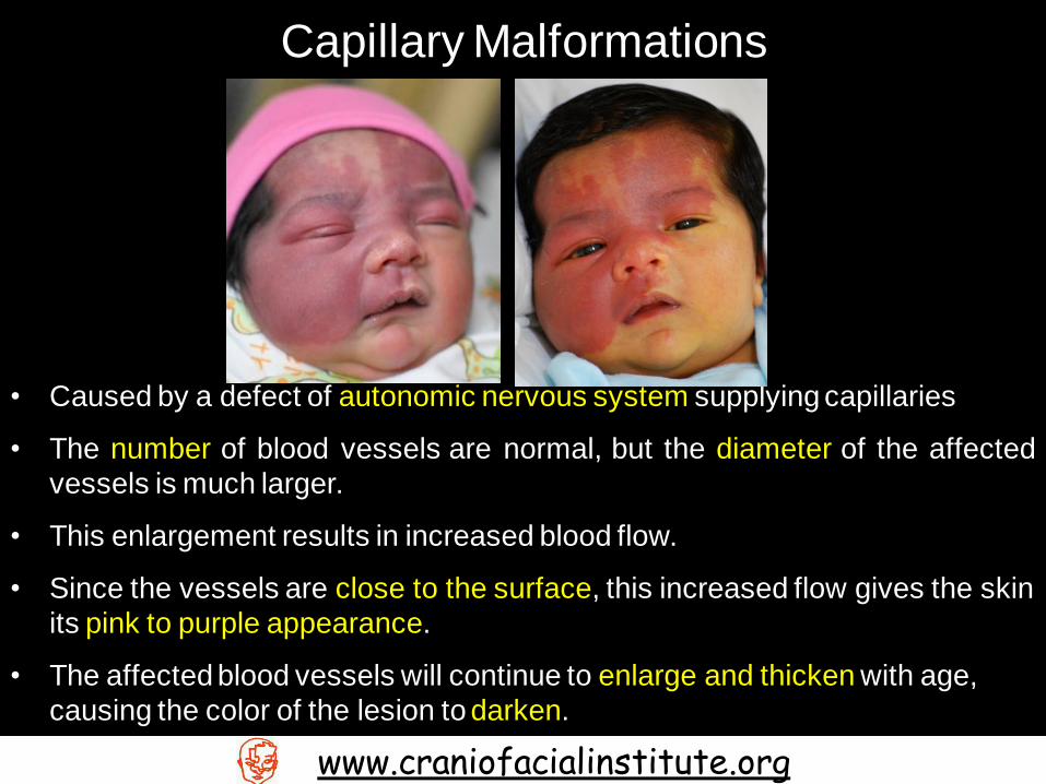
• Caused by a defect of autonomic nervous system supplying capillaries
• The number of blood vessels are normal, but the diameter of the affected
vessels is much larger.
• This enlargement results in increased blood flow.
• Since the vessels are close to the surface, this increased flow gives the skin
its pink to purple appearance.
• The affected blood vessels will continue to enlarge and thicken with age,
causing the color of the lesion to darken.
Capillary Malformations
www.craniofacialinstitute.org

Venous Malformations
• Made up of malformed veins
• Vary in color from blue to dark purple, depending on how deep the
malformation extends.
• Tend to swell with activity/exercise
• The mass is usually soft and compressible and then refills when released.
• There may be small hard masses palpable in the lesion, called phleboliths,
which are small collections of calcium that have resulted from slow blood
flow and blood clots.
www.craniofacialinstitute.org
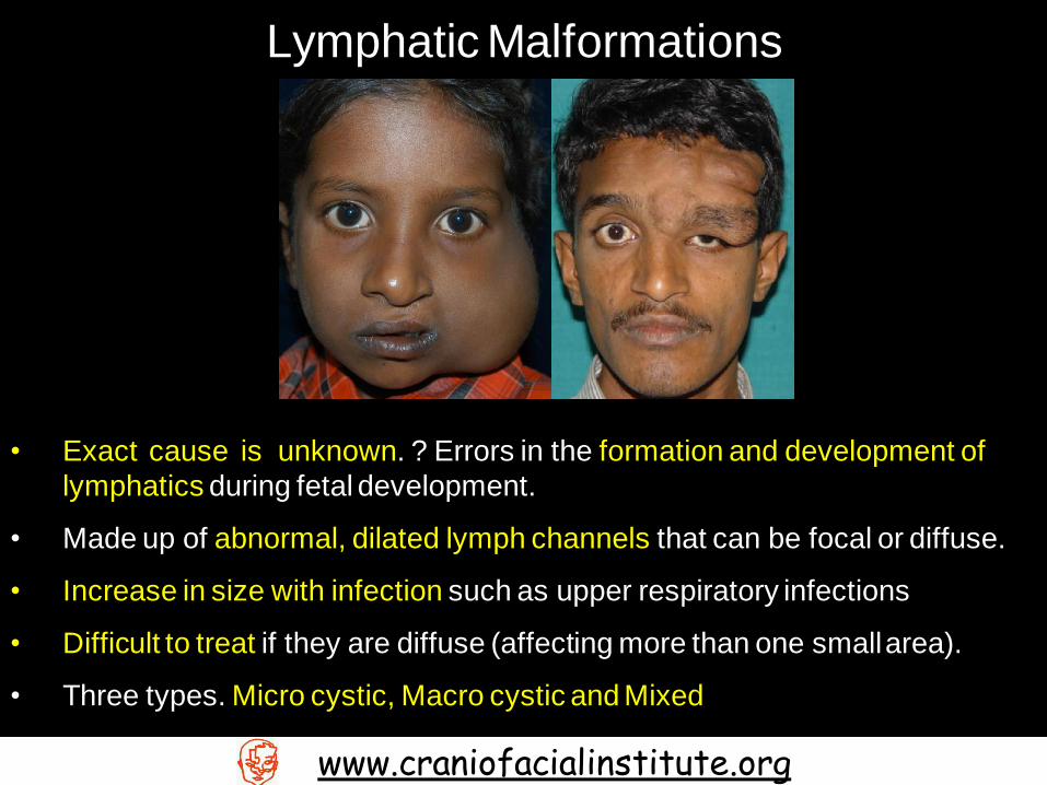
Lymphatic Malformations
• Exact cause is unknown. ? Errors in the formation and development of
lymphatics during fetal development.
• Made up of abnormal, dilated lymph channels that can be focal or diffuse.
• Increase in size with infection such as upper respiratory infections
• Difficult to treat if they are diffuse (affecting more than one small area).
• Three types. Micro cystic, Macro cystic and Mixed
www.craniofacialinstitute.org
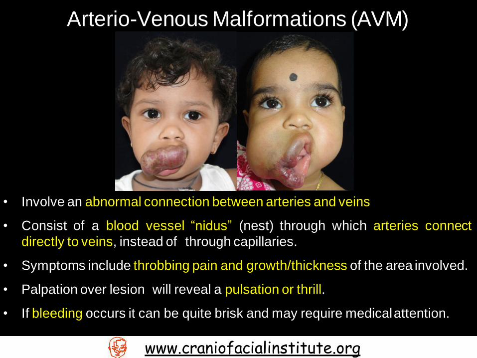
Arterio-Venous Malformations (AVM)
• Involve an abnormal connection between arteries and veins
• Consist of a blood vessel “nidus” (nest) through which arteries connect
directly to veins, instead of through capillaries.
• Symptoms include throbbing pain and growth/thickness of the area involved.
• Palpation over lesion will reveal a pulsation or thrill.
• If bleeding occurs it can be quite brisk and may require medical attention.
www.craniofacialinstitute.org
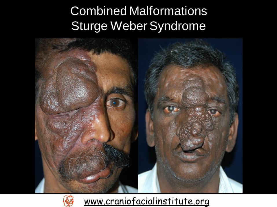
Combined Malformations
Sturge Weber Syndrome
www.craniofacialinstitute.org

Sturge-Weber syndrome consists of
Tortuous slow-flow vessels
involving the conjunctiva, episclera, retina or choroid.
Glaucoma is the most common and serious ophthalmological
complication; the prevalence is 60% (Sujansky and
Conradi, 1995a).
Sudden corneal clouding is the pathognomonic sign of acute
glaucoma; this is an emergency.
www.craniofacialinstitute.org

Ultrasound…
Arterial flow Venous Flow
Ultrasonography of AV malformation of
upper lip
Note the arterial flow, venous flow and nidus
of capillaries
Nidus
www.craniofacialinstitute.org
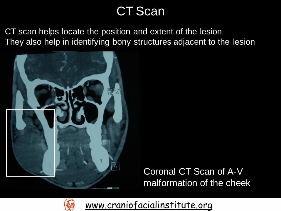
CT Scan
CT scan helps locate the position and extent of the lesion
They also help in identifying bony structures adjacent to the lesion
Coronal CT Scan of A-V
malformation of the cheek
www.craniofacialinstitute.org
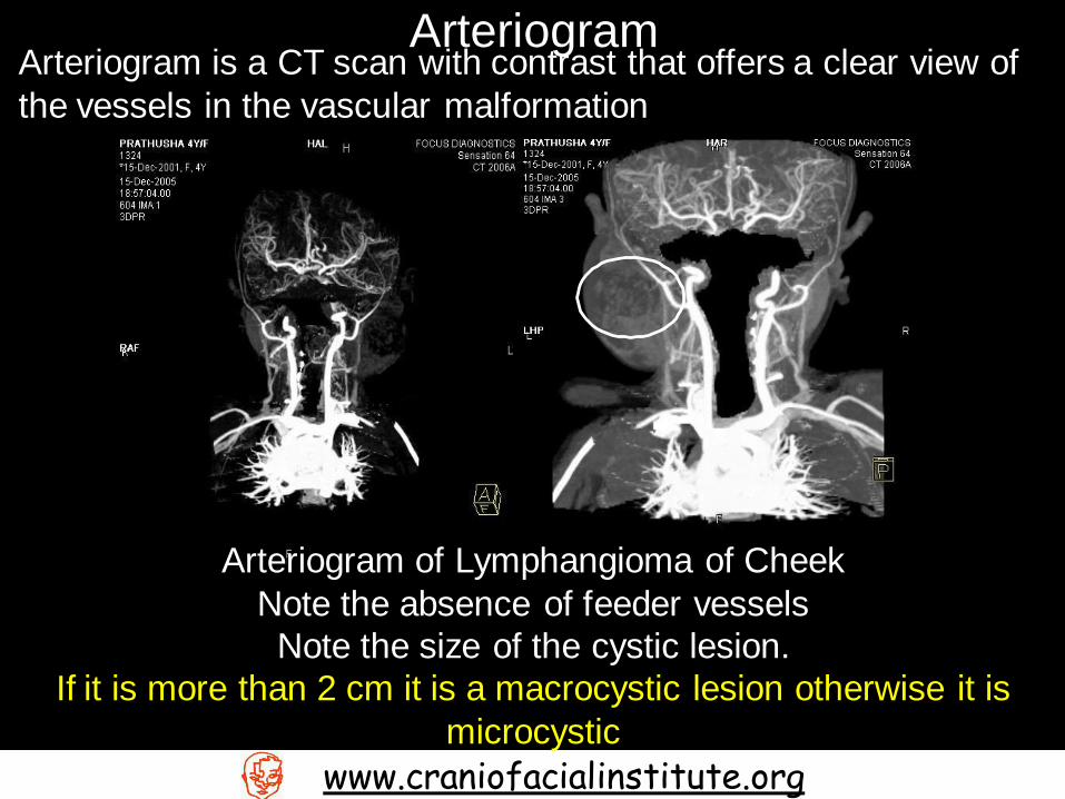
Arteriogram Arteriogram is a CT scan with contrast that offers a clear view of
the vessels in the vascular malformation
Arteriogram of Lymphangioma of Cheek
Note the absence of feeder vessels
Note the size of the cystic lesion.
If it is more than 2 cm it is a macrocystic lesion otherwise it is
microcystic
www.craniofacialinstitute.org
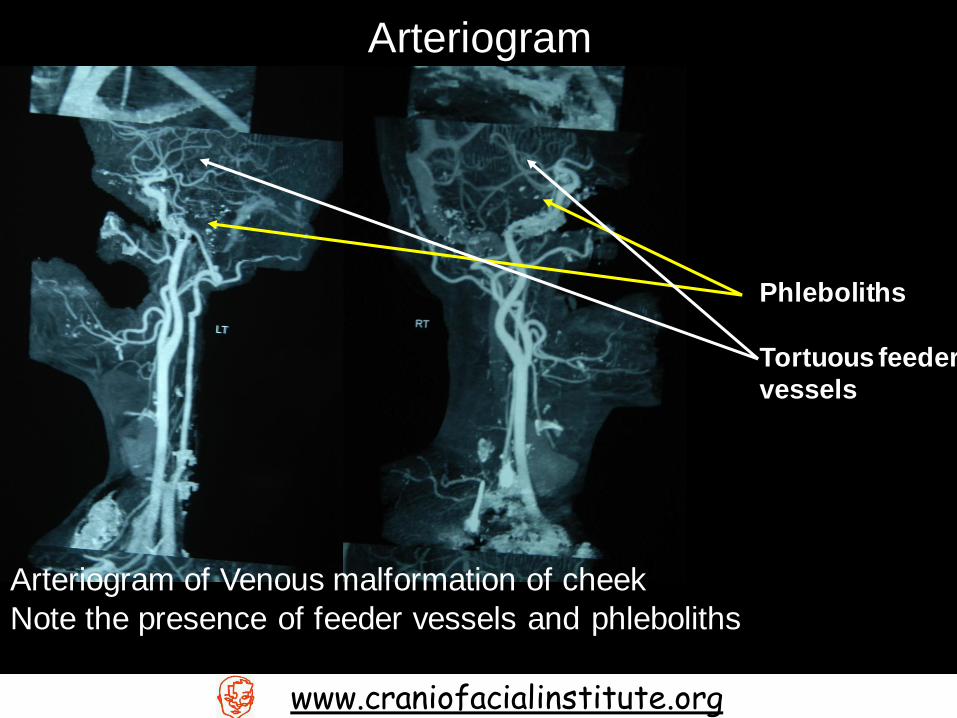
Arteriogram
Arteriogram of Venous malformation of cheek
Note the presence of feeder vessels and phleboliths
Phleboliths
Tortuous feeder
vessels
www.craniofacialinstitute.org
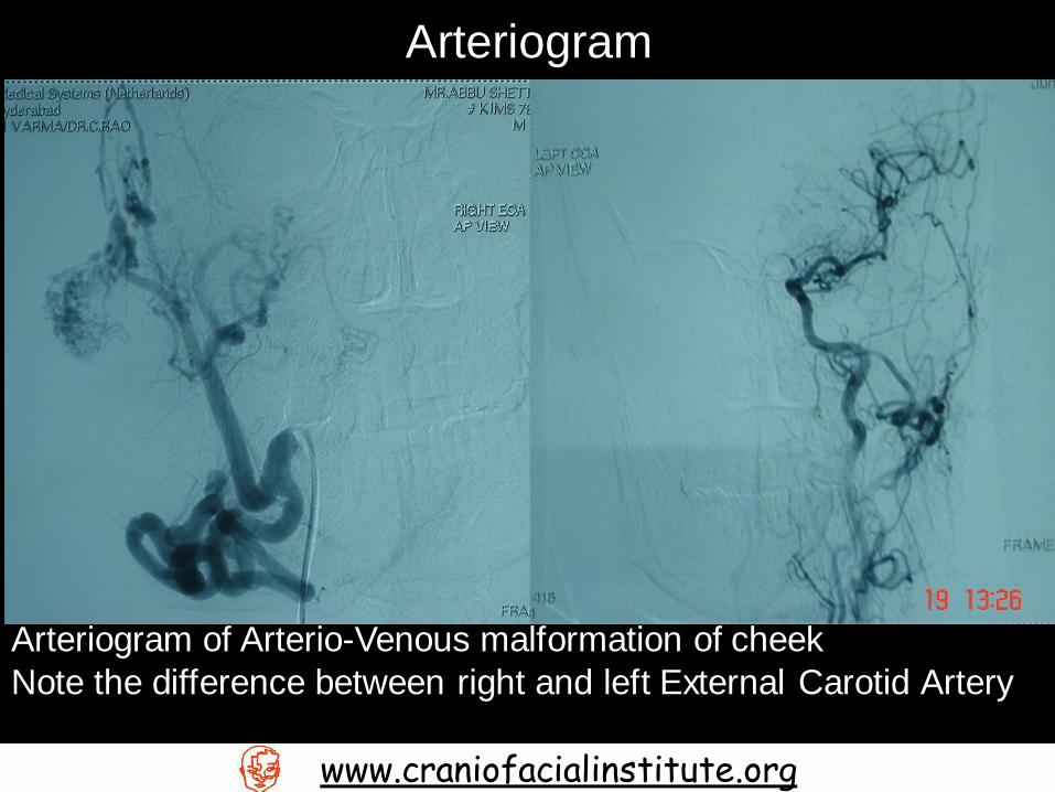
Arteriogram
www.craniofacialinstitute.org
Arteriogram of Arterio-Venous malformation of cheek
Note the difference between right and left External Carotid Artery
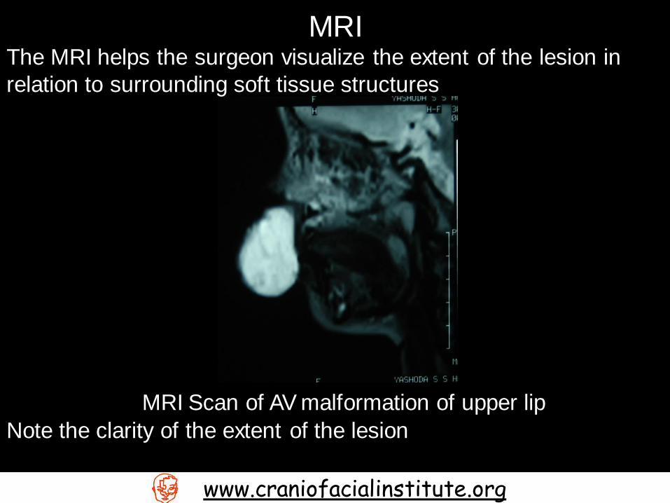
MRI The MRI helps the surgeon visualize the extent of the lesion in
relation to surrounding soft tissue structures
www.craniofacialinstitute.org
MRI Scan of AV malformation of upper lip
Note the clarity of the extent of the lesion
Treatment
www.craniofacialinstitute.org
Treatment for Vascular Malformations and Hemangiomas is
usually as follows
For Low Flow Superficial Lesions
Sclerotherapy followed by
Conventional surgery
For High Flow Lesions
Subtraction angiography with embolization with gel foam or
stents followed by
Conventional surgery within 72 hours
In India Angiography is beyond the capacity of most patients.
Therefore angiography is considered only if any great vessel is
involved.
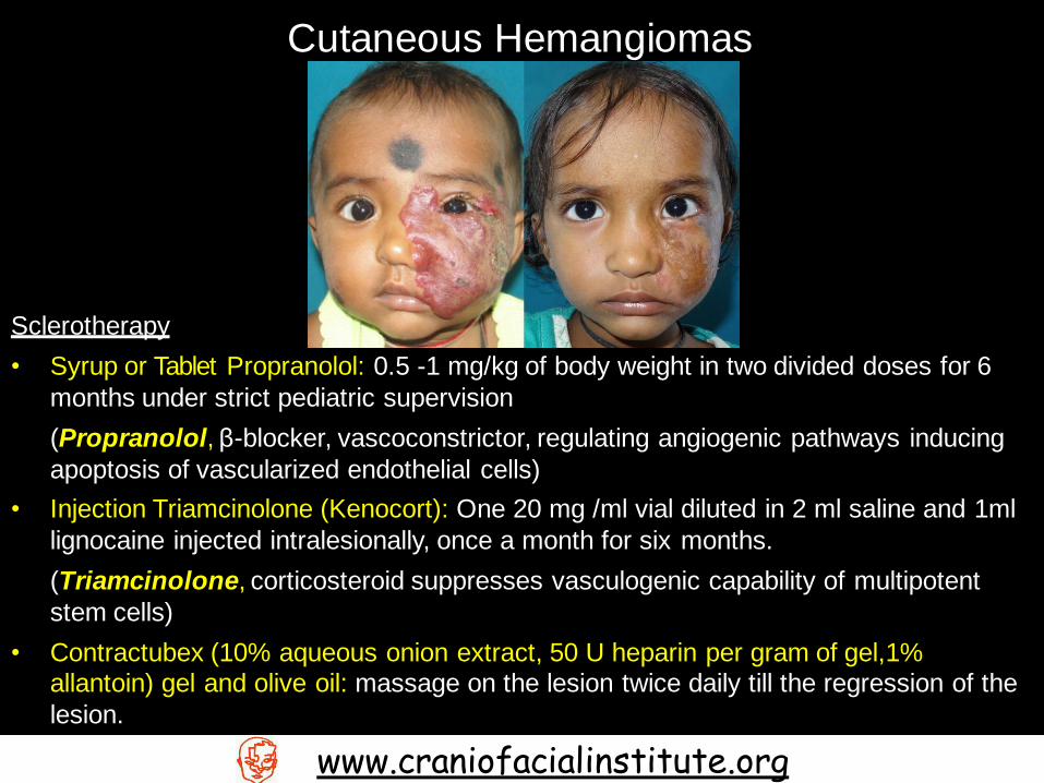
Cutaneous Hemangiomas
www.craniofacialinstitute.org
Sclerotherapy
• Syrup or Tablet Propranolol: 0.5 -1 mg/kg of body weight in two divided doses for 6
months under strict pediatric supervision
(Propranolol, β-blocker, vascoconstrictor, regulating angiogenic pathways inducing
apoptosis of vascularized endothelial cells)
• Injection Triamcinolone (Kenocort): One 20 mg /ml vial diluted in 2 ml saline and 1ml
lignocaine injected intralesionally, once a month for six months.
(Triamcinolone, corticosteroid suppresses vasculogenic capability of multipotent
stem cells)
• Contractubex (10% aqueous onion extract, 50 U heparin per gram of gel,1% allantoin) gel and olive oil: massage on the lesion twice daily till the regression of the
lesion.

Bleomycin Treatment
• Pingyangmycin (Bleomycin A5) : 2-6 ml (0.5 -4 mg/ml concentration) given intralesionally and
repeated every 4 weeks for a maximum of 12 sessions. OR
• Bleomycin: 0.5 – 1.0mg/kg body weight up to a maximum of 6mg (0.5-105 mg/ml
concentration) given intralesionally and repeated every 4 weeks for a maximum of 12 sessions.
• Bleomycin acts by producing a sclerosing effect due to its direct action on the endothelial cells
of the lesion producing non-specific inflammatory reaction
• Can be given in Capillary, Venous, Arterio-venous and Lymphatic malformation and
Hemagiomas.
All Vascular Malformations and Hemangiomas
www.craniofacialinstitute.org

• Key is Accessibility
Accessible = Surgery
Inaccessible = Embolisation and surgery
• Ligation of all possible blood vessels in the vicinity of the lesion
• Aim of surgery
• HARMONIC SCALPEL is used to radically excise all
affected tissue as remnants of necrotic tissue can form a
focus of a granuloma or further infection.
• Reconstruct what ever possible
• Post operative maintenance with steroidal injections intra-
lesionally
www.craniofacialinstitute.org
Surgical Protocol
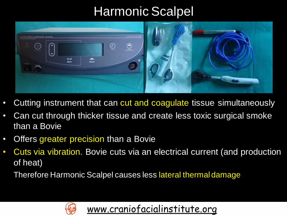
• Cutting instrument that can cut and coagulate tissue simultaneously
• Can cut through thicker tissue and create less toxic surgical smoke
than a Bovie
• Offers greater precision than a Bovie
• Cuts via vibration. Bovie cuts via an electrical current (and production
of heat)
Therefore Harmonic Scalpel causes less lateral thermal damage
www.craniofacialinstitute.org
Harmonic Scalpel
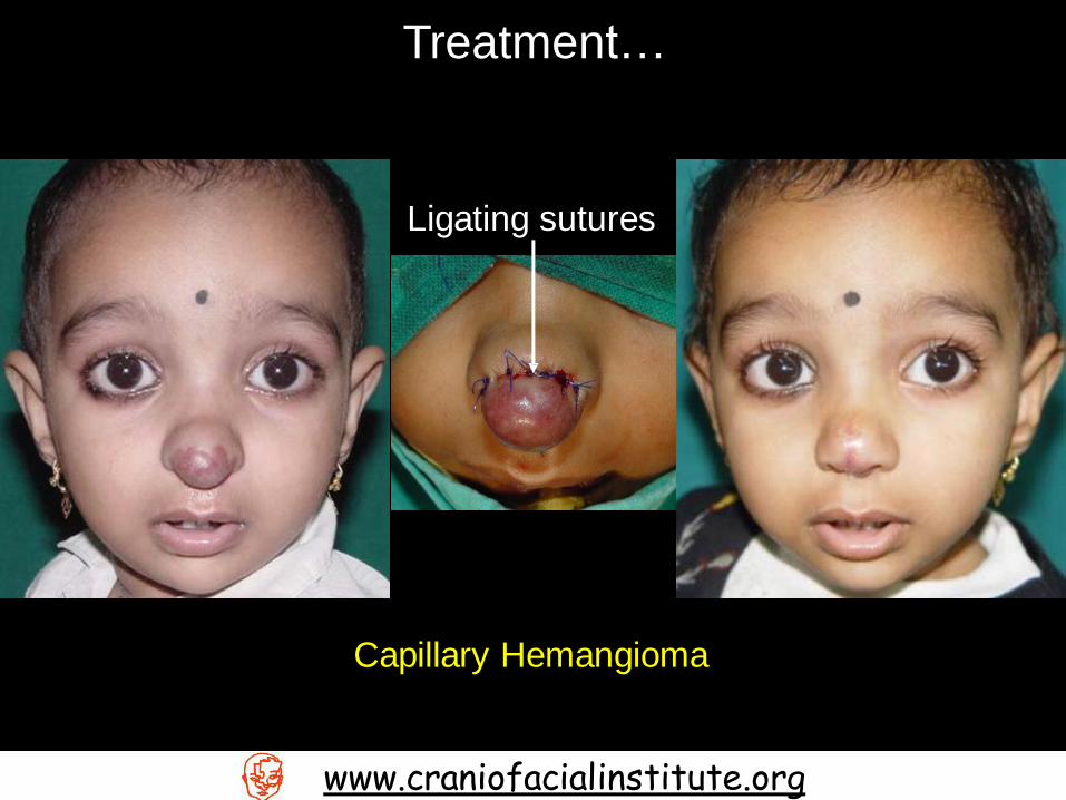
Treatment…
Capillary Hemangioma
Ligating sutures
www.craniofacialinstitute.org
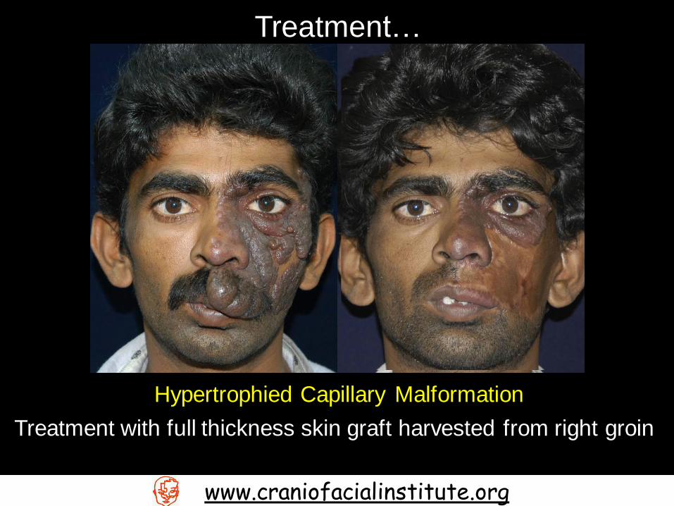
Treatment…
Hypertrophied Capillary Malformation
Treatment with full thickness skin graft harvested from right groin
www.craniofacialinstitute.org
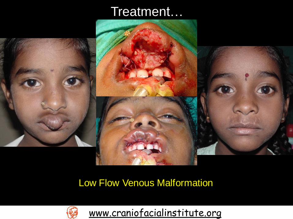
Treatment…
Low Flow Venous Malformation
www.craniofacialinstitute.org
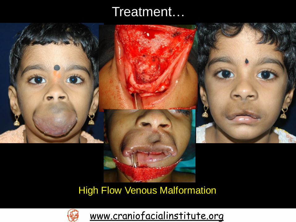
Treatment…
High Flow Venous Malformation
www.craniofacialinstitute.org
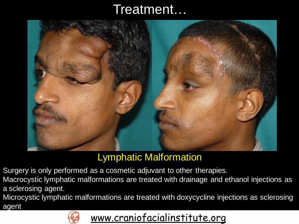
Treatment…
Lymphatic Malformation
Surgery is only performed as a cosmetic adjuvant to other therapies.
Macrocystic lymphatic malformations are treated with drainage and ethanol injections as
a sclerosing agent. Microcystic lymphatic malformations are treated with doxycycline injections as sclerosing
agent
www.craniofacialinstitute.org

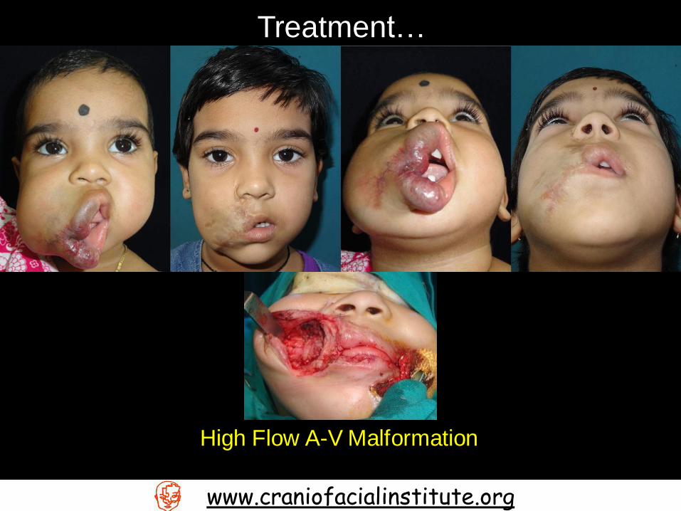
Treatment…
High Flow A-V Malformation
www.craniofacialinstitute.org
Treatment…
High Flow A-V Malformation
www.craniofacialinstitute.org
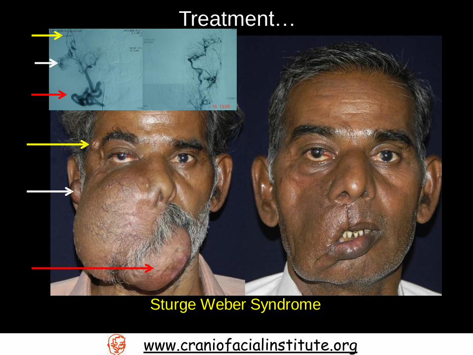
Treatment…
Sturge Weber Syndrome
www.craniofacialinstitute.org

Treatment…
Sturge Weber Syndrome
www.craniofacialinstitute.org

Complications
www.craniofacialinstitute.org
Hemangioma
• Very problematic, interfering with eating, breathing, seeing,
hearing, and speaking.
Vascular malformations: capillary, venous and arterio-venous
• Patients with port-wine stains should be evaluated and
monitored for a larger syndromic entity.
• Malformations that are part of the Klippel-Trenaunay-Weber
syndrome can be located on the lungs, spleen, liver, bladder, or
colon. Visceral involvement can often lead to substantial
morbidity in the form of internal hemorrhage.
Vascular malformations - Lymphatic malformations
• Diffuse cervicofacial disease can result in mandibulomaxillary
hypertrophy because of direct invasion of the bone and growth
of the malformation within the bone..
• Lymphangiomas often swell with the onset of general viral
infection or remote bacterial infection. This typically resolves
with the resolution of the infection.
• Lymphangiomas can become infected
www.craniofacialinstitute.org
Complications…

Do not confuse a Vascular malformation with…
www.craniofacialinstitute.org
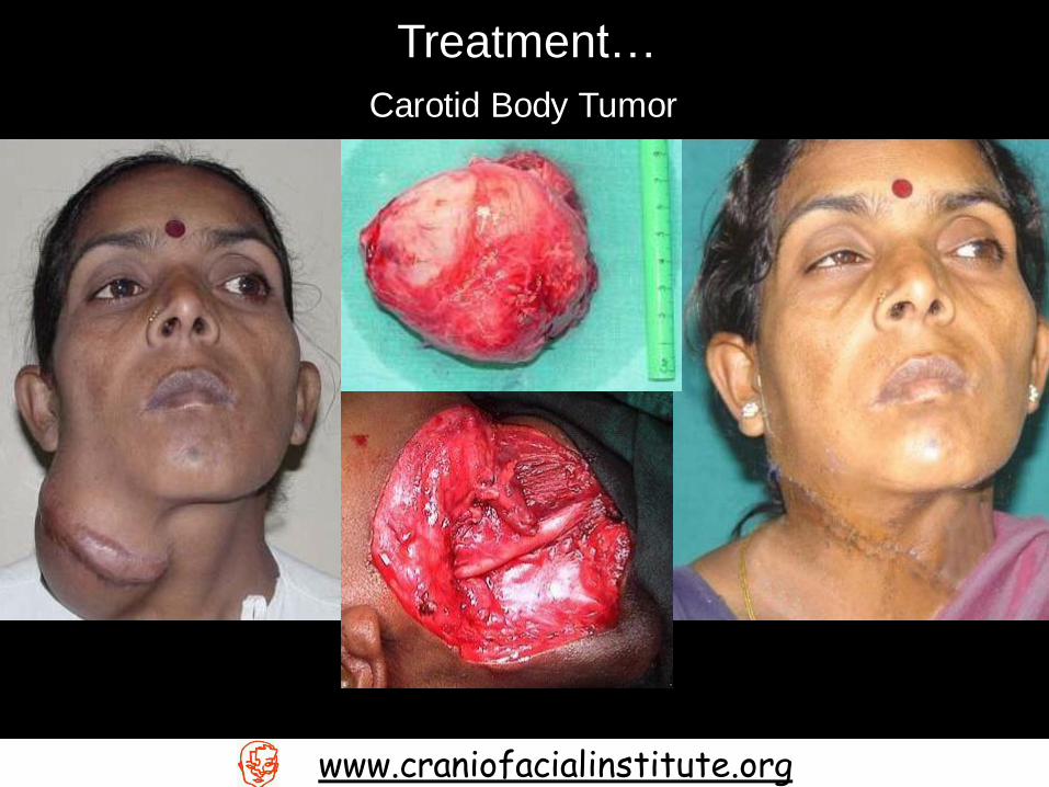
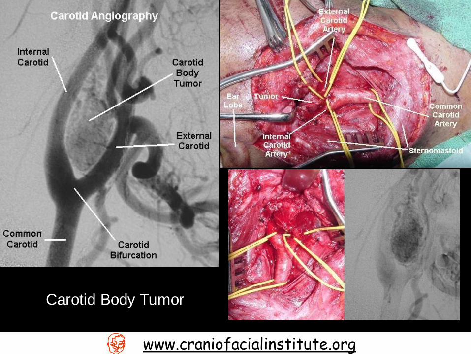
Treatment… Carotid Body Tumor
Slowly enlarging(~5mm per year), non-tender neck masses
located just anterior to the sternocleidomastoid muscle at the level
of the hyoid.
The mass may transmit the carotid pulse or demonstrate a bruit or
thrill, which might confuse the clinician to think it is a vascular
malformation.
As these tumors enlarge, progressive symptoms of dysphagia,
odynophagia, hoarseness and other cranial nerve(IX-XII) deficits
appear.
Carotid angiography is by far the most useful diagnostic test for
paragangliomas.
Biopsy, including finewwnewe.cdrleanaiosfpaircaitaiolinnsistiutuntnee.coersgsary,
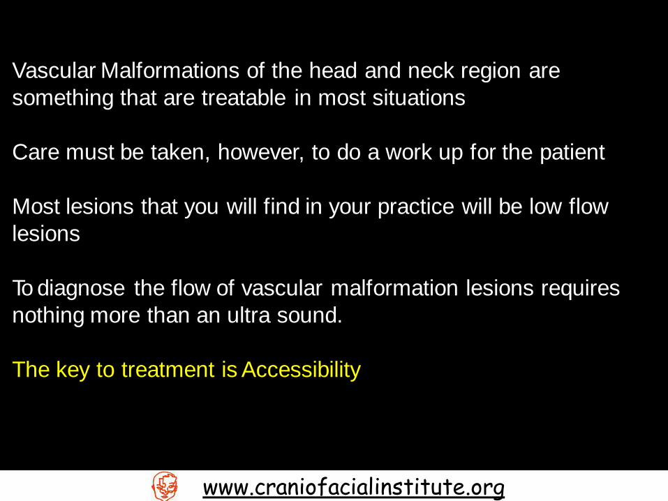
Vascular Malformations of the head and neck region are
something that are treatable in most situations
Care must be taken, however, to do a work up for the patient
Most lesions that you will find in your practice will be low flow
lesions
To diagnose the flow of vascular malformation lesions requires
nothing more than an ultra sound.
The key to treatment is Accessibility
www.craniofacialinstitute.org