


Idiomas
Páginas
Jurídico

UNIVERSIDADAUTÓNOMADEMADRIDFACULTADDEMEDICINA
DepartamentodeAnatomíaPatológica
Alteracioneshistológicasymecanismosdefibrosisenlaslesionesvaricosasdelavenasafenainterna:
Estudioinmunohistoquímico,molecularydemicroscopíaconfocal.
TESISDOCTORAL
DoctoradoenMedicinayCirugía
JuanPedroVelascoMartín
Madrid,2015

JAVIER FRANCISCO REGADERA GONZÁLEZ, MÉDICO PATÓLOGO Y CATEDRÁTICO DEHISTOLOGÍAHUMANADELAFACULTADDEMEDICINADELAUNIVERSIDADAUTÓNOMADEMADRIDyGABRIEL ESPAÑA CAPARRÓS, JEFE DE SERVICIO DE CIRUGÍA VASCULAR DEL HOSPITALMONCLOA,UNIVERSIDADEUROPEADEMADRIDCERTIFICAN QUE: D. JUAN PEDRO VELASCO MARTÍN, Licenciado en Biología por la
UniversidadAutónomadeMadrid,harealizadobajonuestradireccióneltrabajodeinvestigación"Alteracioneshistológicaymecanismosdefibrosis en las lesiones varicosas de la vena safena interna: estudioinmunohistoquímico,molecularydemicroscopía confocal.",estudioqueconsideramoscompletamentesatisfactorioparaserpresentadoydefendido como Tesis Doctoral en el Programa de Doctorado deMedicinayCirugíadelaUniversidadAutónomadeMadrid.
LoquefirmamosenMadridel7deseptiembrede2015Fdo.:JavierRegaderaGonzález Fdo.:GabrielEspañaCaparrós
Director Director

AGRADECIMIENTOS:
alProf.Dr.JavierRegadera,porsusenseñanzasdelaHistología,porladireccióndeestaTesisyporsucontribuciónenmiformacióncientíficayacadémica.
alDr.GabrielEspañaCaparrós,porlatrasmisióndelosconceptosclínicosdelapatologíavascularduranteladireccióndeestaTesisyporpermitirmeobtenerel
materialquirúrgicoencondicionesidóneas.
alProf.Dr.LuisSantamaría,porsusenseñanzasdelaHistologíayporsuapoyopararealizarlasestanciascientíficasenMcGillUniversitydeMontreal.Tambiénmi
reconocimientoalProf.Dr.ÁngelNúñez,DirectordelDpto.deAnatomía,HistologíayNeurociencia,porsuapoyoyfacilidadesparamiformacióncientífica.
alaDra.AndreaAguado,unadelasmejorescientíficosqueheconocidoyconla
queaprendílarealizacióneinterpretacióndediversastécnicasmoleculares,alaProfªDra.AnaBriones,porsuconocimiento,interés,ayudaylaaportaciónnuevasideasy
futurosexperimentosparamiproyectoyalaProfª.Dra.MercedesSalaices,porabrirmedesinteresadamentesulaboratorio.NoquieroolvidarmedeMarisol,Laura,
Sonia,Rosa,Ana,MaríayRoberporcompartirtantosmomentos.SindudamiestanciaenFarmacologíahasidocientíficamenteyhumanamentemuygratificante.
alProf.Dr.DavidHardisson,porsusvaliososconsejos,ayudadesinteresadaytan
agradablesconversacionescientíficasyacadémicas.TambiénagradeceralProf.Dr.ManuelNistal,porsusconsejosyacercarmealmundodelapatología.
alDr.MiguelCampanero,porenseñarmenuevastécnicasenelusódel
microscopioconfocal.TambiénmiagradecimientoalDr.EmilioBurgos,porsuayudaenlainterpretacióndelamicroscopiaelectrónica.
alProf.Dr.CarlosR.Morales,ProfessorofHistologyenlaFacultaddeMedicinade
McGillUniversity,Montreal,poraceptarmeensulaboratorio,porsutratohumano,suamistadyladesufamiliayporsusconsejos.Además,quieroagradeceralaDra.LorenaCarvellisupacienciaparaenseñarmenuevastécnicasysusgrandesdotesdocentes.Perosobretodoquieroagradecerlasuamistad.Sequedondeseaque
estemosyapesardelosocéanosquenosseparensiempretendréunaamiga.Montrealnohubierasidolomismosinustedes.
aDña.CarmenSánchezPalomoyaDña.MartaCorreaVárez,TécnicossuperioresdelLaboratoriodeHistologíadelaUAM,porelprocesamientodelasmuestrashistológicassincuyalabornohubierasidoposiblelarealizacióndeestaTesis;a
Dña.NatiMuñozporlacalidaddelosmétodosdemicroscopíaelectrónica.
alaDra.AnaAranda,alaDra.OlaiaMartínez‐IglesiasyaElviraAlonsoMerino,porlaformaciónenmétodosmolecularesdurantemiestanciaensulaboratoriodel
InstitutodeInvestigacionesBiomédicasCSIC‐UAM
alosnumerososdoctoresconlosquehetenidolagransuertedecolaborarensuslíneasdeinvestigación,realizadasparalelamentedurantelosexperimentosdela
presenteTesis.LosDrs.Boscá,Fernández‐Velasco,Alemany,Aller,Cuadrado,Botella,Padin,Rada,ArribasyM.C.Gonzálezhancontribuidoaabrirmelucesenestecaminocientífico.QuieroespecialmentehacerreconociendoaIago,Pilar,PerlayDavidporsu
conversaciónycompañíadurantelosCongresosCientíficos.
alDr.LuisReparaz,quetantomeanimoyquienconsiguióunabecaquepermitiólarealizacióndelapresenteTesis;alDr.LuisFelipeRieraporpermitirmecolaborar
científicamenteconélyalaDra.InmaculadaSantosÁlvarez,porsuvaliosaexperienciaenlaedicióndetextosmédicos.
tambiénquieroacordarmeenestosmomentosdemicarreracientíficadeaquellos
amigosqueempezaronconmigoenlaFacultaddeBiologíadelaUAM.Graciasatodosportanbuenosmomentosdentroyfueradelafacultad.Sequeconvosotrostengo
verdaderosamigos.
TambiénmegustaríaacordarmedelaDra.HelenaRomoquetantomeenseñoyconlaquepubliquemiprimerartículocientífico.
DEDICATORIA:
Yporúltimodedicárselaamispadres,PedroyPilar,yamihermanoDavid.Quedecirquenosesepa.Sinellosnoseríaquiensoyynohabríallegadohastaaquí.Quiero
darleslasgraciasporsupacienciayapoyo,perosobretodoporhaberformadounabuenapersona,loqueestáporencimadetodoslostítulosacadémicos.
Aleaiactaest
Índice
i
INTRODUCTION 1
Clinical,Etiology,Anatomic,Pathophysiology(CEAP)classificationof
chronicvenousinsufficiency 4
Pathophysiolgyofveinwallalterationinvaricosediseaseofleg 5
Venousanatomyandbloodflowintheleg 7
Histology,immunohistochemistryandmolecularbiologyofvein 9
Cellularandmolecularpathologyofvaricosevein 14
InflammationandROSmediatorsinvascularsystem 21
Venousanatomyandbloodflow 21
HIPHOTESISANDOBJETIVES 29
MATERIALANDMETHODS 31
MATERIAL 32
METHODS 33
Ethicalaspects 33
Tissueprocessing 33
Histologicalmethods 33
Immunohistochemistry(StreptavidinBiotinPeroxidaseMethod) 34
Electronmicroscopymethods 35
Fluorescencemethodsforelasticfibers 36
Confocalmicrocopymethodsforintotostudyofelasticfibersandcollagen
tissue,usinganendovascularveinwallreconstruction 36
Histologicalquantification 39
Molecularmethods 40
Microscopyphotography 43
Dataanalysisandstatistics 43
RESULTS 44
CollagentypeIhistometryquantifyinginvenousintima 76
CollagentypeIIIhistometryquantifyinginvenousintima 77
SMAhistometryquantifyinginvenousintima 79
Elasticfibershistometryquantifyinginvenousintima 81
Índice
ii
MolecularquantificationofcollagentypeI,collagentypeIII,
elastinandSMAinproximalanddistalsegmentsofvaricose
veins 83
MolecularstudyofinflammatoryandROSmechanismsrelates
tovaricoselesions 84
DISCUSIÓN 86
CONCLUSIONS 104
CONCLUSIONES 106
RESUMEN 108
SUMMARY 112
REFERENCES 116

Introduction
1
INTRODUCTION
Introduction
2
Varicoseveins(tortuous,twisted,orlengthenedveins123isacommonvasculardisease
ofthelowerextremity.Varicesaresometimesclassifiedintotrunk,reticularandhyphenweb
typesbasedontheirsizeandanatomicaldistribution.75ChronicVenousInsufficiency(CVI)is
acommonproblemwithasignificantimpactonbothafflictedindividualsandthehealthcare
system.73 In this sense, varicose veins affectingmore than 25million adults in theUnited
States,andmorethan6millionsufferingmoreadvancedvenousdisease.27
Venouspathologydevelopswhenvenouspressureisincreasedandreturnofbloodis
impairedthroughseveralmechanisms.41Theoverallincreasedvenouspressureasaresultof
bloodstasismayresultinveinwalldilatationandthecharacteristicCVIdermalchangeswith
hyperpigmentation, venous eczema, subcutaneous tissue fibrosis and ultimately
ulceration.72,225 One general functional consequence of varicose vein formation and/or
venousinsufficiencyisadecreaseinvenousreturnandanincreaseinthefillingpressureof
theassociatedvenousnetwork–aphenomenonthatisreferredtoas’venoushypertension‘.
Althoughmanyriskfactorshavebeenproposed,thereisstillsomecontroversyabout
theetiologyandpathogenesisofthevaricosedisease.77Theprevalenceofvaricoseveinsis
higher in developed, industrial countries than in underdeveloped countries. Risk factors
found to be associated with CVI include age, sex, and family history of varicose veins,
obesity,pregnancy,phlebitisandprevious leg injury.120,150,248Therearealsoenvironmental
orbehavioralfactorsassociatedwithCVI,suchasprolongedstandingandperhapsasitting
posture at work.120,145 Like we had said, varicose vein disease is a frequently occurring
pathologywithmultifactorialcausesandageneticcomponent,239asiscorroboratedbythe
frequent clinical observation that the distensibility of arm veins in patients with varicose
veinsisincreasedabnormally,suggestingasystemicdiseaseofthevenouswall.306
Many clinical studies have shown that the prevalence of varicose veins is
approximatelytwiceashighinwomenasmen,andincreaseswithadvancingage.49,75,150,253
TheEdinburghVeinStudyscreened1566subjectswithduplexultrasoundforreflux,finding
CVIin9.4%ofmenand6.6%ofwomenafterageadjustment,whichrosesignificantlywith
age(21%inmenover50yearsand12%inwomenover50years).233Also,theFramingham
Introduction
3
Studydemonstratedaprevalenceof1%inmenand10%inwomenagedlessthan30years,
comparedwith57percentinmenand77percentinwomenover70yearsold.34
Varicose veins aremore common inwomenwho have had several pregnancies and
had had hemorrhoids and vulvar varicosities during and after pregnancy.100 The
development of new varicose veins occurs in up to 28 % of pregnancies.258 During
pregnancy,weightgainfromincreasedtotalbodyfluidandraisedintra‐abdominalpressure
may predispose varicose vein formation in woman. Furthermore, upregulation of certain
hormones, such as relaxin, oestrogen and progesterone, causes venous relaxation and
increasesveincapacitance.199
Familyhistoryisanotherdescribedriskfactor.AstudyinFrancereportedthatahistory
ofvaricoseveinsinafirst‐degreerelativeisthemostimportantriskfactorinbothmenand
women.46Patientswithvaricoseveinswere21.5timesmorelikelytoreportapositivefamily
history.248 Another study in Japan found that 42 % of patients with varicose veins had a
positive family history compared with only 14 % in those without varicosities.105 In
conclusion,epidemiologicalstudieshavedemonstratedaninvolvementofhereditaryfactors
forthetransmissionofvaricoseveins.61,81FiebigA.etal.201078concludethattheadditive
geneticcomponentofCVIisapproximately17%.
Finally,theTamperestudyinterviewed3,284randomlychosenmenand3,590women
40to60yearsofage.150Thequestionnairecoveredfamilystatus,sex,age,professionand
weight.With theseparameters itwaspossible tocorrelate the incidenceofvaricoseveins
withspecificlifestylefactors.Fromthedataonecanconcludethatvaricoseveinscorrelate
with female sex, obesity, extensive standing type of work, parity (proportional to the
numberofgivenbirths)andafamilyhistoryofvaricosedisease.Thelatterrelationpointsto
apossiblegeneticpredispositiontodevelopvaricoseveins. Interestingly,mostrisk factors,
such as Body Mass Index, parity and prolonged standing, lead to a rise in hydrostatic
pressureandmaythereforeaugmentvenousremodeling.216

Introduction
4
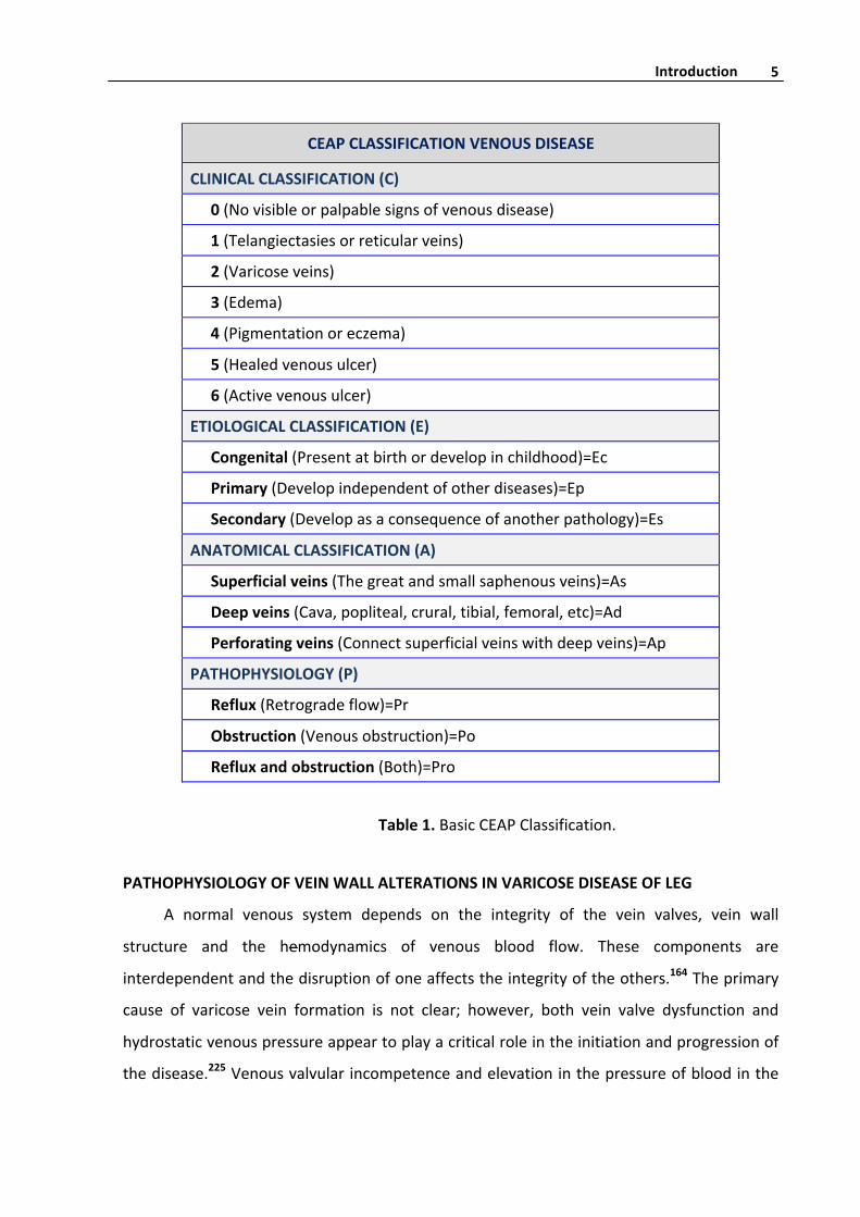
CLINICAL,ETIOLOGY,ANATOMIC,PATHOPHYSIOLOGY(CEAP)CLASSIFICATIONOFCHRONIC
VENOUSINSUFFICIENCY
The CEAP classification was introduced in 1996, defining clinical (C), etiological (E),
anatomical(A)andpathophysiological(P)aspectsofCVI.ThemanifestationsofCVImaybe
viewed in termsofawell‐establishedclinical classificationscheme.TheCEAPclassification
wasdevelopedbyaninternationalconsensusconferencetoprovideabasisofuniformityin
reporting,diagnosing,andtreatingCVI(Table1).218Theclinicalclassificationhas7categories
(0–6)andisfurthercategorizedbythepresenceorabsenceofsymptoms.Theclassification
isavaluable tool in theobjectiveevaluationofCVI,providingasystemtostandardizeCVI
classification with emphasis on themanifestations, cause, and distribution of the venous
disease.249 Ithasbecomewidelyacceptedas thestandardclassificationsystemforvenous
disorders.131AndtheCEAPclassificationshowsastatisticallysignificantassociationbetween
ahigherCEAPgradeandanoldercurrentageofpatients.46
Introduction
5
CEAPCLASSIFICATIONVENOUSDISEASE
CLINICALCLASSIFICATION(C)
0(Novisibleorpalpablesignsofvenousdisease)
1(Telangiectasiesorreticularveins)
2(Varicoseveins)
3(Edema)
4(Pigmentationoreczema)
5(Healedvenousulcer)
6(Activevenousulcer)
ETIOLOGICALCLASSIFICATION(E)
Congenital(Presentatbirthordevelopinchildhood)=Ec
Primary(Developindependentofotherdiseases)=Ep
Secondary(Developasaconsequenceofanotherpathology)=Es
ANATOMICALCLASSIFICATION(A)
Superficialveins(Thegreatandsmallsaphenousveins)=As
Deepveins(Cava,popliteal,crural,tibial,femoral,etc)=Ad
Perforatingveins(Connectsuperficialveinswithdeepveins)=Ap
PATHOPHYSIOLOGY(P)
Reflux(Retrogradeflow)=Pr
Obstruction(Venousobstruction)=Po
Refluxandobstruction(Both)=Pro
Table1.BasicCEAPClassification.
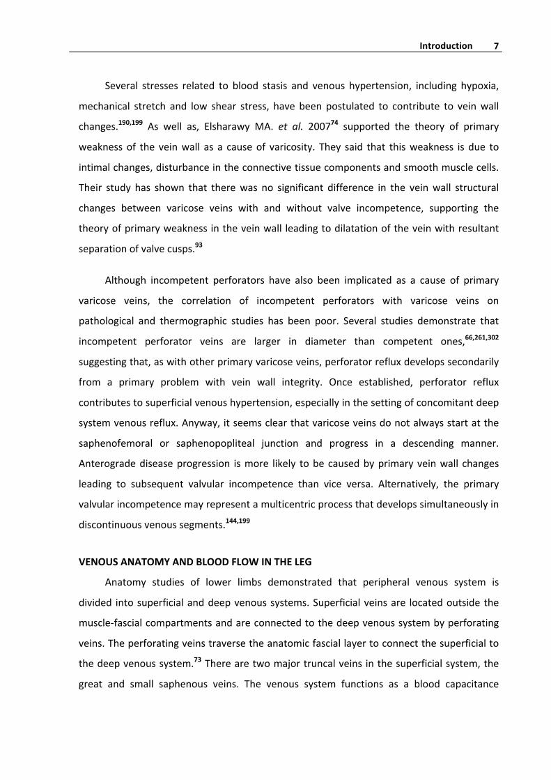
PATHOPHYSIOLOGYOFVEINWALLALTERATIONSINVARICOSEDISEASEOFLEG
A normal venous system depends on the integrity of the vein valves, vein wall
structure and the hemodynamics of venous blood flow. These components are
interdependentandthedisruptionofoneaffectstheintegrityoftheothers.164Theprimary
cause of varicose vein formation is not clear; however, both vein valve dysfunction and
hydrostaticvenouspressureappeartoplayacriticalroleintheinitiationandprogressionof
thedisease.225Venousvalvularincompetenceandelevationinthepressureofbloodinthe
Introduction
6
lower‐limb superficial venous system is considered contributory to vein dilatation and
tortuosityseesinvaricoseveins.73
Two principal theories, the so‐called valvular hypothesis172,173 and the vein wall
hypothesis,232 have been put forward to explain the pathogenesis of varicose veins and
chronic venous insufficiency (CVI). One hypothesis proposes that valvular dysfunction
causingreflux is the initialpathologicalchangethatoccurs invaricoseveins.Venousreflux
thencausesbloodstasisandvenoushypertension,whichdamagestheveinwall leadingto
weaknessanddilatation.Venousdilatation separates thevalve cusps furtherandworsens
the valvular incompetence, triggering a vicious cycle. Varicose veins also demonstrate
hypotrophyofthevalvesandwideningofthevalvularannuluscomparedwithnon‐varicose
veins.59,60,207
The theoryofprimaryvenousdilatation leading to secondaryvalvular incompetence
has received more attention nowadays. This plausible hypothesis has been challenged
recently by several common ultrasonography and histological findings relating to varicose
veins.Varicositiesareoftenobservedbelowcompetentvalves,andnotuncommonlyfound
toprecedevalvularincompetence.Venousdilatationisalsofrequentlyseendistaltoavalve
rather than proximal, which one would expect if valvular dysfunction is the initial
event.143,199,232 It has been reported that varicose veins can develop without valvular
incompetence.58,297 Although valve reflux may precede vein‐dilatation,246 there is a
significant body of evidence supporting the view that vein dilation can precede venous
reflux,andthatvalvulardysfunctionmaybeanepiphenomenonofveinwalldilation.84,130,225
Also,findingsfromduplexultrasonographyonthepatternandprogressionofvenous
dilatation and reflux strongly support vein wall changes as the primary event, although
isolatedprimaryvalvulardysfunctionmaystillsometimescontribute.Thevalvesofvaricose
veinscontainlesscollagenandlosethenormalviscoelasticfeaturestypicalofnon‐varices.221
Monocyteandmacrophage infiltration intothevalvularsinuses isalsogreaterthanthat in
the distal vein wall of varicose veins, indicating increased inflammatory activity in the
valves.207Theaccordtothistheory,distalvalvesmayalsobecomeincompetentsecondaryto
theproximalrefluxanddilatation,leadingtoaretrogradeprogressionofdisease.186,225,255
Introduction
7
Several stresses related to blood stasis and venous hypertension, including hypoxia,
mechanical stretch and low shear stress, have been postulated to contribute to veinwall
changes.190,199 As well as, Elsharawy MA. et al. 200774 supported the theory of primary
weaknessof the veinwall as a causeof varicosity. They said that thisweakness is due to
intimalchanges,disturbanceintheconnectivetissuecomponentsandsmoothmusclecells.
Their study has shown that therewas no significant difference in the veinwall structural
changes between varicose veins with and without valve incompetence, supporting the
theoryofprimaryweaknessintheveinwall leadingtodilatationoftheveinwithresultant
separationofvalvecusps.93
Although incompetent perforators have also been implicated as a cause of primary
varicose veins, the correlation of incompetent perforators with varicose veins on
pathological and thermographic studies has been poor. Several studies demonstrate that
incompetent perforator veins are larger in diameter than competent ones,66,261,302
suggestingthat,aswithotherprimaryvaricoseveins,perforatorrefluxdevelopssecondarily
from a primary problem with vein wall integrity. Once established, perforator reflux
contributestosuperficialvenoushypertension,especiallyinthesettingofconcomitantdeep
systemvenousreflux.Anyway,itseemsclearthatvaricoseveinsdonotalwaysstartatthe
saphenofemoral or saphenopopliteal junction and progress in a descending manner.
Anterogradediseaseprogression ismore likely tobecausedbyprimaryveinwall changes
leading to subsequent valvular incompetence than vice versa. Alternatively, the primary
valvularincompetencemayrepresentamulticentricprocessthatdevelopssimultaneouslyin
discontinuousvenoussegments.144,199
VENOUSANATOMYANDBLOODFLOWINTHELEG
Anatomy studies of lower limbs demonstrated that peripheral venous system is
divided intosuperficialanddeepvenoussystems.Superficialveinsare locatedoutside the
muscle‐fascialcompartmentsandareconnectedtothedeepvenoussystembyperforating
veins.Theperforatingveinstraversetheanatomicfasciallayertoconnectthesuperficialto
thedeepvenoussystem.73Therearetwomajortruncalveinsinthesuperficialsystem,the
great and small saphenous veins. The venous system functions as a blood capacitance
Introduction
8
reservoirandalsoachanneltoreturnthebloodtotheheart.Asveinsareexposedtolower
pressure, it is logical to expect less mechanical stretch and shear stress in veins in
comparison to arteries. In the erect position, blood that enters into the lower extremity
venous system must travel against gravity and other pressures to return to the central
circulationandpreventretrogradeflowintothelegscalledvenousreflux).73Thereisaseries
of 1‐way bicuspid valves located throughout the deep and superficial veins that open to
allowflowtowardtheheartbutclosetopreventthereturnofbloodtowardthefeet.174
Thevalvesfunctioninconcertwithvenousmusclepumpstoallowthereturnofblood
againstgravitytotheheart.209Contractionofthemusclepumps,primarilyinthecalf,force
bloodoutof thevenousplexus toascendup thedeepvenoussystem.Thevalvesprevent
blood frombeing forcedmoredistallywithin thedeepsystemor throughperforatorveins
intothesuperficialsystem.73Inaddition,perforatingveinsalsocontainvalvesthatonlyallow
blood flow from the superficial to the deep veins.73 From the clinical perspective, the
peripheralvenoussystem is important,because leads to increasedpoolingofblood in the
legsandhighvenouspressureexertingastaticstretchontheveinwall.72Failofthissystem
isassociatedwithchronicvenousinsufficiency(CVI).
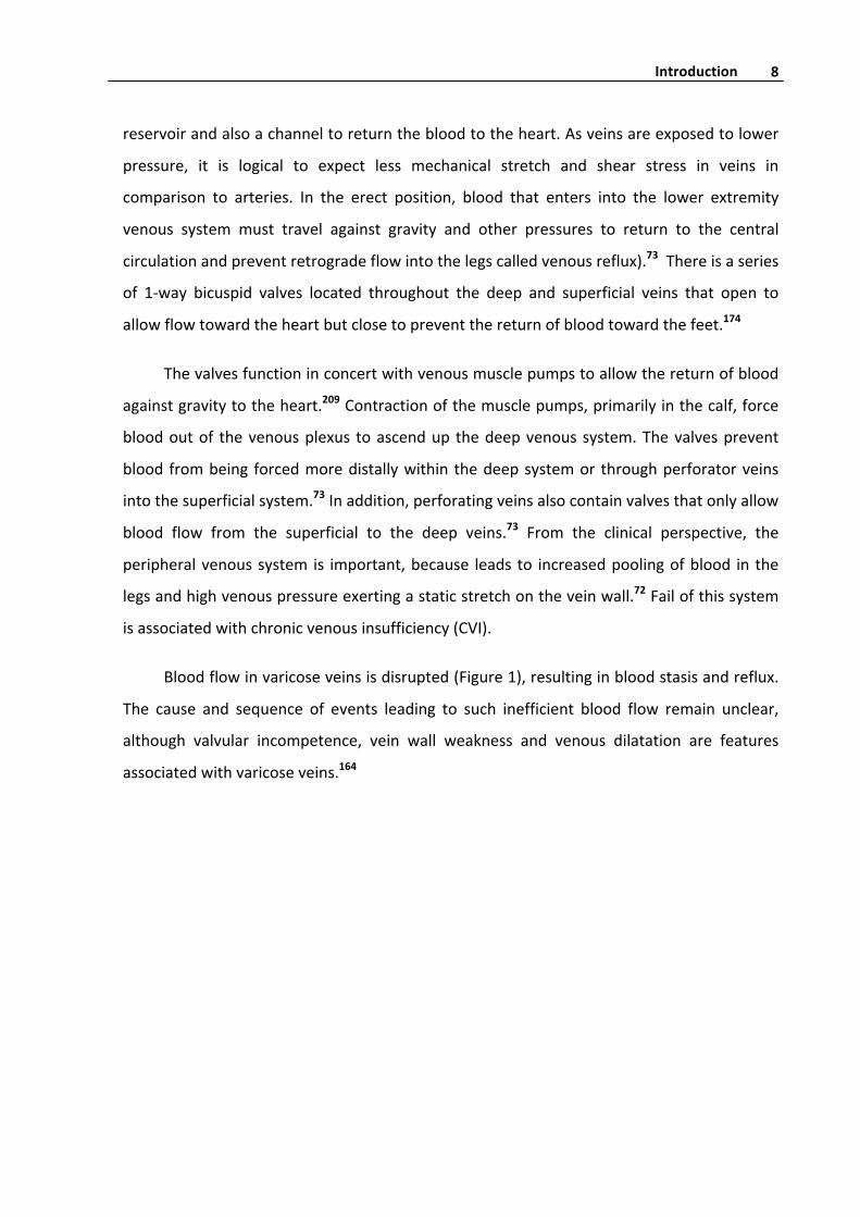
Bloodflowinvaricoseveinsisdisrupted(Figure1),resultinginbloodstasisandreflux.
The cause and sequence of events leading to such inefficient blood flow remain unclear,
although valvular incompetence, vein wall weakness and venous dilatation are features
associatedwithvaricoseveins.164
Introduction
9
Figure1.Diagramofbloodflowatanormalveinandatavaricosevein.
Likewehadsaid,theanatomicclassificationoftheperipheralvenoussystemdescribes
thesuperficial,deep,andperforatingvenoussystems,withmultiplevenoussegmentsthat
maybeinvolved.Thefailureinthevenouscirculationcanbelocalizedtosuperficial,deep,or
perforatingveinsofthelowerextremities.27Therearevariouscausesofvaricoseveinsinthe
lowerextremities.
HISTOLOGY,IMMUNOHISTOCHEMISTRYANDMOLECULARBIOLOGYOFVEIN
Veins are histological and functionally organized in three layers: intima, media and
adventitia (Figure2).Thedifferent layersof thevascularwallexert theirown influenceon
boththevasomotorcontrolandthevascularstructure,beingthefinaleffecttheresultofthe
interrelatedparticipationof the three layers. Endothelial, smoothmuscle cells (SMCs)and
different immunocompetent cell types embedded in the extracellular matrix (ECM) are
observedinsaphenousveinwall.
The tunica intima delimits the vessel wall towards the lumen of the vessel and
comprisestheendothelium,consistingofasimplelayerofepithelium,thebasallaminaand
Introduction
10
the intimal connective tissue (Figure 2). Under the basement membrane, normal vessels
contain a layer of elastic fibers, called the internal elastic lamina, composed of SMCs
separated by interlaminarmatrix collagens,microfibrils, proteoglycans, glycoproteins, and
ground substance.287 Although themedia components can be different between different
typesofbloodvessels.Veins,forexample,havelesscollagensandelastinthanarterieshave.
The endothelium is not only a mechanical barrier but also acts as receptor and
transmitterofsignalsbetweenbloodandothercomponentsofthevascularwall.Endothelial
cells (ECs) have exocrine, paracrine and autocrine functions, and they are involved in the
regulationofvasculartone,vasculogenesisandangiogenesis,bloodcoagulation,fibrinolysis,
and inflammation.188ECsaresensitive tohemodynamicchangessuchaspressureorshear
stress forces and to circulating chemical messengers. ECs respond to these signals by
secreting different growth factors and vasoactive substances, including vasodilator factors
such as NO and prostaglandin (PG) I2 or prostacyclin (PGI2) which also inhibits platelet
aggregation.83Mainvasoconstrictorfactorsreleasedfromtheendotheliumareendothelin‐1
(ET‐1),ReactiveOxygenSpecies(ROS)andvasoconstrictorprostanoidssuchasthromboxane
A2(TXA2)andPGE2.83,270
ThetunicamediaisformedbyalayerofcircumferentiallyarrangedvascularSMCsand
variable amounts of ECM. The tunicamedia is separated from the tunica adventitia by a
secondlayerofelasticfibers,theexternalelasticlamina,whichislocatedabovethevascular
SMCs (Figure 2). Elastic fibers are found throughout the vessel wall in the medial layer,
wheretheyarrange inconcentric fenestratedelastic laminae.VascularSMCscanreleasea
varietyofvasoconstrictorandvasodilatorsubstancesincludingprostanoidsandROS,among
others.155 The tunica media respond to the action of different vasoactive factors and
hemodynamicforcesbycontractingordilatingthevesselsthusbeingessentialinthecontrol
ofvasculartone.
The tunica adventitia is mainly composed of connective tissue, fibroblasts,
immunocompetentcellsandperivascularadiposetissue(Figure2).Dependingonthevessel
type, a number of small arteries termed vasa vasorum can be found to facilitate blood
irrigationtothevesselwall.Adventitiaalsoreceivestheneuronalaxonsthat innervatethe

Introduction
11
musculartissue.229 Inthe lastyears ithasbecomeevidentthattheadventitia isnotonlya
mechanical support for thevesselbutalsohasanactive role in the regulationof vascular
tone and structure259 by releasing different factors such as free fatty acids, adipokines,208
adipose‐derivedrelaxingfactor,169ROS104,228andCOX‐2derivedprostanoids.7,28
Figure2.Generalstructureofthevesselwall.
Extracellularmatrixinvascularwall
The ECM is an important structural and functional scaffolding made up of proteins
(including collagen, elastin, fibronectin, growth factors, proteoglycans and
glycosaminoglycans).36ThesemoleculesareproducedbyECs,SMCsandadventitialcellsand
are necessary for a variety of cell functions, including cell differentiation and signaling,
cellular migration, angiogenesis, blood vessel support, epithelialization, and wound
repair.36,129,287Inthissense,theECMcanreleasearepertoireofinsolubleligandsthatinduce

Introduction
12
cellsignalingtocontrolproliferation,migration,differentiationandsurvival.36ThesameECM
proteinsatdifferentregionsofthebloodvesselwallmaycomefromdifferentcellsandbe
regulatedbydifferentmodulatorsundercertaincircumstances. Inthemedia,collagenand
elastinareproducedprimarilybySMCs. Intheadventitia,however,ECMproteins ‐suchas
collagen, osteopontin, and fibronectin‐ primarily come from fibroblasts, as in other
connectivetissues.299
Collagen is a very stiff protein that has the physiological role to limit vessel
distension.36Thepolypeptideprecursorof thecollagenmolecules,procollagen, is secreted
to the extracellular compartment where it transforms in tropocollagen. After enzymatic
modification, the mature collagen monomers aggregate and become crosslinked to form
collagenfibers.ThepredominantvascularcollagensaretypeIcollagenandtypeIIIcollagen,
whichcompriseupto80‐90%ofthetotalbloodvesselwallcollagens(TypeIcollagenabout
60%andtypeIIIabout30%ofthetotalcollagen.114TypeIVcollagenisamajorcomponentof
thebasallaminaofbloodvessels,whichplaysanimportantroleinregulatingpro‐andanti‐
angiogenicevents.175
Elastin is themajor protein that imparts the property of elasticity to blood vessels.
Elastinfunctionsasacross‐linkedpolymeraspartofanelasticfiberanditsassemblyoutside
the cell requires an association with numerous other extracellular proteins such as
microfibrils.15,36,287Elastinrepresents90%oftheelasticfibers.Elastinisnormallyproduced
by SMCs in the media and by fibroblasts in the adventitia. Elastin deposition in normal
vascularwall is limited to themedia layer extending from the internal to external elastin
laminae.Undernormalconditions,elastogenesisisrestrictedmainlytofetallifeandinfancy,
andmatureelasticfiberslastfortheentirelifespan.Thehalf‐lifeofelastinfibersisabout40
years; elastic fibers are considered themost durable element of ECM.15 Elastic fibers are
degradedandfragmentedwithageanddisease,leadingtoincreasedstiffnessofthevessel
wall.98Underpathologicalconditions,vascular (SMCs,ECs,andfibroblasts)makeelastinas
partofthereactiontoincreasedmechanicalstress.126
Theprecursorofelastin,tropoelastin,isahighlyhydrophobicprotein,whichissoluble
insaltsolution.Matureelastinfibrilsareinsolubleandintertwinedwithcollagen,fibrilinand
Introduction
13
fibulin proteins, and alsowith carbohydrates.213,286 Elastin comprises repetitive sequences
and multiple chains cross‐linked by desmosine linkages formed among lysine residues
modifiedbylysyloxidase.213,286Althoughcollagenandelastinarethemajorcontributorsto
the visco‐elastic characteristics of the vascular wall, a number of additional ECM
componentsaffectboththecompliancecharacteristicsandvasoregulatoryabilitiesofblood
vessels.
Fibronectin is a multi‐domain ECM protein that interacts with multiple integrins,
heparan sulfate proteoglycans, collagens, and fibrins tomediate cellular behaviors.211 The
contentoffibronectininbloodvesselsisimportantbecauseitmodifiesthemeanstressand
elasticmodulusofthewall.197Fibronectinisproducedandsecretedbynumerouscelltypes
includingSMCs,fibroblastsandmyofibroblasts,andiswidelydistributedinECM.Fibronectin
functioninthevasculatureismediatedbyα5β1integrin,whichisexpressedbyECs,SMCs,
and fibroblasts, and is widely distributed in ECM.299 In addition, fibronectin controls the
deposition, organization, and stability of othermatrixmolecules including type I collagen,
type III collagen and thrombospondin‐1, as well as, fibronectin modulates leukocyte
infiltration, expression of adhesion molecules, cell proliferation, and vascular SMCs
phenotypicdifferentiation.Allfactorsinvolvedinvascularremodelingprocesses.55
IntegrinsaretransmembraneproteinsthatmediateattachmentofthecelltotheECM
andhave a role in signal transduction from the ECM to the cell.86 Integrins represent the
largestfamilyofcellsurfacereceptors.Ithasbeensuggestedthatchangesinintegrinprofile
are important for processes leading to more chronic structural rearrangement of the
vascularwallandECMmaterial.197
Matrixmetalloproteinases (MMPs) are a family of endopeptidases which require a
zincionattheiractivesiteforproteolyticactivity.47Theseenzymesareabletodegrademost
constituents of the ECM.225 They are mainly responsible for the degradation and
reorganization of ECM that can lead to physiological or pathological processes. Thus, in
normal physiological vascular remodeling, MMP activity is tightly controlled at different
levels.However,factorsthatpromotesvesselremodelingupregulateMMPactivities.Lossof
control of MMP activity could result in degradation of ECM, enabling vascular SMCs to

Introduction
14
migrateandproliferate,aswellas inflammatorycellsto infiltratethevesselwall.31,47Thus,
partial degradation of the ECM surrounding vascular SMCs is likely a necessary step for
allowing repositioningof cellsduring remodeling.197MMPsare inactivatedbyendogenous
tissue inhibitors TIMPs. So, homoeostasis of the ECM is regulated by MMPs and
TIMPs.108,165,225,255
Plasminogen/plasminogen activator system. Plasmin can degrade ECM directly or
indirectlyviaMMPactivation.31Thereforealteredplasminactivitycanhavehighimpacton
vascular fibrosis.Plasmin is releasedasa zymogencalledplasminogen.Theactivityof this
system is tightlycontrolledbyplasminogenactivator inhibitor type1 that functionsas the
principalinhibitoroftissueplasminogenactivator.
TenascinsareECMglycoproteins.TenascinCismainlyfoundinvesselsanditwasthe
firstmemberofafamilyoffourstructurallysimilarproteinsidentifiedincludingtenascinR,
W and X.92 Tenascin C has diverse functions including weakening of cell adhesion, up‐
regulationoftheexpressionandactivityofMMPs,modulationof inflammatoryresponses,
promotionofmyofibroblastsrecruitmentandenhancementoffibrosis.110
CELLULARANDMOLECULARPATHOLOGYOFVARICOSEVEIN
Likewehave said, recent studiesof varicoseveinpathogenesishave focusedon the
structural and biochemical changes in the vein wall.186,225,255 Zsoter T. et al. 1966306 has
suggested that veins frompatientswith varicosities aremore distensible than those from
patientswithnormalveins,indicatingaprobablesystemicbasisfortheabnormality.
It iswellknownthatvesselwall remodelingoccursasanadaptationtopressureand
flow (e.g., vein graft) or to mechanical (e.g., angioplasty) or biochemical (e.g.,
atherosclerosis) injuries, all of which promote ECM‐regulated SMCs migration and
proliferation.202 Evidence has shown that the tension generated by intraluminal pressure
affects the thickness and compositionof the vesselwall.147Mechanical stretchexertedby
increased intraluminal pressure induces vascular smooth muscle hypertrophy and
hyperplasia and changes in contractile and matrix proteins.157 Localised haemodynamic
stresses in vessels are considered to play a role in attracting inflammatory cells into the
Introduction
15
vesselwall.4
Areas of intimal hyperplasia with associated collagen deposits, SMC infiltration,
subendothelial fibrosis and luminal dilation are common in varicose veins.74,231,255,290
Changes in the media, including SMC proliferation, ECM degradation, fragmentation of
elastic lamellae and loss of circular and longitudinalmuscle fibers are seenmoreoften in
varicose than in non‐varicose veins.19,74,289 Similarly, the adventitia of varicose veins
demonstrates areas of increased SMCs, fibroblasts, and connective tissue with others
regions of atrophy anddevoid ofvasa vasora.19,199 Also, focal aggregates ofmacrophages
weredescribedwithin themediaandadventitiaofbothnormalandvaricoseveins.298The
low‐magnificationmicroscopicappearanceofavaricoseveinsectionconsistsofathickened
veinwallwithdisruptionof thenormalorganizationof theextracellularmatrix (ECM)and
SMCs.199 Previous studies221,242,276 shown that there were significant changes in collagen,
elastin and SMCs contents of the wall of varicose veins compared to the normal, even
withoutsaphenofemoralvalveincompetence.
Themediaof varicoseveinsexhibit areasof SMChypertrophyandproliferation,but
alsoregionsofatrophyarealsopresent.170,199,255,276,289,290Rearrangementandmigrationof
SMCs intothe intimamayalsobeseen.74,255,290Somestudieshavereportedan increase in
amounts of SMCs or their activity in varicose veins,220 whereas, others found reduced
amountsofSMCsduetoreplacementbyconnectivetissue.19,232
TheECMisadynamicstructurethatmaintainstheintegrityandhomoeostasisofthe
vein through interactionswith cellular components suchas theendotheliumandSMCs.108
Cellular interactionwith ECM regulates cell adhesion,migration, proliferation, phenotype,
and tissue architecture under different circumstances.299 ECM remodeling, precede the
progression of varicosities. Its degradation is likely to contribute to the weakening and
dilatation of veins. ECM, containing mainly elastin and collagen, is essential for vessel
homeostasis and contributes to the strength, flexibility, and structural integrity of the
vascularwall.162DegradationofECM ismainly causedbyanarrayofproteolyticenzymes,
including MMPs and serine proteases, which are produced by both vascular ECs and
fibroblasts cells as well as white blood cells,178,179 in particular during inflammation.109,192

Introduction
16
Disruptionoftheelasticfibers,includingfragmentationoftheelasticlaminas,andthickening
ofindividualcollagenfibershaveoftenbeenobservedinvaricoseveins.219,289,290
The overall collagen content in varicose compared with non‐varicose veins remains
unclear, with some studies reporting an increase,84,241,289,290 but some describing no
change284andsomeshowingareduction.8Additionally,thesubtypesofcollageninvaricose
veinsaredifferentfromthoseinnon‐varicoseveins.IthasbeenshownthattypeIcollagenis
significantly increased in segments of varicose veins compared with control saphenous
veins.288Also,KirschD.etal.2000128haveshownthatthereissignificantincreaseinmatrix
proteins suchas type IV collagenand laminin in thewallof varicoseveins comparedwith
normal veins. Moreover, a dysregulation in collagen synthesis is described in varicose
veins.240,242 Immunohistological and Transmission electron microscopy results showed
alterations in the distributionpattern of type I, III, and IV collagen in thewall of varicose
veins compared with control saphenous veins.90 Ghaderian & Khodaii tagged the
immunohistochemical pattern of type I collagen revealed strong staining in the
subendothelial region of most varicose vein specimens as did control saphenous vein
specimenswhichisanindicationofanincreaseofthistypeofcollageninthesubendothelial
region.90
Indeed,othersinvestigatorshavequantifiedtheoverproductionofSMCsderivedfrom
varicoseveinsof type I collagen,whichprovides tensile strength,decreasedproductionof
typeIIIcollagen,whichcontributestoelasticity,andsimilarquantitiesoftypeVcollagenin
varicoseveinscomparedwithcontrolsaphenousveins.242,243,288This imbalancemighthave
consequences for themechanicalpropertiesof thetissue.Moreover, the imbalance in the
synthesis of type I collagen and type III collagen can affect veinwall function in varicose
veinsasdescribed in“theweakwallhypothesis”.However, theirobservationswerebased
oncollagenfibersderivedfromculturesofvaricoseveinSMCsdonotexplainthedistribution
ofcollagentypesinthethreelayersofveins.
AlterationofSMCsbehaviorfromquiescentor“contractilestate”typicalofthenormal
vessel phenotype to a proliferative or “synthetic state” characteristic of the varicose
phenotypeincreasescollagensynthesis.9ThesesyntheticSMCssynthesizelargeramountsof

Introduction
17
the ECM components and lose the expression of the contractile filaments leading to the
thickeningofthevenouswallandlossofthecontractility inthevaricosevein.189However,
others believe there is a reduction in the cellularity of the smooth muscle layer with
replacement by collagen or a significant increase in collagen content of varicose veins.184
Anyway, development of vascular stiffening, at least in arterial hypertension, has always
beenlinkedtoexcessivedepositionofcollageninthevesselwalls.111,206
Elastin,theothermainproteinoftheECMinvessel,actsasmorethanavenouswall
structuralprotein for thestorageof recoilingenergy. Invitro,manycellsexhibitmigration
and proliferation in response to tropoelastin, elastin degradation products, and elastin
peptides.299 Development of varicose veins therefore involves elastin component
reconstruction,evenwhenelastinexpressionisturnedoffinadults;39however,anincrease
of elastin is found in arterial an d venous diseases. In vivo, elastic fibers and laminae
prohibited SMC proliferation and prevented intimal hyperplasia.158 In a rat model of
adventitial implantation of collagen, basal lamina, and elastic laminae patches, elastic
laminaepatches,butnotlamininorcollagenpatches,areassociatedwithreducedneointima
formation and SMC proliferation.167 In fact,mature elastin fibers are key elements in the
maintenance of acquiescent vascular SMC phenotype by providing a physical barrier for
cellularmigration.299
During the development of varicose veins, the elastic network is damaged and
deregulated, showing reduced and fragmented elastin along with disordered collagen
distribution.3,241 At damaged during aging or tissue injury, elastic fibers are generally not
replaced, because elastin expression is turned off in adults. Instead, more collagens are
made, shifting the arterial wall toward a stiff arrange of collagen fibers.299 Other authors
marked that varicose veins show high latent TGF‐β) binding protein (LTBP)‐2 and TGFβ
expression,particularlyinthesubendotheliumandmedia,andinareaswithmarkedinjury.
However, the intimalmechanisms implicated inmolecular alterations of varicose vein are
notwellestablishedandseveralstudiesaboutgeneexpressionweredone.Itisshowedthe
upregulatedgenesincludethoseofECMmolecules,cytoskeletalproteinsandmyofibroblasts
suchastransforminggrowthfactorβ‐inducedgene(BIGH3),tubulin,lumican,actinin,typeI

Introduction
18
collagen,versican,actinandtropomyosin.154
In primary varicose veins compared to normal veins, some authors demonstrated a
reduction incollagenandelastincontent.74,284 Incontrast,somehavefoundan increase in
thecollagencontentwithoutchange276or reduction84,242 inelastincontent in segmentsof
varicose veins compared to normal. It seems probable that alteration in the balance of
elastinandcollagencontent is likely tocontribute tovaricoseveinwallweakening.74,289,290
The diffusely disorganised architecture and distribution of collagen and elastin fibres
correspondstofibroticdegradationoftheparietalwallandalossofmechanicalproperties
invaricoseveins.40,134Therefore, thepathologicalabnormalities invaricoseveinswerenot
duetodeficiencyofsmoothmuscleslayers,butcouldbereferredtotheinabilityofSMCsto
provide the necessary tone in the vessel wall leading to vein wall dilatation.232 This SMC
dysfunction38maybeduetothebreak‐upofitsregulararrangementbyfibroustissue289as
effective contraction cannot occur when individual cells are not in direct communication
witheachother.232Previouslyitisreferredthattheformationofvaricoseveinsissecondary
to defects in cellular and ECM components, causing weakness and altered venous
tone.225,255,283 The triggers for these changes remain unclear, although several factors
associated with hemodynamic abnormality are likely to be involved, including hypoxia,
mechanicalstretchandlowshearstress.190,199
The involvement ofMMPand TIMP in vascular diseases is amatter of a strong and
continuous scientific interest, especially in the study of effective MMP modulators that
would be important in themanagement of patients with venous diseases.164 It has been
widelydocumentedthattheeffectsofMMPandTIMPonECMdegradationmayresultina
significantvenous tissue remodeling,225,272degenerativeandstructural changes in thevein
wall,115,290leadingtovenousdilationandvalvedysfunction.225,254Takentogether,increased
MMPactivityandalteredMMP/TIMPbalance21,224mayalsoinduceearlymodificationsinthe
endothelium and venous SMC function in the absence of significant ECM degradation or
structuralchangesintheveinwall.Inparticular,forwhatconcernsCVIithasbeensuggested
thatthebalancebetweenMMPandTIMPplayacrucialroleinearlystepsofvaricosevein
formationinthelowerextremities.223 Inaddition,evidencesuggeststhat increasedactivity
Introduction
19
of MMP is also present in the advanced stages of CVI encompassing skin changes and
chronicvenousulceration,215,235,305aswellasinthewoundfluidmicroenvironment.263,279
Amongproteolysisevents,MMPsand theirTIMPshavegarnered themostattention
and have been linkedwith the pathological events of varicose veins, but different groups
havereportedcontradictoryobservations.141,164Sansilvestri‐MorelP.etal.2007239showed
increasedMMP‐1,MMP‐2,MMP‐3,MMP‐7,TIMP‐1,andTIMP‐3,anddecreasedTIMP‐2,in
varicose saphenous vein tissues; Ishikawa Y. et al. 2000112 found reduced expression of
MMP‐1 and MMP‐2 in human varicose veins. Different research groups also found
discrepant expression patterns of MMP‐9, TIMP‐1, and TIMP‐2.12,112,139,298 Therefore,
although a counter balance between MMPs and TIMPs may affect vein structural
integrity.225Other families of proteolytic enzymesmay alsoparticipate in varicosedisease
development.
Also, Raffetto JD. et al.2008226 shown than an increase in the level anddurationof
stretchalsoevokesadaptationoftheSMCphenotypeenablingareorganizationoftheECM,
e. g. by altering the expression and activity of MMPs. Consequently, an increase in wall
stressstimulatesexpressionofMMP‐2andelevatesgelatinaseactivitybothinthemediaof
stretched mouse veins and in human venous SMCs.77,187 Likewise, MMP‐9 activity is
increased in varicose veins of human patients116, and in rat veins upon exposure to an
increasedtransmuralpressurelevel.224Thismayexplainforthefactthattheabundanceof
collagentypesIandIIIisalteredinvaricoseveinsascomparedtohealthyveins.241,243Infact,
theamountofrigidity‐mediatingcollagentypeIisincreasedinthevaricosevesselwallwhile
the distensible collagen type III fiber network is degraded. Recently it is reported that
regulatorygenesofcollagenproductionaredown‐regulatedinveinsaffectedbysuperficial
reflux disease.180 In addition, MMPs involved in vascular diseases are the interstitial
collagenase,MMP‐1,whichcleavesfibrillarcollagens,whicharesubsequentlydegradedby
the gelatinases,MMP‐2 andMMP‐9.114,201 There are few studies in human tissues which
havedemonstratedtheroleofPGE2ontheexpression/activationofMMPs.151,304
On the other hand, MMP activities are also under control of endogenous tissue
inhibitorofmetalloproteinase(TIMP)andchangesinMMP/TIMPratioareprobablyinvolved
Introduction
20
in vascular wall remodeling and in varicose vein formation.139,141,239 Another group found
thanactiveMMP‐1andtotalMMP‐2concentrationsweresignificantlydecreasedinvaricose
veins while the TIMP ‐1 and ‐2 tissue inhibitors of metalloproteinases, were significantly
increased.95Inconclusion,suchareorganizationoftheECMisahallmarkoftheremodeling
processes in varicose veins and associatedwith an increase in rigidity enabling it towith
standachronicincreaseinwallstress.216Aproactiveapproachtothetreatmentoftheearly
and late stages of CVImay be focused on the inflammation‐related andMMP‐dependent
proteolysis.225 In fact,actually the inhibitionofMMPsmayrepresenta realistic,noveland
possible therapeutic intervention to limit the progression of varicose vein to CVI and leg
ulceration.223 The therapeutic hypothesis is based on the well known role of
glycosaminoglycans(speciallydermatansulfate)inhealthanddisease,inwoundhealingand
veinremodeling.176,275,277
ECsandSMCsparticipate in remodelingprocessof thevesselwall to counteractan
increase inwall stress.Dysregulatedapoptosisandcell cycledysfunctionoccur invaricose
veins.16,70,71,280 The overall number of apoptotic cells and activity are reduced in varicose
compared with non‐varicose veins.16,17,40 Dedifferentiation of SMCs maybe due to
dysregulatedapoptosis.124
Besides,abnormalitiesofthevenousendotheliummaycontributetovenousdilatation
andthepathogenesisofvaricoseveins.93Essentiallyadaptationsofthevesselwall,suchas
an enlarged lumen diameter and remodelling of the ECM,may be evoked by changes in
hemodynamic forces such as shear stress and/or (circumferential) wall stress, hence
biomechanicalstretch.Ontheotherhand,iswellstablishthattheirchronicalterationoften
promotes cardiovascular pathologies such as hypertension,195,196 atherosclerosis138 and
venousvalvedysfunction.18
ECs of varicose veins appear desquamated and degenerated under electron
microscopy.19 Such injured cells are activated and known to release various types of
inflammatory mediator and growth.190,199 Increased expression of inflammatory markers,
suchasvascularcelladhesionmolecule1, intercellularadhesionmolecule1andvonWille
brand factor, by the endotheliumof varicose comparedwith non‐varicose veins has been
Introduction
21
recorded.19,255 Inaddition,significantlymoremastcells,macrophagesandmonocyteshave
beenobserved invaricosecomparedwithnon‐varicoseveins.125,244,301Activated leucocytes
mayalsoreleaselargeamountsofsuperoxideanionsandproteasesthatareabletodegrade
theECM.190,199ThesefindingspointtotheroleofROSinthepropagationofvaricosedisease
becausetheirproductionseemstobefurtherenhancedbylocalhemodynamicfactors.101
Another hallmark of both remodeling processes pointing to an involvement of wall
stress is the proliferation ofmedial SMCs. Besides spontaneous responses (contraction or
relaxation of SMCs) to temporary changes in blood flow or pressure, a chronic rise in
transmuralpressure(e.g.duringvenoushypertension)elicitsadaptivevascularremodeling
foremosttonormalizewallstress.Inthecomparativelythin‐walledandSMC‐poorveins,the
corresponding structural adaptations of the vessel wall preferentially lead to the typical
corkscrew‐like morphology of remodeling (varicose) veins that rather points to a
(longitudinal)growthbetweenfixedends.77
Endothelial cells (ECs) aredirectly affectedby changes inbothhemodynamic forces,
whereas only biomechanical stretch stimulates vascular SMCs in the media. Under
physiological conditions, both forces stabilize the function of veins and maintain a
normotensive blood pressure. In this situation, laminar shear stress‐mediated expression
and activity of endothelial nitric oxide synthase (eNOS) stimulate the production of nitric
oxide (NO). NO as a freely diffusing and membrane‐penetrating signaling molecule‐
transmits the rise in laminar shear stress to the SMCs, inhibiting their proliferation and
triggering their production of cyclic guanosine 3’,5’‐monophosphate (cGMP), which
subsequentlypromotesrelaxationofthesecellsand,asaconsequence,vasodilatation.33,198
Furthermore,NObearsseveralanti‐inflammatoryeffects,as itmayreactwithROSsuchas
superoxide to form peroxynitrite, thereby inactivating these well‐characterized
proinflammatorymediators. These findingspoint to the roleofROS in thepropagationof
varicose disease because their production seems to be further enhanced by local
hemodynamicfactors.101
Introduction
22
INFLAMMATIONANDROSMEDIATORSINVASCULARSYSTEM
Growing evidences suggest that an imbalance between pro‐ and anti‐inflammatory
mediators is a common pathophysiological mechanism in different cardiovascular
diseases.135,285 Inflammationoftenbeginswithendothelialactivationthroughseveralsignal
transductionmechanisms, leading to the expression of adhesionmoleculeswhich attracts
different immune cells.161 These immune cells, aswell as the resident cells, participate as
donors and recipients of cytokine signals, amplifying the inflammatory activity.135,161 This
inflammatory environment has deleterious consequences in different functions of the
vasculatureincludingcontraction/dilation,stiffnessandvascularstructure.51,52Wewillfocus
ontwopro‐inflammatoryproteins,andtheirrespectivemediators:COX‐2andprostanoids,
andNADPHoxidaseandROS.
Biosynthesis of prostanoids begins with the formation of PGG2/PGH2 through the
action of COXs on arachidonic acid released by phospholipases from the membrane
phosphoglycerides. Different prostaglandin synthases (PGS) metabolize immediately the
PGH2intospecificprostanoids.Prostanoidsplayanimportantroleininflammation,platelet
aggregation,vasoconstriction/relaxationandvascularremodeling.Aftertheinitialsynthesis
of theshort‐livedbutbiologicallyactivePGH2, theproductionof thedifferentprostanoids
(theprostaglandinsPGE2,PGD2,PGF2α,PGI2andTXA2)dependsontheactivityofspecific
synthases;thus,prostanoidproductiondependsonbothCOXandPGSactivities.
ThreedifferentCOX isoformshavebeendescribed:COX‐1, COX‐2 andCOX‐3,where
COX‐3isasplicevariantofCOX‐1,thus,COX‐3issometimesnamedasCOX‐1borCOX‐1v.293
InmosttissuesCOX‐1isconstitutivelyexpressedunderphysiologicalconditions.COX‐2isan
inducible isoenzyme and the dominant source of PGs in inflammation. While COX‐1 is
localizedintheendoplasmicreticulum,COX‐2actspredominantlyatendoplasmicreticulum
andnuclearenvelope.194 Invascularcells,COX‐2expressionis inducedbyawidevarietyof
stimuli such as IL‐1β, ROS, lipopolysaccharide or TNF‐α142,181,269 and it is up‐regulated in
vascular inflammatory diseases such as aortic aneurysms127 and balloon‐injured
arteries.291,303
Introduction
23
NADPH oxidases (NOX) are the major source of ROS in the vascular wall in both
physiological and pathological conditions.10,68,69,148 NOX are a family of transmembrane
oxidases that reduce molecular oxygen to superoxide using energy derived from the
oxidationofNADPH/NADHtoNADP/NAD.NOXenzymefamilyconsistsoffiveNOXenzymes
(NOX1‐5) and two Dual oxidases (DUOX1‐2). NOX isoforms differ in their inducibility,
expression pattern and they also require different sets of accessory proteins for their
enzymaticactivity.26
NOX2complexisthefirstlyidentifiedandbeststudiedmemberoftheNOXfamily.Itis
expressed by phagocytes (granulocytes,monocytes, dendritic cells) and its expression has
alsobeenreported inothercellsofthe immunesystem,suchasnaturalkillercells,Bcells
and mast cells. Lower levels of NOX2 expression has also been detected in T cells.113,282
Interestingly, recent published data report that neuronal expression of NOX2 regulates
stress‐responsebehaviorinratsandmice.245,256
NOX1 was the first homologue that was described for NOX2.264 NOX organizer 1
(NOXO1,NCF1homolog),theorganizersubunitofNOX1andNOXactivator1(NOXA1,NCF2
homolog)arethecytosolicaccessorymoleculesneededforsuperoxideproductionbyNOX1
complex.22,88,268 High expression of NOX1 has been detected on colon epithelium.266
Additionally, lymphocytes,266 vascular SMCs,149 ECs,133 uterus,264 placenta,63 osteoclasts152
and retina177 are reported to express NOX1. NOX1 deficient mouse has decreased blood
pressure183 and develop enhanced hyperoxia induced acute lung injury,45 suggesting an
inflammationregulatingroleforNOX1derivedROS.
NOX3isexpressedintheinnerearandcanproduceROS53usingbothNCF1andNOXO1
organizer subunits.23,54 In addition, NOX3 expression has been reported to take place on
hepatocytes.159
NOX453,87,252 was originally identified from kidney, but it is also expressed in many
other tissues including ECs2 and fibroblasts.57 Interestingly, upon heterologous expression
NOX4 is active without cell stimulation and does not require cytosolic subunits for its
activity.182

Introduction
24
NOX5 was identified as calcium activated NADPH oxidase23 in various fetal tissues,
lymphocyterichareasinspleenandlymphnodesandinuterusandintestis.24,25,53Thelast
twoenzymesbelongingtotheNOXfamilyarethethyroiddualoxidase1and2(DUOX1and
2).64 Contrary to otherNOX enzymes and despite of some disagreement in the field, it is
likelythatdualoxidasesconverttheproducedsuperoxidedirectlyintohydrogenperoxide.26
Inactivating mutations in DUOX2 constitute the causative reason for congenital
hypothyroidism,193 highlighting the importance of these enzymes to thyroid function. In
addition,thereisalsoevidencethatmucosalsurfacehostdefenseissupportedbyhydrogen
peroxidederivedfromtheDUOXenzymes.89
Inconclusions,ROSareaheterogeneousgroupofoxygenradicalsandotherstrongly
oxidizing molecules and share common features with closely related reactive nitrogen
species. After generation, ROS are further converted into other oxidative species or
neutralizedbycarefullyregulatedenzymaticandnon‐enzymaticantioxidativereactions.Free
radicalssuchasROSareatomsorgroupsofatomswithanunpairednumberofelectrons.
Examplesoffreeradicalsarehydrogenperoxide,hydroxylradical,nitricoxide,peroxynitrite,
singletoxygen,superoxideanionandperoxylradical.Freeradicalformationisincreasedby
immunecellactivation,inflammation,ischemia,infection,cancer,andchronicheartdisease.
TheradicalsreactwithvariouscellularcomponentsincludingDNA,proteins,andlipid/fatty
acidswhich leadstoDNAdamage,mitochondrialmalfunction,cellmembranedamageand
eventuallycelldeath(apoptosis).Molecularoxygen(O2)isconvertedintosuperoxideanion
(O2−) by the action of specialized enzymes, inmitochondria during cellular respiration, by
ionizing and UV radiation and during the metabolism of a wide range of xenobiotic
substancesanddrugs.296Superoxidecaneitherspontaneouslyorbytheactionofoneofthe
threesuperoxidedismutases(SOD1‐3)befurtherconvertedintohydrogenperoxide(H2O2).
The reaction between superoxide and nitric oxide produces peroxynitrite (ONOO−),
convergingreactivenitrogenandoxygenspeciesmetabolism.
InrelationtoinflammatorymechanismsandROSproductioninvaricoseveins,stretch‐
stimulatedECsorSMCsincreasetheexpressionandactivityofcertainNADPHoxidasesand
thereby production of ROS, which in low to intermediate amounts trigger signalling
Introduction
25
pathways sensitive to oxidativemodification of pivotal effector proteins.18 This is quite in
analogytoprocesseselicitedbyarterialhypertension,whereROSareamajorcomponentin
theinitiationofSMCdedifferentiation,214partiallybytheincreaseinH2O2formation,262and
additionallysupportapro‐migratoryandgrowthpromotingphenotype.42,153
Interestingly,activationofthetranscriptionfactoractivatorprotein1(AP‐1)appeared
to be a prerequisite for venous remodeling, proliferation and MMP‐2 expression in this
context,asblockadeofitsactivityessentiallyabolishedalltheseprocesses.Therelevanceof
wallstressforvenousremodelingisfurtherunderlinedbythefactthatAP‐1isactivatedby
ROS, namely hydrogen peroxide (H2O2),whose intracellular concentration is controlled by
biomechanical stretch.216 Stretch‐stimulated ECs or SMCs increase the expression and
activity of certain NADPH oxidases and thereby production of ROS, which in low to
intermediate amounts trigger signaling pathways sensitive to oxidative modification of
pivotaleffectorproteins.18,30
During varicose vein development, the release of NO would inhibit the pro‐
inflammatory activation and proliferation of ECs and SMCs, which is promoted by an
increase in wall stress. Moreover, NO would neutralize ROS such as superoxide anions,
whose production is elevated by a wall stress‐dependent stimulation of NADPH oxidase
expression and activity. In addition, prevention of superoxide dismutation to H2O2 would
simultaneously limit the activity of AP‐1, as has been shown to be the case in arterial
hypertension.216,271 Infact,enhancedROSproductionhasrecentlybeenverifiedinvaricose
veins,140suggestingthatthismechanismsmayplayacrucialroleinthepathogenesisofthe
disease.
Inflammatorycellinfiltrationintothevascularwallisanimportantfeatureofvaricose
veins,300 and its central roles are broadly evident in vascular diseases,32,162,257 that was
principally demonstrated in arterial pathology, characterized by the presence of
macrophages,166 T cells,132 and mast cells265 in the intimal arteriosclerotic lesions. In the
adventitia of varicose veins Xu N. et al. 2014300 demonstrates increased tryptase‐positive
mastcells infiltration.Several studieshavedemonstratedthatmastcellsandtheir specific
proteases,tryptasesandchymases,activateMMPs;234convertangiotensin‐Itoangiotensin‐
Introduction
26
II;163degradeECMfibronectin;273andinduceSMCdetachmentandapoptosis.273Also,XuJ&
Shi GP found that T‐cell numbers significantly increased in varicose veins compared with
non‐varicose veins,299 although Sayer GL & Smith PD showed no differences at T‐cells
number in varicose veins.244 But,when they studiedmacrophagesCD68+, theydidn’t find
significantdifferences inmacrophagesatvaricoseveins.Oncontrary,NicolaidesAN found
infiltration of valve leaflets and the venous wall by leukocytes (monocytes and tissue
macrophages) and also an increased number of mast cells and neutrophils).203 Over
expressionof induciblenitricoxidesynthaseandtransforminggrowthfactor‐β1,aswellas
increased presence of CD68+ monocyte/macrophages, has also been documented in
patientswithvaricoseveins.117
Insummary,thisincreasednumberofinflammatorycellsthatbothinflammationand
degradation of ECM proteins would be crucial in the early etiopathogenesis of CVI.267
Independentlyofthecellulartypeinvolved,agenerallyacceptedtheoryisthatinflammatory
process and weakness or alteration of the matrix components of the vein wall play an
important role in the pathogenesis of CVI.171,204,242 The primary CVI is an inflammatory
pathology that involves an erratic structural remodeling in the venous well leading to
vascular incompetence and the development of varicose vein, characterized by altered
collagenandelastincontent.Intheearlystepsofvaricoseveinformationiscrucialtherole
ofMMP/TIMPbalance,duetotheincreasedsecretionofproteolyticenzymes,implicatedin
bothECMandvasculardegradationduringinflammationprocesses,fromvascularcellsand
inflammatorycells(likemacrophages,neutrophilsandmastcells).115,118,203Theinflammatory
mechanismsarealso corroboratedbyanimalmodels inwhichacuteandhigh intravenous
pressure levels appear to promote pro‐inflammatory responses,102,103,146 suggesting that
theymaybeaconsequenceratherthanthecauseofadvancedvaricoseveindevelopment.
However, new research are necessary in relation to the participation of inflammatory
mechanismsinthepathophysiologyofvaricosevein,becauseinarecentpublication,Gomez
I.etal.201394foundtheabsenceofCOX‐2inthevaricoseveins.
Prostanoids, including prostaglandins (PG) and thromboxane, has been rarely
investigated inthecontextofvaricoseveins.Prostanoidsareproducedbymostbloodand

Introduction
27
vascularcelltypes.79PGE2viaselectiveactivationofEP1‐4receptorsubtypesis involvedin
the control of vascular tone,80 inflammation,43,96 pain260 and vascular wall remodeling.151
PGE2playsamajorroleinvascularwallremodelingandincollagenoverexpression.95PGE2is
synthesized from arachidonic acid (AA) through the enzymatic activities of two
cyclooxygenases(COX‐1and/orCOX‐2)andthreePGEsynthases(PGES).212Amongthethree
PGES that specifically catalyze the final step of PGE2 biosynthesis; two are constitutive:
microsomal (mPGES‐2) and cytosolic (cPGES).43,99 The third,mPGES‐1,43,99 is quantitatively
themostimportantenzymaticactivityforPGE2production.mPGES‐1andCOX‐2expression
aregenerallyco‐inducedbyinflammatorycytokinessuchasIL‐1b.119
Invaricoseveins,thisreducedPGE2contentandthelowerdensityofitsreceptor(EP4)
areresponsibleforthedown‐regulationoftheMMP/TIMPratio.95Theconsequenceofthis
biological cascade is a reduction of active collagenase content and an accumulation of
collageninthevascularwallofvaricoseveinsandcouldexplaintheintimahyperplasiaand
the thickening observed in the varicose wall.20,205 This phenomenon is in complete
accordancewitha recentpublicationwhere selectivedeletionofmPGES‐1 inbothECand
vascularSMCresultedinhyperplasiabyenhancingtheneointimalproliferativeresponseto
vascular injury inmice.52 Also,Gomez I.et al. 201495 showed that cPGESproteinwas not
foundinallsampleandwasonlydetectableatthemRNAlevel.Incontrast,mPGES‐2protein
was foundatsimilar levels inallvenouspreparations.Theseresults95 concerningmPGES‐1
were unexpected since they found this enzyme in the SV while mPGES‐1 expression is
classically inducedby inflammatory conditions viaNF‐kB.96 In conclusion, the reductionof
PGE2 concentrations in human varicose veins is due to a decrease in mPGES‐1 and an
increase in 15‐PGDH. These effects lead to the imbalance of vascular wall remodeling by
decreasingtheMMP/TIMPratioandcouldresultintheaccumulationofcollageninvaricose
veins. This endogenousmechanism could be a protective effect of the saphenous vein in
ordertorestrainthebloodstasisbyreinforcingthevascularwall,avoidingectasicsegment
formationandvenouswallrupture.95
The aim of the present Doctoral Thesis is the study of histological,
immunohistochemical, ultrastructural and morphometrical alterations that success at

Introduction
28
varicoseveinsandtriestoconnectwiththeprimaryeventsthattriggerswiththeevolution
of thedisease. In thepresentThesisweproposeanewprocedure toevaluateofconfocal
microscopestructureoftheendovascularsurfaceandintimalsurfaceofintotofixedsurgical
specimensof saphenousveinwithvaricose lesions.Thismethodcanbedemonstrated the
panoramicreconstructionofdifferentgradesof intimalvaricose lesions. Inourknowledge,
this procedure has not been previously apply to normal and pathological vein research.
Nowadays, as we have shown, literature presents controversy about the role of
inflammation,ROSorthemechanismofgenesinvolveinECMproduction.Wewouldliketo
clarify,oratleast,supportwithargumentstheimplicationorabsentofthishypothesisinthe
evolution of varicose veins. For that intention, we have used histological, biological and
molecularmethodsinourstudyforaglobalvisionofthevaricosedisease.

HypothesisandObjectives
29
HIPHOTESISANDOBJECTIVES

HypothesisandObjectives
30
HYPHOTESIS
¿Arethehistologicalandmolecularchangesofvaricoseveinregulatedbymoleculesinvolved
atoxidativestresspathways?
OBJECTIVES
1st.Todescribehistologicalandimmunohistochemicaldistributionofelasticfibers,smooth
muscle actin, collagen type I and collagen type III at the proximal and distal segment of
varicosesaphenousveinswithintimallesions.
2nd. To analyze the evolution of the proportion of the different proteins; elastic fibers,
smoothmuscleactin,collagentypeIandcollagentypeIII,inrelationtofibroticmechanisms
developintheintimaofvaricosevein.
3rd. To study the confocal microcopy reconstruction of the intimal distribution of elastic
fibers and collagen in varicose veins, and their possible correlation to intimal fibrosis
demonstratedbyultrastructuralstudy.
4rd.ToevaluatethemolecularmechanismofoxidativestressbytheH2O2production,NADPH
activityand themRNAexpressionofNOX‐1,NOX‐4,COX2,mPGES,MAC3, collagen type I,
collagentypeIII,elastinandSMA.
MaterialandMethods
31
MATERIALANDMETHODS

MaterialandMethods
32
MATERIAL
The aim of the studywas evaluated immunochemistry andmolecular expression of
molecules related to extracellularmatrix, ROS stress and inflammation in human varicose
veins. 20 saphenous varicose veins removed by phlebectomy from patients with chronic
venous insufficiency (CVI)and10saphenousnormalveins frompatientswhosuffera limb
amputate were studied. Veins were divided in two groups: proximal segment and distal
segment (Figure 3). The proximal segments were extracted near the saphenofemoral
junction,and thedistal segmentsnear themalleolusarea.Subjectswerematched forage
and sex, known from previous studies to affect vascular oxidative stress and superoxide
production.
Figure3.Histologicalandmolecularprocessingofbothproximalanddistalsaphenous
veinsegmentsrealizedbyJuanVelascointosurgicalroom.

MaterialandMethods
33
METHODS
Ethicalaspects
Use of human varicose veins and normal veins samples, had been approved by the
EthicsCommitteeofClinical Investigationsof theLaPazHospitalandMoncloaClinic,with
fullinformedconsentfromthepatients.
TissueprocessingVeins full removed were sectioned perpendicular to main axis for extracted three
pieces of approximately 5 mm each from proximal and distal segment. Pieces were
dehydratedbyimmersionatincreasingconcentratedalcohols(70%,96%and99%)andafter
that into butyl acetate two times per one hour each. Finally, tissues were embedded in
paraffinwaxfortwohours.
Histologicalmethods
Veinswere fixed in4%buffered formalinduring48hoursandembedded inparaffin
wax. 5 slices from deparaffinized tissue sections were stained with hematoxylin‐eosin,
Masson´s trichrome and orcein (for elastic fibers detection), using standard procedures
(Figure 4). As mounting methods, the water‐free mounting mediumDPX (Probus,
Badalona)wasused.
Figure4.Panoramicviewofatransversalsectionofavaricosesaphenousveinwith
thickenedoftheintimalayerandmoderatefibrosisofthemedialayer.Hematoxylin‐eosin
stain

MaterialandMethods
34
Immunohistochemistry(StreptavidinBiotinPeroxidaseMethod)
Humanveinswerefixedin4%formalinfor48h,embeddedinparaffinandpreparedin
5 μm cross sections with a microtome. Sections from saphenous veins were de
deparaffinizedinxyleneandrehydratedingradedethanolsolutions.Slideswerethenrinsed
in distilledwater and treatedwith 3% hydrogen peroxide in distilledwater for 10min to
removeendogenousperoxidaseactivity.SliceswerewashedatPBSatroomtemperaturefor
5minutes.Then,sliceswereimmerseatcitratebuffer(pH7,6)andintroducedtwotimesfor
2.5minutes intoamicrowave forepitopedetection.After that,whensliceswereagainat
room temperature, they werewashedwith distilled water. Sections were blockedwith a
10%ofnormalseruminPBSfor20minandincubatedwithanti‐SMA(1/400),anti‐collagen‐I
(1/400), anti‐collagen‐III (1/400), anti‐collagen‐IV (1/400) and anti‐vimentin antibodies
overnight at 4°C at a humidity chamber (Table 2). Antibodies were diluted at PBS+BSA
solution (1%). After washing 3 times in PBS samples were incubated for 1 h with a
biotinylatedsecondaryantibody.Afterrinsing3timesinPBS,streptavidin‐biotin‐peroxidase
complexwasapplied,andtheslideswereincubatedfor30min.Colorwasdevelopedusing
3,3’‐diaminobenzidine and sections were counterstained with hematoxylin before
dehydration, clearing, and mounting with DPX (Figure 5). Negative controls in which the
primaryantibodywasomittedwereincludedtotestfornon‐specificbinding.
Table2.Antibodiesused.
Antibody Company Dilution
SMA(monoclonalmouseanti‐smoothmuscleactin) DAKO 1:400
CollagenI(monoclonalmouseanti‐humanCollagenI) ABCAM 1:400
CollagenIII(monoclonalmouseanti‐humanCollagenI) ABCAM 1:400
Vimentin(monoclonalmouseanti‐humanVimentin) ABCAM 1:200
MaterialandMethods
35
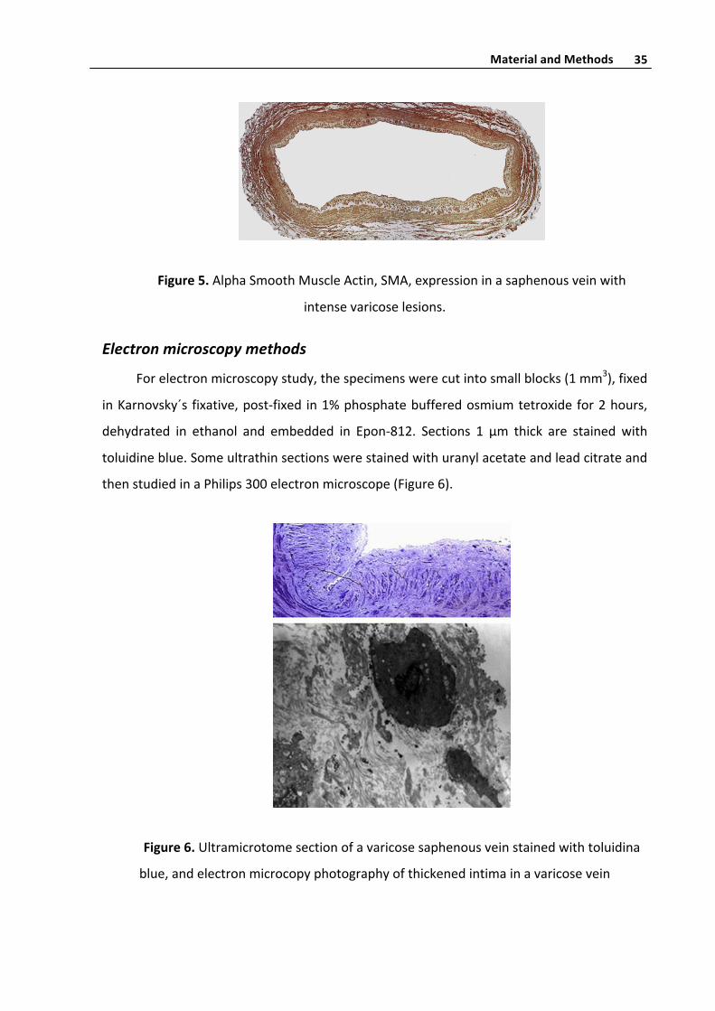
Figure5.AlphaSmoothMuscleActin,SMA,expressioninasaphenousveinwith
intensevaricoselesions.
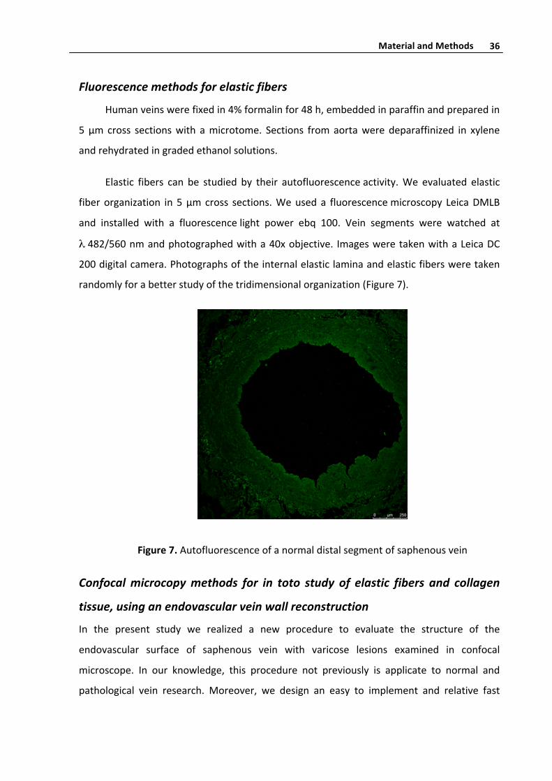
Electronmicroscopymethods
Forelectronmicroscopystudy,thespecimenswerecutintosmallblocks(1mm3),fixed
inKarnovsky´s fixative,post‐fixed in1%phosphatebufferedosmiumtetroxidefor2hours,
dehydrated in ethanol and embedded in Epon‐812. Sections 1 µm thick are stained with
toluidineblue.Someultrathinsectionswerestainedwithuranylacetateandleadcitrateand
thenstudiedinaPhilips300electronmicroscope(Figure6).
Figure6.Ultramicrotomesectionofavaricosesaphenousveinstainedwithtoluidina
blue,andelectronmicrocopyphotographyofthickenedintimainavaricosevein
MaterialandMethods
36
Fluorescencemethodsforelasticfibers
Humanveinswerefixedin4%formalinfor48h,embeddedinparaffinandpreparedin
5 μm cross sectionswith amicrotome. Sections fromaortawere deparaffinized in xylene
andrehydratedingradedethanolsolutions.
Elastic fibers can be studied by their autofluorescenceactivity.We evaluated elastic
fiberorganization in 5μmcross sections.Weuseda fluorescencemicroscopy LeicaDMLB
and installed with a fluorescencelight power ebq 100. Vein segments were watched at
λ 482/560nmandphotographedwitha40xobjective. ImagesweretakenwithaLeicaDC
200digitalcamera.Photographsoftheinternalelasticlaminaandelasticfibersweretaken
randomlyforabetterstudyofthetridimensionalorganization(Figure7).
Figure7.Autofluorescenceofanormaldistalsegmentofsaphenousvein
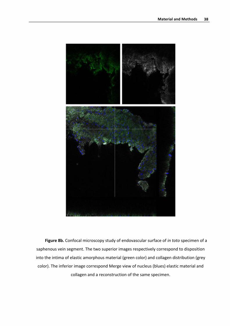
Confocalmicrocopymethods for in toto study of elastic fibers and collagen
tissue,usinganendovascularveinwallreconstruction
In the present study we realized a new procedure to evaluate the structure of the
endovascular surface of saphenous vein with varicose lesions examined in confocal
microscope. In our knowledge, this procedure not previously is applicate to normal and
pathological vein research. Moreover, we design an easy to implement and relative fast

MaterialandMethods
37
methods forvaluationof intimalsurface innormalendovascularareasand inothersareas
that present intimal thickening secondary to chronic varicose disease. Conventional
stereoscopic microcopy permit visualized the endovascular surface of vein using surgical
specimens open longitudinally, and permits distinguish the normal intima to the fibrotic
intimalareas.Howeverthemorphological informationobtainedwiththisclassicmicrocopy
method is poor. In the present Thesis, the use of confocal microcopy demonstrated the
different grades of intimal varicose lesions (Figure 8a). In the fibrotic areas associated to
intimalvaricoselesions,wemergeconfocalimagefromZaxisatthesuperiorlevel(80‐90µm
deep) from the intimal thickening. In thiswayweevaluated thehistological correlationof
elastic autofluorescence, with light ray lase reflection for collagen detection and nuclear
DAPI stain for distribution of nuclei into the vein intima. In a determinate confocal Z axis
section,wemergeimagefromelasticautofluorescence,lightrayreflectionandDAPI.Image
canberotatedtoaplanperpendiculartotheluminalsurface.Thisimagealsodemonstrated
thehistologicalstructurefromtworandomlyperpendicularplansrespecttheintimalsurface
view(Figure8b). Inconclusion,theautofluorescenceproprietiesofelasticfiberobservedin
LeicaSP5LaserMicroscopy,usingalaserrayat488λ,combinedwiththeapplicationupthe
sameveinspecimenofreflexedlaserray,forviewthedistributionofcollagenfibersintothe
connectivetissuepresentinthelaminapropriaoftheintimadeterminateanewdiagnostic
methods for intimal fibrosis evaluation in toto of surgical specimens of veinswith intimal
lesions(Figure8b).
Figure8a.Confocalmicroscopystudyofendovascularsurfaceofintotospecimenofa
saphenousveinsegment.Inthescreen,viewtheimageconfocalcapture.
MaterialandMethods
38
Figure8b.Confocalmicroscopystudyofendovascularsurfaceofintotospecimenofa
saphenousveinsegment.Thetwosuperiorimagesrespectivelycorrespondtodisposition
intotheintimaofelasticamorphousmaterial(greencolor)andcollagendistribution(grey
color).TheinferiorimagecorrespondMergeviewofnucleus(blues)elasticmaterialand
collagenandareconstructionofthesamespecimen.
MaterialandMethods
39
Histologicalquantification
Vein slices were previously examined by two pathologists for select slices without
artifacts.Afterwards,veinwallareasateachslicewererandomlyselected.10microscopic
fieldsperveinwallareaweredelimited.
For fields selection,weused amethod called “randomly selection”, that it is a non‐
biased systematic random samplingmethod. A grid divided at same 100 areaswas used.
Eachgaphadassignedanumberbetween00and99andthenweusedarandomnumber
table for select 10 numbers. Those methods guarantee a non‐bias selection. Each field
selectedwasphotographedwiththe40xobjective.Ateveryfieldselected,intimalayerwere
morphometrically studied from slices immunochemistry labelled with anti‐SMA antibody,
anti‐collagen‐I antibody, anti‐collagen‐III antibody, and anti‐collagen‐IV antibody and also
fortheelasticfibersstudy,orceinstainorautofluorescenceactivitywereused.
All imageswereacquiredatroomtemperatureusingamicroscope(LeicaEclipse55i)
mountedwithadigitalcamera(LeicaDC200)andphotographswereacquiredbyImage‐Pro
PlussoftwareatTIFFformat.Allimagesweredonewitha40xobjective.Atallselectedfields
were photographed intima and media tunica. Afterwards, images were processed with
ImageJ (http://rsb.info.nih.gov/ij) software. ImageJ is a public domain, Java‐based image
processing program developed at the National Institutes of Health. We used ImageJ for
quantifyexpressionofdifferentproteinsatintimalayer.
ImageJ was designed with an open architecture that provides extensibility via Java
pluginsandrecordablemacros.ImageJhadbeenpreviouslyusedbyotherinvestigatorsfor
histologicalquantifications.Nowadays,ImageJpluginsmakeitpossibletosolvemanyimage
processingandanalysisproblems, fromthree‐dimensional live‐cell imaging to radiological
image processing, multiple imaging system data comparisons to automated hematology
systems. ImageJ'spluginarchitectureandbuilt‐indevelopmentenvironmenthasmade ita
popular platform for teaching imageprocessing.Nowadays, thousandsof newplugins are
develop forusers for solvenewproblems.Also, ImageJcandisplay,edit,analyze,process,
save,andprint8‐bitcolorandgrayscale,16‐bit integer,and32‐bitfloatingpointimages.It
canreadmanyimagefileformats,includingTIFF,PNG,GIF,JPEG,BMP,DICOM,andFITS,as

MaterialandMethods
40
well as raw formats. ImageJ supports image stacks, a series of images that share a single
window,anditismultithreaded,sotime‐consumingoperationscanbeperformedinparallel
onmulti‐CPUhardware.ImageJcanbeusedatWindows,OSXorLinuxoperatingsystems.
To a correct morphometric study of area occupied by SMA, collagen‐I, collagen‐III,
collagen‐IVandelasticfiberscomparedtototalintimalareaatvaricoseandnormalveinsat
bothsegmentsweusedthe ImageJplugincolordeconvolution,developbyGabrielLandini
(Landini G: Colour deconvolution plugin 1.5. http://www.dentistry.bham.ac.uk/
landinig/software/cdeconv/cdeconv.html).Histologicalstainsare“lightabsorbingdyes”so
canbeconsideredasbeingsubtractivecolor.Thepluginrequires imagestohaveaneutral
background to work properly. Vectors should be worked out from single‐stained control
slides. So, we could separate original images into three different channels, in our case
Hematoxylinchannel,DABchannelandacomplementaryone.WestudiedDABchannel(8
bitsimage)forquantificationofimmunochemicalpositiveareaofSMA,collagen‐I,collagen‐
III,collagen‐IVandforelasticfibersstudyweused.Then,wesplitupimageintointimalayer
and rest of vascularwall, sowe studiedonly intima layer.Afterplied colordeconvolution
andselectintimalayer,wedecidedathresholdbetween0and255(because8bits images
worksbetween0(minimumlight,black)and255(maximalight,white)),andweusedsame
thresholdateverymarker.Subsequently,wequantifiedimmunochemistrypositiveareafor
eachprotein,SMA,collagen‐I,collagen‐III,collagen‐IVatintimallayer.Wedidalsothesame
for positive stain for elastic fibers. We measure as well total intimal area at each
photography.Resultswereexpressedasproportionbetweenpositivestainareaandintimal
totalarea(%).
Molecularmethods
RNAAnalysis
For varicose veinsmRNA extraction, veins were homogenized on ice in 1mL of Tri
Reagent using a Polytron PT‐20 (Kinematica AG, Lucerna, Switzerland). Samples were
centrifuged5min12,000gtoremovethedebrisoftissue,supernatantsweretransferredto
afreshtubeandtotalRNAwasobtainedaccordingtothemanufacturer’srecommendations.
MaterialandMethods
41
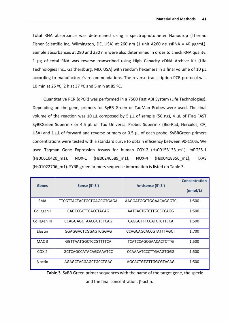
Total RNA absorbance was determined using a spectrophotometer Nanodrop (Thermo
FisherScientific Inc,Wilmington,DE,USA)at260nm (1unitA260de ssRNA=40μg/mL).
Sampleabsorbancesat280and230nmwerealsodeterminedinordertocheckRNAquality.
1 μg of total RNA was reverse transcribed using High Capacity cDNA Archive Kit (Life
TechnologiesInc.,Gaithersburg,MD,USA)withrandomhexamersinafinalvolumeof10μL
accordingtomanufacturer’srecommendations.ThereversetranscriptionPCRprotocolwas
10minat25ºC,2hat37ºCand5minat85ºC.
QuantitativePCR(qPCR)wasperformedina7500FastABISystem(LifeTechnologies).
Depending on the gene, primers for SyBR Green or TaqMan Probeswere used. The final
volumeof the reactionwas10μLcomposedby5μLof sample (50ng),4μLof iTaqFAST
SyBRGreen Supermix or 4.5 μL of iTaq Universal Probes Supermix (Bio‐Rad, Hercules, CA,
USA)and1μLofforwardandreverseprimersor0.5μLofeachprobe.SyBRGreenprimers
concentrationsweretestedwithastandardcurvetoobtainefficiencybetween90‐110%.We
used Taqman Gene Expression Assays for human COX‐2 (Hs00153133_m1), mPGES‐1
(Hs00610420_m1), NOX‐1 (Hs00246589_m1), NOX‐4 (Hs00418356_m1), TXAS
(Hs01022706_m1).SYBRgreenprimerssequenceinformationislistedonTable3.
Genes Sense(5'‐3') Antisense(5'‐3')Concentration
(nmol/L)
SMA TTCGTTACTACTGCTGAGCGTGAGA AAGGATGGCTGGAACAGGGTC 1:500
CollagenI CAGCCGCTTCACCTACAG AATCACTGTCTTGCCCCAGG 1:500
CollagenIII CCAGGAGCTAACGGTCTCAG CAGGGTTTCCATCTCTTCCA 1:500
Elastin GGAGGACTCGGAGTCGGAG CCAGCAGCACCGTATTTAGCT 1:700
MAC3 GGTTAATGGCTCCGTTTTCA TCATCCAGCGAACACTCTTG 1:500
COX2 GCTCAGCCATACAGCAAATCC CCAAAATCCCTTGAAGTGGG 1:500
βactin AGAGCTACGAGCTGCCTGAC AGCACTGTGTTGGCGTACAG 1:500
Table3.SyBRGreenprimersequenceswiththenameofthetargetgene,thespecie
andthefinalconcentration.β‐actin.

MaterialandMethods
42
PCRcyclesproceededas follows: initialdenaturation for30sat95°C followedby40
cyclesat95°Cfor5sand60°Cfor30s.MeltingcurveanalysiswasperformedinSYBRgreen
reactionstoshowPCRproductspecificity.Relativeexpressionwasdeterminedusing2‐ΔΔCt
method(whereCtisthresholdcycle),168usingβ‐actinrRNAastheinternalcontrol.Variations
of mRNA levels were calculated as fold increase over controls or over controls with the
correspondinginhibitor.
NADPHoxidaseactivity
The O2•− production generated by NADPH oxidase activity was determined by a
chemiluminescenceassayusinglucigenin.Lucigeninisanaromaticcompound,whichcanbe
reducedbyO2•−producinglight.Thislightisreadbyaluminometer.
H2O2release
TheAmplexRedreagentincombinationwithhorseradishperoxidase(HRP),hasbeen
used to detect H2O2 released from biological samples. In the presence of peroxidase, the
AmplexRedreagentreactswithH2O2ina1:1stoichiometrytoproducethered‐fluorescent
oxidation product, resorufin. Resorufin has excitation and emission maxima of
approximately 571 nm and 585 nm respectively and the signal can be measured
fluorometricallyorspectrophotometrically.
Tissues were homogenates in phenol red‐free medium. In order to prevent
interference with the resorufin measurement, we used phenol red‐free medium. The
homogenatewere used to determineH2O2 release and tomeasure total protein content.
Amplex Red (100 μmol/L; Sigma‐Aldrich) and horseradish peroxidase type II (0.2 U/mL;
Sigma‐Aldrich)wereadded to50μLof supernatants. Fluorescence readingsweremade in
duplicate in a 96‐well plate at Ex/Em = 530/580 nm. H2O2 concentration was estimated
usingastandardcurvebetween0‐4.8μmol/LofH2O2.Totalproteinofcelllysatesaswellas
thevolumeofthesupernatantswasmeasuredinordertonormalizeH2O2values.
MaterialandMethods
43
Microscopyphotography
MicroscopyphotographsweredonebyadigitalcameraLeicaDC200,savedasTIFFand
processwithAdobe PhotoshopCS4. Photographpieceswere donewith programQuark X
PressPassport6.0software.
Dataanalysisandstatistics
DataanalysiswasperformedusingGraphPadPrismsoftware(GraphPadSoftware,San
Diego, CA, USA). All values are expressed as mean ± SEM of the samples used in each
experiment.StatisticalanalysiswasdonebyStudent’sttest,Mann‐Whitneytestorbyone‐
wayANOVA followedbyaBonferroni test.Valueswereconsidered tobe significantwhen
P<0.05.

Results
44
RESULTS

Results
45
Allvaricoseveinssamplesstudiedwerefrompatientsat2‐3CEAPstage,whosewent
to surgical procedure. For histological study, we divided the vein in a proximal segment
(saphenous‐femoral junction) and in a distal segment (near ankle). Each segment was
preferentially evaluated by transversal sections, but also few of themwere evaluated by
longitudinalsections,forabettermappingoftheintimalvaricosealterations.Obviously,due
tothevaricoselesionismultiplealongthelengthofthevein,areaswithoutlesionorwitha
minimallesionwerealsoevaluated.
Those cases with a well‐established varicose lesion always have a moderate or intense
intimal thickening. These areas are associated to an increase of connective tissue in the
medialayerandtomultipleareasofmuscularatrophy(Figure9).Depositsattheintimaof
denseirregularconnectivetissuesurroundtheSMCsandnestsofspindleorcirclecellsare
locatedinthesuperficialregionoftheintima.TheECsfromtheseveinsarenormal,although
inmultiplehistologicalsectionstheendothelialepitheliumhasdisappeared.(Figure10).
Our immunohistochemical studyshows twocellpopulationsat the intima.Thebasal
regionoftheintimahasastrongstainofSMAatthecytoplasmofthetransversecutSMCs.
Likewise theundifferentiatedcells fromthe topregionhasastrongstain forSMAat their
cytoplasm, although most of them are small cells and present a thin line around their
cytoplasm (Figure 11). In addition, the immunoexpresion of SMA is strong at the
leiomyocytes in the medial layer that allows us to see the limit of the SMCs and to
differentiate them from the interstice of the connective tissue (Figure 10). The use of a
monoclonal antibody against vimentin allow us to identify the presence of intermediate
vimentinfilamentsstainthescantcytoplasmofthespindleorroundedcellspresentatthe
surface of the intimal layer (Figure 12). The use of seriated slices allows observing
undifferentiated cells nests that co‐expressed both vimentin and SMA molecules. It is
important to point out the high number of vimentin + cells at the intima and the limited
numberofvimentin+ fibroblastorganizedat the intersticeof theconnective tissueat the
medialayer(Figure12).
To identify thehistological typeof theproliferative cells from the intimawestudied
theultrastructurefromtheproximalanddistalsegmentsofthevaricoseveins.Thestudyof
Results
46
thesemithincutsconfirmthewidedepositionofECMinallcaseswithintimalthickeningand
thepresenceof undifferentiated cells andnests of spindle and round cells,most of them
localizedatthethirdtopoftheintima.Theultrastructureoffewofthesecellscorresponds
tofibroblast,butmostofthemaremyofibroblast,duetotheircytoplasmisshorterthanthe
fibroblast cytoplasm.Also, at theperiphery,weobserve focal areaswithhigherelectronic
density associated to the cytoplasmic membrane that can be interpret as anchor
electrodense deposits near to the cytoplasmic membrane, characteristic feature of
leiomyocytesandnotobservedinfibroblast(Figure13y14).
Theuseofconfocalmicroscopy,athistologicalsectionandalsofromtheendovascular
surface to connective tissue under the endothelium (in toto vein samples), allow us to
organizethespatialdispositionofthefibersandtheECMatdifferentsectionsoftheintimal
lesion. The laser emission (λ=488nm) over the intimal wall from veins in toto and in
microtomesectionspermittoidentifytheautofluorescencesignalfromelasticfibers.Atall
ourmaterialwehaveconfirmedthattheelasticfibersarescantatthethickeningintimabut
withsignalsofamorphouselasticmaterial(Figure15).Thesematerialsareimmersedatthe
ECM with the presence of thin collagen fibers visualized by the refraction of the laser
emission (Figure 15 and 16). The study of different level from the confocal microscopy
permittoconfirmtherelationbetweentheelasticmaterialwiththeECMandtheexistence
ofthinandshortcollagenfiberswhosearewidelydistributedforalltheintimalarea(Figure
17).
The expression of collagen type III is different between the proximal and the distal
segment in the intima from varicose veins. The proximal segment has a higher intimal
thickening than the distal segment with a strong stain for collagen type III. Also, at the
mediallayerexistsaweakstainforcollagenIII,wherethecollagenfiberssurroundtheSMCs
(Figure 18). The distal segment has also shown stain for collagen type III, although the
intimalthickeningissmallerthanintheproximalsegment.Themediallayerhasaweakstain
for collagen III that it is deposited in the connective tissue near the SMCs (Figure 19).
Additionally,theyaredifferencesatcollagentypeIexpressionbetweentheproximalandthe
distalsegment.TheexpressionofcollagentypeIismoreintenseattheintimallevelthanat
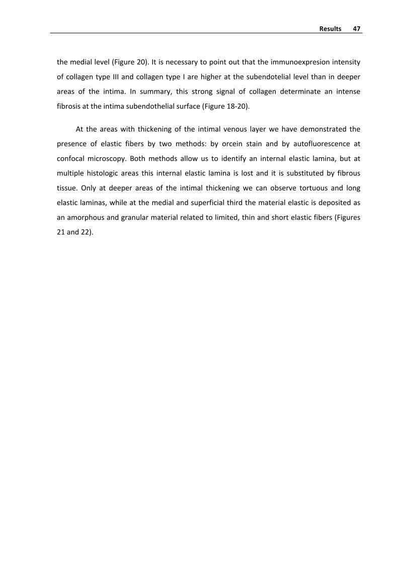
Results
47
themediallevel(Figure20).Itisnecessarytopointoutthattheimmunoexpresionintensity
ofcollagentypeIIIandcollagentypeIarehigheratthesubendotelial levelthanindeeper
areas of the intima. In summary, this strong signal of collagen determinate an intense
fibrosisattheintimasubendothelialsurface(Figure18‐20).
At the areaswith thickening of the intimal venous layerwe have demonstrated the
presence of elastic fibers by two methods: by orcein stain and by autofluorescence at
confocalmicroscopy. Bothmethods allow us to identify an internal elastic lamina, but at
multiple histologic areas this internal elastic lamina is lost and it is substituted by fibrous
tissue. Only at deeper areas of the intimal thickening we can observe tortuous and long
elasticlaminas,whileatthemedialandsuperficialthirdthematerialelasticisdepositedas
anamorphousandgranularmaterialrelatedtolimited,thinandshortelasticfibers(Figures
21and22).
Results
48
Fig.9.Saphenousvaricoseveinwithdifferentfibrosisgradesatthewall
a: Histologicalimagefromatransversesectionoftheproximallevelinsaphenousvein.We
canshowabigthickeningoftheintima.H‐E.
b: At the same segment can be seen a rise of the connective tissue that causemultiple
fibroses nodules at the intimal wall. Also, the medial wall presents an irregular
thickness,withdepositoffibrotictissueandadecreaseofthemuscularwall.Masson's
trichrome.
c: Detailfromtheimagebefore.Wecanseethedifferentgradeofintimalfibrosisandthe
atrophyfromdeSMCsatthemediallayer.Masson'strichrome.
d&e:Thefibrosisnodulesfrombothsidesoftheveincausesanevidentnarrowingofthe
vascular lumen.This fibrous tissue ismainly formbydense irregularconnective tissue
withdominationoffibersandECM.Also,thecellulardensityattheintimallayerisbig.
Masson'strichrome.
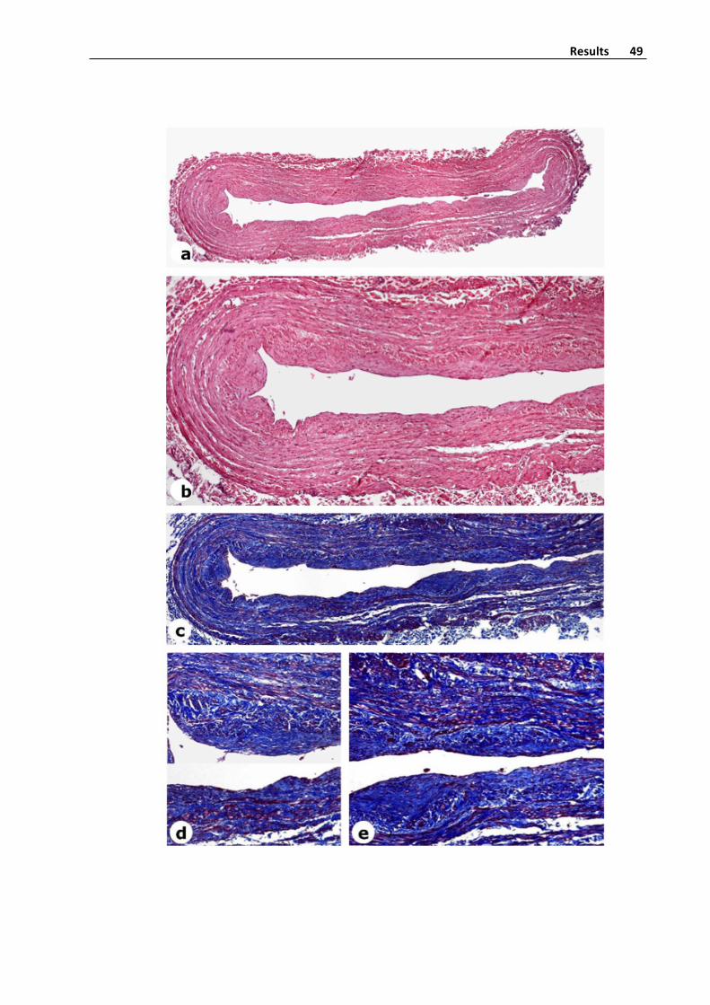
Results
49

Results
50
Fig.10.Fibrosisandcellularproliferationattheintimallayer,associatedtomuscular
atrophyatthemediallayerinasaphenousveinwithmoderatevaricoselesions
a: Longitudinalsectionfromasaphenousvein. Itobservesamoderateriseofthe intimal
thickening.Theendotheliallayerhasdisappeared.Also,itisseemadepositionofdense
irregular connective tissue and rise of the intimal cells at the intimal layer.Masson's
trichrome.
b: Detail from the image before.We can identify few oval or spindle cells with limited
fibrillarycytoplasmattheintimallayer.Thesecellsareimmersedatadenseconnective
tissuewherewecanseefibrillarycomponentsandabundantECM.Atthemediallayer,
the fascicules of the SMCs are very irregular and the leiomyocytes presents a small
diameter. Themuscular fascicules from themedial layer are also circle by connective
tissue.Masson'strichrome.
c: Parallelsectionfromtheimagebefore.Thethicknessoftheintimalismoderatedandit
is formbyECManda lowdensityofcollagenfibers.Weshouldfocustheexistenceof
spindlecellssimilartofibroblastwithsmallandthinnucleusandscantcytoplasm.The
limitbetweentheintimaandthemediaisevident.Muscularintimalatrophyandfibrosis
aroundthemuscularfasciculesfromthemediallayerarevisible.H‐E.
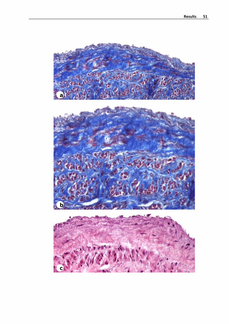
Results
51
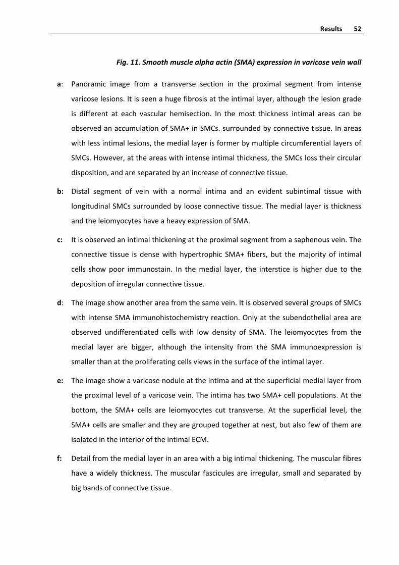
Results
52
Fig.11.Smoothmusclealphaactin(SMA)expressioninvaricoseveinwall
a: Panoramic image from a transverse section in the proximal segment from intense
varicoselesions.Itisseenahugefibrosisattheintimallayer,althoughthelesiongrade
is different at each vascular hemisection. In themost thickness intimal areas can be
observedanaccumulationofSMA+inSMCs.surroundedbyconnectivetissue.Inareas
withlessintimallesions,themediallayerisformerbymultiplecircumferentiallayersof
SMCs.However,attheareaswithintenseintimalthickness,theSMCslosstheircircular
disposition,andareseparatedbyanincreaseofconnectivetissue.
b: Distal segment of vein with a normal intima and an evident subintimal tissue with
longitudinalSMCssurroundedbylooseconnectivetissue.Themedial layeristhickness
andtheleiomyocyteshaveaheavyexpressionofSMA.
c: Itisobservedanintimalthickeningattheproximalsegmentfromasaphenousvein.The
connective tissue is densewith hypertrophic SMA+ fibers, but themajority of intimal
cells show poor immunostain. In themedial layer, the interstice is higher due to the
depositionofirregularconnectivetissue.
d: Theimageshowanotherareafromthesamevein.ItisobservedseveralgroupsofSMCs
withintenseSMAimmunohistochemistryreaction.Onlyatthesubendothelialareaare
observed undifferentiated cells with low density of SMA. The leiomyocytes from the
medial layer are bigger, although the intensity from the SMA immunoexpression is
smallerthanattheproliferatingcellsviewsinthesurfaceoftheintimallayer.
e: Theimageshowavaricosenoduleattheintimaandatthesuperficialmediallayerfrom
theproximallevelofavaricosevein.TheintimahastwoSMA+cellpopulations.Atthe
bottom, the SMA+ cells are leiomyocytes cut transverse. At the superficial level, the
SMA+cellsaresmallerandtheyaregroupedtogetheratnest,butalsofewofthemare
isolatedintheinterioroftheintimalECM.
f: Detailfromthemediallayerinanareawithabigintimalthickening.Themuscularfibres
haveawidely thickness.Themuscular fasciculesare irregular, smallandseparatedby
bigbandsofconnectivetissue.
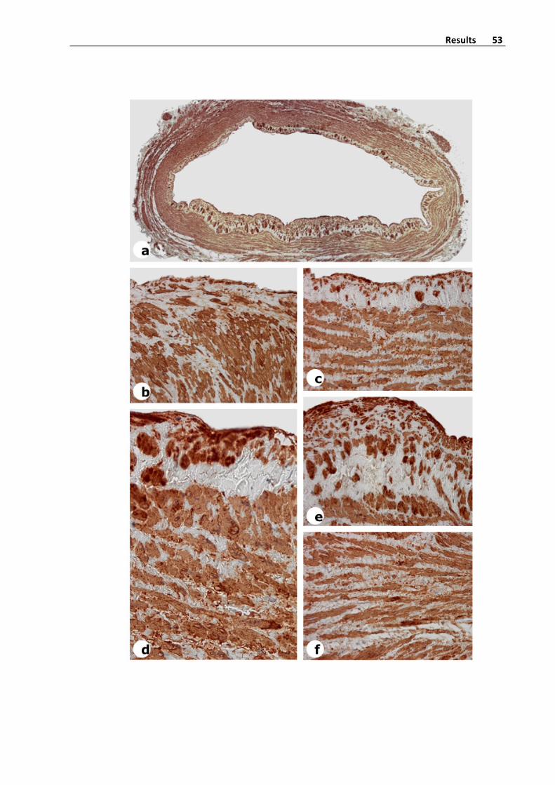
Results
53
Results
54
Fig.12.Vimentinexpressionfromthewallofasaphenousveinwithvaricoselesions
a: Itexistsaprogressive fibrosisat theproximalsegment fromthesaphenousveins.The
noduleofintimalthicknesscontainsnumerouscellswithsmallcytoplasmandanintense
vimentin expression. These cells are surrounded by ECM with low collagen fibers
deposits.Thelimitbetweenthemediaandtheintimaisclearandshowsathininternal
elastic membrane. At the medial layer, the vimentin+ cells are poor and they are
disposedroundedthefasciculesofSMC.
b: It isobservedamoderatethickeningofthe intima.The intimapresentsfewvimentin+
spindlecellsthattheyaremorenumerousatthesubendothelialarea.Themediallayer
hasareaswithinitialfibrosisthatcontentsabiggernumberofvimentin+fibroblast.
c: A huge proliferation of vimentin+ cells are situated in an area of initial fibrosis, sited
under the endothelium. The connective tissue from the medial layer is not very
abundantwithpresentsalimitednumberofvimentin+cells.
d: An intimal nodule in an areawith intense intimal fibrosis is shown. The immunostain
demonstrated the existence of transversal fascicules of vimentin+ cells. At the
superficiallevelexistisolatedcellswithlessexpressionofvimentin.
e: Anodulewith intensefibrosisshowssmall fasciculesofvimentin+SMCsatthe intima,
surroundedbyECM.Atthesuperior leveloftheintima, isolatedandspindlecellswith
scantcytoplasmareobserved.Theseundifferentiatedcellsarerecognizedbyavimentin
stainlowerthantheintensevimentinstainofthecytoplasmoftheendothelialcells.
f: The image included the interphase between the intima and the medial layer at the
proximal venous segment. It is evident the bigger number of vimentin+ cells in the
medial layer fibrotic tissue. The intimapresents perpendicular fascicules of vimentin+
cells.
g: The imagehaveahuge intimalthicknessthatcontainsan intenseproliferationofcells
with scant cytoplasm that showed an irregular vimentin stain distribution into their
cytoplasm.Aroundthesecells,thinbandsofECMaredeposited.Atmostoftheareas,
theendothelialcellshavebeendisappearedandsubstitutedbyfibroustissue.
Results
55
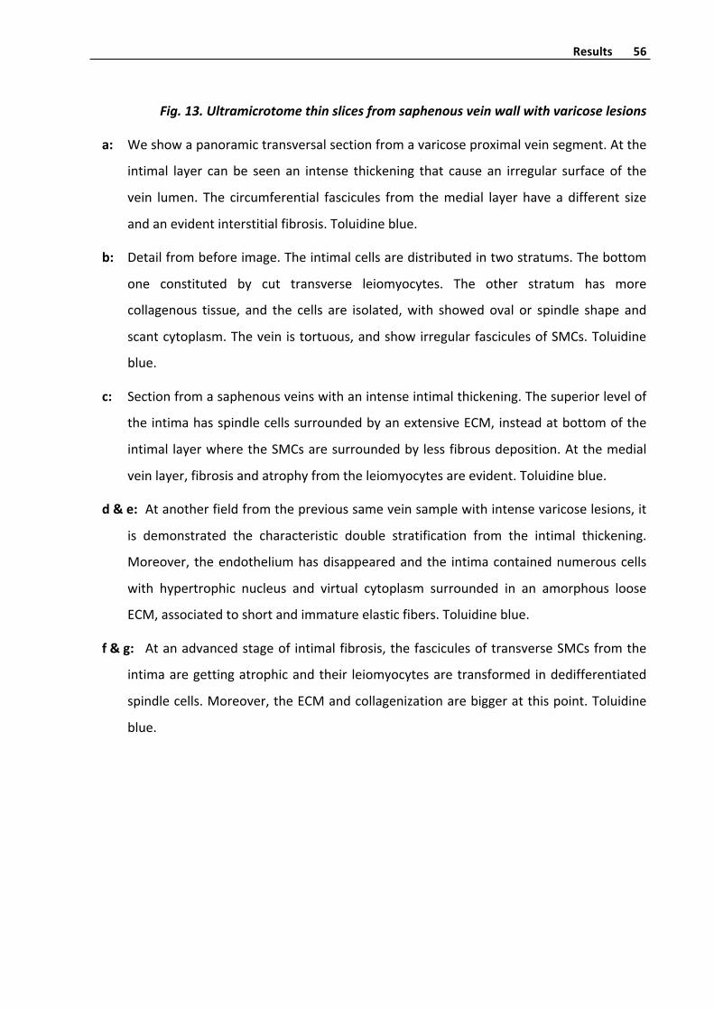
Results
56
Fig.13.Ultramicrotomethinslicesfromsaphenousveinwallwithvaricoselesions
a: Weshowapanoramictransversalsectionfromavaricoseproximalveinsegment.Atthe
intimal layer canbe seen an intense thickening that cause an irregular surfaceof the
vein lumen. The circumferential fascicules from themedial layer have a different size
andanevidentinterstitialfibrosis.Toluidineblue.
b: Detailfrombeforeimage.Theintimalcellsaredistributedintwostratums.Thebottom
one constituted by cut transverse leiomyocytes. The other stratum has more
collagenous tissue, and the cells are isolated,with showed oval or spindle shape and
scantcytoplasm.Theveinistortuous,andshowirregularfasciculesofSMCs.Toluidine
blue.
c: Sectionfromasaphenousveinswithanintenseintimalthickening.Thesuperiorlevelof
theintimahasspindlecellssurroundedbyanextensiveECM,insteadatbottomofthe
intimal layerwheretheSMCsaresurroundedbylessfibrousdeposition.Atthemedial
veinlayer,fibrosisandatrophyfromtheleiomyocytesareevident.Toluidineblue.
d&e:Atanotherfieldfromtheprevioussameveinsamplewithintensevaricoselesions,it
is demonstrated the characteristic double stratification from the intimal thickening.
Moreover, theendotheliumhasdisappearedandthe intimacontainednumerouscells
with hypertrophic nucleus and virtual cytoplasm surrounded in an amorphous loose
ECM,associatedtoshortandimmatureelasticfibers.Toluidineblue.
f&g: Atanadvancedstageof intimalfibrosis,thefasciculesoftransverseSMCsfromthe
intimaaregettingatrophicandtheir leiomyocytesaretransformed indedifferentiated
spindlecells.Moreover,theECMandcollagenizationarebiggeratthispoint.Toluidine
blue.
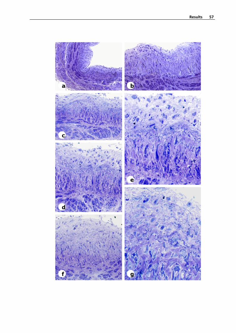
Results
57

Results
58
Fig.14.Ultrastructuralchangesinthesaphenousveinwithvaricoselesions
a&b: Intheintima,theinternalelasticfiberissubstitutedbyfibroustissueandsmall,short
and thin elastic fibers intermixed with the amorphous ECM. In some areas exist a
progressive transformation from the intimal SMCs to oval or spindle dedifferentiates
cells,someofthisshowedahypertrophicnucleus.Theluminalareaisirregularandthe
endothelialcellsaredenuded.Toluidineblue.
c: Ultrastructuralimagefromavaricoseintimallesion.Atthebottomoftheimagehasbe
seen a flexuous elastic membrane characterized by a central core of amorphous
material with low electronic density. Also, some small spindle cells with circular or
elongatednucleusandheterochromaticmaterialcanbeseenunderthekaryotheca.The
cytoplasmofthiscellsisdenseandfibrillary.
d: ThisultrastructuralimageshowntheintimalsurfacewhereECMdepositionroundedthe
irregular collagen fibers. Moreover, we can observe two intimal cells with virtual
cytoplasmanddevelopmentofcytoplasmicprolongationsthataresurroundedbyECM.
e: This image contains a fragment of a cell in the intimal layer. The nucleus is
hyperchromaticandwithandshowedanirregularshape.Thecytoplasmiswide,witha
polygonal shape and contains round mitochondria and fascicles of intermediate
filaments. Additionally, it canbe seen somedensedisc blockingbymyofilaments and
desmosomes. Also, in the interstice near the cytoplasmic membrane, deposition of
amorphousmaterial,ECMandirregularelongatedcollagenfibersaredemonstrated.
f: Canbeobservedthecytoplasmofthesomaandtheprolongationsfromacell located
intotheintimalfibrotictissueofavaricosevein.Insidethecytoplasmcanbeseenabig
numberofintermediatefilaments.Also,canbeseendensediscswithmyofilamentsand
few desmosomes. The cell presents small vesicles, but no caveolae, so probably it is
diagnosed as a myofibroblast. Near to the external lamella of the cytoplasmic
membrane,anelectrodensematerialthatgetspolymertocreateisolatedthinandshort
collagenfibers,surroundedbyECMareidentified.
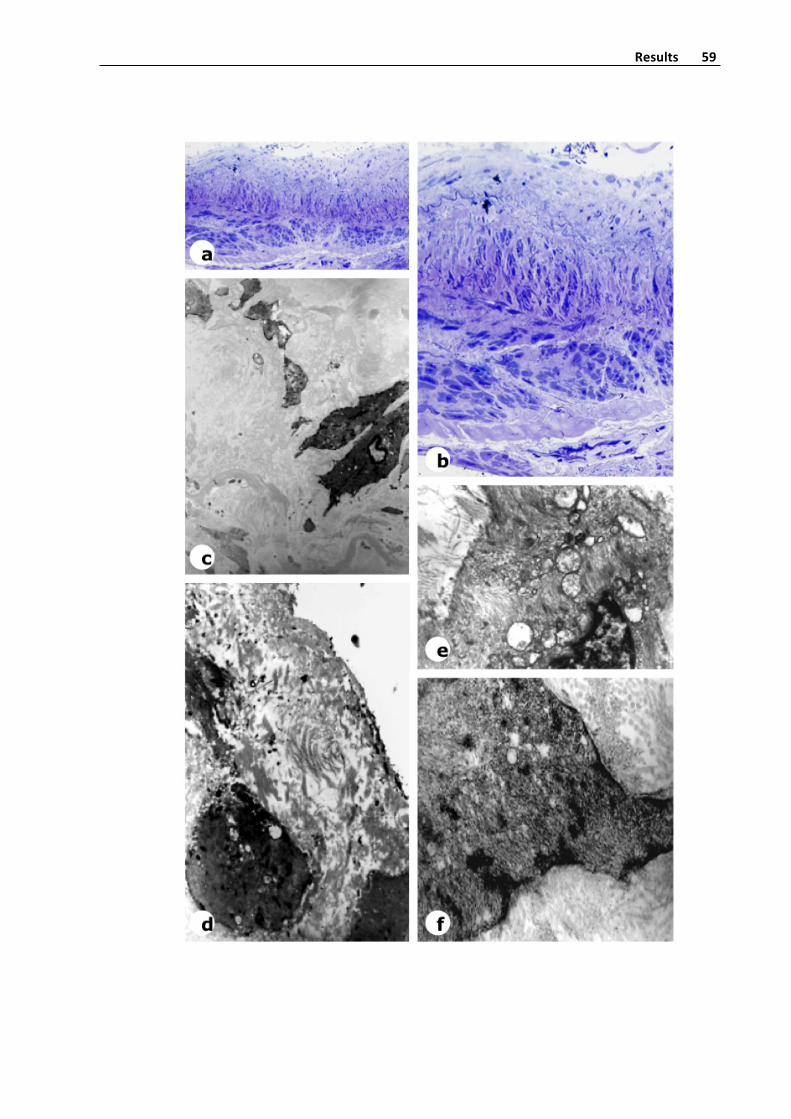
Results
59
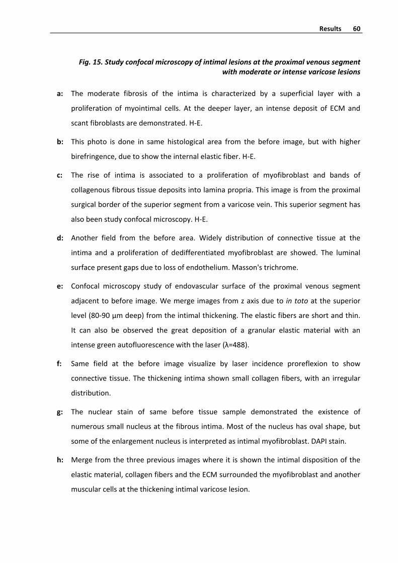
Results
60
Fig.15.Studyconfocalmicroscopyofintimallesionsattheproximalvenoussegmentwithmoderateorintensevaricoselesions
a: The moderate fibrosis of the intima is characterized by a superficial layer with a
proliferation ofmyointimal cells. At the deeper layer, an intense deposit of ECM and
scantfibroblastsaredemonstrated.H‐E.
b: This photo is done in same histological area from the before image, but with higher
birefringence,duetoshowtheinternalelasticfiber.H‐E.
c: The rise of intima is associated to a proliferation of myofibroblast and bands of
collagenousfibroustissuedepositsintolaminapropria.Thisimageisfromtheproximal
surgicalborderofthesuperiorsegmentfromavaricosevein.Thissuperiorsegmenthas
alsobeenstudyconfocalmicroscopy.H‐E.
d: Another field from the before area. Widely distribution of connective tissue at the
intima and a proliferation of dedifferentiatedmyofibroblast are showed. The luminal
surfacepresentgapsduetolossofendothelium.Masson'strichrome.
e: Confocal microscopy study of endovascular surface of the proximal venous segment
adjacenttobefore image.Wemerge imagesfromzaxisdueto intotoatthesuperior
level(80‐90µmdeep)fromtheintimalthickening.Theelasticfibersareshortandthin.
It can also be observed the great deposition of a granular elastic material with an
intensegreenautofluorescencewiththelaser(λ=488).
f: Same field at the before image visualize by laser incidence proreflexion to show
connective tissue.Thethickening intimashownsmallcollagen fibers,withan irregular
distribution.
g: The nuclear stain of same before tissue sample demonstrated the existence of
numeroussmallnucleusatthefibrousintima.Mostofthenucleushasovalshape,but
someoftheenlargementnucleusisinterpretedasintimalmyofibroblast.DAPIstain.
h: Mergefromthethreepreviousimageswhereitisshowntheintimaldispositionofthe
elasticmaterial,collagenfibersandtheECMsurroundedthemyofibroblastandanother
muscularcellsatthethickeningintimalvaricoselesion.

Results
61

Results
62
Fig.16.Studybyconfocalmicroscopyofintimallesionsatthedistalvenoussegmentwithmoderatevaricoselesions
a: Panoramicimagefromthesuperioredgeofthedistalsegmentfromavaricosevein.
Itcanbeseenaninitial intimalthickeningatthesuperficialarea.Atadeeper intimalarea,
thetissueshowedstronglycollagenizationandcanbe identifydense fasciculesofcollagen
fibers.Atthisarea,thepresencesofspindlecellslooklikefibroblastarescant.H‐E.
b: Detail from thebefore image. It is shown theconnective tissue situatedbelow the
disappeared endothelium. The ECM is abundant and it surrounded the immature
myofibroblast.Atthedeepintimalareathecollagenizationisbigger.H‐E.
c: Photo takenata close field from thebefore image.A loose connective tissuewith
abundantECMisshown.Mostcellsareundifferentiated,butsomepresentaspindlenucleus
andafibrillarycytoplasm,thanlooklikemyofibroblast.Atthedeepestarea,theintimahasa
more intense collagenization, with thick collagen fibers in an irregular disposition. This
fibroustissuealsohasisolatedSMCsofcirculardisposition.Masson'strichrome.
d: Confocalmicroscopy study of in toto intimal surface of the distal venous segment
adjacent to image before.Wemerge images from Z axis at the superior level (80‐90 µm
deep)fromtheintimalthickening,withanabundantdepositionofelasticmaterial.Itcanbe
seenfewsmall,shortelasticfiberswithspiralshapewithanintensegreenautofluorescence
observedinconfocallaser(λ=488).
e: The reflectionof the laser light at the samemicroscopic fieldallow toobserve the
deposition of thin collagen fibers, randomly distributed, into the intima. The darker areas
correspondtofoldsoftheintimalsurfaceduetothecircularnormalshapeofthevein.
f: Abigproliferationofcellsisfound.Thenucleusissmallandclosebetweenthemwith
alimitedinterstitialtissue.Mostofthesecellsarefibroblastandmyofibroblast.DAPIstain.
g: Merge imagefromelasticautofluorescence, lightrayreflectionandDAPIsee inthe
three previous images that demonstrated the intense connective tissue deposition and
collagenfasciculesthatsurroundedtheelasticmaterial.
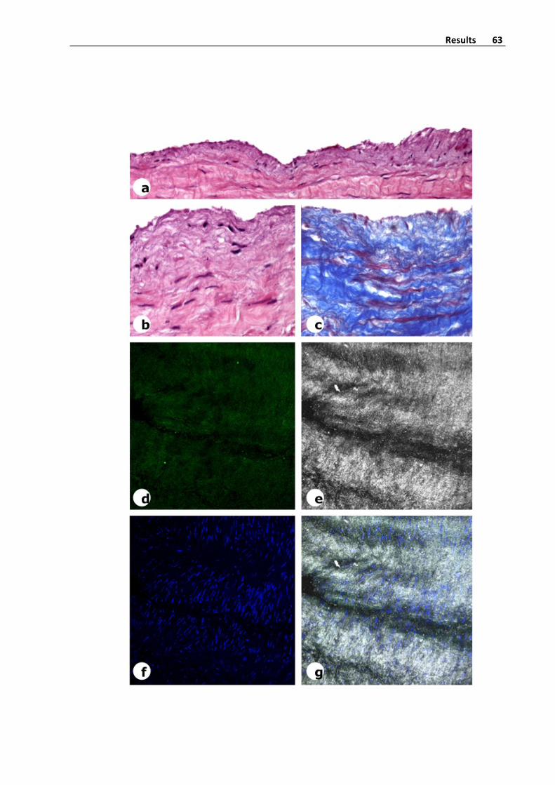
Results
63
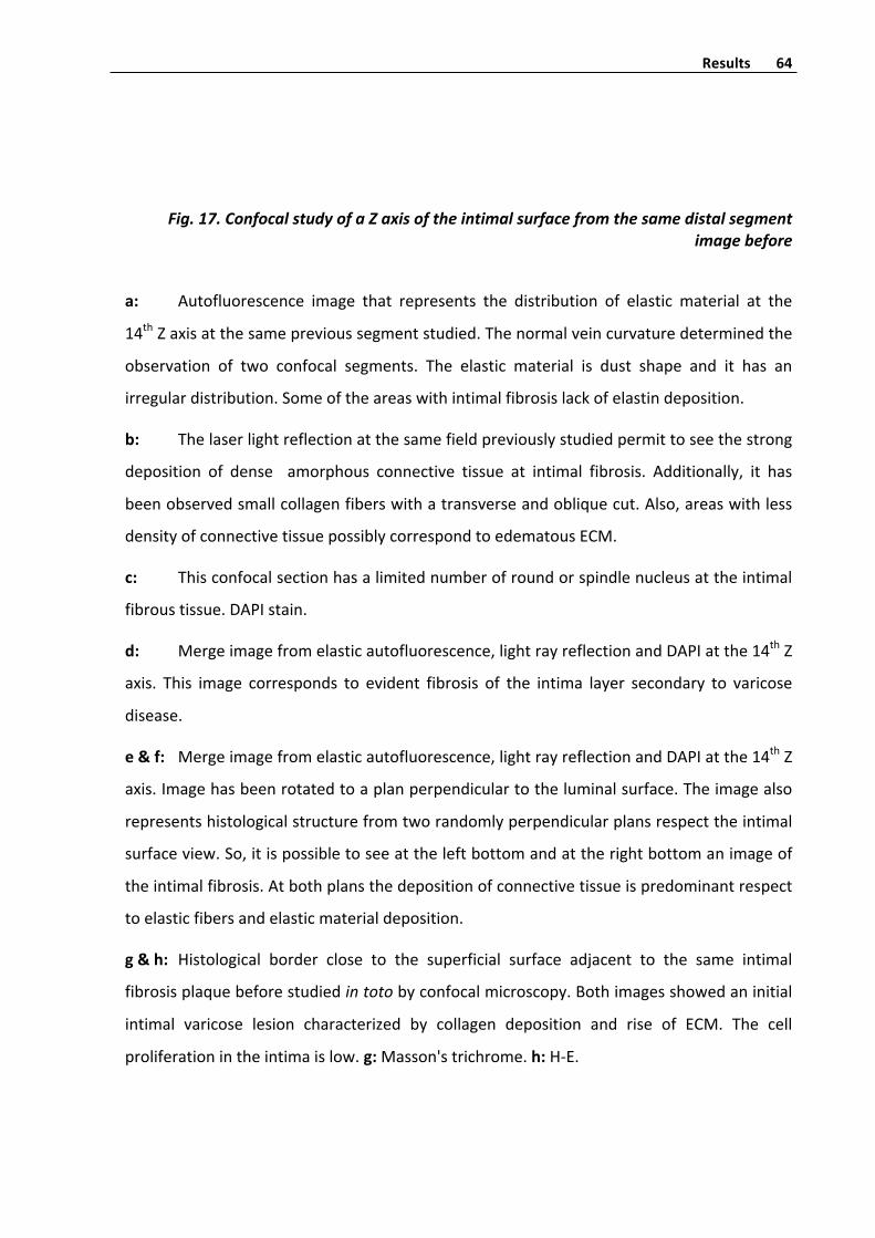
Results
64
Fig.17.ConfocalstudyofaZaxisoftheintimalsurfacefromthesamedistalsegmentimagebefore
a: Autofluorescence image that represents the distribution of elastic material at the
14thZaxisatthesameprevioussegmentstudied.Thenormalveincurvaturedeterminedthe
observation of two confocal segments. The elastic material is dust shape and it has an
irregulardistribution.Someoftheareaswithintimalfibrosislackofelastindeposition.
b: Thelaserlightreflectionatthesamefieldpreviouslystudiedpermittoseethestrong
deposition of dense amorphous connective tissue at intimal fibrosis. Additionally, it has
beenobservedsmallcollagenfiberswithatransverseandobliquecut.Also,areaswithless
densityofconnectivetissuepossiblycorrespondtoedematousECM.
c: Thisconfocalsectionhasalimitednumberofroundorspindlenucleusattheintimal
fibroustissue.DAPIstain.
d: Mergeimagefromelasticautofluorescence,lightrayreflectionandDAPIatthe14thZ
axis. This image corresponds to evident fibrosis of the intima layer secondary to varicose
disease.
e&f: Mergeimagefromelasticautofluorescence,lightrayreflectionandDAPIatthe14thZ
axis.Imagehasbeenrotatedtoaplanperpendiculartotheluminalsurface.Theimagealso
representshistologicalstructurefromtworandomlyperpendicularplansrespecttheintimal
surfaceview.So,itispossibletoseeattheleftbottomandattherightbottomanimageof
theintimalfibrosis.Atbothplansthedepositionofconnectivetissueispredominantrespect
toelasticfibersandelasticmaterialdeposition.
g&h:Histological border close to the superficial surface adjacent to the same intimal
fibrosisplaquebeforestudiedintotobyconfocalmicroscopy.Bothimagesshowedaninitial
intimal varicose lesion characterized by collagen deposition and rise of ECM. The cell
proliferationintheintimaislow.g:Masson'strichrome.h:H‐E.

Results
65
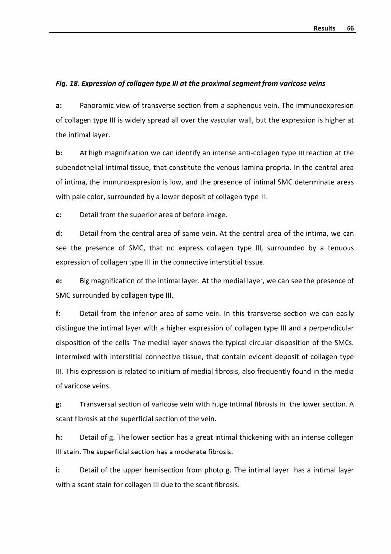
Results
66
Fig.18.ExpressionofcollagentypeIIIattheproximalsegmentfromvaricoseveins
a: Panoramicviewoftransversesectionfromasaphenousvein.Theimmunoexpresion
ofcollagentypeIIIiswidelyspreadalloverthevascularwall,buttheexpressionishigherat
theintimallayer.
b: Athighmagnificationwecanidentifyanintenseanti‐collagentypeIIIreactionatthe
subendothelialintimaltissue,thatconstitutethevenouslaminapropria.Inthecentralarea
ofintima,theimmunoexpresionislow,andthepresenceofintimalSMCdeterminateareas
withpalecolor,surroundedbyalowerdepositofcollagentypeIII.
c: Detailfromthesuperiorareaofbeforeimage.
d: Detailfromthecentralareaofsamevein.Atthecentralareaoftheintima,wecan
see the presence of SMC, that no express collagen type III, surrounded by a tenuous
expressionofcollagentypeIIIintheconnectiveinterstitialtissue.
e: Bigmagnificationoftheintimallayer.Atthemediallayer,wecanseethepresenceof
SMCsurroundedbycollagentypeIII.
f: Detail from the inferior areaof samevein. In this transverse sectionwe caneasily
distinguetheintimallayerwithahigherexpressionofcollagentypeIIIandaperpendicular
dispositionofthecells.ThemediallayershowsthetypicalcirculardispositionoftheSMCs.
intermixedwith interstitialconnectivetissue,thatcontainevidentdepositofcollagentype
III.Thisexpressionisrelatedtoinitiumofmedialfibrosis,alsofrequentlyfoundinthemedia
ofvaricoseveins.
g: Transversalsectionofvaricoseveinwithhugeintimalfibrosisinthelowersection.A
scantfibrosisatthesuperficialsectionofthevein.
h: Detailofg.Thelowersectionhasagreatintimalthickeningwithanintensecollegen
IIIstain.Thesuperficialsectionhasamoderatefibrosis.
i: Detailoftheupperhemisectionfromphotog.Theintimal layer hasa intimal layer
withascantstainforcollagenIIIduetothescantfibrosis.

Results
67
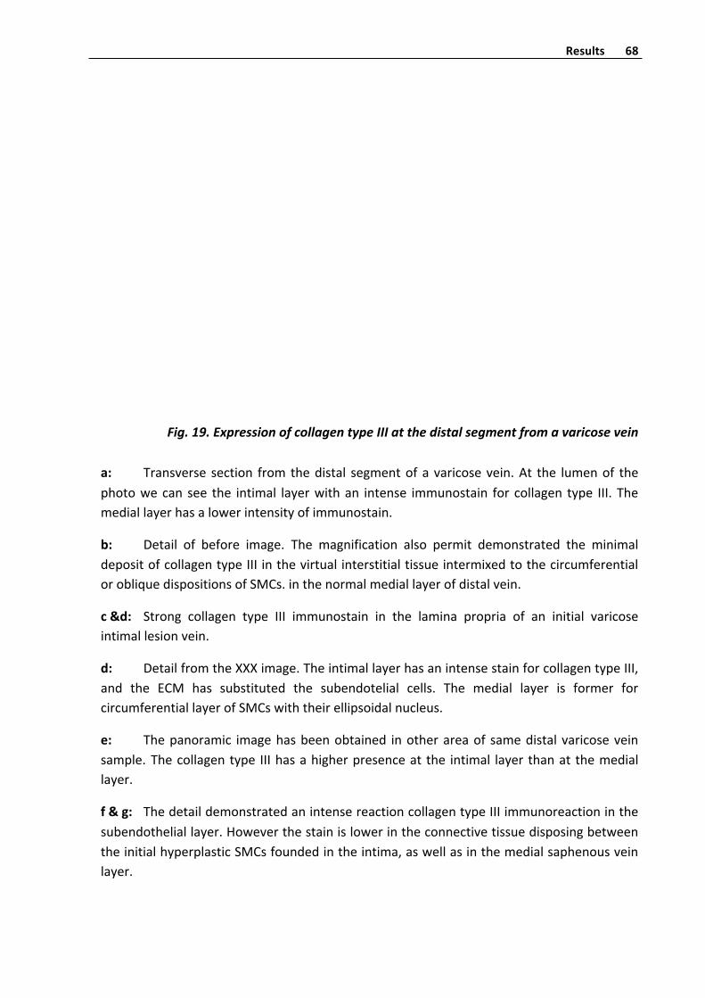
Results
68
Fig.19.ExpressionofcollagentypeIIIatthedistalsegmentfromavaricosevein
a: Transverse section from thedistal segmentofa varicosevein.At the lumenof thephotowecan see the intimal layerwithan intense immunostain for collagen type III. Themediallayerhasalowerintensityofimmunostain.
b: Detail of before image. The magnification also permit demonstrated the minimaldepositofcollagentypeIII inthevirtualinterstitialtissueintermixedtothecircumferentialorobliquedispositionsofSMCs.inthenormalmediallayerofdistalvein.
c&d: Strong collagen type III immunostain in the lamina propria of an initial varicoseintimallesionvein.
d: DetailfromtheXXXimage.TheintimallayerhasanintensestainforcollagentypeIII,and the ECM has substituted the subendotelial cells. The medial layer is former forcircumferentiallayerofSMCswiththeirellipsoidalnucleus.
e: Thepanoramic imagehasbeenobtained inotherareaof samedistal varicoseveinsample.Thecollagentype IIIhasahigherpresenceat the intimal layerthanat themediallayer.
f&g: ThedetaildemonstratedanintensereactioncollagentypeIIIimmunoreactioninthesubendotheliallayer.HoweverthestainislowerintheconnectivetissuedisposingbetweentheinitialhyperplasticSMCsfoundedintheintima,aswellasinthemedialsaphenousveinlayer.
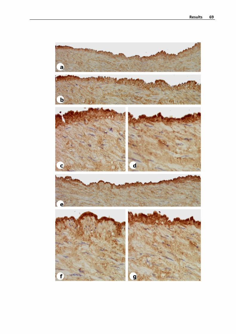
Results
69

Results
70
Fig.20.ExpressionofcollagentypeIattheproximalanddistalsegmentfromavaricosevein
a: The proximal segment of vein shows an intense thickened of the intimal layer
characterized by a strong deposit of Collagen type I upper in the subendothelial lamina
propria.ThediffusecollagentypeIdepositextendstotheintestinaltissuethatsurrounded
theSMCfascicleslocatedatintimaandmedialayers.
b: The intima of this proximal vein segment is extraordinary thickened and the
immunostainingwithcollagentypeIantibodydemonstratesan intensefibrillaranddiffuse
pattern.Theintensityofimmunostainingislowerinthemedialayer.
c: Detailfromthebeforeimagethatdemonstratedastrongbandofimmunoexpression
ofcollagentypeIatthelaminapropriainvicinitytoendothelialepithelium.Thefibroblasts
and myofibroblasts of the intima are embedded in the ECM and rich collagen type I
connectivetissueoftheintima
d: This image obtained in the proximal segment of a saphenous vein shows the
transition of a normal intima to an initial varicose lesion of the intima, characterized by
hyperplasia of SMCs. and irregular deposits of collagen type I that intermixed with non‐
immunostaining ECM. Noted the high immunostaining seen in the interstitial connective
tissueofmediallayer.
e: Thisdistalsaphenousveinsegmentshowsasoftanddiffusedepositofcollagentype
I.
f: RobuststainofcollagentypeIinlaminapropria,ifcomparedwiththelowexpression
inthedeepintima.Notedanareawithevidentproliferationofintimalcellsassociatedtothe
fibrillarydistributionofcollagentypeI.
g: ThegradeofcollagentypeIexpressionishighertheintimasurfaceandprogressively
decreaseintheprofoundintimaandinthevascularmedia.
h: Extensive fibrosis is found in the superficial area of the saphenous intima, due to
strongimnunoexpressionofcollagentypeI.However,thestainislowerintheprofoundarea
ofthisvaricosevein.
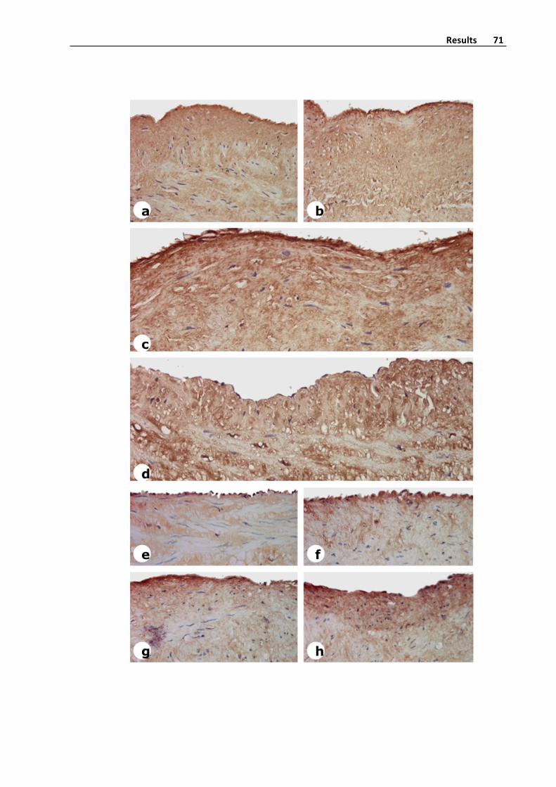
Results
71
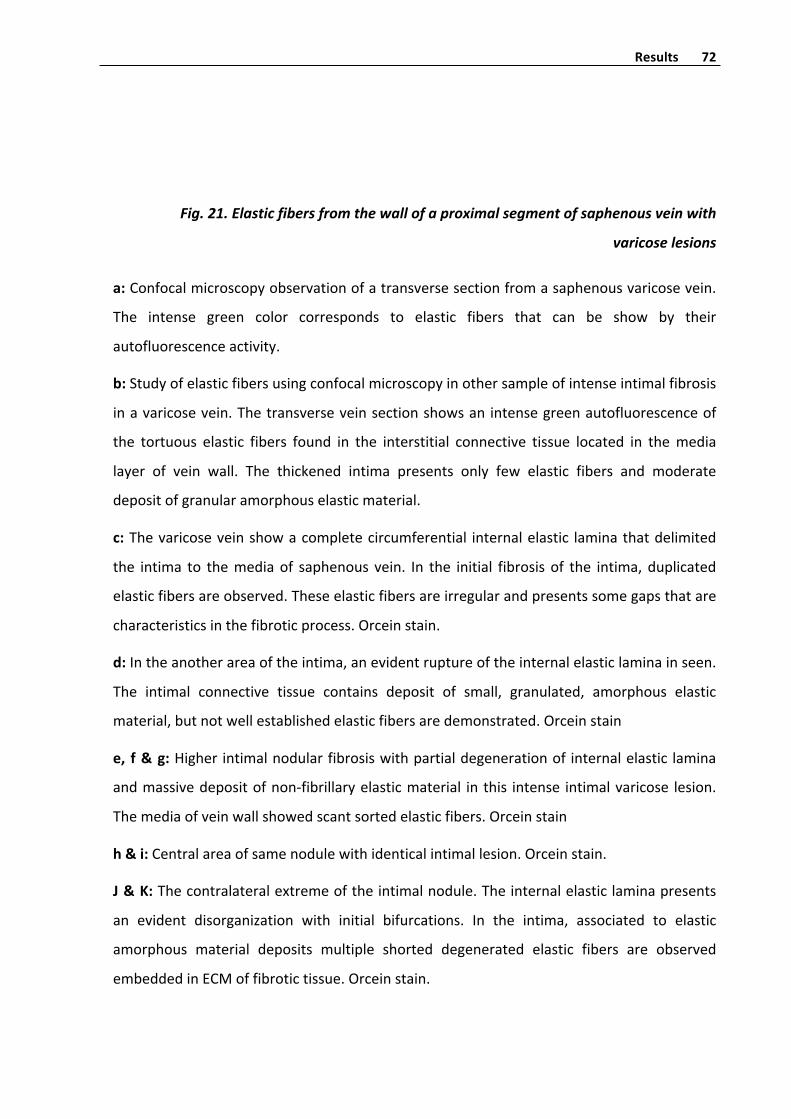
Results
72
Fig.21.Elasticfibersfromthewallofaproximalsegmentofsaphenousveinwith
varicoselesions
a:Confocalmicroscopyobservationofatransversesectionfromasaphenousvaricosevein.
The intense green color corresponds to elastic fibers that can be show by their
autofluorescenceactivity.
b:Studyofelasticfibersusingconfocalmicroscopyinothersampleofintenseintimalfibrosis
inavaricosevein.Thetransverseveinsectionshowsan intensegreenautofluorescenceof
the tortuous elastic fibers found in the interstitial connective tissue located in themedia
layer of vein wall. The thickened intima presents only few elastic fibers and moderate
depositofgranularamorphouselasticmaterial.
c:Thevaricoseveinshowacompletecircumferential internalelastic laminathatdelimited
the intima to themediaof saphenous vein. In the initial fibrosis of the intima,duplicated
elasticfibersareobserved.Theseelasticfibersareirregularandpresentssomegapsthatare
characteristicsinthefibroticprocess.Orceinstain.
d:Intheanotherareaoftheintima,anevidentruptureoftheinternalelasticlaminainseen.
The intimal connective tissue contains deposit of small, granulated, amorphous elastic
material,butnotwellestablishedelasticfibersaredemonstrated.Orceinstain
e, f&g:Higher intimalnodularfibrosiswithpartialdegenerationof internalelastic lamina
andmassivedepositofnon‐fibrillaryelasticmaterial inthis intense intimalvaricose lesion.
Themediaofveinwallshowedscantsortedelasticfibers.Orceinstain
h&i:Centralareaofsamenodulewithidenticalintimallesion.Orceinstain.
J&K:Thecontralateralextremeoftheintimalnodule.Theinternalelasticlaminapresents
an evident disorganization with initial bifurcations. In the intima, associated to elastic
amorphous material deposits multiple shorted degenerated elastic fibers are observed
embeddedinECMoffibrotictissue.Orceinstain.
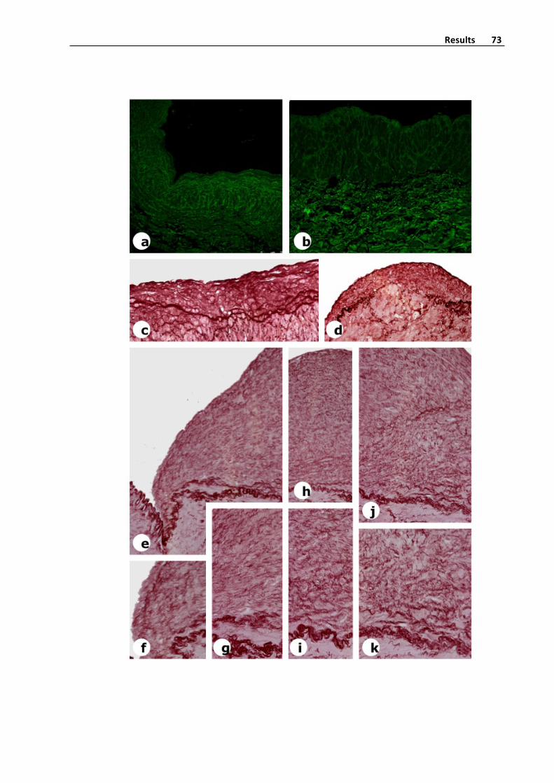
Results
73

Results
74
Fig.22.Elasticfibersfromthewallofadistalsegmentofsaphenousveinwithinitial
varicoselesionsintheintima
a:Transversesectionfromadistalsegmentofsaphenousvein.Theelasticautofluorescent
permittoidentifythethreeveinlayers:intimal,medialandadventitiallayers.Theintimais
virtual.Theendothelialcellsapparentlyaredirectlycloselytoelasticinternallamina.
b:Aninitialgrowthoftheintima,anevidentdespoticofelastinfibers(greencolor)intothe
subendotheliallaminapropriaarefound.Thisnewelasticfibersarethinandirregular.
c, d & e: Different grade of initial varicose alterations. The normal intima progressively
presents EMC deposits, reduplication of elastic internal lamina and a new formed intimal
elasticfiberpopulation.Shortandscantelasticfibersdepositsintotheinterstitialconnective
tissuethatsurroundthecircumferentialfasciclesofSMCarefound.Orceinstain.
f: Normal distal segment of saphenous vein. Noted the direct transition to an initial
progressivelydepositof connective tissueandelastic amorphousmaterial into the intima.
Theinternalelasticlaminaisnormal.Inset:thelaminapropriaisvirtual.Orceinstain.
g: Transition of normal intima tominimal intimal fibrosiswithout deposit of new training
elasticfibers.Orceinstain.
h:Disorganizationoftheinternalelasticlaminaandthedustshapeelasticfibersdeposition.
Anincreaseofelasticfibersisobservedinthemediallayerofthisdistalveinsegment.
i:Moderatethickeningoftheintimaandruptureofinternalelasticlamina.Thisnoduleifstill
growingcanproduceamoderatepartial stenosisof thevein lumen. In the intima,wecan
appreciatethedifferentdepositionofimmatureelasticfibers,characterizedbythepresence
ofmultipleshortedelasticfibersembeddedbyconnectivetissue.Orceinstain.
j:Transmuralvaricose lesions.Disorganizationof the internalelastic laminaanddepositof
multiples elastic fibers into the intima were associated to atrophy of SMC fascicles,
interstitialfibrosisandadegenerationofelasticfibersinthemediallayer.

Results
75

Results
76
COLLAGENTYPEIHISTOMETRYQUANTIFYINGINVENOUSINTIMA
Table 4 shown histometric data from the area occupied by collagen type I
immunoreactionpositive in relation to intimal totalareaof thevein.Weconsidered three
groupsinrelationtoheightofveinintimallayer.
Table4.CollagentypeIarea.
Intimalheightthickness(µm)
Intimaltotalarea(µm2)
CollagenIarea(µm2)
ProportioncollagenIarea/totalarea
1to39.9 21742±8153 5374±3088 0.24±0.10
39.9to89.9 60923±13890a 26032±9577a 0.42±0.10a
>90 149382±38622a 53194±14958a 0.35±0.02
a:p<0.05significanttobeforegroup
Intimal area and Col I area
1-39.9 40-89.9 >900
50000
100000
150000
200000
250000
Col I areaIntimal area
Intimal height (µm)
µm2
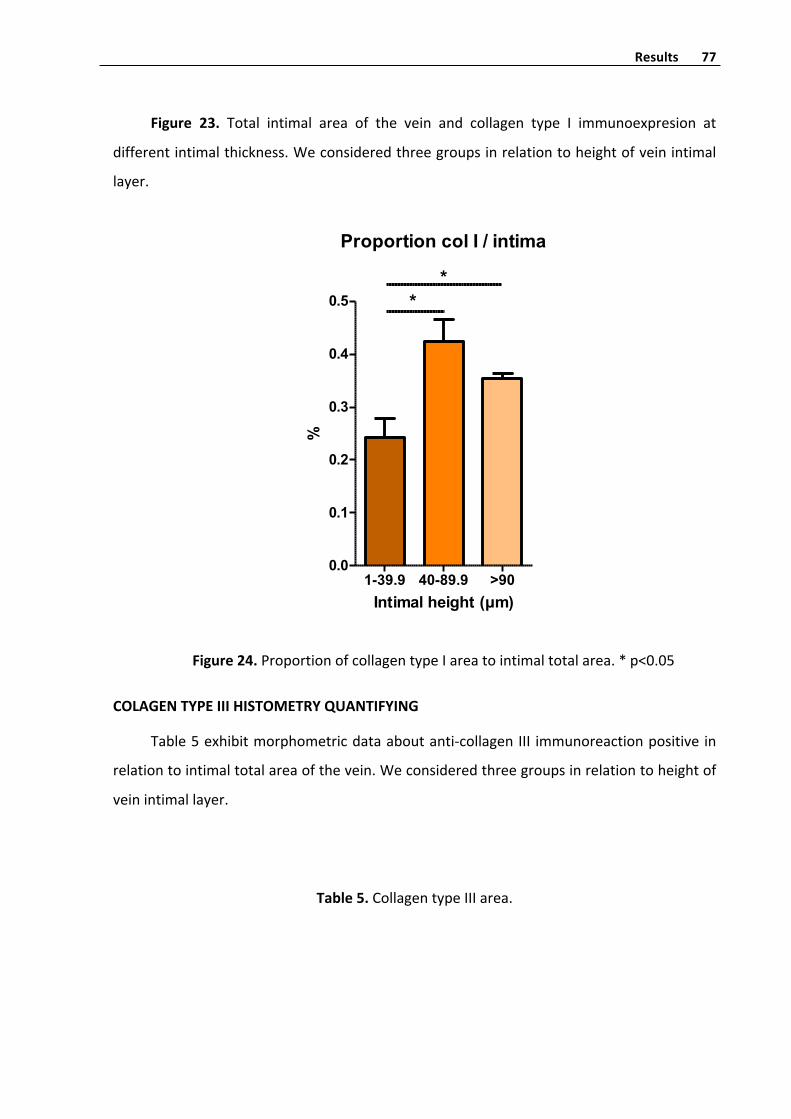
Results
77
Figure 23. Total intimal area of the vein and collagen type I immunoexpresion at
differentintimalthickness.Weconsideredthreegroupsinrelationtoheightofveinintimal
layer.
Figure24.ProportionofcollagentypeIareatointimaltotalarea.*p<0.05
COLAGENTYPEIIIHISTOMETRYQUANTIFYING
Table5exhibitmorphometricdataaboutanti‐collagenIII immunoreactionpositive in
relationtointimaltotalareaofthevein.Weconsideredthreegroupsinrelationtoheightof
veinintimallayer.
Table5.CollagentypeIIIarea.
Proportion col I / intima
1-39.9 40-89.9 >900.0
0.1
0.2
0.3
0.4
0.5 **
Intimal height (µm)
%
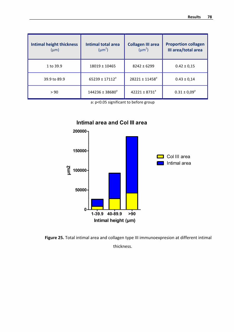
Results
78
Intimalheightthickness(µm)
Intimaltotalarea(µm2)
CollagenIIIarea(µm2)
ProportioncollagenIIIarea/totalarea
1to39.9 18019±10465 8242±6299 0.42±0,15
39.9to89.9 65239±17112a 28221±11458a 0.43±0,14
>90 144236±38680a 42221±8731a 0.31±0,09a
a:p<0.05significanttobeforegroup
Figure25.TotalintimalareaandcollagentypeIIIimmunoexpresionatdifferentintimal
thickness.
Intimal area and Col III area
1-39.9 40-89.9 >900
50000
100000
150000
200000
Col III areaIntimal area
Intimal height (µm)
µm2
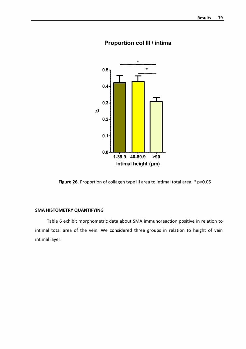
Results
79
Figure26.ProportionofcollagentypeIIIareatointimaltotalarea.*p<0.05
SMAHISTOMETRYQUANTIFYING
Table6exhibitmorphometricdataaboutSMAimmunoreactionpositiveinrelationto
intimal total area of the vein. We considered three groups in relation to height of vein
intimallayer.
Proportion col III / intima
1-39.9 40-89.9 >900.0
0.1
0.2
0.3
0.4
0.5 **
Intimal height (µm)
%
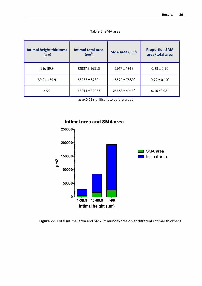
Results
80
Table6.SMAarea.
Intimalheightthickness(µm)
Intimaltotalarea(µm2) SMAarea(µm2) ProportionSMA
area/totalarea
1to39.9 22097±16113 5547±4248 0.29±0,10
39.9to89.9 68983±8739a 15520±7589a 0.22±0,10a
>90 168011±39963a 25683±4943a 0.16±0.03a
a:p<0.05significanttobeforegroup
Figure27.TotalintimalareaandSMAimmunoexpresionatdifferentintimalthickness.
Intimal area and SMA area
1-39.9 40-89.9 >900
50000
100000
150000
200000
250000
SMA areaIntimal area
Intimal height (µm)
µm2
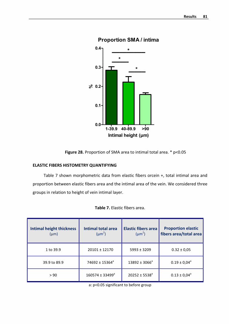
Results
81
Figure28.ProportionofSMAareatointimaltotalarea.*p<0.05
ELASTICFIBERSHISTOMETRYQUANTIFYING
Table7shownmorphometricdatafromelasticfibersorcein+,total intimalareaand
proportionbetweenelasticfibersareaandtheintimalareaofthevein.Weconsideredthree
groupsinrelationtoheightofveinintimallayer.
Table7.Elasticfibersarea.
Intimalheightthickness(µm)
Intimaltotalarea(µm2)
Elasticfibersarea(µm2)
Proportionelasticfibersarea/totalarea
1to39.9 20101±12170 5993±3209 0.32±0,05
39.9to89.9 74692±15364a 13892±3066a 0.19±0,04a
>90 160574±33499a 20252±5538a 0.13±0,04a
a:p<0.05significanttobeforegroup
Proportion SMA / intima
1-39.9 40-89.9 >900.0
0.1
0.2
0.3
0.4
*
**
Intimal height (µm)
%
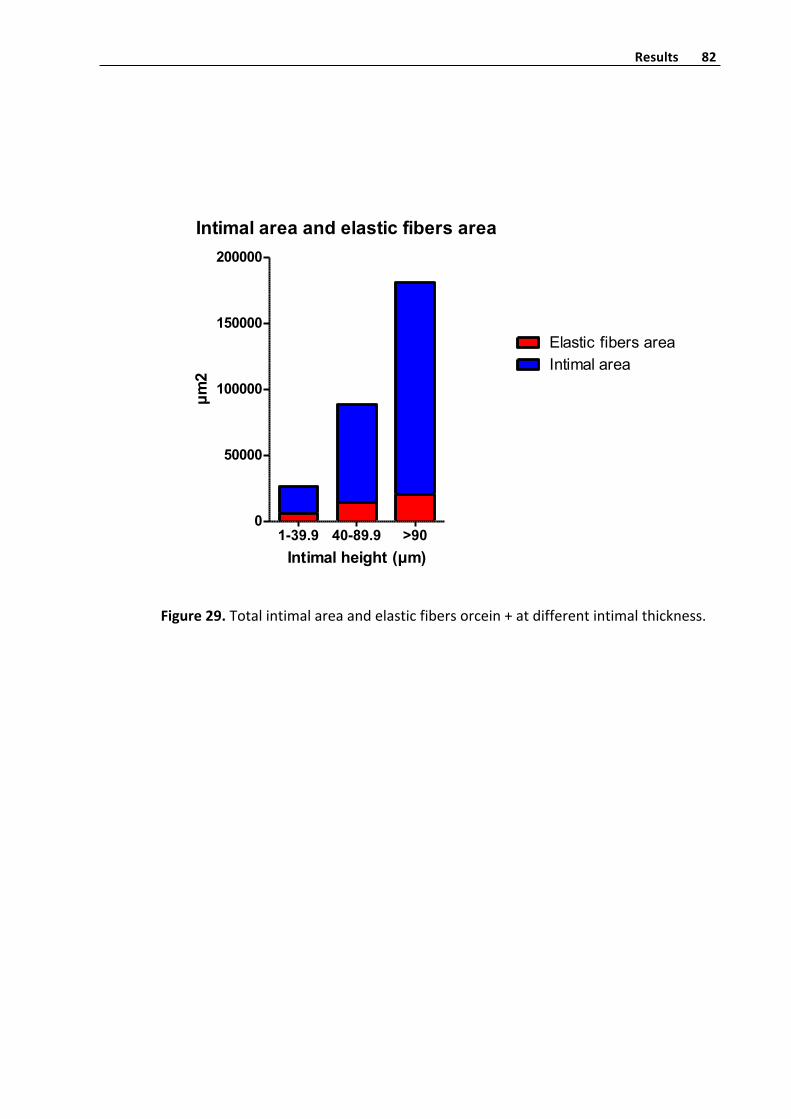
Results
82
Figure29.Totalintimalareaandelasticfibersorcein+atdifferentintimalthickness.
Intimal area and elastic fibers area
1-39.9 40-89.9 >900
50000
100000
150000
200000
Elastic fibers areaIntimal area
Intimal height (µm)
µm2
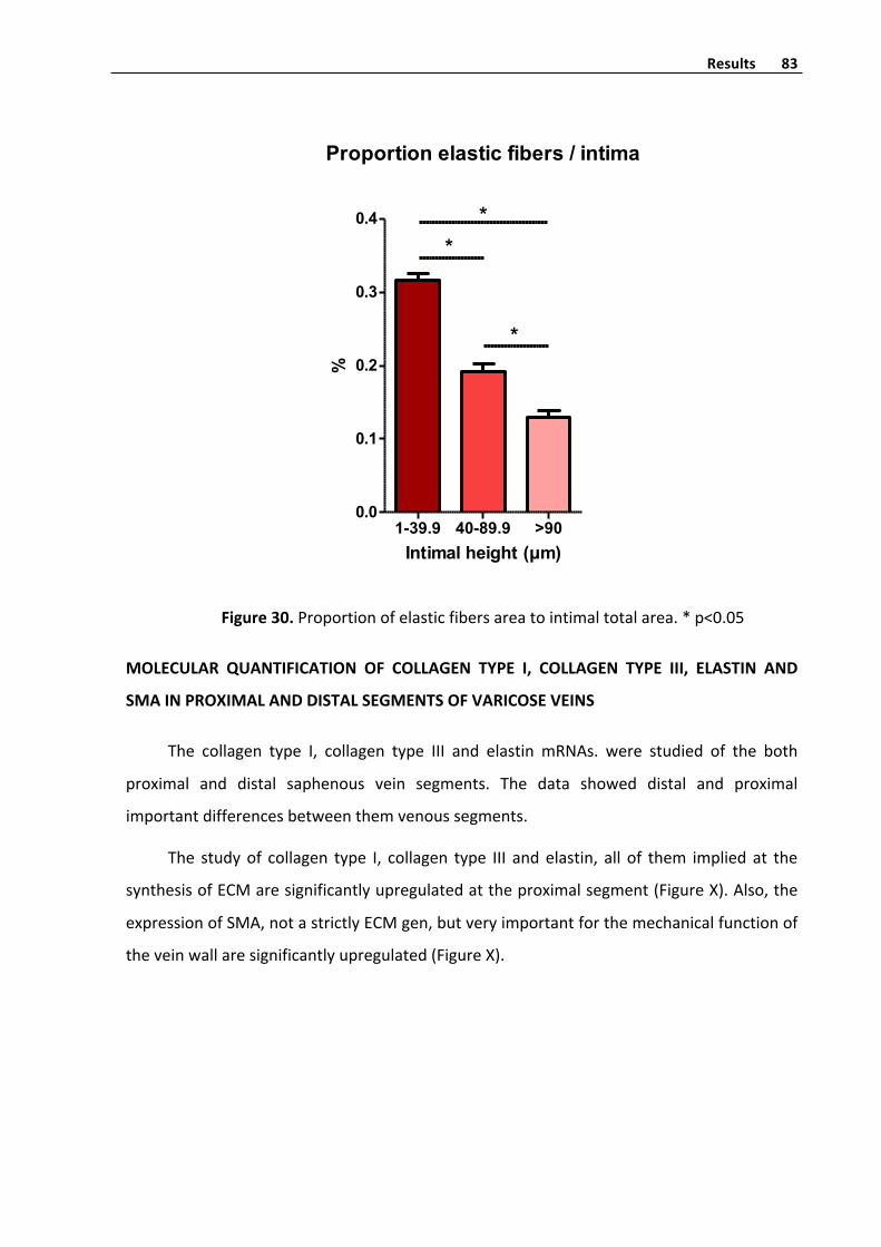
Results
83
Figure30.Proportionofelasticfibersareatointimaltotalarea.*p<0.05
MOLECULAR QUANTIFICATION OF COLLAGEN TYPE I, COLLAGEN TYPE III, ELASTIN AND
SMAINPROXIMALANDDISTALSEGMENTSOFVARICOSEVEINS
The collagen type I, collagen type III and elastin mRNAs. were studied of the both
proximal and distal saphenous vein segments. The data showed distal and proximal
importantdifferencesbetweenthemvenoussegments.
The studyof collagen type I, collagen type III andelastin, all of them implied at the
synthesisofECMaresignificantlyupregulatedattheproximalsegment(FigureX).Also,the
expressionofSMA,notastrictlyECMgen,butveryimportantforthemechanicalfunctionof
theveinwallaresignificantlyupregulated(FigureX).
Proportion elastic fibers / intima
1-39.9 40-89.9 >900.0
0.1
0.2
0.3
0.4
*
**
Intimal height (µm)
%
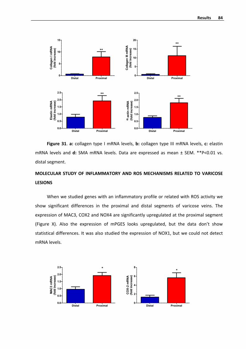
Results
84
Figure 31.a: collagen type ImRNA levels,b: collagen type IIImRNA levels,c:elastin
mRNA levels andd: SMAmRNA levels. Data are expressed asmean ± SEM. **P<0.01 vs.
distalsegment.
MOLECULARSTUDYOF INFLAMMATORYANDROSMECHANISMSRELATEDTOVARICOSE
LESIONS
WhenwestudiedgeneswithaninflammatoryprofileorrelatedwithROSactivitywe
show significant differences in the proximal and distal segments of varicose veins. The
expressionofMAC3,COX2andNOX4aresignificantlyupregulatedattheproximalsegment
(Figure X). Also the expression of mPGES looks upregulated, but the data don’t show
statisticaldifferences. ItwasalsostudiedtheexpressionofNOX1,butwecouldnotdetect
mRNAlevels.
Distal Proximal0
5
10
15
**C
olla
gen
I mR
NA
(fold
incr
ease
)
Distal Proximal0
5
10
15
20**
Col
lage
n III
mR
NA
(fold
incr
ease
)Distal Proximal
0.0
0.5
1.0
1.5
2.0
2.5 **
Elas
tin m
RN
A(fo
ld in
crea
se)
Distal Proximal0.0
0.5
1.0
1.5
2.0
2.5**
α-a
ctin
mR
NA(f
old
incr
ease
)
Distal Proximal0.0
0.5
1.0
1.5
2.0
2.5 *
MAC
3 m
RN
A(fo
ld in
crea
se)
Distal Proximal0
2
4
6
8*
CO
X-2
mR
NA
(fold
incr
ease
)

Results
85
Figure 32. a: MAC3 mRNA levels, b: COX2 mRNA levels, c: NOX4 mRNA levels, d:
mPGESmRNAlevels.Dataareexpressedasmean±SEM.*P<0.05vs.distalsegment.
In relationwithROSpathways, itwas also studied theH2O2productionbyAMPLEX‐
RED,due toNOX‐4 isoneof theenzymes impliesatH2O2production.Moreover,ourdata
showed a significant increase production of H2O2 at the proximal level compared to the
distalsegment(FigureX).
Finally, we studied the NAPDH oxidase activity due to NADPH oxidase generates
superoxide by transferring electrons from NADPH and can be implied at ROS stress. The
NADPHoxidaseactivitywassignificantlyhigherat theproximal segment thanat thedistal
segment(FigureX).
Figure 33.a:H2O2productionandb:NAPDHoxidaseactivity.Dataareexpressedas
mean±SEM.*P<0.05,**P<0.01vs.distalsegment.
Distal Proximal0.0
0.5
1.0
1.5
2.0
2.5 *N
OX-
4 m
RN
A(fo
ld in
crea
se)
Distal Proximal0.0
0.5
1.0
1.5
2.0
mPG
ES-1
mR
NA
(fold
incr
ease
)
Amplex Red
Distal Proximal0.0000
0.0002
0.0004
0.0006
0.0008*
Ampl
ex: µ
mol
H2O
2/µg
pro
tein
Distal Proximal0
1
2
3
4
**
NAD
PH o
xida
se a
ctiv
ity(fo
ld in
crea
se)

Discusión
86
DISCUSIÓN
Discusión
87
LapresenteTesissecentraenlavaloracióninmunohistoquímica,morfométricaymolecularde
las mecanismos iniciales involucrados en el desarrollo de varices en la extremidad inferior.
Estudiosprevioshanseñaladoquemuyprobablementelaenfermedadvaricosaseaelresultadode
alteracionesprimariasdelaparedvascularysecundariamenteaparezcaunainsuficienciavalvular,
determinandoambosprocesostrastornoshemodinámicos,conaumentodelapresiónhidrostática
intraluminaldebidaalreflujosanguíneo.19Sesabequelaaltapresiónsanguíneaesresponsablede
loscabiosmorfológicosyfuncionalesdelosvasossanguíneos,demodoquelaaltapresiónvenosa
causaríalaformacióndevenasvaricosasydelaICV.11
Descripcionesclásicashistológicasdelaslesionesvaricosasdelavenasafenasehancentrado
en la existencia de engrosamiento de la íntima, y alteraciones de la capamedia vascular. Estas
lesionessecaracterizanporlapresenciadehipertrofiaoatrofiadelasfibrasmusculareslisasdela
capamedia,ademásmuy frecuentementeseproduceen lacapamediaun incrementodeECM,
unaalteracióndelasfibraselásticasyunafibrosisintersticial;demodoquetodosestoscambios
determinanunaalteracióndeltonovenoso.74,225,289
Enpacientesconenfermedadvaricosabienestablecida,estudiosclínicosyhemodinámicosde
handemostradoque la formacióndevaricesesmuchomás frecuentey las lesiones sonmucho
más relevantes en el segmento proximal que en el segmento distal de la vena safena interna,
segmentoenelqueloscambioshemodinámicossonmenosimportantes.Comobiensabemos,la
principal fuente de reflujo en venas varicosas primarias en la unión safeno‐femoral o la unión
safeno‐poplítea, así que el reflujo puede ser considerado como un cambio de vía de la sangre
venosadesdeelmuslohacialaparteinferiordelapierna.227
Si bien los cambios hemodinámicos y las lesiones morfológicas elementales de las varices
estánbienestablecidas;sinembargo,ennuestroconocimiento,noexisteunestudiosistematizado
decomparacióndelaslesioneshistológicasinícialessituadasenelsegmentodistal,conrespectoa
laslesionesbienestablecidasdevaricessituadasenelsegmentoproximal.Porello,enlapresente
Tesissehaevaluadounaseriedevenassafenaextirpadasenpacientescon IVCcon lesionesde
enfermedad varicosas nomuy evolucionadas, pero en los que la flebectomía está clínicamente
indicada(pacientesenestadioCEAP2).91
Endefinitiva,esteestudionosólotieneuninterésacadémico,sinoque,aldefinirlospatrones
moleculares y morfológicos de las lesiones varicosas ayudara en el diseño de nuevos estudios
Discusión
88
clínicos,histopatológicosymoleculares invitro.Cabe laposibilidaddequeestas investigaciones
permitan la identificación de posibles dianas terapéuticas que eviten o disminuyan el daño
vascular, loqueredundaráenunbeneficioparaelpacientealpoderalterarelprocesoevolutivo
delaIVCyenunadisminucióndeloscostessanitarios.
En el presente estudio, hemos comparado los progresivos cambios histológicos que
experimenta la íntima vascular de la vena safena, desde los momentos iniciales con mínimo
engrosamientohastalaslesionesbienestablecidasdondeseproducenengrosamientosintímales
superioresa las200µmdeespesor. Seexcluyeronparaesteestudioaquellos casosen losque,
ademásdelalesiónintimal,aparecíanevidentesalteracionesdelacapamediavascular,incluidas
áreasdefibrosisintersticial;deestemodoalestarloscambioshistológicosmayoritariamenteenla
íntima, se pudieron comparar los datos inmunohistoquímicos, histológicos ymorfométricos con
loscambiosmolecularesdelaparedvaricosa,ycotejarestosdatosconelgradodeengrosamiento
intimal, esto es con las alteraciones de la ECM y de las células intimales de los segmentos
varicosos.Además,paraunconocimientoenmayorprofundidaddelaestructuraycomposiciónde
laparedvenosavaricosaseutilizoporprimeravezenpatologíavenosaunatécnicabasadaenla
refracción de las fibras de colágeno y la autofluorescencia de las fibras elásticas a través del
microscopioconfocalyasíestudiarladisposicióndelcolágenoydelasfibraselásticas.236
Enelmaterialestudiadohemosobservadoque,enlossegmentosdepareddevenasafenade
apariencia macroscópica normal, el estudio histológico detallado permite visualizar áreas de
íntima rigurosamente normal, revestidas por un endotelio plano, un tejido conjuntivo
subendotelialyunláminapropia,dondeelgrosordelacapaíntimaesmínimo;adyacentemente
sepuedenobservarpequeñas irregularidadesyengrosamientos intímales,caracterizadosporun
finodepósitode tejidoconjuntivoentreelendotelioy la láminaelástica interna.Enestasáreas
conmínimos cambios también sepuedenencontrar pequeñas fibrasmusculares lisas intimales,
dispuestaslongitudinalmente,lascualesexpresanSMAensuescasocitoplasma.
Amedidaquevaavanzandolaenfermedadvaricosa,laláminaíntimadelasvenascomienzaa
experimentarunengrosamientoylaestructuraycomposicióndeestasufrecambiosimportantes
y,enprincipionoreversibles.Enlossegmentosconunapatologíamásinicial,esdecir,enaquellos
conunaalturadelaíntimade40µmomenor,laproporcióndeáreaqueocupaelcolágenotipoI,
principalcolágenoestructuraldeltejidoconjuntivo intimal juntoalcolágenoIII,299 respectoesal
Discusión
89
área total de la íntima es de 0,24. Pero a medida que aumenta el engrosamiento intimal, la
cantidad de colágeno tipo I relativa aumenta, al principio muy rápidamente (grupo de altura
intimal entre 40 y 90 µm) hasta alcanzar una proporción 0,42 y luego en los segmentos más
comprometidosseestabilizaentornoal0,35.Esinteresantecompararestosdatosconloquele
sucedealotrocolágenomásabundanteenlasvenas,elcolágenotipoIII.299Observamoscomoen
condicionessincasipatologíaounapatologíainicial,laproporcióndeáreaqueocupaelcolágeno
esde0,42,manteniéndoseenelsiguientegrupodealturaenelmismorango(proporción0,43),
perodisminuyeenaquelloscasosconunalesiónbienestablecida.
Esinteresantedestacarque,sibienambostiposdecolágenoseexpresanentodalaalturade
laíntima,esmuchomásevidenteenlaporciónsuperficialeintermediadeloscasosconevidente
engrosamiento intimal. Además, la expresión de colágeno III es muy intensa en el tercio más
superficialdelengrosamiento intimal, formandounabandade tejido fibrosohomogéneoymuy
densodecolágenoIIIsobrelaqueseapoyaelendoteliovenoso(figura18y19),loqueimpideun
correcto intercambio de metabolitos en la pared venosa varicosa. Sin duda, estas alteraciones
determinancambioshemodinámicosirreversiblesdelflujovenosoenlospacientesconIVC.240
Asimismo,secuantificaronlosnivelesdeexpresióndemRNAparaelcolágenotipoIyelmRNA
elcolágenotipoIII,conelfindeobtenerdatosmolecularesdelapatologíavaricosadelasvenas.
Secomparólaexpresióndelosdoscolágenosentodalaparedvascularentreelsegmentodistaly
el proximal, ante la imposibilidad física de separar la íntima venosa de la túnica media. Esta
limitaciónnosimpideunacomparaciónocorrelacióndirectaconlosdatoshistomorfometricosde
las lesiones intimales. Pero sí nos permite una inferencia de la posible patogénesis de la
enfermedadvaricosa.Nuestrosdatosmolecularesmostraron,alcompararlaexpresióndemRNA
de colágeno tipo I y colágeno tipo III, una sobreexpresión de ambos tipos de colágenos en el
segmentoproximal,elmásdañadoenlaenfermedadvaricosa,conrespectoalsegmentodistal.
Se sabe que la proporción de los diferentes tipos de colágenos estructurales (sobre todo
colágenoIycolágenoIII)varíandeunórganoaotroy,sobretodo,cambiansustancialmenteen
situacionespatológicasrelacionasconmecanismosdefibrosisyreparacióntisular.136Enlasvenas
varicosas estudiadas, si comparamos los dos colágenos entre sí, los datos morfométricos
obtenidos nos demuestran una progresiva modificación de la ECM de la íntima vascular,
sustituyéndoseelcolágenoIIIporelcolágenoI.Estosdatoscoindicen,enparte,conlosobtenidos

Discusión
90
porXuJ.etal.2014299,quienestambiénencuentranunadisminucióndecolágenoIIIenlaíntima,
evaluada mediante técnicas histológicas. Además, el aumento de la expresión de mRNA de
colágenoIqueobservamosennuestromaterialesrefrendadoporelgrupodeSansilvestri‐Morel
P. et al. 2001241. Otros autores también describen un incremento en los niveles de mRNA de
colágenoI,peronoencuentrandiferenciasenlosnivelesdecolágenoIII.121
En este sentido, la actividad de síntesis de colágeno en la íntima venosa es distinta,
dependiendodelnúmeroydeladistribucióndelosfibroblastosydelosmiofibroblastosasociados
alasáreasdefibrosisintimal.Estudiosdecultivosdefibroblastosenratahanencontradoque,el
colágeno Iyel colágeno III inducen laproliferaciónde fibroblastos,mientrasqueel colágeno IV
induce la desdiferenciación de los fibroblastos en miofibroblastos.200 También, en el estudio
ultraestructuralquehemosrealizadodescribimoslapresenciaenlaíntimadeundepositoamorfo
de ECM y finos fascículos de colágeno y una irregular distribución de células fusiformes que
correspondenafibroblastosymiofibroblastos.Losfibroblastostienenunnúcleoelectrodensoyun
citoplasma con abundantes filamentos intermedios de vimentina; mientras que, los
miofibroblastos presentan pequeñas vesículas en vez de caveolas (lo que los diferencia de
verdaderos leiomiocitos)yabundantesprolongacionesdel citoplasmaquecontienennumerosos
miofilamentos relacionados con cuerpos densos y placas de anclaje, pero carecen de conos
sarcoplásmicos.Todosestoshallazgosultraestructuralessoncaracterísticosdelosmiofibroblastos.
Además,elcolágenoI,peronoelcolágenoIII,activaría lavíaERK1/2enfibroblastos.62Detodas
formas, según nuestros datos histológicos, la disminución de colágeno III y el aumento de
colágenoIprovocaríanunadisminucióndelratiodecolágenoIIIrespectoalcolágenoI,locualque
afectaría a determinaría un comportamientodistintode la respuesta en la pared venosas de la
resistencia a la tensión. Esta sustitución de colágeno III, que contribuye a la elasticidad, por
colágeno I, un colágeno más duro, provocaría mayor fibrosis y una mayor rigidez en la vena
varicosa. Como previamente han señalado Fan CM 200576, la síntesis anormal de colágeno
provocaría un debilitamiento y expansión de la base de las válvulas venosas, lo que muy
probablemente determinaría una aposición incompleta de estas y, por tanto, el consiguiente
reflujo, aunque las válvulas no tuvieran un especial daño. Está claro, pues, que todos estos
mecanismos apoyarían la hipótesis de la debilidad primaria de la pared venosa, frente a la
hipótesisvalvular.
Discusión
91
En la primera fase de engrosamiento intimal, tanto en los segmentos distales, como en los
proximales, se observa un progresivo incremento de SMCs SMA+, las cuales se disponen en
fascículos longitudinales, situados por dentro de la lámina elástica interna. En el segmento
proximal, cuando el engrosamiento intimal esmoderado, lamayor parte de la superficie de la
íntimaestáocupadaporcélulasSMA+,lascualesmuestransignosdehipertrofia.Sinembargo,a
medida que se va incrementándose el engrosamiento intimal, se observan SMCs atróficas
localizadas cerca del endotelio; mientras que otras SMCs son hipertróficas y se disponen en
posiciónmás basal. Asociados a estos cambios que experimentan los leiomiocitos de la íntima,
progresivamentesevaincrementandoeláreaocupadaporcolágenoIyIII,siendomásabúndateel
colágeno Ien lazonamássuperficialde la íntimamuyengrosada, formandounazonadetejido
fibrosomáscompactodebajodelendoteliovascular,talcomopreviamentehemoscomentando,
produciéndoserugosidadesirregularesdelasuperficieendovasculardelavenaymodificandolas
característicasdelflujosanguíneocirculante,talcomohandemostradoestudioshemodinámicos
previosenvenasafenasvaricosas.199
Enestos segmentos venosos conmínimos cambios intímales,nuestrosdatosmorfométricos
cuantitativos revelan que la superficie ocupada por todas las SMCs inmunomarcadas con
anticuerpoanti‐SMAesbaja,sisecomparaconlasuperficietotalintimal,evaluadaencadacampo
microscópico.Sinembargo,enesteestadiomásinicialdelesionessecundariasaIVC,cuandolos
datosseexpresanenvaloresrelativosdeláreaocupadaporlasSMCsenrelaciónconeláreatotal
intimal, se demuestra que aproximadamente el 29% del engrosamiento intimal corresponde a
SMCs;mientrasqueelrestodelasuperficieintimalestáocupadaporeldepósitodefinasláminas
elásticas, de material elástico granular y amorfo, y por la ECM virtual subendotelial.
Contrariamente, histológicamente hemos encontrado que en los casos de lesiones varicosas
completamente establecidas y con evidente engrosamiento intimal, se aprecia una importante
heterogeneidaddeltamaño,orientaciónydensidaddelasSMCspresentesenlaíntimavascular,
demodoque,existenáreasenlasquesevenabundantesleiomiocitoshipertróficos,conextensa
expresióndeSMA,ydispuestoslongitudinalmente,hastaotroscamposmicroscópicosenlosque
las SMCsSMA+ sonmenosabundantes, tienenescaso citoplasmayestán rodeadasporamplias
áreasdelaECMdeltejidoconjuntivo.Asimismo,elestudiomorfológicodeloscortessemifinosy
ultrafinos de la pared venosa varicosa y las células contenidas en ella realizado, nos permitió
Discusión
92
analizar con más profundidad los cambios de la íntima vascular. Es evidente en los cortes
semifinosquelascélulasobservadaspresentesenlaíntimapresentanenlaporciónmásapicalde
lamisma un núcleo redondeado, escaso citoplasma y están aisladas entre si.Mientras que, las
célulasintimalessituadasenlaporciónmásbasalpresentanunnúcleoconformadehusoymenor
cantidaddecitoplasma.Además,amedidaqueseengruesa la íntima, lacapadeECsempiezaa
desaparecer,yaparecencélulasconnúcleoshipertróficosyescasocitoplasma.Asimismo,lasSMCs
localizadas en la íntima sufrendesdiferenciación como señalan sumorfologíamásestrelladaen
lugardesuformaalargadamáshabitual.
Endefinitiva, amedidaque la lesiónvaricosaevolucionay la íntima seengrosa, seproduce
unaproliferación,entérminosabsolutos,deSMCs,aunquelaproporcióndeláreaintimalocupada
por laexpresiónSMAen la íntimavenosadecreceprogresivamente segúncrece la íntimahasta
alcanzarunárearelativadel16%.
Lasfibraselásticasdelaíntimatambiénexperimentancambiosmuyimportantesenrelación
con el progresivo engrosamiento intimal de los segmentos venosos con lesiones varicosas. En
nuestro conocimiento, después de una amplia revisión bibliográfica no hemos encontrado
estudiospreviosqueespecíficamentesecentrenenlasposiblesalteracionesdelasfibraselásticas,
tanto de la lámina elástica interna comode las fibras elásticas presentes en la ECMde la capa
íntima.Losdatoshistológicosobtenidosen lapresenteTesisdemuestranqueel incrementodel
material elásticos es muy evidente en la capa íntima, pero no se forman verdaderas fibras
elásticas,sinoqueelmaterialelásticodepositadoesmuyirregular,asociándosemuchasvecesla
rotura y desaparición de la lámina elástica interna de la pared venosa. El significado
histofisiológicodeestoshallazgosacercadeladegeneracióndelasfibraselásticasdelaíntimade
venasvaricosassondifícilesdeinterpretar,dadoqueenvenasnoexistenpublicacionesprevias,y
todoloquesesabedealteracióndefibraselásticasvascularessehaobtenidodesuestudioenel
territorio arterial, cuyas condiciones hemodinámicas y de tono vascular son bien diferentes al
lecho venoso.147,306 Está claro que las fibras elásticas han sido ampliamente estudiadas en el
sistema vascular arterial normal, sobre todo en segmentos de aorta y arterias musculares en
pacientes con arteriosclerosis5,85 y también en las arterias de fino calibre y arteriolas que
constituyen la microcirculación o en pacientes con diabetes mellitus222 en los que se han
encontradoalteracionesdelosmecanismosderemodelacióndelasfibraselásticas.

Discusión
93
Enrelaciónconlosmecanismosdeelastogénesis,sehademostradoque,enlaparedarterialy
también en la pared venosa, las fibrasmusculares lisas tienen la capacidad de sintetizar fibras
elásticas;14porello,laelastogénesisobservadaenlaslesionesintímalesdevenasvaricosasesun
procesoque,enlasfasesmásiniciales,suelerelacionarseconlahipertrofiadefibrasmusculares
lisasintímales,talcomosehademostradoenelpresentetrabajo.Sinduda,lasalteracionesdelas
fibras elásticas de la íntima venosa sonmuy importantes, ya que lo que se observa son fibras
elásticasmuycortasydelgadas,asociadasaundepósitodifusodematerialelásticoamorfo.Estos
cambios que experimentan las fibras elásticas en los segmentos venosos contrastan con los las
lesionesdescritaspreviamentedelafibraselásticasdelasarteriasmuscularesdepequeñocalibre
enmodelos exprímeteles de hipertensión arterial, en los que las láminas elásticas permanecen
circunferenciales concéntricas, pero lo que se incrementa es el tamaño de las fenestras.13 De
hecho,enlasarteriasderesistencia,estoes,enarteriasmuscularesfinasyarteriolasdepacientes
o de modelos experiméntales de hipertensión, se han demostrado lesiones degenerativas
irreversibles de las láminas elásticas de su pared.15 Estos hallazgos morfológicos de la fibras
elásticasenestasarteriascoinciden,enparte,conloquetambiénhemosencontradoenlaíntima
engrosadadevenasconvarices,enlasqueseveundepósitodematerialelásticoyfibrascortas,
tortuosasydefinocalibre.
Estos cambios de elastólisis de la lámina elástica interna a nivel de las lesiones de
engrosamiento intimal varicosoy sobre todoeldepositodematerialelásticodegenerandoyde
fibras entrecortadas y finas embebidas en la ECMamorfade la íntima son lesioneshistológicas
bastantesemejantesalosobservadasenotrosórganosdurantelosprocesosdegenerativosdelas
fibraselásticasamedidaqueavanza laedad.292,295Enefecto,enpacientesdemedianaedad,en
losquelaIVCesfrecuente,ysobretodoenlasenectudaparecencambiosdelaorganizaciónde
lasfibraselásticasdemúltiplesórganos(piel,vasossanguíneos,tejidoelásticoasociadoalmúsculo
visceral, etc.), provocándose una elastólisis progresiva, caracterizada ultraestructuralmente por
una degeneración del centro claro de elastina, la cual puede contener depósito de material
electrodenso muy irregular, y evidente disminución del manto periférico de fibrillas.210 Estos
cambios determinan el entrecortamiento de las fibras elásticas, las cuales están rotas,
dicotomizadasyfrecuentementeestánsustituidasporunmaterialelásticodeaspectogranular.210
Discusión
94
Así, losdatosdenuestroestudiomorfométricode lasuperficiede la íntimaocupadapor las
fibraselásticasdemuestranque,amedidaqueaumentalaalturadelacapaíntimalaproporción
de las fibras elásticas disminuye. En el primer grupo conmínimo engrosamiento intimal, estas
fibraselásticasocupanun32%deláreadelacapaíntima,disminuyendosignificativamenteenel
segundogrupo,representandosoloel19%delasuperficieintimal.Además,lacantidaddefibras
elásticas desciende muy notablemente en el grupo de lesión intensa, con alturas de la íntima
superioresa las90µm;eneste caso losdatosmorfométricosquehemosobtenidodemuestran
que las fibras elásticas solo representan un 13%. Aun así, en estos casos de importante
engrosamiento intimal, las fibras elásticas sintetizadasde novo estánmuy desorganizadas y no
forman láminas circunferenciales, sino que se ven pequeños discos de fibras de elastina y un
materialamorfodistribuidoirregularmenteporlaECM.Dehecho,elgrupodeXuN.etal.2014,300
utilizando una tinción de van‐Gieson orceína, una técnica histológica similar a la usada por
nosotros, encuentran una obvia disminución, fragmentación y desestructuración de las fibras
elásticasenvenasvaricosas.
En la presenta Tesis hemos efectuado un estudio mediante microscopia confocal de las
lesionesintímalesdelavena,conelfindeevaluarladisposiciónyrelaciónentrelasfibraselásticas
ylasfibrasdecolágeno.Paraello,hemosobtenidopequeñossegmentosdelaporciónproximaly
distaldelavena,loscualeshansidofijadosintotoorientandolasuperficieendovascular,queesel
primer estrato sobre el que incidió el rayo láser del microscopio confocal. En nuestro
conocimiento, esta metodología no ha sido previamente usada en otros estudios de la
histopatologíavenosa.Nuestrosresultadosmuestranunaíntimarelaciónentreelmaterialelástico
depositadoenla íntimaengrosaday lasfibrasdecolágenoy laECM.Cuandoseevalúaunúnico
plano,mediantemicroscopiaconfocal,sedemuestraque la fibrasdecolágenosonmuycortasy
delgadas y el material elástico aparece como un deposito granular amorfo y más
infrecuentementecomopequeñasfibraselásticasautorefringentes.Estoshallazgos,enparte,son
semejantes a las áreas de fibrosis intimal en modelos experimentales de arterioesclerosis,
evaluados también in toto por microscopia confocal; sin embargo, en estos modelos de
ateroesclerosis,lasplacasdeateromacontienenlípidosycélulasinflamatorias.236
Paracomprendermejor losmecanismos íntimosqueexperimenta laparedvenosayadesde
losprimerosestadiosdeldesarrollodelaenfermedadvaricosaesconvenientecotejarloscambios
Discusión
95
hemodinámicosconlaslesioneshistológicasdelaíntimadescritasenelpresenteestudio.Resulta
verosímilsuponerquecronobiológicamenteloprimeroqueseproduzcaseanpequeñoscambios
hemodinámicos,pequeñasturbulenciasdelflujovenososendeterminadospuntosdelasuperficie
endovascular, como consecuencia de las lesiones de engrosamiento mínimo intimal; estas
pequeñas lesiones iniciales también se pueden observar en las zonas de la íntima en donde se
implanta la basede las valvasde las numerosas válvulas que tiene la vena safena interna. Este
reflujovenoso,asícomoeléstasissanguíneoasociado,determinanun incrementode lapresión
hidrostáticaque,asuvez,determinaunaumentodelaincompetenciadelasválvulasvenosas.
Comorespuestaaestoscambiosfuncionales,enlosprimerosmomentosdeldesarrollodela
enfermedadvaricosalasSMCsdelaparedexperimentanunaumentodeltonovascular,locualva
acompañado de una hiperplasia de SMCs en la íntima vascular, aunque la proporción de SMA
disminuyasegúnaumentalaalturadelacapaíntima.Estahiperplasiacelularenlatúnicaíntima,
como ya hemos dicho, se acompaña de un incremento también progresivo de la superficie
ocupadaporcolágenosIy III,aumentandolaproporcióndecolágenoIsegúnaumenta laaltura,
mientrasqueel colágeno III aumentaenunprincipiopara luegoestabilizarse.Además,aparece
una disminución en la proporción de por fibras elásticas a medida que aumenta la íntima
fibrosada. Esta correlación morfométrica que hemos encontrado cuando evaluamos
histológicamente laexpresióndediferentesmoléculasen la íntimavaricosaes lógica,yaquese
sabe que las SMCs son capaces de sintetizar colágenos, degradar elastina y participar en los
mecanismosdeelastogénesisyremodeladovascular.36,129,287
Además,estosdatosestánenconsonanciaconestudios invitroquehanencontradoquelas
SMCsderivadasdevenassafenasvaricosasestánmásdesdiferenciadasymuestranunaumentode
lacapacidadproliferativaydesíntesisdeproteínas,encomparaciónconlaobservadaenlasSMCs
devenasnormales,pasandodeunfenotipocontráctilaunosecretor.164Sehasugeridoqueestos
mecanismospudierancontribuira la remodelaciónde laparedvenosayaldebilitamientodesu
capacidadfuncional,debidoalestrésmecánicoquesufrelavenasafenaenpacientesconIVC.74,199
Apesardelosestudiosrealizadoscentradosenmecanismosimplicadosenproliferaciónenvenas
safenas,16,71 no se conocen losmecanismos íntimosdeestaproliferaciónydiferenciaciónde las
SMCsintímales.35,50,93,97
Discusión
96
Estudiosrecienteshandemostradolaparticipacióndelendoteliovascularenlaregulaciónde
mecanismos de estrés oxidativo frente a alteraciones locales del flujo hemodinámico y de la
presión intraluminal, los cuales pueden modificar la composición de la pared venosa.147,306
MoléculasROSrelacionadasconelestrésoxidativoesposiblequepudieranregularlaproliferación
y diferenciación de células intímales de la pared venosa, las cuales se transformarían en
miofibroblastos y SMCs, presentes en las fases iniciales de la fibrosis intimal varicosa, tal como
hemos demostrado inmunohistoquímica y ultraestructuralmente en la presente Tesis. Pero
también cabe especular que la presencia de SMCs SMA+ encontradas en la íntima engrosada
procedieranenpartedelamigracióndelosleiomiocitosdelatúnicamediavascular.
Está establecidoque las fuerzas biomecánicas son, de unaparte, la fuerza de fricciónde la
sangrequeactúasobreelendotelioy,deotra,latensióncircunferencialqueactúatantosobreel
endotelio,comosobrelasSMCsdelaíntimaydelamediaysobrelosfibroblastosdelaadventicia
CITAS. Estas fuerzas biomecánicas inducen el inicio de mecanismos de inflamación, como
consecuencia de la interacción entre el endotelio y los leucocitos circulantes. Es más, estas
modificaciones del tono vascular producen cambios de señalización de las ECs y liberación de
moléculasbioactivashaciael tejidoconjuntivo intimal.190,199Sinduda,estoscambiosfuncionales
queejercenmodificaciones inicialesdel flujoyde lapresión intraluminalposiblemente sean los
que desencadenan los primeros cambios de la ECM de la íntima venosa (tal como se ha
demostradoenlaíntimaenmodelosdehipertensiónarterial,156,157ysubsecuentementeel inicio
del incremento del espesor intimal y la diferenciación de SMCs SMA+. Por ello, ya desde estas
fases inicialessubclínicasde IVCseproduceunaprogresivadesestructuraciónmorfológicayuna
modificaciónmoleculardelossegmentosdeparedvenosaconlesionesvaricosas.147,306
Esposiblequemínimoscambioshemodinámicoslocales,derivadosdeléstasisvenosoydela
hipertensiónsecundaria, seansuficientesparaprovocarunestrésen laparedde lavenasafena
varicosa, lo cual pudieran determinar cambios fisiopatológicos que den lugar al inicio de
mecanismosderemodelacióndelatúnicaíntima.Sesabequeestoscambioshemodinámicosdan
lugar a unas fuerzas biomecánicas determinadas por el flujo sanguíneo, la presión intra y
extraluminal y las diferentes cargas tensiónales que actúan en cada segmento venoso
comprendidoentredosválvulas;todosestosmecanismossonlosresponsablesdequelaslesiones
varicosasalolargodelamismavenaseanmultifocalesydediferenteintensidad.274Laexposición

Discusión
97
a altas presiones en el extremo inferior de la pierna provocaría cambios en la propiedades
estructuralesyvenosasqueconllevatodounremodelamientovascular.Enesteremodelamiento
vascularestáimplicadalamigración,proliferaciónyapoptosisdeECsySMCs,asícomolasíntesisy
degradacióndeECM.156
Porotro lado, en la formaciónde la neoíntima, tantoen las patologías arteriales y venosas
primarias y también en los injertos vasculares autólogos,230 intervienen procesos de óxido
reducción, dando lugar a una activación de mecanismos enzimáticos que regulan facilitando o
inhibiendo la formacióndedichaneoíntima.230Lasalteracionesde laparedvenosaenpacientes
conIVCpuedenestardesencadenadaspormecanismosdeestrésoxidativo,loscuales,talcomose
ha demostrado en arterias de elevada resistencia, determinarían alteraciones estructurales y
moleculares de las fibras elásticas, que dificultarían los mecanismos de reparación y de
remodelaciónvascular.56
Enrelaciónconelestrésoxidativo,lasECsparticipanenlaregulacióndeROSydeestemodo
participan en los mecanismos de tipo inflamatorio que experimentan las ECs y las SMCs de la
pared vascular.106 Está demostrado que en los pacientes con IVC se produce un estado de
hipertensión vascular venosa, secundario al estasis venoso, lo cual puede desencadenar
mecanismos proinflamatorios y liberación de interleucinas, tal y como se ha señalado en otras
situacionesdealteracioneshemodinámicasvascularesquecursanconhipertensión.6Portanto,el
endotelio experimenta cambios funcionales y estructurales secundarios al estrés oxidativo y al
procesoinflamatorio.
Estasalteracionesendotelialeshansidoampliamenteestudiadasen las fases inicialesde las
lesionesateroscleróticasytambiénenlosmecanismosdeendotelizacióndelaneoíntimapresente
en los injertos vasculares;294 así mismo, en modelos experimentales de hipertensión venosa
crónica(hipertensiónportal)sehademostradoeldesencadenamientodeunestrésoxidativodela
paredvascular.65
En nuestro conocimiento la pormenorizada revisión de la literatura que realizamos en el
momento de comenzar la presente Tesis no existían estudios previos que compararan las
alteraciones histológicas y cuantitativas de la íntima de las venas varicosa con métodos
molecularesqueexploraranmecanismosdeestrésoxidativoyde inflamación.Porello,creemos
que el planteamiento de objetivos de la presente Tesis en los que se cotejan los hallazgos

Discusión
98
histológicos con datosmoleculares de estrés oxidativo y demecanismos inmunológicos aporta
nuevosconocimientosenlahistofisiologíadelaslesionesinicialesdelaenfermedadvaricosa.De
hecho,eléstasisvenosoenestospacientes,yadesdelosprimerosestadiosdeldesarrollodeIVC,
producedañoendotelial,aumentodeROSy liberacióndemoléculasproinflamatorias, lascuales
tienenlacapacidaddeatraerhacialaparedvenosalesionadamonocitosymacrófagos,capacesde
degradarlaECM.
EnnuestroestudiohemosencontradounasignificativamayorexpresióndemRNAdeNOX‐4,
así comounamayor producción deH2O2 y, asimismo, una significativamayor actividadNADPH
oxidasaenelsegmentovenosoproximal,conevidenteslesionesintimalesdevarices,conrespecto
alsegmentodistaldelavenasafena,quehabitualmentepresentamínimaslesiones.Sinembargo,
nos encontramos expresión demRNA de NOX‐1 en las muestras de venas safenas que hemos
estudiado.Enestudios realizadosenarterias,de las7 isoformasdeNOXconocidas, seexpresan
NOX1,NOX2,NOX4yNOX5.37,48LaexpresióndeNOX‐4essignificativamentemayorquelasotras
isoformas en arterias.37,191 Con todo, consideramos que nuestros datos de mRNA de NOX‐4,
produccióndeH2O2yactividadNADPHoxidasasondegraninterés,yaquelasobreexpresiónenel
segmentoproximaldeNOX‐4,unadelasprincipalesenzimasimplicadasenlaproduccióndeH2O2,
secorrelacionaconunamayorproduccióndeH2O2yconunasignificativamayoractividadNADPH
oxidasa, loquesugiereunmayorestrésoxidativoenel segmentoproximalqueenel segmento
distaldelasvenasvaricosas.Enestesentido,estudiosprevioshandemostradoquelospacientes
conIVCpadecenunincrementoenelestrésoxidativo,44,140aunquenolorelacionanconelgrado
de alteraciones de la pared venosa. Estosmecanismos sugeridos en la regulación de la fibrosis
progresiva de la íntima venosa pueden ser verosímiles, dado que está comprobado que la
activacióndeNADPHoxidasaincrementalaproduccióndeROSprocedentedelasmitocondrias,y
viceversa, que las ROS procedentes de la mitocondria son capaces de activar NADPH oxidasa,
especialmenteencondicionespatológicas;67además, lasROSmitocondrialesy laNADPHoxidasa
participan en la regulación de la expresión y actividad de la xantina oxidasa247 y el
desacoplamientodeeNOSenlasECs.247TambiénelH2O2escapazdeactivarlaNADPHoxidasay
provocarunaumentodelaproduccióndeO2‐,además,elH2O2escapazdeinducirlatransiciónde
laxantinadeshidrogenasaenxantinaoxidasa,queescapazdegenerartantoH2O2comoO2‐.,185lo
cualdeterminaaúnunmayordañocelular.82
Discusión
99
Se sabe que la enzima COX‐2 responde a estímulos pro‐inflamatorios catalizando el primer
pasodelabiosíntesisdeprostanoides:prostaglandinasytromboxano,siendolosmacrófagosyECs
la fuente principal de las dos isoformas de COX en la IVC.1 En los tejidos de la pared venosa
varicosaquehemosevaluadosencontramosqueCOX‐2estásignificativamentesobreexpresadaen
elsegmentoproximalvenoso,tambiénlaexpresióndemRNAdem‐PGES‐1tiendeasermayoren
el segmento proximal que en el segmento distal de las venas estudiadas, aunque el análisis
estadístico no encontró diferencias significativas. Estudios recientes de otros autores han
establecidoquelaformainducibledelaciclooxigenasaestradicionalmenteconsideradacomoun
biomarcadortempranodeinflamación.238Además,laactividaddelaperoxidasadelaCOXcataliza
lageneracióndeunproductointermedioinestable,laprostaglandina(PG)H2.Unaprostaglandina
sintasaespecifica,lamPGES,conviertePGH2aPGE2,queeslamásabundanteprostaglandinapro‐
inflamatoriaasociadaencondicionesinflamatorias.Algunosestudioshandemostradoelpapelde
PGE2enlaexpresión/activacióndemetaloproteinasasentejidoshumanos,fundamentalmenteen
el estudio de SMCs provenientes de aorta,304 pero en nuestro conocimiento no han sido
estudiadasenlaparedvenosavaricosa.
Al igualqueennuestroestudio,el grupodeBertrand‐ThiebaultC.etal. 200429 identifica la
expresión de COX‐2mRNA en venas varicosas. Sin embargo, en una publicación reciente se ha
señalado la ausencia de COX‐2 en venas varicosas.95 Aunque la expresión de COX‐2 ha sido
considerada un importante factor de la citotoxicidad asociada con inflamación,250 los factores
responsablesdelacitotoxicidaddeCOX‐2nohansidocompletamentedefinidos.Eneldañotisular
presente en las venas varicosas, podría estar implicada la producción de prostanoides pro‐
inflamatorios,250 de modos que la producción de ROS y su relación con mecanismos de
peroxidación, cuando PGG2 es convertida en PGH2,278 aumentarían los radicales libres que
inducirían apoptosis y daño tisular crónico.107 Estos mecanismos también pudieran estar
potenciadosporlarelaciónentreCOX‐2ylasNADPHoxidasas,yaquelasNADPHoxidasaspueden
ser una fuente de ROS, mediado por la sobrerregulación de COX‐2;160 existiendo además una
regulaciónrecíprocaentreproteínasNOXyCOX‐2.237
Teniendo en cuenta nuestros resultados, el desencadenamiento de mecanismos pro‐
inflamatorios, el aumento de ROS y la mayor expresión de COX‐2 podrían explicar la
sobreexpresión que encontramos de MAC‐3 (una proteína presente en el fagosoma de

Discusión
100
macrófagos251 en el segmento proximal de las venas varicosas. Nuestros datos están en
consonanciaconestudiospreviosquehandescritolapresenciade infiltradosdemacrófagosen
lasválvulasdevenasvaricosasenlapareddevenasvaricosas.244
Una vez que se desencadenan estos mecanismos oxidativos e inmunológicos en la pared
venosa,secundariosaléstasissanguíneo,comienzaelincrementodelaECMenlaíntimavascular,
talcomohemosdemostradoenlapresenteTesis,dandoorigenaunahiperplasiadeSMCsyauna
progresivafibrosisintimalcondepósitodefibraselásticasyconincrementodecolágenotipoIy
colágeno tipo III mucho mayor en la íntima del segmento proximal que del distal. Como
previamente se ha comentado, todos estos hallazgos histológicos han sido corroborados en
nuestroestudiointotodelaslesionesendovascularesrealizadomediantemicroscopiaconfocal.
LaexpresióndemRNAdeSMA,elastina,colágenoIycolágenoIIIestásobreexpresadoenlos
segmentosproximales,dondelalesiónesmásimportanteyexisteunaproliferacióndeSMCsenla
capa íntima. Estos resultados coinciden con las observaciones realizadas por el grupo Cario‐
Toumaniantz C. et al. 200744, que mediante RT‐PCR también encuentran un aumento en la
expresióndecolágenoIycolágenoIIIenmuestrasdetejidodevenasvaricosas,comparadocon
venas normales, aunque en cultivos de SMC procedentes de venas varicosas solo el mRNA de
colágenotipoIseencuentrasobreexpresado,mientrasqueelcolágenoIIInomuestradiferencias.
Comoessabido,lasSMCstienelacapacidadderemodelarelcolágenodelamatrizextracelular.281
También el estudio realizado por Lee S. et al. 2005154 muestra sobreexpresión de genes
relacionadosconmoléculasdelaECM.Sinembargo,otrosgruposmuestranunadisminuciónenel
contenido de proteínas colágeno III,mientras que elmRNAde colágeno III no está alterado en
cultivosdeSMCyfibroblastosdérmicosderivadosdepacientesconvenasvaricosas.241
En definitiva, en la presente Tesis se ha demostrado que existen cambios
inmunohistoquímicosycuantitativosdelaproporcióndeSMA,colágenotipoI,colágenotipoIIIy
fibras elásticas a medida que aumenta el engrosamiento de la íntima de la vena safena en
pacientesconIVC.Estoscambiosseasociantambiénconunincrementodelaelastogénesisdela
íntimavenosa,aunquelasuperficieocupadaporlasfibraselásticasenrelaciónalasuperficietotal
de la íntima disminuye a medida que aumenta el engrosamiento intimal. Todos estos datos
sugieren que las lesiones de fibrosis intimal en las venas varicosas son irreversibles cuando
progresaelengrosamientointimalenpacientesconvarices.
Discusión
101
Nuestrosresultadosindicanqueesenlasfasesiniciales,cuandoelengrosamientoesmínimo,
elmomentodeexplorarnuevosmecanismosonuevasmoléculasqueeviten laprogresiónde la
enfermedad. Hasta elmomento presente no se han encontrado tratamientosmédicos idóneos
paraprevenir la IVCypara impediroremodelar loscambios iniciales intímales,aunqueestudios
comoel nuestromuestran la implicación demecanismos de estrés oxidativo e inflamación que
podríanayudarenlabúsquedadedianasterapéuticasqueeviteneliniciodelalesiónvaricosa.
Así mismo, es necesario diseñar nuevas investigaciones de correlación de los cambios
histopatológicosydelosmecanismosmolecularesconestudioshemodinámicosyfuncionalesde
las venas con diferentes grados de enfermedad varicosa; sin embargo, estas correlaciones
morfofuncionales han sido ampliamente evaluadas en el territorio arterial. En este sentido,
aunqueenlasarteriassehaestudiadoelefectoquetienenlasfuerzasbiomecánicasproducidas
por las alteraciones hemodinámicas, aún son pocos los trabajos en venas, pero es sugestivo
considerarqueelfalloenlarespuestamiogénicasecundarioaalteracioneshemodinámicastiene
unpapelimportanteeneldesarrollodelasvenasvaricosas.
Aunasí,esteestudiopresenta limitacionespotenciales.Elestudiohistológicode laparedde
venas completamente normales enmaterial humano es complicado, dado que la obtención de
muestrasenpacientessanosnoeséticamenteposible.Loquesíesposible,unavezobtenidoel
consentimientoinformadoparainvestigación,esestudiarsegmentosdevenasafenaprocedentes
de pacientes sometidos a cirugía coronaria que no son utilizados en el bypass coronario.122,137
Estos segmentos podrían ser en el futuro una fuente de material del grupo Control para
desarrollar nuevos estudios histológicos y moleculares relacionados con los mecanismos de
formación de varices. Por último, comomaterial control también podrían ser usados pequeños
segmentos de vena aparentemente normal que sobran en la realización de fístulas arterio‐
venosas, como acceso vascular para los tratamientos de diálisis o aféresis, o de vena safena
aparentemente normal no empleada en bypass de extremidades o en revascularización
coronaria.217
Endefinitiva,losdatosobtenidosenlapresenteTesissugierenquelaactivacióndemoléculas
relacionadas con mecanismos de estrés oxidativo desencadenan el inicio de procesos
proinflamatoriosquedeterminancambiosenlaECM,produciéndoseunprogresivoaumentodela
ECM,condepósitoenlaíntimadeelastina,eincrementodelratiocolágenoI/colágenoIII,loque

Discusión
102
origina una fibrosis intimal intensa e irreversible que provoca irregularidades en la superficie
luminal,condisminucióndeldiámetrodeluz.Estaslesionesintímalesydelasuperficieluminalse
podríancorrelacionarconlasimportantesalteracioneshemodinámicasdelflujovascular;demodo
que,progresivamentesepotenciaríalaformacióndelesionesvaricosascrónicasatodololargode
todalavenasafena.

Conclusions
104
CONCLUSIONS
Conclusions
105
CONCLUSIONS
FIRST. The saphenousvaricosevein showsaprogressive intimal thickeningwithmoderate
muscular atrophy at the medial layer. The intima presents a collagen I and collagen III
deposition and disorganized elastic fibers. Also, SMA, elastin, collagen I and collagen III
mRNA are upregulated at the proximal segment respect to the distal segment. However,
whentheintimalrises,theproportionofcollagenIrisestoo,buttheproportionofcollagen
III,SMAandelasticfibersdecrease.
SECOND.TheintimallayerfromvaricoseveinshasSMApositiveSMCsandothersellipsoidal
shape cells that are vimentin and SMA positive and their ultrastructure correspond to
myofibroblast.
THIRD. Intimal fibrosis studied in toto by confocalmicroscopy showan amorphous elastic
materialdepositionandthinelasticfiberssurrenderbycollagenfibers.
FOURTH.Theproximalsegmenthasahigheroxidativestress thanthedistal segment.The
proximalsegmenthasahigherH2O2production,NADPHoxidaseactivityandNOX‐4mRNAis
upregulated.
FIFTH.Ourdatademonstratedthedevelopofstressoxidativemechanisminrelationtothe
fibrosisoftheintimallayerofvaricosesaphenousvein.Theprogressivedepositofcollagen
andelasticfibersdeterminateanintensefibrosisoftheintimalsurface.

Conclusiones
106
CONCLUSIONES

Conclusiones
107
CONCLUSIONES
PRIMERA.Lavenasafenavaricosapresentaunaumentoprogresivodelatúnicaíntimacon
moderada atrofia de la capa muscular media. En la íntima se observa el depósito de
colágeno I, colágeno III y de fibras elásticas desorganizadas. La cuantificación de mRNA
muestra un aumento de SMA; elastina, colágeno I y colágeno III en el extremo venoso
proximalrespectoaldistal.Además,morfometricamentesedemuestraque,amedidaque
aumenta la superficie intimal, también aumenta la proporción de colágeno I, aunque
disminuyelaproporcióndecolágenoIII,SMAyfibraselásticas.
SEGUNDA. En la lesión varicosa intimal se identifican SMCs SMA positivas y acúmulos de
células fusiformes vimentina y SMA positivas, que ultraestructuralmente corresponden a
miofibroblastos.
TERCERA.Lalesióndefibrosisintimalsehaestudiadointotomediantemicroscopiaconfocal
evidenciándose una estrecha relación entre el depósito de material elástico amorfo y
pequeñasfibraselásticasrodeadasporfibrasdecolágeno.
CUARTA. Existe un mayor estrés oxidativo en el extremo proximal que en el distal,
evidenciándoseunasignificativamayorproduccióndeH2O2,un incrementode laactividad
NADPHoxidasaylasobreexpresióndeNOX‐4.
QUINTA.Nuestrasdatosdemuestranel desarrollodemecanismosdeestressoxidativoen
relaciónconfibrosisdelacapaíntimadelasvenassafenasvaricosas.Eldepósitoprogresivo
defibrasdecolágenoyelásticasdetermianunafibrosisintensadelasuperficieintimal.

Resumen
108
RESUMEN
Resumen
109
LaInsuficienciaVenosaCrónica(IVC)delasextremidadesinferioresesunaenfermedad
muyfrecuentequecausaunagranmorbilidadenpersonasdemedianayavanzadaedad.Los
mecanismos fisiopatológicos y los datos hemodínamicos de esta enfermedad están bien
establecidos.Tambiénseconocenlaslesionesgeneralesdefibrosisdelacapaíntimaydela
capa media de las venas con varices. Sin embargo, la histogénesis y los mecanismos de
progresión de las lesiones de varices no están completamente establecidos y tampoco se
hanevaluadopormenorizadamentelasalteracionesmorfométricas,celularesymoleculares
a nivel del segmento proximal (con evidente lesión varicosa) y del segmento distal (con
menorlesiónvaricosa).
En la presente Tesis realizamos un estudio morfométrico de la expresión
inmunohistoquímica de alfa actinamuscular (SMA), colágeno tipo I, colágeno tipo III y de
fibraselásticasenlosdiferentesgradosdeengrosamientointimal.Asimismo,seharealizado
unavaloracióndesegmentosdevenaestudiadosintotomediantemicroscopiaconfocal,con
el fin de establecer las relaciones de las fibras elásticas y del colágeno en la porción
superficial de la íntima lesionada, método que no ha sido aplicado previamente en la
investigacióndevenas.Tambiénseharealizadounestudioultraestructuralparavalorar la
matrizextracelular(ECM)y laproliferacióncelularde la íntima lesionada.Porúltimoseha
estudiadoladiferenteexpresióngénicademoléculasimplicadasenprocesosdeinflamación,
ROSydesíntesisdecolágeno,tantoenelsegmentoproximalcomoeneldistaldelasvenas
varicosas.
Se estudiaron 20 venas safenas de pacientes en estadio clínico CEAP 2. Las venas
safenas fueron extirpadas e inmediatamente procesadas dentro del quirófano para la
realización de los experimentos histopatológicos y moleculares. Se usaron anticuerpos
monoclonales para identificar inmunohistoquímicamente SMA, colágeno tipo I, colágeno
tipo III y vimentina, así como el uso de tinción de orceina para la detección de fibras
elásticas. La cuantificacióndeestasmoléculas se realizómedianteelprograma ImageJ. Se
obtuvieronpequeñosfragmentosparaelestudiodemicroscopiaelectrónicadetransmisión.
También pequeños segmentos de la región proximal y distal, después de fijarse, se
estudiaronintotopormicroscopiaconfocal,identificandolacapaendoluminal,paravalorar
Resumen
110
las fibras elásticas y de colágenode la porción superficial de la capa íntima. Losmétodos
molecularesqueserealizaronfueronRT‐PCR,lamedidadelaactividadNADPHoxidasayde
laproduccióndeperóxidodehidrogenomedianteAmplexRed.
Nuestrosresultadosdemuestranqueelengrosamientointimalenlasáreasconlesión
varicosaesprogresivoysedebefundamentalmentealdepósitodeECMycolágenotipoIy
III. También existe un depósito de material elástico, pero la presencia de fibras elásticas
completasesmínima.Enlaíntima,hemosidentificadocélulasproliferantesvimentinaySMA
positivas,yhemosdemostradoqueultraestructuralmentecorrespondenamiofibroblastos.
Losdatosdelacuantificacióndelasuperficieinmunomarcadaconelanticuerpoanti‐
colágeno Ipermitencomprobarque,enunprimermomentodeengrosamiento intimal, la
proporcióndecolágenoIaumentaenelgrupoconlesiónintimalvaricosamoderada(39,9a
89,9 µm) y luego se estabiliza en torno al 35% en le grupo con una mayor lesión. La
morfometríarealizadaparacuantificarlasuperficieintimalocupadaporcolágenoIIImuestra
unaproporciónentornoal40%enlosdosprimerosgrupos(engrosamientointimaliniciale
intermedio), para descender bruscamente en el tercer grupo de engrosamiento intimal
intensohastavaloresentornoal30%.
El estudio morfométrico de la superficie marcada inmunohistoquímicamente con
anticuerposanti‐SMAdemuestraquelasfibrasmusculareslisasdelacapaíntimaaumentaa
medida que se produce el engrosamiento intimal. Sin embargo, cuando se calcula la
proporcióndel área SMA+, con respecto a la superficie total de la íntima, seobservauna
progresivadisminucióndelmúsculolisoenlosgruposconmayorengrosamientointimal.
La morfometría de las fibras elásticas orceína positivas muestra que la superficie
ocupada por las fibras elásticas va aumentando progresivamente y paralelamente al
incrementodelasuperficiedelaíntima.Sinembargo,apesardeldesarrollodeesteproceso
de elastogénesis intimal, la proporción que ocupan estas fibras elásticas en la íntima,
respectoaláreatotalintimal,sufreunaprogresivaysignificativadisminuciónamedidaque
aumentalahiperplasiaintimal.
Resumen
111
En la presente Tesis hemos evaluado por métodos moleculares las alteraciones
existentestantoenelsegmentodistalcomoenelsegmentoproximaldevenassafenascon
lesionesvaricosas,explorandoalgunasmoléculasimplicadasenmecanismosdeinflamación
yestrésoxidativo.Enprimerlugar,secuantificoporRT‐PCRlaexpresióndecolágenotipoI,
colágenotipoIII,elastinaySMA.Estosgenesestánsignificativamentesobreexpresadosenel
segmento proximal respecto al distal. También se estudio la expresión de MAC3, COX2,
NOX4,NOX1ymPGES.NuestrosdatosdemuestranqueelmRNAparaMAC3,COX2yNOX4
seencuentrasignificativamentesobreexpresadoenelextremoproximal.Sinembargo,nose
encontrarondiferenciassignificativasenelmRNAparamPGES‐1ynosedetectóexpresión
paraNOX1(Figura32).Porúltimo,seestudiótambiénladiferenteproduccióndeperóxido
dehidrógeno y la actividadNADPHoxidase, observándoseunamayor actividadde ambas
moléculasenelextremodelavenassafenaconlesionesvaricosas.

Summary
112
SUMMARY
Summary
113
TheChronicVenous Insufficiencyof the lower limbs (CVI) is a very commondisease
thatcauseshighmorbidity inmiddleandelderlypeople.Thepathophysiologymechanisms
andhemodynamicdataarewellestablished.Also,thegeneralfibrosislesionsattheintima
andmedialayerfromvaricoseveinsareknown.However,thehistogenesisandprogression
mechanisms of varicose veins are notwell established. It has been neither evaluated the
morphometric, cellular andmolecular alterations at proximal segment (with high varicose
lesion)comparedtodistalsegment(withsmallvaricoselesion).
In this Thesis, we made a morphometric study of the immunohistochemistry
expressionofsmoothmuscleactin(SMA),collagentypeI,collagentypeIIIandelasticfibers
at thedifferent intimal thickeninggrades.Additionally, itwasdonea study in totoofvein
segments by confocalmicroscopy, to know better the relation between elastic fibers and
collagen fibersat the superficial levelof the intima layer.Thismethodhasnotbeendone
previously at vein research. It was effectuated an ultrastructural study to valuate
extracellularmatrix (ECM)andcellularproliferationat the intima injured.Lastofall, ithas
been studied the genetic expression of molecules implicated at inflammation, ROS and
collagensynthesis,inbothproximalanddistalsegments.
Ithasbeenstudied20saphenousveinsfrompatientsatCEAP2clinicstage.Theveins
wereremovedandimmediatelywereprocessedinsidethesurgicalroomfortherealization
of thehistological andmolecular experiments. It hasbeenusedmonoclonal antibodies to
identify SMA, collagen type I, collagen type III and vimentin, and use of orcein stain for
elasticfibersdetection.ThequantificationofthesemoleculeswasdonebyImageJsoftware.
It was also obtained small segments from the distal and proximal fixed vein regions, for
confocal microscopy study in toto. It was identified the endoluminal surface and it was
valuated elastic fibers and collagen fibers at the superficial part of the intimal layer.
Moreover, it was done RT‐PCR, measure of NADPH oxidase and peroxide hydrogen by
AmplexRedformolecularmethodsinproximalanddistalveinsamples.
Ourresultsdemonstratedthatthe intimalthickeningatareaswithvaricose lesions is
progressive and it duemainly to ECMand collagen type I and III deposits. It also exist an
elasticdeposition,buttheexistenceofcompleteelasticfibersisminimum.Attheintima,we
Summary
114
have identified proliferative cell that they are positive to vimentin and SMA, that
ultrastructurallycorrespondtomyofibroblast.
The data of the immunostain area quantification with antibody anti‐collagen type I
allows to confirm that in a firstmoment of intimal thickening, the proportion of collagen
type I increase in the groupwithmoderate intimal lesion (39,9 a 89,9 µm) and then it is
stabilizearound35%at thegroupwithahigher lesion.Themorphometricdataabout the
areaoccupiedbycollagentypeIIIhasaproportionaround40%atthetwofirstgroups(initial
andintermediateintimalthickening),buttheproportionhasanintensedecreaseto30%at
thegroupwithanintenseintimalthickening.
The morphometric study of the total area stain by anti‐SMA demonstrate that the
muscular fibers fromthe intimariseswhenthe intimathickening increase.However, ifwe
calculatetheproportionoftheareaoccupiedbySMArelatedtothetotal intima layer,we
observeaprogressivereductionoftheproportionofSMAinthegroupswithahigherintimal
layer.
Themorphometric studiedbyorcein+elastic fibers show that theareaoccupiedby
elastic fibers increase when the intimal layer grow. Nevertheless, the proportion of the
elastic fibers compared to the total intimal area suffers a progressive and significant
decreasewhentheintimarises.
In this Thesiswehaveevaluatedbymolecularmethods thealterationspresentedat
the distal and proximal segment from saphenous veins with varicose lesions. We have
exploredfewmoleculesimplicatedatmechanismofinflammationandoxidativestress.First
ofall,wequantifybyRT‐PCRtheexpressionofcollagentypeI,collagentypeIII,elastinand
SMA.Thesegenesaresignificantlyupregulatedattheproximalsegment.Wealsostudythe
expression ofMAC3, COX2, NOX4, NOX1 andmPGES. Our data show that themRNA for
MAC3, COX2 and NOX4 genes are significantly upregulated at the proximal segment.
However,we do not find significant difference at themRNA formPGES‐1 andwe do not
detectedtheexpressionofNOX1.Finally,thedifferentproductionofhydrogenperoxideand

Summary
115
NADPHoxidaseactivitywerestudied.Weobservedahigheractivityofbothmoleculesatthe
proximalsegmentfromsaphenousveinswithvaricoselesions.
LosdatosdelapresenteTesisdemuestranquelaslesioneshistológicasdelaíntima,ya
desde losprimerosestadiosde laenfermedadvaricosa,secaracterizanporun incremento
importantedel tejido conjuntivo asociadoauna proliferación focal demiofibroblastos. En
estas áreas, la capa media muscular sufre atrofia de leiomiocitos y fibrosis intersticial.
Ademásnuestrosdatosmolecularesindicanalteracionesrelacionasconelestrésoxidativoe
infamaciónquedesencadenaríanelinicioylaprogresióndelosprocesosdeengrosamiento
yfibrosisintimal.
ThedatafromthisThesisdemonstratethatthehistologicallesionsattheintimalayer,
sincetheinitialstagesofthevaricosedisease,arecharacterizedforanimportantincreaseof
connective tissueassociated toamyofibroblastproliferation. In theseareas, themuscular
media layer suffers atrophy of leiomyocytes and interstitial fibrosis. Moreover, our
molecular data indicated alterations related to oxidative stress and inflammation that
developthebeginningandprogressionofthefibrosisandthickeningoftheintima.

References
116
REFERENCES
References
117
1. Abd‐El‐AleemSA,FergusonMW,AppletonI,BhowmickA,McCollumCN,IrelandGW.Expression of cyclooxygenase isoforms in normal human skin and chronic venousulcers.JPathol2001;195:616‐623.
2. AgoT, Kitazono T,OoboshiH, IyamaT,Ha, YH, Takada J,WakisakaM, Ibayashi S,
UtsumiH,IidaM.Nox4asthemajorcatalyticcomponentofanendothelialNAD(P)Hoxidase.Circulation2004;109:227‐233.
3. AguileraCM,GeorgeSJ,JohnsonJL,NewbyAC.RelationshipbetweentypeIVcollagen
degradation, metalloproteinase activity and smooth muscle cell migration andproliferationinculturedhumansaphenousvein.CardiovascRes2003;58:679‐688.
4. Ailawadi G, Eliason JL, Upchurch GR Jr. Current concepts in the pathogenesis of
abdominalaorticaneurysm.JVascSurg2003;38:584–588.
5. AlbiniPT,SeguraAM,LiuG,MinardCG,Coselli JS,MilewiczDM,ShenYH,LeMaireSA. Advanced atherosclerosis is associated with increased medial degeneration insporadicascendingaorticaneurysms.Atherosclerosis2014;232:361‐368.
6. AllerMA,De lasHerasN,NavaMP,RegaderaJ,Arias J,LaheraV.Splanchnic‐aortic
inflammatory axis in experimental portal hypertension. World J Gastroenterol2013;19:7992‐7999.
7. ÁlvarezY,BrionesAM,BalfagónG,AlonsoMJ,SalaicesM.Hypertensionincreasesthe
participationofvasoconstrictorprostanoidsfromcyclooxygenase‐2inphenylephrineresponses.JHypertens2005;23:767‐777.
8. AndreottiL,CammelliD.Connectivetissueinvaricoseveins.Angiology1979;30:798–
805.
9. Ang AH, Tachas G, Campbell JH, Bateman JF, Campbell GR. Collagen synthesis bycultured rabbit aortic smooth‐muscle cells. Alteration with phenotype. Biochem J1990;265:461‐469.
10. Anrather J,RacchumiG, IadecolaC.NF‐!B regulatesphagocyticNADPHoxidaseby
inducingtheexpressionofgp91phox.JBiolChem2006;281:5657‐5667.
11. AnwarMA,ShalhoubJ,LimCS,GohelMS,DaviesAH.Theeffectofpressure‐inducedmechanical stretch on vascular wall differential gene expression. J Vasc Res2012;49:463‐478.
12. AravindB, Saunders B,Navin T, SandisonA,MonacoC, Paleolog EM,DaviesAH.
Inhibitory effect of TIMP influences the morphology of varicose veins. Eur J VascEndovascSurg.2010;40:754‐765.
References
118
13. Arribas SM, Briones AM, Bellingham C, GonzálezMC, SalaicesM, Liu K,Wang Y,HinekA.Heightenedaberrantdepositionofhard‐wearingelastininconduitarteriesofprehypertensiveSHRisassociatedwithincreasedstiffnessandinwardremodeling.AmJPhysiolHeartCircPhysiol2008;295:H2299‐H2307.
14. Arribas SM, Hermida C, González MC, Wang Y, Hinek A. Enhanced survival of
vascularsmoothmusclecellsaccountsforheightenedelastindepositioninarteriesofneonatalspontaneouslyhypertensiverats.ExpPhysiol2010;95:550‐560.
15. Arribas SM, Hinek A, González MC. Elastic fibres and vascular structure in
hypertension.PharmacolTher2006;111:771‐791.
16. AscherE,JacobT,HingoraniA,GunduzY,MazzariolF,KallakuriS.Programmedcelldeath(apoptosis)anditsroleinthepathogenesisoflowerextremityvaricoseveins.AnnVascSurg2000;14:24‐30.
17. Ascher E, Jacob T, Hingorani A, Tsemekhin B, Gunduz Y. Expression ofmolecular
mediatorsofapoptosisandtheirroleinthepathogenesisoflower‐extremityvaricoseveins.JVascSurg2001;33:1080‐1086.
18. AttaHM.Varicoseveins:roleofmechanotransductionofvenoushypertension.IntJ
VascMed2012;2012:538627.
19. Aunapuu M, Arend A. Histopathological changes and expression of adhesionmoleculesandlaminininvaricoseveins.Vasa2005;34:170‐175.
20. Badier‐Commander C, Couvelard A, Henin D, Verbeuren T,Michel JB, JacobMP.
Smoothmusclecellmodulationandcytokineoverproductioninvaricoseveins.Aninsitustudy.JPathol2001;193:398‐407.
21. Badier‐Commander C, Verbeuren T, Lebard C, Michel JB, Jacob MP. Increased
TIMP/MMP ratio in varicose veins: a possible explanation for extracellular matrixaccumulation.JPathol2000;192:105–112.
22. Banfi B, Clark RA, Steger K, Krause KH. Two novel proteins activate superoxide
generationbytheNADPHoxidaseNOX1.JBiolChem2003;278:3510‐3513.
23. Banfi B,Malgrange B, Knisz J, Steger K, Dubois‐DauphinM, Krause KH. NOX3, asuperoxide‐generatingNADPHoxidaseoftheinnerear.JBiolChem2004;279:46065‐46072.
24. Banfi B,MolnarG,MaturanaA, StegerK,HegedusB,DemaurexN, KrauseKH. A
Ca(2+)‐activated NADPH oxidase in testis, spleen, and lymph nodes. J Biol Chem2001:276;37594‐37601.
References
119
25. BedardK, JaquetV,KrauseKH.NOX5: frombasicbiology tosignalinganddisease.FreeRadicBiolMed2012;52:725‐734.
26. Bedard, K, Krause KH. The NOX family of ROS‐generating NADPH oxidases:
physiologyandpathophysiology.PhysiolRev2007;87:245‐313.
27. Beebe‐DimmerJL,PfeiferJR,EngleJS,SchottenfeldD.Theepidemiologyofchronicvenousinsufficiencyandvaricoseveins.AnnEpidemiol2005;15:175‐184
28. Beltrán AE, Briones AM, García‐Redondo AB, Rodríguez C, Miguel M, Álvarez Y,
AlonsoMJ,Martínez‐GonzálezJ,SalaicesM.p38MAPKcontributestoangiotensinII‐inducedCOX‐2expression inaorticfibroblastsfromnormotensiveandhypertensiverats.JHypertens2009;27:142‐154.
29. Bertrand‐ThiebaultC,FerrariL,Boutherin‐FalsonO,KockxM,Desquand‐BillialdS,
Fichelle JM, Nottin R, Renaud JF, Batt AM, Visvikis S. Cytochromes P450 aredifferently expressed in normal and varicose human saphenous veins: linkagewithvaricosis.ClinExpPharmacolPhysiol2004;31:295‐301.
30. Birukov KG. Cyclic stretch, reactive oxygen species, and vascular remodeling.
AntioxidRedoxSignal2009;11:1651‐1667.
31. Bobik A, Tkachuk V. Metalloproteinases and plasminogen activators in vesselremodeling.CurrHypertensRep2003;5:466‐472.
32. BoisseauMR. Leukocyte involvement in thesignsandsymptomsofchronicvenous
disease.Perspectivesfortherapy.ClinHemorheolMicrocirc2007;37:277‐290.
33. Bolotina VM, Najibi S, Palacino JJ, Pagano PJ, Cohen RA. Nitric oxide directlyactivatescalcium‐dependentpotassiumchannelsinvascularsmoothmuscle.Nature1994;368:850‐853.
34. Brand FN, Dannenberg AL, Abbott RD, KannelWB. The epidemiology of varicose
veins:theFraminghamStudy.AmJPrevMed1988;4:96‐101.
35. BrandjesDP,BüllerHR,HeijboerH,HuismanMV,deRijkM, JagtH, tenCate JW.Randomised trial of effect of compression stockings in patients with symptomaticproximal‐veinthrombosis.Lancet1997;349:759‐762.
36. Briones AM, Arribas SM, Salaices M. Role of extracellular matrix in vascular
remodelingofhypertension.CurrOpinNephrolHypertens2010;19:187‐194.
37. BrownDI,GriendlingKK.Noxproteins in signal transduction.FreeRadicBiolMed2009;47:1239–1253.
References
120
38. Brunner F, Hoffman C, Schuller‐Petrovic S. Responsiveness of human varicosesaphenousveintovasoactiveagents.BrJClinPharmacol2001;51:219‐224.
39. BujánJ,GimenoMJ, Jiménez JA,KieltyCM,MechamRP,BellónJM.Expressionof
elasticcomponentsinhealthyandvaricoseveins.WorldJSurg2003;27:901‐905.
40. Buján J, Jiménez‐Cossio JA, JuradoF,GimenoMJ,PascualG,García‐HonduvillaN,Dominguez B, Bellón JM. Evaluation of the smooth muscle cell component andapoptosisinthevaricoseveinwall.HistolHistopathol2000;15:745‐752.
41. BurnandKG. The physiology and hemodynamics of chronic venous insufficiency of
the lower limb. In: Gloviczki P, Yao JS, eds. Handbook of Venous Disorders, 2ndEdition.NewYork,NY:ArnoldPublisher;2001:49–57.
42. CaiY,KnightWE,GuoS,LiJD,KnightPA,YanC.Vinpocetinesuppressespathological
vascular remodeling by inhibiting vascular smooth muscle cell proliferation andmigration.JPharmacolExpTher2012;343:479‐488.
43. CamachoM,Gerbolés E, Escudero JR,AntónR,García‐Moll X, Vila L.Microsomal
prostaglandin E synthase‐1, which is not coupled to a particular cyclooxygenaseisoenzyme,isessentialforprostaglandinE(2)biosynthesisinvascularsmoothmusclecells.JThrombHaemost2007;5:1411‐1419.
44. Cario‐ToumaniantzC,BoularanC,SchurgersLJ,HeymannMF,LeCunffM,LégerJ,
Loirand G, Pacaud P. Identification of differentially expressed genes in humanvaricoseveins:involvementofmatrixglaproteininextracellularmatrixremodeling.JVascRes2007;44:444‐459.
45. Carnesecchi S, Deffert C, Pagano A, Garrido‐Urbani S, Metrailler‐Ruchonnet I,
Schappi M, Donati Y, Matthay MA, Krause KH, Barazzone Argiroffo C. NADPHoxidase‐1 plays a crucial role in hyperoxia‐induced acute lung injury inmice.Am JRespirCritCareMed2009;180:972‐981.
46. CarpentierPH,MaricqHR,BiroC,Pon¸cot‐MakinenCO,FrancoA.Prevalence, risk
factors, and clinical patterns of chronic venous disorders of lower limbs: apopulation‐basedstudyinFrance.JVascSurg2004;40:650‐659.
47. CastroMM, Tanus‐Santos JE. Inhibition ofmatrixmetalloproteinases (MMPs) as a
potential strategy to ameliorate hypertension‐induced cardiovascular alterations.CurrDrugTargets2013;14:335‐343.
48. CaveAC, BrewerAC,Narayanapanicker A, Ray R, GrieveDJ,Walker S, ShahAM.
NADPH oxidases in cardiovascular health and disease. Antioxid Redox Signal2006;8:691‐728.

References
121
49. CesaroneMR,BelcaroG,NicolaidesAN,GeroulakosG,GriffinM, Incandela L,DeSM,SabetaiM,GeroulakosG,AgusG,BaveraP,IppolitoE,LengG,DiRA,CazaubonM,VasdekisS,ChristopoulosD,VellerM.‘‘Real’’epidemiologyofvaricoseveinsandchronic venous diseases: the San Valentino vascular screening project. Angiology2002;53:119‐130
50. ChangJW,MaengYH,KimSW.Expressionofmatrixmetalloproteinase‐2and‐13and
tissueinhibitorofmetalloproteinase‐4invaricoseveins.KoreanJThoracCardiovascSurg2011;44:387‐391.
51. ChenL,YangG,MonslowJ,ToddL,CormodeDP,TangJ,GrantGR,DeLongJH,Tang
SY, Lawson JA, Pure E, Fitzgerald GA. Myeloid cell microsomal prostaglandin Esynthase‐1 fosters atherogenesis in mice. Proc Natl Acad Sci USA 2014;111:6828‐6833.
52. Chen L, Yang G, Xu X, Grant G, Lawson JA, Bohlooly‐Y M, FitzGerald GA. Cell
selective cardiovascular biology of microsomal prostaglandin E synthase‐1.Circulation2013;127:233–243
53. ChengG,CaoZ,XuX,vanMeirEG,LambethJD.Homologsofgp91phox:cloningand
tissueexpressionofNox3,Nox4,andNox5.Gene2001;269:131‐140.
54. ChengG,RitsickD,LambethJD.Nox3regulationbyNOXO1,p47phox,andp67phox.JBiolChem2004;279;34250‐34255.
55. Chiang HY, Korshunov VA, Serour A, Shi F, Sottile J. Fibronectin is an important
regulator of flow‐induced vascular remodeling. Arterioscler Thromb Vasc Biol2009;29:1074‐1079.
56. CliffordPS,EllaSR,StupicaAJ,NourianZ,LiM,Martinez‐LemusLA,DoraKA,Yang
Y, Davis MJ, Pohl U, Meininger GA, Hill MA. Spatial distribution and mechanicalfunction of elastin in resistance arteries: a role in bearing longitudinal stress.ArteriosclerThrombVascBiol2011;31:2889‐2896.
57. Colston JT, de la Rosa SD, Strader JR, AndersonMA, FreemanGL. H2O2 activates
Nox4 through PLA2‐dependent arachidonic acid production in adult cardiacfibroblasts.FEBSlett2005;579:2533‐2540.
58. CooperDG,Hillman‐CooperCS,BarkerSG,HollingsworthSJ.Primaryvaricoseveins:
the sapheno‐femoral junction, distribution of varicosities and patterns ofincompetence.EurJVascEndovascSurg2003;25:53‐59.
59. CorcosL,DeAnnaD,DiniM,MacchiC,FerrariPA,DiniS.Proximallongsaphenous
veinvalvesinprimaryvenousinsufficiency.JMalVasc2000;25:27‐36.
References
122
60. Corcos L, Procacci T, Peruzzi G, Dini M, De Anna D. Sapheno‐femoral valves.Histopathological observations and diagnostic approach before surgery. DermatolSurg1996;22:873‐880.
61. Cornu‐ThenardA,BoivinP,Baud JM,DeVincenzi I, CarpentierPH. Importanceof
thefamilialfactorinvaricosedisease.JDermatolSurgOncol1994;20:318‐326.
62. Critser PJ, Kreger ST, Voytik‐Harbin SL, Yoder MC. Collagen matrix physicalproperties modulate endothelial colony forming cell‐derived vessels in vivo.MicrovascRes2010;80:23‐30.
63. Cui XL, Brockman D, Campos B,Myatt L. Expression of NADPH oxidase isoform 1
(Nox1)inhumanplacenta:involvementinpreeclampsia.Placenta2006;27:422‐431.
64. DeDekenX,WangD,ManyMC,CostagliolaS,LibertF,VassartG,DumontJE,MiotF. Cloning of two human thyroid cDNAs encoding new members of the NADPHoxidasefamily.JBiolChem2000;275:23227‐23233.
65. DelasHerasN,AllerMA,MartinFernadezB,MianaM,BallesterosS,RegaderaJ,
Cachoferiro V, Arias J, Lahera V. A wound‐like inflammatory aortic response inchronicportalhypertensiverats.MolImmunol2012;51:177‐187.
66. DelisKT,HusmannM,KalodikiE,WolfeJH,NicolaidesAN.Insituhemodynamicsof
perforatingveinsinchronicvenousinsufficiency.JVascSurg2001;33:773‐782
67. Dikalov S. Cross talk betweenmitochondria and NADPH oxidases. Free Radic BiolMed2011;51:1289‐1301
68. DikalovaA,ClempusR,LassègueB,ChengG,McCoyJ,DikalovS,SanMartinA,Lyle
A,WeberDS,WeissD,TaylorWR,SchmidtHH,OwensGK,LambethJD,GriendlingKK. Nox1 overexpression potentiates angiotensin II‐induced hypertension andvascularsmoothmusclehypertrophyintransgenicmice.Circulation2005;112:2668‐2676.
69. DrummondGR,SelemidisS,GriendlingKK,SobeyCG.Combatingoxidativestressin
vascular disease: NADPH oxidases as therapeutic targets. Nat Rev Drug Discov2011;10:453‐471.
70. DucasseE,GiannakakisK,ChevalierJ,DasnoyD,PuppinckP,SpezialeF,FioraniP,
FaraggianaT.Dysregulatedapoptosisinprimaryvaricoseveins.EurJVascEndovascSurg2005;29:316‐323.
71. Ducasse E, Giannakakis K, Speziale F,Midy D, Sbarigia E, Baste JC, Faraggiana T.
Association of primary varicose veins with dysregulated vein wall apoptosis. Eur JVascEndovascSurg2008;35:224‐229.

References
123
72. EberhardtRT,RaffettoJD.Chronicvenousinsufficiency.Circulation2005;111:2398–
2409.
73. Eberhardt RT, Raffetto JD. Chronic venous insufficiency.Circulation 2014;130:333‐346.
74. ElsharawyMA,NaimMM,AbdelmaguidEM,Al‐MulhimAA.Roleofsaphenousvein
wall inthepathogenesisofprimaryvaricoseveins. InteractCardiovascThoracSurg2007;6:219‐224.
75. EvansCJ,FowkesFG,RuckleyCV,LeeAJ.Prevalenceofvaricoseveinsandchronic
venous insufficiency inmenandwomen in thegeneralpopulation: EdinburghVeinStudy.JEpidemiolCommunityHealth1999;53:149‐153.
76. FanCM.Venouspathophysiology.SeminInterventRadiol2005;22:157‐161.
77. FeldnerA,OttoH,RewerkS,HeckerM,KorffT.Experimentalhypertensiontriggers
varicosislikemaladaptive venous remodeling through activator protein– 1. FASEB J2011;25:3613‐3621.
78. FiebigA,KruscheP,WolfA,KrawczakM,TimmB,NikolausS,FringsN,SchreiberS.
Heritabilityofchronicvenousdisease.HumGenet2010;127:669‐674.
79. Foudi N, Gomez I, Benyahia C, Longrois D, Norel X. Prostaglandin E(2) receptorsubtypesinhumanbloodandvascularcells.EurJPharmacol2012;695:1‐6.
80. Foudi N, Louedec L, Cachina T, Brink C, Norel X. Selective cyclooxygenase‐2
inhibitiondirectlyincreaseshumanvascularreactivitytonorepinephrineduringacuteinflammation.CardiovascRes2009;81:269‐277.
81. FowkesFGR.Epidemiologyofchronicvenousinsufficiency.Phlebology1996;11:2‐5.
82. FrazzianoG,AlGhouleh I,Baust J,ShivaS,ChampionHC,PaganoPJ.Nox‐derived
ROSareacutelyactivatedinpressureoverloadpulmonaryhypertension: indicationsfor a seminal role for mitochondrial Nox4. Am J Physiol Heart Circ Physiol2014;306:H197‐205
83. FurchgottRF,VanhouttePM.Endothelium‐derivedrelaxingandcontractingfactors.
FASEBJ1989;3:2007‐2018.
84. GandhiRH,IrizarryE,NeckmanGB,HalpernVJ,MulcareRJ,TilsonMD.Analysisoftheconnectivetissuematrixandproteolyticactivityofprimaryvaricoseveins.JVascSurg1993;20:814‐20.

References
124
85. GayralS,GarnotelR,Castaing‐BerthouA,BlaiseS,FougeratA,BergeE,MontheilA,MaletN,WymannMP,MauriceP,DebelleL,MartinyL,MartinezLO,PshezhetskyAV,DucaL,LaffargueM.Elastin‐derivedpeptidespotentiateatherosclerosisthroughtheimmuneNeu1‐PI3Kγpathway.CardiovascRes2014;102:118‐127.
86. GeigerB,YamadaKM.Moleculararchitectureandfunctionofmatrixadhesions.Cold
SpringHarbPerspectBiol2011;3:005033.
87. GeisztM,KoppJB,VarnaiP,LetoTL.Identificationofrenox,anNAD(P)Hoxidaseinkidney.ProcNatlAcadSciUSA2000;97:8010‐8014.
88. Geiszt M, Lekstrom K, Witta J, Leto TL. Proteins homologous to p47phox and
p67phox support superoxide production by NAD(P)H oxidase 1 in colon epithelialcells.JBiolChem2003;278:20006‐20012.
89. Geiszt M, Witta J, Baffi J, Lekstrom K, Leto TL. Dual oxidases represent novel
hydrogen peroxide sources supporting mucosal surface host defense. FASEB J2003;17:1502‐1504
90. GhaderianSM,KhodaiiZ.Tissueremodelinginvestigationinvaricoseveins.IntJMol
CellMed2012;1:50‐61.
91. GloviczkiP,ComerotaAJ,DalsingMC,EklofBG,GillespieDL,GloviczkiML,LohrJM,McLafferty RB, Meissner MH, Murad MH, Padberg FT, Pappas PJ, Passman MA,Raffetto JD, VasquezMA,Wakefield TW. The care of patientswith varicose veinsandassociatedchronicvenousdiseases:clinicalpracticeguidelinesoftheSocietyforVascularSurgeryandtheAmericanVenousForum.JVascSurg2011;53:2S‐48S.
92. Golledge J, Clancy P, Maguire J, Lincz L, Koblar S. The role of tenascin C in
cardiovasculardisease.CardiovascRes2011;92:19‐28.
93. Golledge J, Quigley FG. Pathogenesis of varicose veins. Eur J Vasc Endovasc Surg2003;25:319‐324.
94. GomezI,BenyahiaC,LeDallJ,PayréC,LouedecL,LesécheG,LambeauG,Longrois
D,NorelX.Absenceofinflammatoryconditionsinhumanvaricosesaphenousveins.InflammRes2013;62:299‐308.
95. Gomez I, Benyahia C, Louedec L, Leséche G, Jacob MP, Longrois D, Norel X.
DecreasedPGE₂contentreducesMMP‐1activityandconsequentlyincreasescollagendensityinhumanvaricosevein.PLoSOne2014;9:e88021.
96. GomezI,FoudiN,LongroisD,NorelX.TheroleofprostaglandinEinhumanvascular
inflammation.ProstaglandinsLeukotEssentFattyAcids2013;89:55‐63
References
125
97. González JM, BrionesAM, Somoza B, Daly CJ, Vila E, Starcher B,McGrath JC,M.GonzálezMC,ArribasSM.PostnatalalterationsinelasticfiberorganizationprecederesistancearterynarrowinginSHR.AmJPhysiolHeartCircPhysiol2006;291:H804‐H812.
98. GreenwaldSE.Ageingoftheconduitarteries.JPathol2007;211:157‐72.
99. GudisK,TatsuguchiA,WadaK,FutagamiS,NagataK,HiratsukaT,ShinjiY,Miyake
K,TsukuiT,FukudaY,SakamotoC.MicrosomalprostaglandinEsynthase(mPGES)‐1,mPGES‐2 and cytosolic PGESexpression in humangastritis and gastric ulcer tissue.LabInvest2005;85:225‐236
100. GuptaA,McCarthyS.Pelvicvaricesasacauseforpelvicpain:MRIappearance.
MagnResonImaging1994;12:679‐681.
101. GuzikB,ChwałaM,MatusikP,LudewD,SkibaD,WilkG,MrowieckiW,BatkoB,CencoraA,KapelakB,Sadowski J,KorbutR,GuzikTJ.Mechanismsof increasedvascular superoxide production in human varicose veins. Pol Arch Med Wewn2011;121:279‐286.
102. HahnTL,UnthankJL,LalkaSG.Increasedhindlimbleukocyteconcentrationina
chronicrodentmodelofvenoushypertension.JSurgRes1999;81:38‐41.
103. HahnTL,WhitfieldR,SalterJ,GrangerDN,UnthankJL,LalkaSG.Evaluationofthe role of intercellular adhesionmolecule 1 in a rodentmodel of chronic venoushypertension.JSurgRes2000;88:150‐154.
104. Haurani MJ, Pagano PJ. Adventitial fibroblast reactive oxygen species as
autacrine and paracrinemediators of remodeling: bellwether for vascular disease?CardiovascRes2007;75:679‐689.
105. HiraiM,NaikiK,NakayamaR. Prevalenceand risk factorsof varicose veins in
Japanesewomen.Angiology1990;41:228‐232.
106. HiroiY,GuoZ, LiY,BeggsAH, Liao JK.Dynamic regulationofendothelialNOSmediated by competitive interaction with a‐actinin‐4 and calmodulin. FASEB J2008;22:1450‐1457.
107. HoL,OsakaH,AisenPS,PasinettiGM.Inductionofcyclooxygenase(COX)‐2but
not COX‐1 gene expression in apoptotic cell death. J Neuroimmunol 1998;89:142‐149.
108. Hobeika MJ, Thompson RW, Muhs BE, Brooks PC, Gagne PJ. Matrix
metalloproteinasesinperipheralvasculardisease.JVascSurg2007;45:849‐857.
References
126
109. Hu J, Van den Steen PE, Sang QX, Opdenakker G. Matrix metalloproteinaseinhibitors as therapy for inflammatory and vascular diseases.NatRevDrugDiscov2007;6:480‐498.
110. Imanaka‐Yoshida K. Tenascin‐C in cardiovascular tissue remodeling: from
developmenttoinflammationandrepair.CircJ2012;76:2513‐2520.
111. Intengan HD, Schiffrin EL. Vascular remodeling in hypertension: roles ofapoptosis,inflammation,andfibrosis.Hypertension2001;38:581‐587.
112. IshikawaY,AsuwaN, Ishii T, ItoK,AkasakaY,MasudaT, ZhangL,KiguchiH.
Collagenalteration invascularremodellingbyhemodynamicfactors.VirchowsArch2000;437:138‐148.
113. Jackson SH, Devadas S, Kwon J, Pinto LA, Williams MS. T cells express a
phagocytetypeNADPHoxidasethatisactivatedafterTcellreceptorstimulation.NatImmunol,2004;5:818‐827.
114. JacobMP.Extracellularmatrixremodelingandmatrixmetalloproteinasesinthe
vascular wall during aging and in pathological conditions. Biomed Pharmacother2003;57:195‐202.
115. JacobMP,Badier‐CommanderC,FontaineV,BenazzougY,FeldmanL,Michel
JB. Extracellular matrix remodeling in the vascular wall. Pathol Biol (Paris)2001;49:326‐332.
116. JacobMP,CazaubonM,ScemamaA,PriéD,BlanchetF,GuillinMC,MichelJB.
Plasma matrix metalloproteinase–9 as a marker of blood stasis in varicose veins.Circulation2002;106:535‐538.
117. JacobT,HingoraniA,AscherE.Overexpressionof transforminggrowth factor‐
beta1correlateswithincreasedsynthesisofnitricoxidesynthaseinvaricoseveins.JVascSurg2005;41:523‐530.
118. Jacob SS, Shastry P, Sudhakaran PR. Monocyte‐macrophage differentiation in
vitro: modulation by extracellular matrix protein substratum. Mol Cell Biochem2002;233:9‐17.
119. JakobssonPJ,ThorenS,MorgensternR,SamuelssonB.Identificationofhuman
prostaglandin E synthase: amicrosomal, glutathione‐dependent, inducible enzyme,constituting a potential novel drug target. Proc Natl Acad Sci USA 1999;96:7220‐7225.
120. JawienA.Theinfluenceofenvironmentalfactorsinchronicvenousinsufficiency.
Angiology2003;54:S19–S31.
References
127
121. Jeanneret C, Baldi T, Hailemariam S, Koella C, Gewaltig J, Biedermann BC.
Selective loss of extracellularmatrix proteins is linked to biophysical properties ofvaricoseveinsassessedbyultrasonography.BrJSurg2007;94:449‐456.
122. Jones RH, Velazquez EJ, Michler RE, Sopko G, Oh JK, O'Connor CM, Hill JA,
MenicantiL,SadowskiZ,Desvigne‐NickensP,RouleauJL,LeeKL.Coronarybypasssurgery with or without surgical ventricular reconstruction. N Engl J Med2009;360:1705‐1717.
123. Jung SC, LeeW,Chung JW, JaeHJ, Park EA, JinKN, ShinCI, Park JH.Unusual
causes of varicose veins in the lower extremities: CT venographic and Doppler USfindings.Radiographics2009;29:525‐536.
124. JurukovaZ,MilenkovC.Ultrastructuralevidenceforcollagendegradationinthe
wallsofvaricoseveins.ExpMolPathol1982;37:37‐47.
125. Kakkos SK, Zolota VG, Peristeropoulou P, Apostolopoulou A, Geroukalos G,Tsolakis IA. Increasedmast cell infiltration in familial varicose veins: pathogeneticimplications?IntAngiol2003;22:43‐49.
126. KeeleyFW,JohnsonDJ.Theeffectofdevelopinghypertensiononthesynthesis
andaccumulationofelastinintheaortaoftherat.BiochemCellBiol1986;64:38‐43.
127. KingVL, TrivediDB,Gitlin JM, LoftinCD.Selective cyclooxygenase‐2 inhibitionwith celecoxib decreases angiotensin II‐induced abdominal aortic aneurysmformationinmice.ArteriosclerThrombVascBiol2006;26:1137‐1143.
128. Kirsch D, Dienes HP, Kuchle R, Duschner H,WahlW, Böttger T, Junginger T.
Changes intheextracellularmatrixoftheveinwall‐‐thecauseofprimaryvaricosis?Vasa2000;29:173‐7.
129. Kirsch D, Schreiber J, Dienes HP, Bottger T, Junginger T. Alterations of the
extracellularmatrixofvenouswallsinvaricousveins.Vasa1999;28:95‐99.
130. KirschD,WahlW,BottgerT,JungingerT.Primaryvaricoseveins‐‐changesinthevenouswallandelasticbehavior.Chirurg2000;71:300‐305.
131. Kistner RL, Eklof B, Masuda EM. Diagnosis of chronic venous disease of the
lowerextremities:the“CEAP”classification.MayoClinProc1996;71:338‐345.
132. Kitamoto S, SukhovaGK, Sun J, YangM, Libby P, LoveV,DuramadP, Sun C,Zhang Y, Yang X, Peters C, Shi GP. Cathepsin L deficiency reduces diet‐inducedatherosclerosis in low‐density lipoprotein receptor‐knockout mice. Circulation2007;115:2065‐2075.
References
128
133. Kobayashi S, Nojima Y, Shibuya M, Maru Y. Nox1 regulates apoptosis and
potentially stimulates branchingmorphogenesis in sinusoidal endothelial cells. ExpCellRes2004;300:455‐462.
134. Kockx MM, Knaapen MW, Bortier HE, Cromheeke KM, Boutherin‐Falson O,
FinetM.Vascularremodelinginvaricoseveins.Angiology1998;49:871‐877.
135. Kofler S, Nickel T andWeisM. Role of cytokines in cardiovascular diseases: afocusonendothelialresponsestoinflammation.ClinSci(Lond)2005;108:205‐213.
136. Kong CH, Lin XY, Woo CC, Wong HC, Lee CN, Richards AM, Sorokin VA.
Characteristics of aorticwall extracellularmatrix in patientswith acutemyocardialinfarction: tissuemicroarray detection of collagen I, collagen III and elastin levels.InteractCardiovascThoracSurg2013;16:11‐15.
137. KonigG,McAllister TN,DusserreN,GarridoSA, IyicanC,MariniA, FiorilloA,
AvilaH,WystrychowskiW,ZagalskiK,MaruszewskiM, JonesAL,Cierpka L,de laFuente LM, L'Heureux N.Mechanical properties of completely autologous humantissueengineeredbloodvesselscomparedtohumansaphenousveinandmammaryartery.Biomaterials2009;30:1542‐1550.
138. KorffT,AufgebauerK,HeckerM.CyclicstretchcontrolstheexpressionofCD40
in endothelial cells by changing their transforming growth factor‐beta1 response.Circulation2007;116:2288‐2297.
139. KowalewskiR,SobolewskiK,WolanskaM,GackoM.Matrixmetalloproteinases
intheveinwall.IntAngiol2004;23:164‐169.
140. KrzysciakW, KozkaM. Generation of reactive oxygen species by a sufficient,insufficientandvaricoseveinwall.ActaBiochimPol2011;58:89‐94.
141. KucukguvenA,KhalilRA.Matrixmetalloproteinasesaspotential targets in the
venousdilationassociatedwithvaricoseveins.CurrDrugTargets2013;14:287‐324.
142. Kuwano T, Nakao S, Yamamoto H, Tsuneyoshi M, Yamamoto T, KuwanoM,Ono M. Cyclooxygenase 2 is a key enzyme for inflammatory cytokine‐inducedangiogenesis.FASEBJ2004;18:300‐310.
143. LabropoulosN,GiannoukasAD,DelisK,MansourMA,KangSS,NicolaidesAN,
LumleyJ,BakerWH.Wheredoesvenousrefluxstart?JVascSurg1997;26:736‐742.
144. Labropoulos N, Giannoukas AD, Stavridis G, Bailey D, Glenville B, NicolaidesAN. Insights in the development of primary venous reflux. Vasc Endovasc Surg1999;33:191‐196.
References
129
145. LacroixP,AboyansV,PreuxPM,HoulèsMB,LaskarM.Epidemiologyofvenous
insufficiencyinanoccupationalpopulation.IntAngiol2003;22:172‐176.
146. LalkaSG,UnthankJL,NixonJC.Elevatedcutaneousleukocyteconcentrationinarodentmodelofacutevenoushypertension.JSurgRes1998;74:59‐63.
147. LangilleBL,BrownleeRD,AdamsonSL.Perinatalaorticgrowthinlambs:relation
tobloodflowchangesatbirth.AmJPhysiol1990;259:H1247‐H1253.
148. Lassègue B, San Martín A, Griendling KK. Biochemistry, physiology, andpathophysiology of NADPH oxidases in the cardiovascular system. Circ Res2012;110:1364‐1390.
149. LassegueB,SorescuD,SzocsK,YinQ,AkersM,ZhangY,GrantSL,LambethJD,
GriendlingKK.Novelgp91(phox)homologuesinvascularsmoothmusclecells:nox1mediatesangiotensin II‐induced superoxide formationand redox‐sensitive signalingpathways.CircRes2001;88:888‐894.
150. Laurikka JO, Sisto T, Tarkka MR, Auvinen O, Hakama M. Risk indicators for
varicoseveinsinforty‐tosixty‐year‐oldsintheTamperevaricoseveinstudy.WorldJSurg2002;26:648‐651.
151. LeeJ,BanuSK,SubbaraoT,Starzinski‐PowitzA,AroshJASelectiveinhibitionof
prostaglandin E2 receptors EP2 and EP4 inhibits invasion of human immortalizedendometriotic epithelial and stromal cells through suppression ofmetalloproteinases.MolCellEndocrinol2011;332:306‐313.
152. LeeNK,ChoiYG,BaikJY,HanSY,JeongDW,BaeYS,KimN,LeeSY.Acrucialrole
for reactive oxygen species in RANKLinduced osteoclast differentiation. Blood2005;106:852‐859.
153. Lee MY, Griendling KK. Redox signaling, vascular function, and hypertension.
AntioxidRedoxSignal2008;10:1045‐1059.
154. Lee S, LeeW, Choe Y, KimD, Na G, Im S, Kim J, KimM, Kim J, Cho J. GeneexpressionprofilesinvaricoseveinsusingcomplementaryDNAmicroarray.DermatolSurg2005;31:391‐395.
155. LeeCW,LinCC,Lee IT,LeeHC,YangCM.Activationand inductionofcytosolic
phospholipase A2 by TNF‐α mediated through Nox2, MAPKs, NF‐κB, and p300 inhumantrachealsmoothmusclecells.JCellPhysiol2011;226:2103‐14.
156. Lemarie CA, Tharaux PL, Lehoux S. Extracellular matrix alterations in
hypertensivevascularremodeling.JMolCellCardiol2010;48:433‐439.
References
130
157. Levy BI, Michel JB, Salzmann JL, Azizi M, Poitevin P, Safar M, Camilleri JP.
Effects of chronic inhibition of converting enzyme on mechanical and structuralpropertiesofarteriesinratrenovascularhypertension.CircRes1988;63:227–239.
158. Li DY, Brooke B, Davis EC, Mecham RP, Sorensen LK, Boak BB, Eichwald E,
KeatingMT. Elastin is an essential determinant of arterialmorphogenesis.Nature1998;21;393:276‐280.
159. LiL,HeQ,HuangX,ManY,ZhouY,WangS,WangJ,LiJ.NOX3‐derivedreactive
oxygenspeciespromoteTNF‐alpha‐inducedreductionsinhepatocyteglycogenlevelsviaaJNKpathway.FEBSlett2010;584:995‐1000.
160. LinCC,LeeIT,YangYL,LeeCW,KouYR,YangCM.InductionofCOX‐2/PGE(2)/IL‐
6 is crucial for cigarette smokeextract‐inducedairway inflammation:Roleof TLR4‐dependentNADPHoxidaseactivation.FreeRadicBiolMed2010;48:240‐254.
161. Libby P. Inflammation and cardiovascular diseasemechanisms.Am J ClinNutr2006;83:456S‐460S
162. LibbyP,LeeRT.Matrixmatters.Circulation2000;102:1874‐1876.
163. Libby P, Shi GP. Mast cells as mediators and modulators of atherogenesis.
Circulation2007;115:2471‐2473.
164. Lim CS, Davies AH. Pathogenesis of primary varicose veins. Br J Surg2009;96:1231‐1242.
165. Lim CS, Shalhoub J, Gohel MS, Shepherd AC, Davies AH. Matrix
metalloproteinases in vascular disease‐‐a potential therapeutic target? Curr VascPharmacol2010;8:75‐85.
166. LiuJ,SukhovaGK,YangJT,SunJ,MaL,RenA,XuWH,FuH,DolganovGM,Hu
C,LibbyP,ShiGP.CathepsinLexpressionandregulationinhumanabdominalaorticaneurysm,atherosclerosis,andvascularcells.Atherosclerosis2006;184:302‐311.
167. Liu SQ, TiecheC,AlkemaPK. Neointima formationon vascular elastic laminae
and collagen matrices scaffolds implanted in the rat aortae. Biomaterials2004;25:1869‐1882.
168. LivakKJ,SchmittgenTD.Analysisofrelativegeneexpressiondatausingreal‐time
quantitativePCRandthe2(‐DeltaDeltaC(T)).MethodMethods2001;25:402‐408.
169. Löhn M, Dubrovska G, Lauterbach B, Luft FC, Gollasch M, Sharma AM.Periadventitialfatreleasesavascularrelaxingfactor.FASEBJ2002;16:1057‐1063.
References
131
170. London NJ, Nash R. ABC of arterial and venous disease.Varicose veins. BMJ2000;320:1391‐1394.
171. LowellRC,GloviczkiP,MillerVM.Invitroevaluationofendothelialandsmooth
musclefunctionofprimaryvaricoseveins.JVascSurg1992;16:679‐686.
172. Ludbrook J. Valvular defect in primary varicose veins. cause or effect? Lancet1964;ii:1289.
173. LudbrookJ,BealeG.Femoralvenousvalvesinrelationtovaricoseveins.Lancet
1962;1:79‐81.
174. LurieF,KistnerRL,EklofB,KesslerD.Mechanismofvenousvalveclosureandroleofthevalveincirculation:anewconcept.JVascSurg2003;38:955‐961.
175. Madri JA. Extracellular matrix modulation of vascular cell behaviour. Transpl
Immunol1997;5:179‐183.
176. Malavaki C, Mizumoto S, Karamanos N, Sugahara K. Recent advances in thestructuralstudyoffunctionalchondroitinsulfateanddermatansulfateinhealthanddisease.ConnectTissueRes2008;49:133‐139.
177. Manea A, Raicu M, Simionescu M. Expression of functionally phagocyte‐type
NAD(P)H oxidase in pericytes: effect of angiotensin II and high glucose. Biol Cell2005;97:723‐734.
178. MannelloF.Serumorplasmasamples?The"Cinderella"roleofbloodcollection
procedures:preanalyticalmethodologicalissuesinfluencethereleaseandactivityofcirculating matrix metalloproteinases and their tissue inhibitors, hamperingdiagnostictruenessandleadingtomisinterpretation.ArteriosclerThrombVascBiol2008;28:611‐614.
179. Mannello F, Jung K, Tonti GA, Canestrari F. Heparin affects matrix
metalloproteinases and tissue inhibitors of metalloproteinases circulating inperipheralblood.ClinBiochem2008;41:1466‐1473.
180. Markovic JN, Shortell CK. Genomics of varicose veins and chronic venous
insufficiency.SeminVascSurg2013;26:2‐13.
181. Martínez‐RevellesS,AvendañoMS,García‐RedondoAB,AlvarezY,AguadoA,Pérez‐GirónJV,García‐RedondoL,EstebanV,RedondoJM,AlonsoMJ,BrionesAM,Salaices M. Reciprocal relationship between reactive oxygen species andcyclooxygenase‐2 and vascular dysfunction in hypertension.Antioxid Redox Signal2013;18:51‐65.

References
132
182. MartynKD,FrederickLM,vonLoehneysenK,DinauerMC,KnausUG.FunctionalanalysisofNox4revealsuniquecharacteristicscomparedtootherNADPHoxidases.CellSignal2006;18:69‐82.
183. Matsuno K, Yamada H, Iwata K, Jin D, Katsuyama M, Matsuki M, Takai S,
Yamanishi K, Miyazaki M, Matsubara H, Yabe‐Nishimura C. Nox1 is involved inangiotensin II‐mediated hypertension: a study in Nox1‐deficient mice. Circulation2005;112:2677‐2685.
184. Maurel E, Azema C, Deloly J, Bouissou H. Collagen of the normal and the
varicosehuman saphenousvein: abiochemical study.ClinChimActa 1990;193:27‐37.
185. McNally JS, Saxena A, Cai H, Dikalov S, Harrison DG. Regulation of xanthine
oxidoreductase protein expression by hydrogen peroxide and calcium.ArteriosclerThrombVascBiol2005;25:1623‐1628.
186. MeissnerMH,GloviczkiP,BerganJ,KistnerRL,MorrisonN,PannierF,PappasPJ, Rabe E, Raju S, Villavicencio JL. Primary chronic venous disorders. J Vasc Surg2007;46:54S‐67S.
187. Meng X,Mavromatis K, Galis ZS. Mechanical stretching of human saphenous
veingraftsinducesexpressionandactivationofmatrixdegradingenzymesassociatedwithvasculartissueinjuryandrepair.ExpMolPathol1999;66:227‐237.
188. MichielsC.Endothelialcellfunctions.JCellPhysiol2003;196:430‐443.
189. Michiels C, Arnould T, Janssens D, Bajou K, Geron I, Remacle J. Interactions
betweenendothelialcellsandsmoothmusclecellsaftertheiractivationbyhypoxia.Apossibleetiologyforvenousdisease.IntAngiol1996;15:124‐130.
190. MichielsC,BouazizN,RemacleJ.Roleoftheendotheliumandbloodstasisinthe
developmentofvaricoseveins.IntAngiol2002;21:18‐25.
191. Miller FJ Jr. NADPH oxidase 4: walking the walk with Poldip2. Circ Res2009;105:209‐210.
192. MoaliC,HulmesDJ. Extracellular and cell surfaceproteases inwoundhealing:
newplayersarestillemerging.EurJDermatol2009;19:552‐564.
193. MorenoJC,BikkerH,KempersMJE,PaulVanTrotsenburgAS,BaasF,DeVijlderJJ,VulsmaT,Ris‐StalpersC.Inactivatingmutationsinthegeneforthyroidoxidase2(THOX2)andcongenitalhypothyroidism.NEnglJMed2002;347:95‐102.
References
133
194. Morita I, Schindler M, Regier MK, Otto JC, Hori T, DeWitt DL, Smith WL.
DifferentintracellularlocationsforprostaglandinendoperoxideHsynthase‐1and‐2.JBiolChem1995;270:10902‐10908.
195. Mulvany MJ. Small artery remodeling in hypertension. Curr Hypertens Rep
2002;4:49‐55.
196. MulvanyMJ. Small artery remodeling and significance in the development ofhypertension.NewsPhysiolSci2002;17:105‐109.
197. Mulvany MJ. Small artery remodelling in hypertension: causes, consequences
andtherapeuticimplications.MedBiolEngComput2008;46:461‐467.
198. MuradF,RapoportRM, FiscusR. Roleof cyclic‐GMP in relaxationsof vascularsmoothmuscle.JCardiovascPharmacol1985;7:111‐118.
199. Naoum JJ, Hunter GC, Woodside KJ, Chen C. Current advances in the
pathogenesisofvaricoseveins.JSurgRes2007;141:311‐316.
200. Naugle JE, Olson ER, Zhang X,Mase SE, Pilati CF,MaronMB, Folkesson HG,HorneWI,DoaneKJ,Meszaros JG.TypeVI collagen induces cardiacmyofibroblastdifferentiation: implications for postinfarction remodeling.Am J Physiol Heart CircPhysiol2006;290:H323‐330.
201. Newby AC.Matrix metalloproteinase inhibition therapy for vascular diseases.
VasculPharmacol2012;56:232‐244.
202. Newby AC, Zaltsman AB. Fibrous cap formation or destruction—the criticalimportance of vascular smooth muscle cell proliferation, migration and matrixformation.CardiovascRes1999;41:345‐360.
203. Nicolaides AN. Chronic venous disease and the leukocyte‐endothelium
interaction:fromsymptomstoulceration.Angiology2005;56:S11‐19.
204. Norgauer J, Hildenbrand T, Idzko M, Panther E, Bandemir E, Hartmann M,Vanscheidt W, Herouy Y. Elevated expression of extracellular matrixmetalloproteinaseinducer(CD147)andmembrane‐typematrixmetalloproteinasesinvenouslegulcers.BrJDermatol2002;147:1180‐1186.
205. Oklu R, Habito R,MayrM, Deipolyi AR, Albadawi H, Hesketh R,Walker TG,
LinskeyKR, LongCA,Wicky S, Stoughton J,WatkinsMT. Pathogenesis of varicoseveins.JVascIntervRadiol2012;23:33‐39.
References
134
206. OlsenMH,ChristensenMK,WachtellK,TuxenC,FossumE,BangLE,WiinbergN,DevereuxRB,KjeldsenSE,HildebrandtP,Dige‐PetersenH,RokkedalJ, IbsenH.Markersofcollagensynthesisisrelatedtobloodpressureandvascularhypertrophy:aLIFEsubstudy.JHumHypertens2005;19:301‐307.
207. Ono T, Bergan JJ, Schmid‐SchönbeinGW, Takase S.Monocyte infiltration into
venousvalves.JVascSurg1998;27:158‐166.
208. OuwensDM,SellH,GreulichS,Eckel J.Theroleofepicardialandperivascularadipose tissue in the pathophysiology of cardiovascular disease. J Cell Mol Med2010;14:2223‐2234.
209. PadbergF. Thephysiologyandhemodynamicsof thenormalveouscirculation.
In:GloviczkiP,YaoJS,eds.HandbookofVenousDisorders,2ndEdition.NewYork,NY:ArnoldPublisher;2001:25‐35.
210. Paniagua R, Regadera J, Nistal M, Santamaria L. Elastic fibres of the human
ductusdeferens.JAnat1983;137:467‐476.
211. PankovR,YamadaKM.Fibronectinataglance.JCellSci2002;115:3861‐3863.
212. Park JY, PillingerMH,AbramsonSB. ProstaglandinE2 synthesis and secretion:theroleofPGE2synthases.ClinImmunol2006;119:229‐240.
213. PartridgeSM,DavisHF,AdairGS.Thechemistryofconnectivetissues.2.Soluble
proteinsderivedfrompartialhydrolysisofelastin.BiochemJ1955;61:11‐21.
214. Patel R, Cardneau JD, Colles SM, Graham LM. Synthetic smooth muscle cellphenotype is associated with increased nicotinamide adenine dinucleotidephosphateoxidaseactivity:effectoncollagensecretion.JVascSurg2006;43:364‐371.
215. PeschenM.Cytokinesinprogressingstagesofchronicvenousinsufficiency.Curr
ProblDermatol1999;27:13‐19.
216. PfistererL,KönigG,HeckerM,KorffT.Pathogenesisofvaricoseveins‐lessonsfrombiomechanics.Vasa2014;43:88‐99.
217. Polo JR, Ligero JM, Diaz‐Cartelle J, Garcia‐Pajares R, Cervera T, Reparaz L.
Randomizedcomparisonof6‐mmstraightgraftsversus6‐to8‐mmtaperedgraftsforbrachialaxillarydialysisaccess.JVascSurg2004;40:319‐324.
218. Porter JM, Moneta GL. Reporting standards in venous disease: an update
International. Consensus Committee on Chronic Venous Disease. J Vasc Surg1995;21:635‐645.
References
135
219. PortoLC,AziziMA,Pelajo‐MachadoM,MatosdaSP,LenziHL.Elasticfibers in
saphenousvaricoseveins.Angiology2002;53:131‐140.
220. PrerovskyI.Biochemistryofvaricoseveins.Phlebologie1981;34:489‐97.
221. Psaila JV,Melhuish J. Viscoelastic properties and collagen content of the longsaphenousveininnormalandvaricoseveins.BrJSurg1989;76:37‐40.
222. Quattrini C, Tavakoli M, Jeziorska M, Kallinikos P, Tesfaye S, Finnigan J,
MarshallA,BoultonAJ,EfronN,MalikRA.Surrogatemarkersofsmallfiberdamageinhumandiabeticneuropathy.Diabetes2007;56:2148‐2154.
223. Raffetto JD. Dermal pathology, cellular biology, and inflammation in chronic
venousdisease.ThrombRes2009;123:S66‐S71.
224. RaffettoJD,BarrosYV,WellsAK,KhalilRA.MMP–2inducedveinrelaxationviainhibitionof[Ca2+]e‐dependentmechanismsofvenoussmoothmusclecontraction.RoleofRGDpeptides.JSurgRes2010;159:755‐764.
225. RaffettoJD,KhalilRA.Mechanismsofvaricoseveinformation:valvedysfunction
andwalldilation.Phlebology2008;23:85‐98.
226. Raffetto JD, Qiao X, Koledova VV, Khalil RA. Prolonged increases in veinwalltension increase matrix metalloproteinases and decrease constriction in rat venacava:Potentialimplicationsinvaricoseveins.JVascSurg2008;48:447‐456.
227. Recek C. Venous pressure gradients in the lower extremity and the
hemodynamicconsequences.Vasa2010;39:292‐297.
228. Rey FE, Pagano PJ. The reactive adventitia: fibroblast oxidase in vascularfunction.ArteriosclerThrombVascBiol2002;22:1962‐1971.
229. Rhodin JAG. Architecture of the VesselWall. Comprehensive Physiology. John
Wiley&SonsInc.2011:1‐31.
230. RodríguezM,PascualG,CifuentesA,Perez‐KöhlerB,BellónJM,BujánJ.Roleoflysyloxidasesinneointimadevelopmentinvascularallografts.JVascRes2011;48:43‐51.
231. Rosai J,AckermanLV.Ackerman’ssurgicalpathology,8thed.St.Louis:Mosby,
1996.
232. Rose SS, Ahmed A. Some thoughts on the aetiology of varicose veins. JCardiovascSurg(Torino)1986;27:534–543.
References
136
233. RuckleyCV,EvansCJ,AllanPL,LeeAJ,FowkesFG.Chronicvenousinsufficiency:
clinicalandduplexcorrelations:theEdinburghVeinStudyofvenousdisordersinthegeneralpopulation.JVascSurg2002;36:520–525.
234. Saarinen J, Kalkkinen N, Welgus HG, Kovanen PT. Activation of human
interstitialprocollagenasethroughdirectcleavageoftheLeu83‐Thr84bondbymastcellchymase.JBiolChem1994;269:18134‐18140.
235. Saito S, TrovatoMJ, You R, Lal BK, Fasehun F, Padberg FT, Jr., Hobson RW,
Duran WN, Pappas PJ. Role of matrix metalloproteinases 1, 2, and 9 and tissueinhibitorofmatrixmetalloproteinase‐1 in chronic venous insufficiency. JVasc Surg2001;34:930‐938.
236. Sánchez SA, Méndez‐Barbero N, Santos‐Beneit AM, Esteban V, Jiménez‐
Borreguero LJ, Campanero MR, Redondo JM. Nonlinear optical 3‐dimensionalmethodforquantifyingatherosclerosisburden.CircCardiovascImaging2014;7:566‐569.
237. SanchoP,Martín‐Sanz P, Fabregat I. Reciprocal regulationofNADPHoxidases
andthecyclooxygenase‐2pathway.FreeRadicBiolMed2011;51:1789‐9178.
238. Sano H, Hla T, Maier JA, Crofford LJ, Case JP, Maciag T, Wilder RL. In vivocyclooxygenase expression in synovial tissues of patientswith rheumatoid arthritisandosteoarthritisandratswithadjuvantandstreptococcalcellwallarthritis. JClinInvest1992;89:97‐108.
239. Sansilvestri‐Morel P, Fioretti F, Rupin A, Senni K, Fabiani JN, Godeau G,
VerbeurenTJ.Comparisonofextracellularmatrix inskinandsaphenousveins frompatientswith varicose veins: does the skin reflect venousmatrix changes?Clin Sci(Lond)2007;112:229‐239.
240. Sansilvestri‐MorelP,NonotteI,Fournet‐BourguignonMP,RupinA,FabianiJN,
VerbeurenTJ,VanhouttePM.Abnormaldepositionofextracellularmatrixproteinsbyculturedsmoothmusclecellsfromhumanvaricoseveins.JVascRes1998;35:115‐123.
241. Sansilvestri‐Morel P, Rupin A, Badier‐Commander C, Kern P, Fabiani JN,
Verbeuren TJ, Vanhoutte PM. Imbalance in the synthesis of collagen type I andcollagen type III in smoothmuscle cellsderived fromhumanvaricoseveins. JVascRes2001;38:560‐568.
242. Sansilvestri‐Morel P, RupinA, Jaisson S, Fabiani JN, Verbeuren TJ, Vanhoutte
PM.Synthesisofcollagenisdysregulatedinculturedfibroblastsderivedfromskinof
References
137
subjectswithvaricoseveinsasitisinvenoussmoothmusclecells.Circulation2002;106:479‐483.
243. Sansilvestri‐Morel P, Rupin A, Jullien ND, Lembrez N, Mestries‐Dubois P,
Fabiani JN, Verbeuren TJ. Decreased production of collagen Type III in culturedsmoothmusclecells fromvaricoseveinpatients isdue toadegradationbyMMPs:possibleimplicationofMMP–3.JVascRes2005;42:388‐398.
244. SayerGL,SmithPD. Immunocytochemicalcharacterisationofthe inflammatory
cellinfiltrateofvaricoseveins.EurJVascEndovascSurg2004;28:479‐483.
245. Schiavone S, Jaquet V, Sorce S, Dubois‐DauphinM, HultqvistM, Backdahl L,Holmdahl R, Colaianna M, Cuomo V, Trabace L, Krause KH. NADPH oxidaseelevations in pyramidal neurons drive psychosocial stress‐induced neuropathology.TranslPsychiatry2012;2:e111.
246. Schultz‐EhrenburgU,WeindorfN,MatthesU,HircheH.Anepidemiologicstudy
ofthepathogenesisofvarices.TheBochumstudyI–III.Phlebologie1992;45:497‐500.
247. Schulz E, Wenzel P, Münzel T, Daiber A. Mitochondrial redox signaling:Interactionofmitochondrialreactiveoxygenspecieswithothersourcesofoxidativestress.AntioxidRedoxSignal2014;20:308‐324
248. ScottTE,LaMorteWW,GorinDR,MenzoianJO.Riskfactorsforchronicvenous
insufficiency:adualcase‐controlstudy.JVascSurg1995;22:622‐628.
249. Scuderi A, Raskin B, Al Assal F, Scuderi P, Scuderi MA, Rivas CE, Costa DH,Bruginski CG, Morissugui AN. The incidence of disease in Brazil based on CEAPclassification.IntAngiol2002;21:316‐321.
250. SeibertK,ZhangY,LeahyK,HauserS,MasferrerJ,PerkinsW,LeeL,IsaksonP.
Pharmacological andbiochemicaldemonstrationof the roleof cyclooxygenase2 ininflammationandpain.ProcNatlAcadSciUSA1994;91:12013‐12017.
251. SetoS,SugayaK,TsujimuraK,NagataT,HoriiT,KoideY.Rab39ainteractswith
phosphatidylinositol 3‐kinase and negatively regulates autophagy induced bylipopolysaccharidestimulationinmacrophages.PLoSOne2013;8:e83324.
252. ShioseA,KurodaJ,TsuruyaK,HiraiM,HirakataH,NaitoS,HattoriM,SakakiY,
SumimotoH.Anovelsuperoxide‐producingNAD(P)Hoxidaseinkidney.JBiolChem2001;276:1417‐1423.
253. SistoT,ReunanenA,LaurikkaJ,ImpivaaraO,HeliövaaraM,KnektP,AromaaA.
Prevalence and risk factors of varicose veins in the lower extremities:mini‐Finlandhealthsurvey.EurJSurg1995;161:405‐414
References
138
254. Soini Y, Satta J,MäättäM, Autio‐HarmainenH. Expression ofMMP2,MMP9,
MT1‐MMP, TIMP1, and TIMP2 mRNA in valvular lesions of the heart. J Pathol2001;194:225‐231.
255. SomersP,KnaapenM. Thehistopathologyof varicose veindisease.Angiology
2006;57:546‐555.
256. Sorce S, Schiavone S, Tucci P, Colaianna M, Jaquet, V, Cuomo V, Dubois‐Dauphin M, Trabace L, Krause KH. The NADPH oxidase NOX2 controls glutamaterelease:anovelmechanisminvolvedinpsychosis‐likeketamineresponses.JNeurosci2010;30:11317‐11325.
257. Sprague AH, Khalil RA. Inflammatory cytokines in vascular dysfunction and
vasculardisease.BiochemPharmacol2009;78:539‐552.
258. StansbyG.Women,pregnancy,andvaricoseveins.Lancet2000;355:1117‐1118
259. StenmarkKR,YeagerME,ElKasmiKC,Nozik‐GrayckE,GerasimovskayaEV,LiM,RiddleSR,FridMG.Theadventitia:essentialregulatorofvascularwallstructureandfunction.AnnuRevPhysiol2013;75:23‐47.
260. St‐Jacques B, Ma W. Prostaglandin E2/EP4 signalling facilitates EP4 receptor
externalization inprimary sensoryneurons invitroand invivo.Pain 2013;154:313‐323.
261. Stuart WP, Adam DJ, Allan PL, Ruckley V, Bradbury AW. The relationship
betweenthenumber,competence,anddiameterofmedialcalfperforatingveinsandtheclinicalstatusinhealthysubjectsandpatientswithlower‐limbvenousdisease.JVascSurg2000;32:138‐143.
262. Su B, Mitra S, Gregg H, Flavahan S, Chotani MA, Clark KR, Goldschmidt‐
Clermont PJ, Flavahan NA. Redox regulation of vascular smooth muscle cell differentiation.CircRes2001;89:39‐46.
263. SubramaniamK,PechCM,StaceyMC,WallaceHJ.InductionofMMP‐1,MMP‐3
andTIMP‐1 innormaldermal fibroblastsbychronicvenous legulcerwound fluid*.IntWoundJ2008;5:79‐86.
264. SuhYA,ArnoldRS,LassegueB,ShiJ,XuX,SorescuD,ChungAB,GriendlingKK,
LambethJD.Celltransformationbythesuperoxide‐generatingoxidaseMox1.Nature1999;401:79‐82.
265. SunJ,ZhangJ,LindholtJS,SukhovaGK,LiuJ,HeA,AbrinkM,PejlerG,Stevens
RL, ThompsonRW, Ennis TL,GurishMF, Libby P, ShiGP. Critical role ofmast cell
References
139
chymaseinmouseabdominalaorticaneurysmformation.Circulation2009;120:973‐982.
266. Szanto I, Rubbia‐Brandt L, Kiss P, Steger K, Banfi B, Kovari E, Herrmann F,
Hadengue A, Krause KH. Expression of NOX1, a superoxidegenerating NADPHoxidase, in colon cancer and inflammatory bowel disease. J Pathol 2005;207:164‐176.
267. Takase S, Pascarella L, Lerond L, Bergan JJ, Schmid‐Schonbein GW. Venous
hypertension, inflammation and valve remodeling. Eur J Vasc Endovasc Surg2004;28:484‐493.
268. TakeyaR,UenoN,KamiK,TauraM,KohjimaM,IzakiT,NunoiH,SumimotoH.
Novel human homologues of p47phox and p67phox participate in activation ofsuperoxide‐producingNADPHoxidases.JBiolChem2015;290:6003.
269. Tanabe T, Tohnai N. Cyclooxygenase isozymes and their gene structures and
expression.ProstaglandinsOtherLipidMediat2002;68‐69:95‐114.
270. TangEH,VanhouttePM.Prostanoidsandreactiveoxygenspecies:Teamplayersinendothelium‐dependentcontractions.PharmacolTher2009;122:140‐149.
271. Taniyama Y, Griendling KK. Reactive oxygen species in the vasculature:
molecularandcellularmechanisms.Hypertension2003;42:1075‐1081.
272. Tarlton JF, Bailey AJ, Crawford E, Jones D, Moore K, Harding KD. Prognosticvalue of markers of collagen remodeling in venous ulcers.Wound Repair Regen1999;7:347‐355.
273. TchougounovaE,ForsbergE,AngelborgG,KjéllenL,PejlerG.Alteredprocessing
of fibronectin in mice lacking heparin. a role for heparin‐dependent mast cellchymaseinfibronectindegradation.JBiolChem2001;276:3772‐3777.
274. ThulesiusO,GjöresJE.Reactionsofvenoussmoothmuscle innormalmenand
patients with varicose veins. I. Studies on active and passive tension in isolatedhumanveinpreparations.Angiology1974;25:145‐154.
275. TovarAM,deMattosDA,StellingMP,Sarcinelli‐LuzBS,NazarethRA,Mourao
PA.Dermatansulfateisthepredominantantithromboticglycosaminoglycaninvesselwalls:implicationsforapossiblephysiologicalfunctionofheparincofactorII.BiochimBiophysActa2005;1740:45‐53.
276. Travers JP, Brookes CE, Evans J, Baker DM, Kent C,Makin GS,Mayhew TM.
Assessment of wall structure and composition of varicose veins with reference to
References
140
collagen,elastinandsmoothmusclecontent.EurJVascEndovascSurg1996;11:230‐237.
277. Trowbridge JM, Gallo RL. Dermatan sulfate: new functions from an old
glycosaminoglycan.Glycobiology2002;12:117R‐125R.
278. TsaiAL,WeiC,KulmaczRJ.InteractionbetweennitricoxideandprostaglandinHsynthase.ArchBiochemBiophys1994;313:367‐372.
279. UlrichD,LichteneggerF,UnglaubF,SmeetsR,PalluaN.Effectofchronicwound
exudates and MMP‐2/‐9 inhibitor on angiogenesis in vitro. Plast Reconstr Surg2005;116:539‐545.
280. Urbanek T, Skop B, Wiaderkiewicz R, Wilczok T, Ziaja K, Lebda‐Wyborny T,
Pawlicki K. Smooth muscle cell apoptosis in primary varicose veins. Eur J VascEndovascSurg2004;28:600‐11.
281. vandenAkkerJ,TunaBG,PisteaA,SleutelAJ,BakkerEN,vanBavelE.Vascular
smooth muscle cells remodel collagen matrices by long‐distance action andanisotropicinteraction.MedBiolEngComput2012;50:701‐715.
282. vanReykDM,KingNJ,DinauerMC,HuntNH.Theintracellularoxidationof2',7'‐
dichlorofluorescininmurineTlymphocytes.FreeRadicBiolMed2001;30:82‐88.
283. Vanhoutte PM, Corcaud S, de Montrion C. Venous disease: frompathophysiologytoqualityoflife.Angiology1997;48:559‐567.
284. VenturiM,BonavinaL,AnnoniF,ColomboL,ButeraC,PeracchiaA,MussiniE.
Biochemicalassayofcollagenandelastininthenormalandvaricoseveinwall.JSurgRes1996;60:245‐248.
285. VicenováB,VopálenskýV,BurýsekL,PospísekM.Emergingroleofinterleukin‐1
incardiovasculardiseases.PhysiolRes2009;58:481‐498.
286. WagenseilJE,MechamRP.Newinsightsintoelasticfiberassembly.BirthDefectsResCEmbryoToday2007;81:229‐40.
287. WagenseilJE,MechamRP,Vascularextracellularmatrixandarterialmechanics.
PhysiolRev2009;89:957‐989.
288. WaksmanY,MashiahA,HodI,RoseSS,FriedmanA.Collagensubtypepatterninnormalandvaricosesaphenousveinsinhumans.IsrJMedSci1997;33:81‐86.
289. WaliMA,DewanM, Eid RA.Histopathological changes in thewall of varicose
veins.IntAngiol2003;22:188–193.
References
141
290. WaliMA, Eid RA. Changes of elastic and collagen fibers in varicose veins. Int
Angiol2002;21:337‐343.
291. WangK,TarakjiK,ZhouZ,ZhangM,ForudiF,ZhouX,KokiAT,SmithME,KellerBT,TopolEJ,LincoffAM,PennMS.Celecoxib,aselectivecyclooxygenase‐2inhibitor,decreases monocyte chemoattractant protein‐1 expression and neointimalhyperplasia in the rabbit atherosclerotic balloon injury model. J CardiovascPharmacol2005;45:61‐67.
292. Wang Y, Zeinali‐Davarani S, Davis EC, Zhang Y. Effect of glucose on the
biomechanicalfunctionofarterialelastin.JMechBehavBiomedMater2015;49:244‐254.
293. Warner TD, Mitchell JA. Cyclooxygenases: new forms, new inhibitors, and
lessonsfromtheclinic.FASEBJ2004;18:790‐804.
294. Weiss DR, Juchem G, Kemkes BM, Gansera B, Nees S. Extensivedeendothelialization and thrombogenicity in routinely prepared vein grafts forcoronarybypassoperations:factsandremedy.IntJClinExpMed20092:95‐113.
295. Wheeler JB, Mukherjee R, Stroud RE, Jones JA, Ikonomidis JS. Relation of
murinethoracicaorticstructuralandcellularchangeswithagingtopassiveandactivemechanicalproperties.JAmHeartAssoc2015;4:e001744.
296. Winterbourn CC. Reconciling the chemistry and biology of reactive oxygen
species.NatChemBiol2008;4:278‐286.
297. WongJK,DuncanJL,NicholsDM.Whole‐legduplexmappingforvaricoseveins:observations on patterns of reflux in recurrent and primary legs, with clinicalcorrelation.EurJVascEndovascSurg2003;25:267‐275.
298. WoodsideKJ,HuM,BurkeA,MurakamiM,PoundsLL,KillewichLA,DallerJA,
Hunter GC. Morphologic characteristics of varicose veins: possible role ofmetalloproteinases.JVascSurg2003;38:162‐169.
299. Xu J, Shi GP.Vascularwall extracellularmatrix proteins and vascular diseases.BiochimBiophysActa2014;1842:2106‐2119.
300. XuN,ZhangYY,LinY,BaoB,ZhengL,ShiGP,LiuJ.Increasedlevelsoflysosomal
cysteinyl cathepsins in human varicose veins: a histology study. Thromb Haemost2014;111:333‐344.
References
142
301. YamadaT,TomitaS,MoriM,SasatomiE,SuenagaE,ItohT.Increasedmastcellinfiltrationinvaricoseveinsofthelowerlimbs:apossibleroleinthedevelopmentofvarices.Surgery1996;119:494–497.
302. Yamamoto N, Unno N, Mitsuoka H, Saito T, Miki K, Ishimaru K, Kaneko H,
Nakamura S. Preoperative and intraoperative evaluation of diameter‐refluxrelationship of calf perforating veins in patientswith primary varicose vein. J VascSurg2002;36:1225‐1230.
303. YangHM,KimHS,ParkKW,YouHJ,JeonSI,YounSW,KimSH,OhBH,LeeMM,
Park YB, Walsh K. Celecoxib, a cyclooxygenase‐2 inhibitor, reduces neointimalhyperplasiathroughinhibitionofAktsignaling.Circulation2004;110:301‐308.
304. YokoyamaU,IshiwataR,JinMH,KatoY,SuzukiO,JinH,IchikawaY,Kumagaya
S,KatayamaY,FujitaT,OkumuraS,SatoM,SugimotoY,AokiH,SuzukiS,MasudaM,MinamisawaS,IshikawaY.InhibitionofEP4signalingattenuatesaorticaneurysmformation.PLoSOne2012;7:e36724.
305. Zamboni P, Scapoli G, Lanzara V, Izzo M, Fortini P, Legnaro R, Palazzo A,
Tognazzo S, Gemmati D. Serum iron andmatrix metalloproteinase‐9 variations inlimbs affected by chronic venous disease and venous leg ulcers. Dermatol Surg2005;31:644‐649.
306. Zsotér T, Cronin RF. Venous distensibility in patients with varicose veins. Can
MedAssocJ1966;94:1293‐1297.
Top Related