Ch 11 01 Lecture Presentation (1)
46
© 2014 Pearson Education, Inc. PowerPoint ® Lecture Presentations prepared by Leslie Hendon University of Alabama, Birmingham 11 PART 1 Muscles of the Body
-
Upload
darichman5 -
Category
Documents
-
view
13 -
download
0
Transcript of Ch 11 01 Lecture Presentation (1)

© 2014 Pearson Education, Inc.
PowerPoint® Lecture Presentations prepared byLeslie Hendon
University of Alabama, Birmingham
11 PART 1
Muscles of the Body

© 2014 Pearson Education, Inc.
Muscles of the Body
• Skeletal muscles
• Produce movements
• Blinking the eye, standing on tiptoe, swallowing food, etc.
• General principles of leverage
• Muscles act with or against each other
• Criteria used in naming muscles

© 2014 Pearson Education, Inc.
Arrangement of Fascicles in Muscles
• Skeletal muscles—consist of fascicles
• Fascicles—arranged in different patterns
• Fascicle arrangement—tells about action of a muscle

© 2014 Pearson Education, Inc.
Arrangement of Fascicles in Muscles
• Types of fascicle arrangement
• Convergent
• Origin of the muscle is broad
• Fascicles converge toward the tendon of insertion
• Example—pectoralis major

© 2014 Pearson Education, Inc.
Arrangement of Fascicles in Muscles
• Types of fascicle arrangement
• Parallel—fascicles run parallel to the long axis of the muscle
• Straplike—sternocleidomastoid
• Fusiform—biceps brachii

© 2014 Pearson Education, Inc.
Arrangement of Fascicles in Muscles
• Types of fascicle arrangement
• Pennate
• Unipennate—fascicles insert into one side of the tendon
• Bipennate—fascicles insert into the tendon from both sides
• Multipennate—fascicles insert into one large tendon from all sides

© 2014 Pearson Education, Inc.
Arrangement of Fascicles in Muscles
• Circular
• Fascicles are arranged in concentric rings
• Surround external body openings
• Sphincter—general name for a circular muscle
• Examples
• Orbicularis oris and orbicularis oculi

© 2014 Pearson Education, Inc.
Figure 11.1 Patterns of fascicle arrangement in muscles.
Fusiform(biceps brachii)
Parallel(sartorius)
Convergent(pectoralis major)
Circular(orbicularis oris)
Multipennate(deltoid)
Unipennate(extensor digitorumlongus)
Bipennate(rectus femoris)
(a)
(b)(c)
(d)
(e)
(f)
(g)

© 2014 Pearson Education, Inc.
Lever Systems: Bone-Muscle Relationships
• Movement of skeletal muscles involves leverage
• Lever—a rigid bar that moves
• Fulcrum—a fixed point
• Effort—applied force
• Load—resistance
© 2014 Pearson Education, Inc.
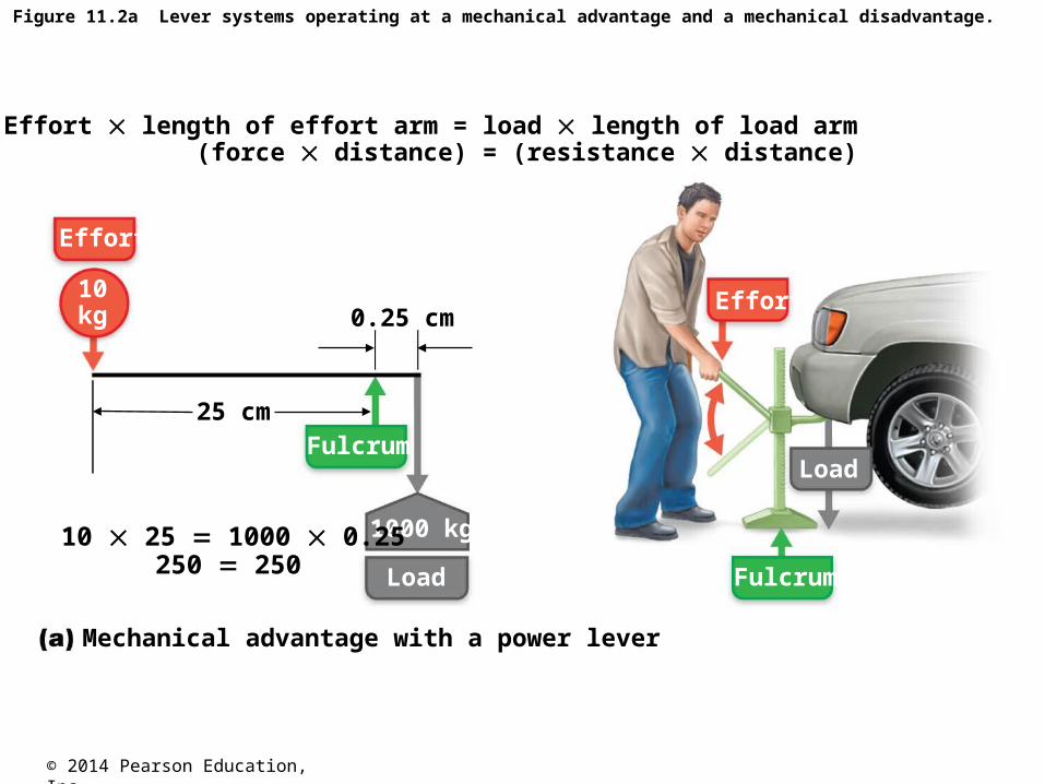
Figure 11.2a Lever systems operating at a mechanical advantage and a mechanical disadvantage.
Effort length of effort arm = load length of load arm (force distance) = (resistance distance)
Mechanical advantage with a power lever
Effort
Effort
1000 kg
Load
LoadFulcrum
Fulcrum
0.25 cm
25 cm
10kg
10 25 1000 0.25 250 250

© 2014 Pearson Education, Inc.
Figure 11.2b Lever systems operating at a mechanical advantage and a mechanical disadvantage.
Mechanical disadvantage with a speed lever
Effort
Effort
50 kg
Load
Load
Fulcrum Fulcrum
25 cm
50 cm
100 kg
100 25 50 50 2500 2500
Effort length of effort arm = load length of load arm (force distance) = (resistance distance)

© 2014 Pearson Education, Inc.
Lever Systems: Bone-Muscle Relationships
• Levers allow a given effort to
• Move a heavier load
• Move a load farther
• Mechanical advantage
• Moves a large load over small distances
• Mechanical disadvantage
• Allows a load to be moved over a large distance

© 2014 Pearson Education, Inc.
Lever Systems: Bone-Muscle Relationships
• Bones—act as levers
• Joints—act as fulcrums
• Muscle contraction
• Provides effort–applied where muscle attaches to bone
• Load
• Bone, overlying tissue, and anything lifted
© 2014 Pearson Education, Inc.
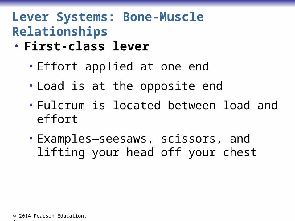
Lever Systems: Bone-Muscle Relationships
• First-class lever
• Effort applied at one end
• Load is at the opposite end
• Fulcrum is located between load and effort
• Examples—seesaws, scissors, and lifting your head off your chest

© 2014 Pearson Education, Inc.
Figure 11.3a Lever systems.First-class lever
Effort
L
Load
Fulcrum
Arrangement of the elements isload-fulcrum-effort.
Effort
L
Load
Fulcrum
Example: scissors
EffortLoad
Fulcrum
In the body: A first-class lever system raises yourhead off your chest. The posterior neck musclesprovide the effort; the atlanto-occipital joint is thefulcrum; and the weight to be lifted is the facialskeleton.

© 2014 Pearson Education, Inc.
Lever Systems: Bone-Muscle Relationships
• Second-class lever
• Effort applied at one end
• Fulcrum is at the opposite end
• Load is between the effort and fulcrum
• Examples—wheelbarrow or standing on tiptoe
• An uncommon type of lever in the body
• Works at a mechanical advantage

© 2014 Pearson Education, Inc.
Figure 11.3b Lever systems.Second-class lever
Effort
L
Load
Fulcrum
Arrangement of the elements isfulcrum-load-effort.
Effort
L
Load
Fulcrum
Example: wheelbarrow
Effort
Load
Fulcrum
In the body: Second-class leverage is exerted whenyou stand on tip-toe. The effort is exerted by the calfmuscles pulling upward on the heel; the joints of theball of the foot are the fulcrum; and the weight of thebody is the load.

© 2014 Pearson Education, Inc.
Lever Systems: Bone-Muscle Relationships
• Third-class lever
• Effort is applied between the load and the fulcrum
• Works speedily
• Always at a mechanical disadvantage

© 2014 Pearson Education, Inc.
Figure 11.3c Lever systems.Third-class lever
Effort
L
Load
Fulcrum
Arrangement of the elements isload-effort-fulcrum.
Effort
L
Load
Fulcrum
Example: tweezers or forceps
Effort
Load
Fulcrum
In the body: Flexing the forearm by the bicepsbrachii muscle exemplifies third-class leverage. Theeffort is exerted on the proximal radius of the forearm;the fulcrum is the elbow joint; and the load is the hand and distal end of the forearm.
© 2014 Pearson Education, Inc.
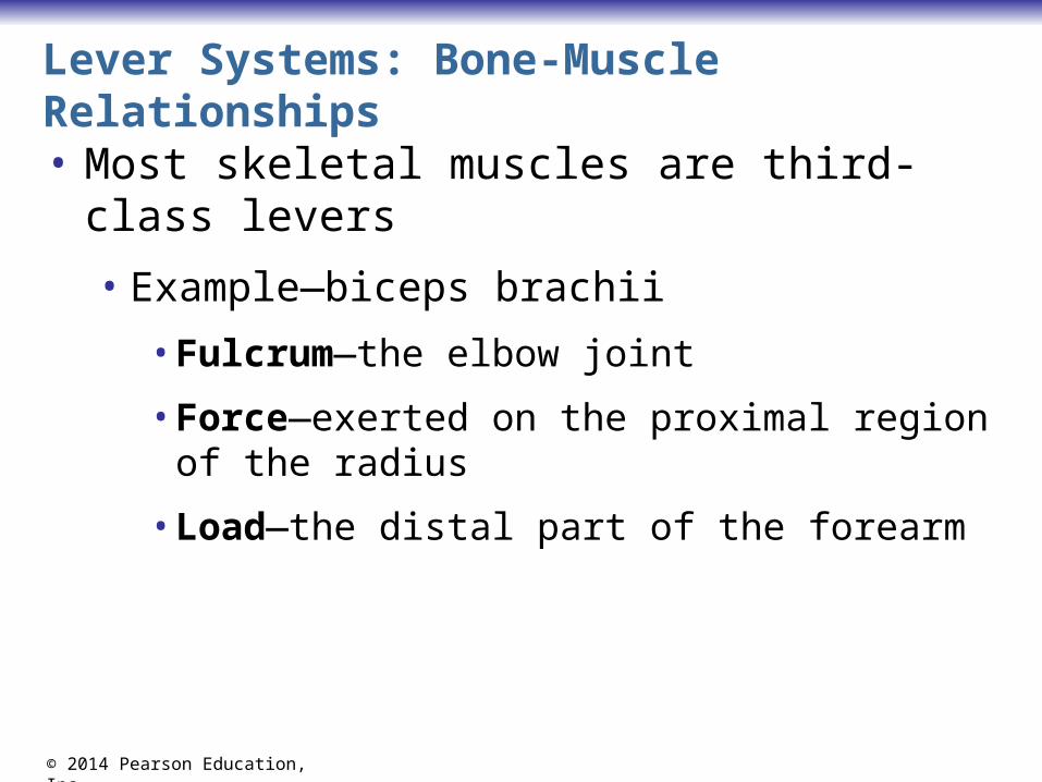
Lever Systems: Bone-Muscle Relationships
• Most skeletal muscles are third-class levers
• Example—biceps brachii
• Fulcrum—the elbow joint
• Force—exerted on the proximal region of the radius
• Load—the distal part of the forearm

© 2014 Pearson Education, Inc.
Organization Scheme Based on Embryonic Development• Groups of muscles organized by
• Embryonic origin
• General function
• Muscles develop from mesoderm
• Myotomes
• Somitomeres
• The first seven myotomes of the head
• Splanchnic mesoderm

© 2014 Pearson Education, Inc.
Figure 11.4a Development and basic organization of the muscles.
Limb bud
Myotomes
6-week embryo
Limb bud
Pharynx
Eye
Somitomeres
First (occipital)myotomes

© 2014 Pearson Education, Inc.
Figure 11.4b Development and basic organization of the muscles.
Cross section at level oflower limb buds
Limb bud
Vertebra
Splanchnicmesoderm
Limb skeleton
Dorsal
Gut tube Limb flexion
Limb extension
Flexormusclesof limbs
Neuraltube
Extensormusclesof limbs
Myotome
Somaticmesoderm

© 2014 Pearson Education, Inc.
Organization Scheme Based on Embryonic Development• Muscles organized into four groups
• Musculature of the visceral organs
• Pharyngeal arch muscles
• Axial muscles
• Limb muscles
© 2014 Pearson Education, Inc.
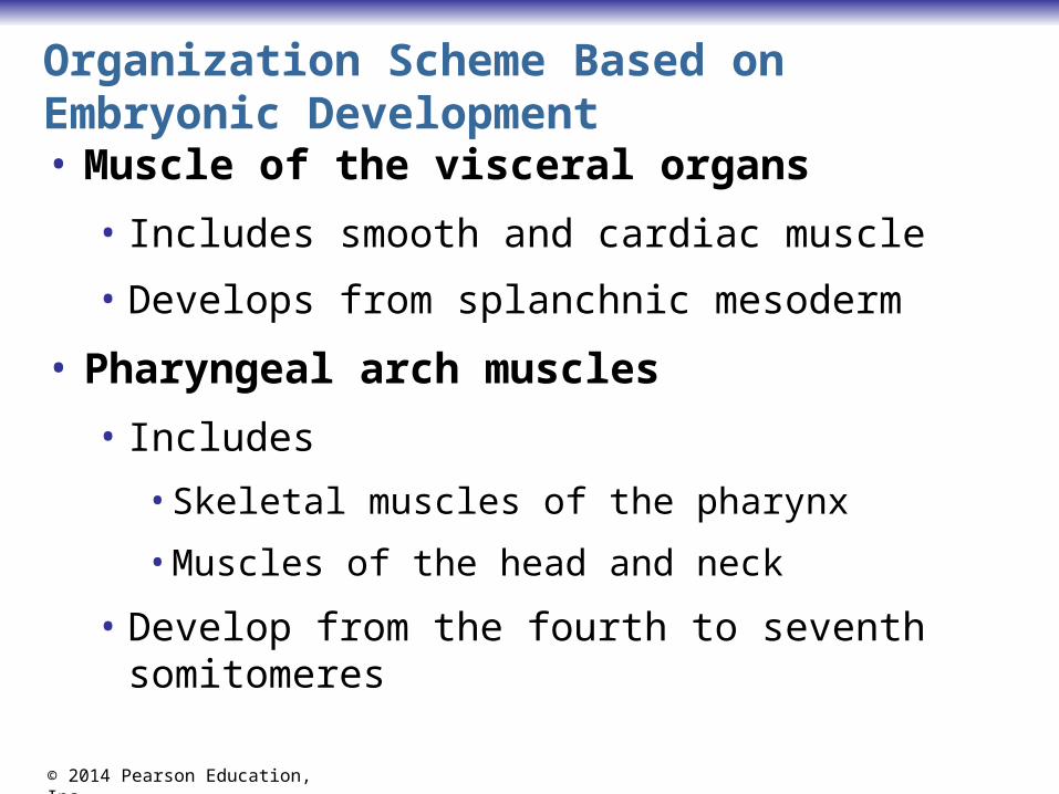
Organization Scheme Based on Embryonic Development• Muscle of the visceral organs
• Includes smooth and cardiac muscle
• Develops from splanchnic mesoderm
• Pharyngeal arch muscles
• Includes
• Skeletal muscles of the pharynx
• Muscles of the head and neck
• Develop from the fourth to seventh somitomeres

© 2014 Pearson Education, Inc.
Figure 11.4c Development and basic organization of the muscles.
Pharynx
Trapezius
Muscles of facial expressione.g., orbicularis oculi
Chewing musclese.g., temporalis, masseter
Suprahyoid muscles (most)
Pharyngeal constrictors(key swallowing muscles)
Pharyngeal arch (branchiomeric) muscles:4th–7th somitomeres

© 2014 Pearson Education, Inc.
Axial Muscles
• Lie anterior and posterior to the body axis
• Muscles of the
• Thorax, abdomen, and pelvis
• Many muscles of the neck
• Some muscles of the head
• Function to move the trunk and maintain posture

© 2014 Pearson Education, Inc.
Axial Muscles
• Develop from myotomes and some somitomeres
• Dorsal regions of myotomes—deep muscles of the back
• Ventral regions of myotomes—muscles of the trunk and neck
• Respiratory muscles
• Anterior abdominal wall muscles
• Muscles of the pelvic floor

© 2014 Pearson Education, Inc.
Figure 11.4d Development and basic organization of the muscles.
Deep muscles of the backe.g., erector spinae
Muscles of the anteriorand lateral trunke.g., 1. infrahyoid muscles (neck) 2. intercostal muscles (thorax) 3. external and internal obliques (abdomen) 4. muscles of the pelvic floor
Axial muscles: 1st–3rd somitomeres and from myotomes
Extrinsic musclesof the eye
Tongue Tonguemuscles

© 2014 Pearson Education, Inc.
Limb Muscles
• Limb muscles arise from lateral parts of nearby myotomes
• Extensors
• Muscle mass dorsal to limb bones
• Flexors
• Muscle mass ventral to limb bones
© 2014 Pearson Education, Inc.
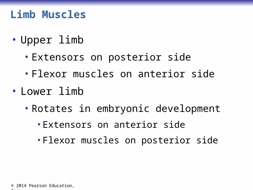
Limb Muscles
• Upper limb
• Extensors on posterior side
• Flexor muscles on anterior side
• Lower limb
• Rotates in embryonic development
• Extensors on anterior side
• Flexor muscles on posterior side

© 2014 Pearson Education, Inc.
Figure 11.4e Development and basic organization of the muscles.
Limb muscles:From myotomes
Extensors
Flexorse.g., hamstrings,gastrocnemius
Extensorse.g., quadricepsfemoris, tibialisanterior
Flexorse.g., biceps brachii,flexor carpi radialis
Extensorse.g., triceps brachii,extensor digitorum
Flexors

© 2014 Pearson Education, Inc.
Muscle Actions and Interactions
• A muscle cannot reverse the movement it produces
• Another muscle must undo the action
• Muscles with opposite actions lie on opposite sides of a joint

© 2014 Pearson Education, Inc.
Muscle Actions and Interactions
• Prime mover (agonist)• Has major responsibility for a certain movement
• Antagonist
• Opposes or reverses a movement
• Synergist—helps the prime mover • By adding extra force
• By reducing undesirable movements
• Fixator
• A type of synergist that holds a bone firmly in place

© 2014 Pearson Education, Inc.
Figure 11.5 The action of a muscle can be inferred by the position of the muscle relative to the joint it crosses.

© 2014 Pearson Education, Inc.
Muscle Compartments of the Limbs
• Dense fibrous connective tissue divides limb muscles into compartments
• Muscles in the same compartment
• Have similar actions; act as synergists
• Muscles in opposing compartments are
• Agonist and antagonist pairs
• Each compartment is innervated by a single nerve

© 2014 Pearson Education, Inc.
Muscle Compartments the Upper Limb
• The upper limb has anterior and posterior compartments
• Anterior arm compartment muscles
• Flex the shoulder or arm
• Innervation is the musculocutaneous nerve
• Anterior forearm compartment muscles
• Flex the wrist and digits
• Innervation is the median or ulnar nerve
© 2014 Pearson Education, Inc.
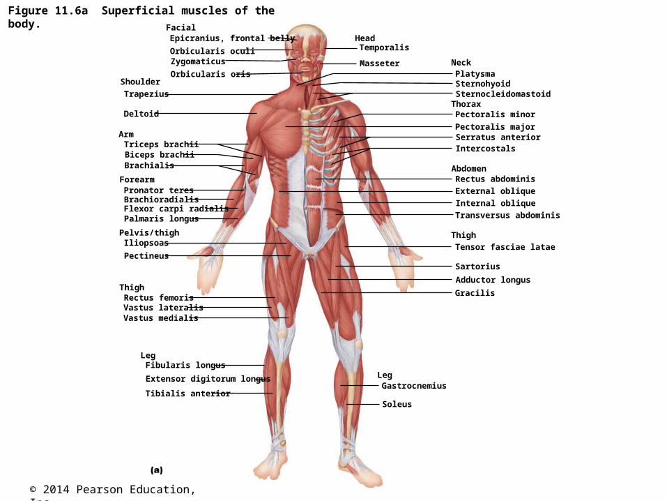
Figure 11.6a Superficial muscles of the body.
Facial
Forearm
Arm
Shoulder
Head
Deltoid
Zygomaticus
Epicranius, frontal belly
Orbicularis oculi
Orbicularis oris
Temporalis
Masseter
Trapezius
Triceps brachiiBiceps brachiiBrachialis
Pronator teres
Palmaris longus
BrachioradialisFlexor carpi radialis
Thigh
Leg
Pelvis/thigh
Tibialis anterior
Iliopsoas
Pectineus
Rectus femoris
Vastus medialis
Fibularis longus
Extensor digitorum longus
Vastus lateralis
Neck
SternohyoidPlatysma
AbdomenRectus abdominis
External oblique
ThoraxSternocleidomastoid
Pectoralis minor
Pectoralis majorSerratus anteriorIntercostals
Internal oblique
Transversus abdominis
ThighTensor fasciae latae
Leg
Sartorius
Adductor longus
Gracilis
Gastrocnemius
Soleus
© 2014 Pearson Education, Inc.
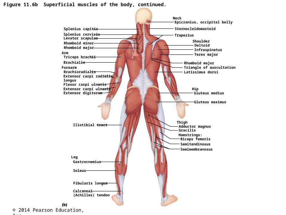
Forearm
Arm
Shoulder
Hip
DeltoidRhomboid minor
Splenius capitis
Levator scapulae
Epicranius, occipital belly
InfraspinatusTeres major
Trapezius
Triceps brachii
Biceps femoris
Brachialis
Extensor digitorum
Brachioradialis
Flexor carpi ulnaris
Iliotibial tract
Fibularis longus
Calcaneal(Achilles) tendon
Neck
Semitendinosus
Sternocleidomastoid
Latissimus dorsi
Rhomboid majorTriangle of auscultation
Gluteus medius
Thigh
Leg
Hamstrings:
Adductor magnusGracilis
Gastrocnemius
Soleus
Extensor carpi ulnaris
Extensor carpi radialislongus
Splenius cervicis
Rhomboid major
Gluteus maximus
Semimembranosus
Figure 11.6b Superficial muscles of the body, continued.
© 2014 Pearson Education, Inc.
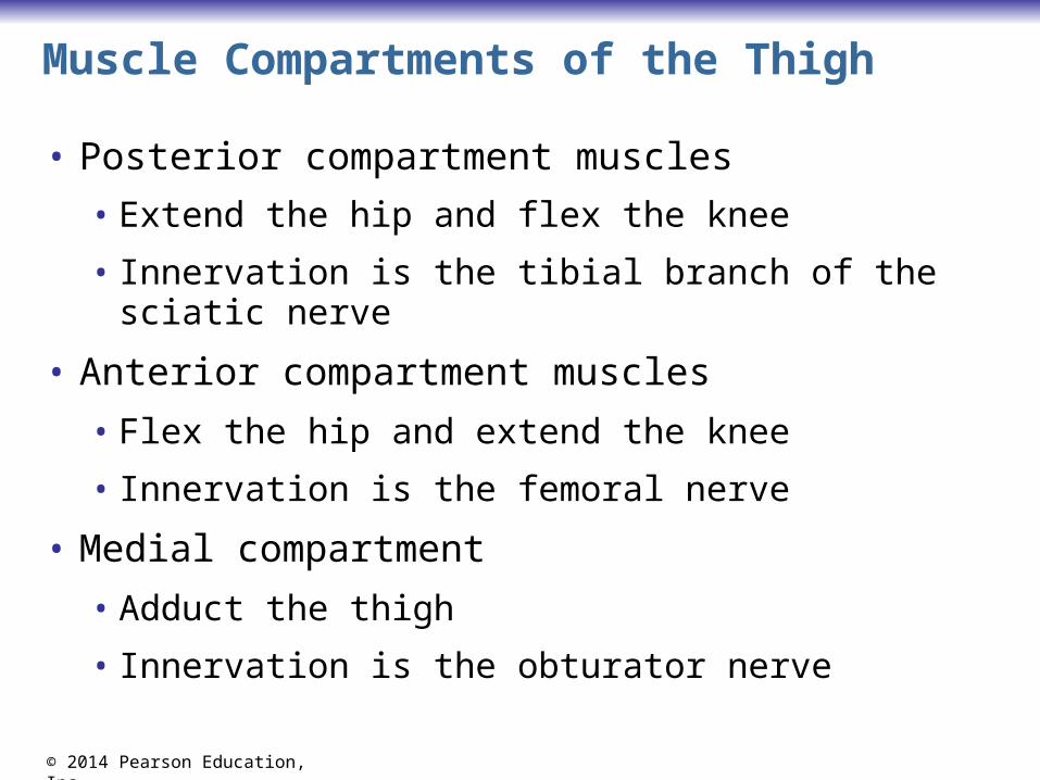
Muscle Compartments of the Thigh
• Posterior compartment muscles
• Extend the hip and flex the knee
• Innervation is the tibial branch of the sciatic nerve
• Anterior compartment muscles
• Flex the hip and extend the knee
• Innervation is the femoral nerve
• Medial compartment
• Adduct the thigh
• Innervation is the obturator nerve

© 2014 Pearson Education, Inc.
Figure 11.7a Muscle compartments in the arm and forearm.
Muscles of the arm
Tricepsbrachii
BrachialisExtensors
Lateral head
Flexors
Long head
Medial head
Posterior compartment of arm(extends forearm at elbow);innervation: radial nerve
Humerus
Biceps brachiiShort headLong head
(a)
Anterior compartment of arm(flexes forearm at elbow);innervation: musculocutaneous nerve

© 2014 Pearson Education, Inc.
Muscle Compartments of the Leg
• Posterior compartment muscles
• Contains digital and plantar flexors
• Innervation is the tibial nerve
• Anterior compartment muscles
• Contains digital extensors and dorsiflexors
• Innervation is the deep fibular nerve
• Lateral compartment muscles
• Plantar flex and evert the foot
• Innervation is the superficial fibular nerve

© 2014 Pearson Education, Inc.
Figure 11.7b Muscle compartments in the arm and forearm.
Extensors
Pronator teres
Extensors
Abductor pollicislongus
Flexors
Posterior compartment of forearm(extends wrist and fingers);innervation: radial nerve
Radius
Brachioradialis(elbow flexor)
Muscles of the forearm
Flexors
Ulna
Anterior compartment of forearm (flexes wrist and fingers);innervation: median or ulnar nerve
(b)

© 2014 Pearson Education, Inc.
Naming the Skeletal Muscles
• Location
• Example—the brachialis is located on the arm
• Shape
• Example—the deltoid is triangular
• Relative size
• Maximus, minimus, and longus indicate size
• Example—gluteus maximus and gluteus minimus

© 2014 Pearson Education, Inc.
Naming the Skeletal Muscles
• Direction of fascicles and muscle fibers
• Name tells direction in which fibers run
• Example—rectus abdominis and transversus abdominis
• Location of attachments—name reveals point of origin and insertion
• Example—brachioradialis

© 2014 Pearson Education, Inc.
Naming the Skeletal Muscles
• Number of origins
• Two, three, or four origins
• Indicated by the words biceps, triceps, and quadriceps
• Action
• The action is part of the muscle’s name
• Indicates type of muscle movement
• Flexor, extensor, adductor, or abductor