CURSO/TALLER DE PATOLOGÍA DIGITAL - Inicio · Integración de sistemas de trazabilidad y...
26
Integración de sistemas de trazabilidad y escáneres de preparaciones Marcial García Rojo Hospital General Universitario de Ciudad Real. Spain. [email protected] Tracking and scanning systems integration
Transcript of CURSO/TALLER DE PATOLOGÍA DIGITAL - Inicio · Integración de sistemas de trazabilidad y...

Integración de sistemas de
trazabilidad y escáneres de
preparaciones
Marcial García Rojo Hospital General Universitario de
Ciudad Real. Spain. [email protected]
Tracking and scanning systems integration
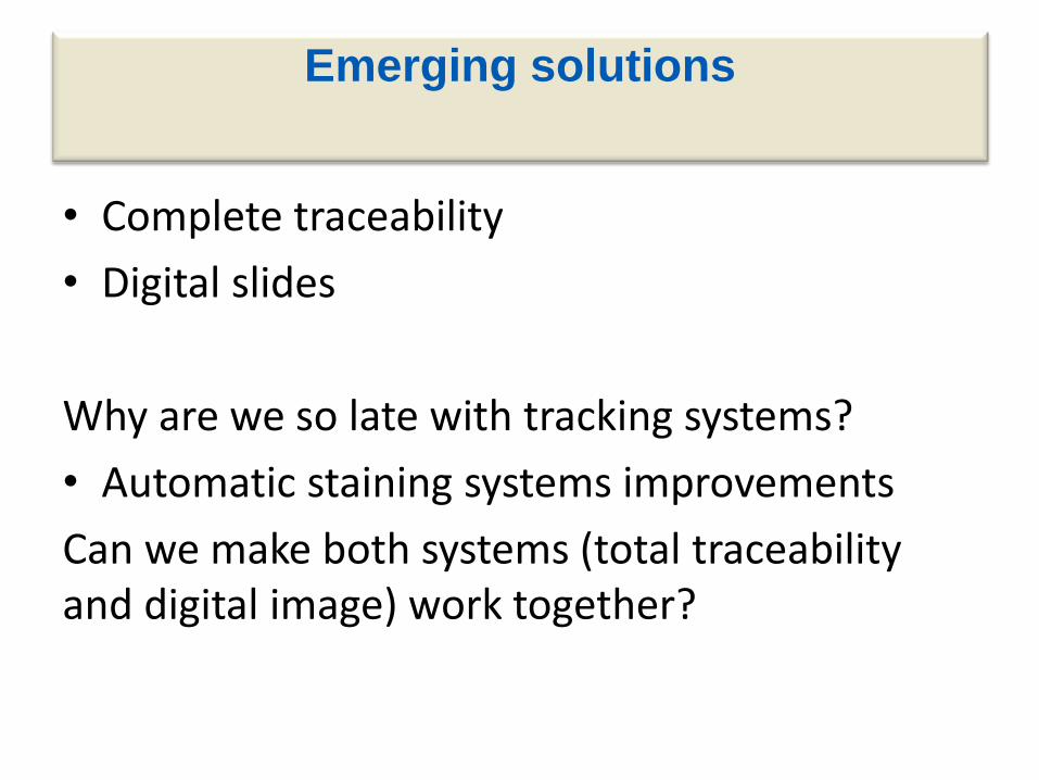
Emerging solutions
• Complete traceability
• Digital slides
Why are we so late with tracking systems?
• Automatic staining systems improvements
Can we make both systems (total traceability and digital image) work together?

Anatomic Pathology Information
Systems-SIAP (I)
Efficient management of data and images to generate final pathology report. Classic functions: • Patient identification and management • Accessioning and specimen management • Gross and microscopic descriptions • Management of special techniques requests…. New functions: • Tracking systems and workflow control • Integration with digital image • Telepathology, ….

Tracking in pathology
• Correct Identification
• Workflow management
• Time management (LEAN)
• Bottlenecks identification
• Where is located each object

Slide and cassettes labelling:
interpretation errors
SOS
50S
505
5O5
S05
1405
1705
1905
14OS
17OS
84
B4
89
B7
87
BY
Fuente: Dako y Leica

Possible scenarios
• AP study request: Electronic, paper based or mixed
• Specimen accession number: does it come from clinical information system or from pathology I.S.
• Accessioning secretary technicians
• Tracking systems: Part of the SIAP or special software
• Laboratory workflow: Batch mode, continuous workflow or mixed
• Digital slides: Partial or total scanning
• Autostainers: Integrated or not
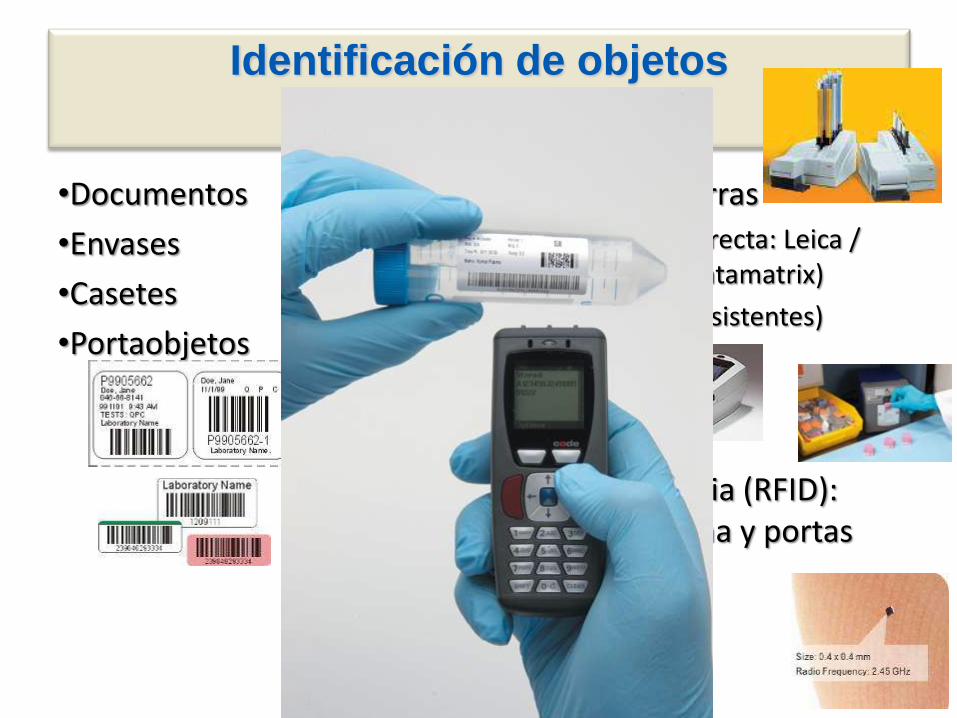
Identificación de objetos
•Documentos
•Envases
•Casetes
•Portaobjetos
•Códigos de barras
–Impresión directa: Leica / Sakura (2D: datamatrix)
–Etiquetas (resistentes)
•Radiofrecuencia (RFID): Bloques parafina y portas

Petición electrónica

Día 1 09:15
Circuito de trabajo RECEPCIÓN Y REGISTRO DE MUESTRAS
Petición electrónica Registro Macro y
fijación Inclusión
Microtomía y H&E
Micro IHQ, SS,
ISH Diagnóstic
o Informe
Fuente: Dako

Ventana Vantage Dako TruePositiveId
Leica Cerebro
Vitro VTS

ESTACIÓN 2:
Macro/Sala de Tallado. Impresión y verificación de casetes
Escanear etiqueta de
informe de petición.
Seleccionar color
cassette desde
Patwin.
Imprimir y verificar
Fuente: Dako


Etiquetar portas
• Sin código de barras: Escasa trazabilidad (posible OCR – Hologic). Se traminte muy poca información. Requiere más trabajo manual
• Doble etiquetado: Queda muy poco espacio para datos como nombre paciente, hospital, nombre patólogo, etc.
• Re-Etiquetado: Añadir 8,5 sec/porta manipulación para colocar etiqueta. Son muchas horas y posibles errores
13
Sin código de
barras, sólo datos
Sitio solo para
3 líneas
Solo código de
barras del
instrumento

Identificación portaobjetos

LIS/TPID – DakoLink – TPID: affects the
whole pathology laboratory workflow
Case information entered into LIS or TPID. Labeling of specimens.
Accessioning
Grossing of the case. Cassettes produced.
Grossing
Processing of cassettes. Tracking of case and cassettes.
Processing
Embedding of cassettes. Tracking of case and blocks.
Embedding
Tracking of blocks. Slide labeling.
Cutting
Tracking of archived blocks and slides.
Archiving
Processing of slides through staining process with Dako Link.
IHC/SS Staining laboratory From
Microtome to
Microscope
Use the same
label! Slide scanning
Slide reading and reporting


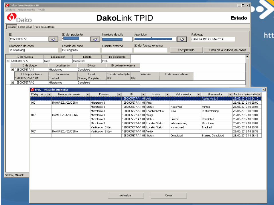

Control de preparaciones. Estado
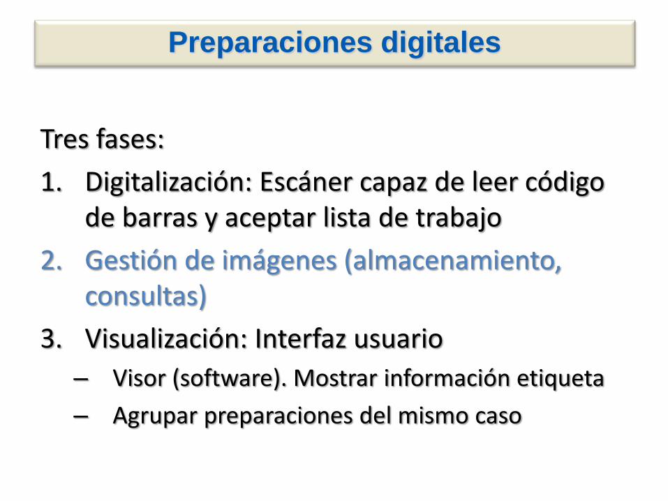
Preparaciones digitales
Tres fases:
1. Digitalización: Escáner capaz de leer código de barras y aceptar lista de trabajo
2. Gestión de imágenes (almacenamiento, consultas)
3. Visualización: Interfaz usuario
– Visor (software). Mostrar información etiqueta
– Agrupar preparaciones del mismo caso

Escáneres de preparaciones
Más de 25 sistemas disponibles:
• 3DHistech Panoramic (4) • Aperio ScanScope (6) • Olympus VS (3) • Hamamatsu Nanozoomer (2) • Claro (2) • Leica SCN400 (2) • Menarini D.Sight(2) • Omnyx (2) • Philips (1) • Roche Ventana (2)
Aperio Scanscope AT / XT / CS
3D Histech
Hamamatsu
Nanozoomer 2 Olympus
VS800, VS120, VS110
.slide Philips
Leica SCN400 Omnyx VL4
Menarini
D.Signt

Supplement 122 in DICOM
• Attributes for the identification and description of specimens:
– (1) identify the specimen (within a given institution and across institutions)
– (2) identify and describe the container in which the specimen resides
– (3) describe specimen collection, sampling, and processing
– (4) describe the specimen or its ancestors when these descriptions help with the interpretation of the image.

Antes de TPID

Con TPID

Conclusions
• Tracking systems are designed to improve laboratory workflow ( “prediagnosis phase”), but not department workflow, but we must learn to use them in diagnosis phase (pathologist and cytotechnician)
• An integrated tracking solution improves workflows and response time.
• Integrated solutions in digital pathology with electronic health record, tracking and central image repository, will improve collaborative work between technicians and pathologists to improve security for patients and quality in health providing.