The Pathogenicity of 2019 Novel Coronavirus in hACE2 … · epithelial cells. The phenomenon was...
15
The Pathogenicity of 2019 Novel Coronavirus in hACE2 Transgenic Mice 1 2 Linlin Bao† ,1 , Wei Deng† ,1 , Baoying Huang† ,2 , Hong Gao† ,1 , Lili Ren 3 , Qiang Wei 1 , Pin 3 Yu 1 , Yanfeng Xu 1 , Jiangning Liu 1 , Feifei Qi 1 , Yajin Qu 1 , Wenling Wang 2 , Fengdi Li 1 , Qi 4 Lv 1 , Jing Xue 1 , Shuran Gong 1 , Mingya Liu 1 , Guanpeng Wang 1 , Shunyi Wang 1 , Linna 5 Zhao 1 , Peipei Liu 2 , Li Zhao 2 , Fei Ye 2 , Huijuan Wang 2 , Weimin Zhou 2 , Na Zhu 2 , Wei Zhen 2 , 6 Xiaojuan Zhang 2 , Zhiqi Song 1 , Li Guo 3 , Lan Chen 3 , Conghui Wang 3 , Ying Wang 3 , 7 Xinming Wang 3 , Yan Xiao 3 , Qiangming Sun 4 , Hongqi Liu 4 , Fanli Zhu 4 , Chunxia Ma 4 , 8 Lingmei Yan 4 , Mengli Yang 4 , Jun Han 2 , Wenbo Xu 2 , Wenjie Tan 2 , Xiaozhong Peng 4 , Qi 9 Jin 3 , Guizhen Wu* ,2 , Chuan Qin* ,1 10 11 1 Key Laboratory of Human Disease Comparative Medicine, Chinese Ministry of Health, 12 Beijing Key Laboratory for Animal Models of Emerging and Remerging Infectious 13 Diseases, Institute of Laboratory Animal Science, Chinese Academy of Medical Sciences 14 and Comparative Medicine Center, Peking Union Medical College, Beijing, China. 15 2 MHC Key Laboratory of Biosafety, National Institute for Viral Disease Control and 16 Prevention, China CDC, Beijing, China. 17 3 Institute of Pathogen Biology, Chinese Academy of Medical Sciences, Beijing, China. 18 4 Institute of Medical Biology, Chinese Academy of Medical Sciences, Beijing, China. 19 20 †These authors contributed equally to this work. 21 *Correspondence should be addressed to Chuan Qin, Email: [email protected], or 22 Guizhen Wu, Email: [email protected]. 23 author/funder. All rights reserved. No reuse allowed without permission. The copyright holder for this preprint (which was not peer-reviewed) is the . https://doi.org/10.1101/2020.02.07.939389 doi: bioRxiv preprint
Transcript of The Pathogenicity of 2019 Novel Coronavirus in hACE2 … · epithelial cells. The phenomenon was...

The Pathogenicity of 2019 Novel Coronavirus in hACE2 Transgenic Mice 1
2
Linlin Bao†,1, Wei Deng†,1, Baoying Huang†,2, Hong Gao†,1, Lili Ren3, Qiang Wei1, Pin 3
Yu1, Yanfeng Xu1, Jiangning Liu1, Feifei Qi1, Yajin Qu1, Wenling Wang2, Fengdi Li1, Qi 4
Lv1, Jing Xue1, Shuran Gong1, Mingya Liu1, Guanpeng Wang1, Shunyi Wang1, Linna 5
Zhao1, Peipei Liu2, Li Zhao2, Fei Ye2, Huijuan Wang2, Weimin Zhou2, Na Zhu2, Wei Zhen2, 6
Xiaojuan Zhang2, Zhiqi Song1, Li Guo3, Lan Chen3, Conghui Wang3, Ying Wang3, 7
Xinming Wang3, Yan Xiao3, Qiangming Sun4, Hongqi Liu4, Fanli Zhu4, Chunxia Ma4, 8
Lingmei Yan4, Mengli Yang4, Jun Han2, Wenbo Xu2, Wenjie Tan2, Xiaozhong Peng4, Qi 9
Jin3, Guizhen Wu*,2, Chuan Qin*,1 10
11
1 Key Laboratory of Human Disease Comparative Medicine, Chinese Ministry of Health, 12
Beijing Key Laboratory for Animal Models of Emerging and Remerging Infectious 13
Diseases, Institute of Laboratory Animal Science, Chinese Academy of Medical Sciences 14
and Comparative Medicine Center, Peking Union Medical College, Beijing, China. 15
2 MHC Key Laboratory of Biosafety, National Institute for Viral Disease Control and 16
Prevention, China CDC, Beijing, China. 17
3 Institute of Pathogen Biology, Chinese Academy of Medical Sciences, Beijing, China. 18
4 Institute of Medical Biology, Chinese Academy of Medical Sciences, Beijing, China. 19
20
†These authors contributed equally to this work. 21
*Correspondence should be addressed to Chuan Qin, Email: [email protected], or 22
Guizhen Wu, Email: [email protected]. 23
author/funder. All rights reserved. No reuse allowed without permission. The copyright holder for this preprint (which was not peer-reviewed) is the. https://doi.org/10.1101/2020.02.07.939389doi: bioRxiv preprint

Abstract 24
2019-nCoV caused pneumonia cases in China has become a public health emergency of 25
international concern (PHEIC). The first priority for prevention and treatment of the 26
disease is to find the pathogenicity of 2019-nCoV in vivo. Weight loss and virus replication 27
were detected in infected-hACE2 mice. The typical histopathology was interstitial 28
pneumonia with significant inflammatory cells infiltration around the bronchioles and 29
blood vessels, and viral antigens were observed in bronchial epithelial cells and alveolar 30
epithelial cells. The phenomenon was not found in wild type mice infected with 2019-31
nCoV and the mock-infected hACE2 mice. The pathogenicity of 2019-nCoV in hACE2 32
mice was clarified and the Koch's postulates was fulfilled as well, and the model may 33
facilitate the development of therapeutics and vaccines against 2019-nCoV. 34
35
author/funder. All rights reserved. No reuse allowed without permission. The copyright holder for this preprint (which was not peer-reviewed) is the. https://doi.org/10.1101/2020.02.07.939389doi: bioRxiv preprint

In late December of 2019, a cluster of severe pneumonia cases caused by 2019 novel 36
coronavirus (2019-nCoV), linked to a seafood market in which exotic animals were also 37
sold and consumed, were identified and reported from Wuhan City, Hubei Province, 38
China1,2. The number of infections has since soared, with almost 10,000 cases reported and 39
over 200 deaths as of January 31, 2020 in China3, and imported cases from travelers of 40
mainland China in several other countries. It is critical to find the pathogenicity and biology 41
of the virus for prevention and treatment of the disease. 42
Because 2019-nCoV was highly homologous with Severe acute respiratory syndrome 43
coronavirus (SARS-CoV), human Angiotensin-converting enzyme 2 (hACE2), which was 44
the entry receptor of SARS-CoV, was also considered to have a high binding ability with 45
the 2019-nCoV by molecular biological analysis4,5. Therefore, we used the hACE2 46
transgenic and wild type mice infected with or without 2019-nCoV infection to study the 47
pathogenicity of the virus. Specific pathogen-free, 6-11-month-old, male and female 48
hACE2 mice and wild type mice (n=7) were inoculated intranasally with 2019-nCoV stock 49
virus at a dosage of 105 TCID50 per mouse, and the mock-infected hACE2 mice (n=3) were 50
used as control. Weight loss of up to 5% was observed for 10 dpi only in the 2019-nCoV-51
infected hACE2 mice (Figure 1a), and other clinical symptoms were not observed. Major 52
organs were harvested at 3 dpi and 5 dpi to assess for biodistribution of 2019-nCoV in 53
infected hACE2 mice. Viral RNA was positive by RT-PCR (Figure 1b) and identified by 54
sequencing only in the lung of infected-hACE2 mice at 3 dpi and 5 dpi. Meanwhile, the 55
virus was successfully isolated by Vero cells culture (Figure 1d) and observed by an 56
electron microscope (Figure 1e). However, the virus was not found in the PBS-infected 57
hACE2 mice or infected-wild type mice (data not shown). 58
author/funder. All rights reserved. No reuse allowed without permission. The copyright holder for this preprint (which was not peer-reviewed) is the. https://doi.org/10.1101/2020.02.07.939389doi: bioRxiv preprint
The typical pneumonia was demonstrated as bilateral ground-glass opacity and 59
subsegmental areas of consolidation by image in patient6, but no histopathological results 60
were reported until now. The major organs of mice were examined by histopathology and 61
immunofluorescence. Compared to PBS-infected hACE2 mice or 2019-nCoV-infected-62
wild type mice (Figure 2a and b), focal lesions were observed in the dorsal of the right 63
middle lobe in 2019-nCoV-infected hACE2 mice (Figure 2c). Consistently, lung tissues 64
from 2019-nCoV-infected hACE2 mice had multifocally mild or moderate pneumonia with 65
interstitial hyperplasia. And significant inflammatory cells infiltration around the 66
bronchioles and blood vessels (Figure 2f) were found. The alveolar interstitium is also 67
expanded with inflammatory cells, and the alveolar lumen contains cell debris mixed with 68
leukocytes. Bronchial epithelial cells showed swelling, degeneration, and some of them 69
dissolved and necrotic foci (Figure 2g, h and i). Meanwhile, 2019-nCoV antigens were 70
observed in the bronchial epithelial cells (Figures 3h and i) and alveolar epithelial cells 71
(Figures 3n, o, p and q) of lungs in 2019-nCoV-infected hACE2 mice. In addition, the co-72
localization of 2019-nCoV S protein and hACE2 receptor was demonstrated in alveolar 73
epithelial cells of infected-hACE2 mice by immunofluorescence (Figures 3n, o, p and q). 74
The phenomenon was not found in the PBS-infected hACE2 mice (Figures 3k, i and m) or 75
infected-wild type mice (data not shown). 76
The speed of geographical spread of severe viral pneumonia disease caused by 2019-77
nCoV has been declared as public health emergency of international concern (PHEIC), 78
with cases reported on multiple continents only weeks after the disease was first reported7. 79
Although it has been determined by bioinformatics that the pathogen of this epidemic is a 80
novel coronavirus, it is necessary to be confirmed by animal experiments following Koch's 81
author/funder. All rights reserved. No reuse allowed without permission. The copyright holder for this preprint (which was not peer-reviewed) is the. https://doi.org/10.1101/2020.02.07.939389doi: bioRxiv preprint

principles. After experimental infection of transgenic hACE2 mice with one of the earliest 82
known isolates of 2019-nCoV, the mice lost weight and showed interstitial pneumonia, 83
which are comparable with initial clinical reports of pneumonia caused by 2019-nCoV6. 84
In addition, the 2019-nCoV S protein and hACE2 receptor were found to co-localize in 85
alveolar epithelial cells, supporting that the 2019-nCoV, similar to SARS-CoV, also 86
utilizes the hACE2 as a receptor for entry4. Therefore, the present study clarified the 87
pathogenicity of 2019-nCoV in hACE2 mice and fulfills the Koch's postulates as well, and 88
the model may facilitate the development of drugs and vaccines against 2019-nCoV. 89
90
Materials and methods 91
Ethics statement 92
Murine studies were performed in an animal biosafety level 3 (ABSL3) facility using 93
HEPA-filtered isolators. All procedures in this study involving animals were reviewed and 94
approved by the Institutional Animal Care and Use Committee of the Institute of 95
Laboratory Animal Science, Peking Union Medical College (BLL20001). 96
97
Viruses and cells 98
The 2019-nCoV (strain HB-01) was kindly provided by Professor Wenjie Tan1, from the 99
China Centers for Disease Control and Prevention (China CDC). The complete genome for 100
this 2019-nCoV was submitted to GISAID (BetaCoV/Wuhan/IVDC-HB-101
01/2020|EPI_ISL_402119), and deposited in the China National Microbiological Data 102
Center (accession number NMDC10013001 and genome accession numbers 103
MDC60013002-01). Seed 2019-nCoV stocks and virus isolation studies were performed 104
author/funder. All rights reserved. No reuse allowed without permission. The copyright holder for this preprint (which was not peer-reviewed) is the. https://doi.org/10.1101/2020.02.07.939389doi: bioRxiv preprint

in Vero cells, which are maintained in Dulbecco's modified Eagle's medium (DMEM, 105
Invitrogen, Carlsbad, USA) supplemented with 10% fetal bovine serum (FBS), 100 IU/ml 106
penicillin, and 100 µg/ml streptomycin, and incubated at 37°C, 5% CO2. Titers for 2019-107
nCoV were determined using a standard 50% tissue culture infection dose (TCID50) assay. 108
109
Animal experiments 110
For the animal experiments, specific pathogen-free, 6-11-month-old, male and female 111
transgenic hACE2 mice were obtained from the Institute of Laboratory Animal Science, 112
Peking Union Medical College, China. Transgenic mice were generated by microinjection 113
of the mice hACE2 promoter driving the human ACE2 coding sequence into Institute of 114
Cancer Research (ICR) or C57BL/6J mice; the presence of human ACE2 in the mice used 115
for these experiments was confirmed by PCR (data not shown). The hACE2 mice (n=7) 116
or ICR mice (n=5) were respectively inoculated intranasally with 2019-nCoV stock virus 117
at a dosage of 105 TCID50 per mouse. As a control, hACE2 mice (n=3) were mock-infected 118
with an equivalent challenge volume of PBS. PBS- and 2019-nCoV-infected animals were 119
continuously observed daily to record body weights, clinical symptoms, decreased 120
responsiveness to external stimuli and death. Two mice from the 2019-nCoV-infected 121
group were dissected at 3 days post-infection (dpi) and at 5 dpi to collect trachea, lung, 122
kidney, brain, spleen, and liver tissues for the determination of viral load after 2019-nCoV 123
infection. One mouse from the PBS-infected group was dissected at 3 dpi as a control. One 124
mouse from the ICR mice group inoculated with 2019-nCoV was dissected at 3 dpi as the 125
receptor control. 126
127
author/funder. All rights reserved. No reuse allowed without permission. The copyright holder for this preprint (which was not peer-reviewed) is the. https://doi.org/10.1101/2020.02.07.939389doi: bioRxiv preprint

Preparation of Homogenate Supernatant 128
Tissues homogenates were prepared by homogenizing perfused whole tissue using an 129
electric homogenizer for 2 min 30 s in 1 ml of DMEM. The homogenates were centrifuged 130
at 3,000 rpm for 10 min at 4°C. The supernatant was collected and stored at −80°C for viral 131
isolation and viral load detection. 132
133
RNA extraction and RT-PCR 134
Total RNA was extracted from organs using the RNeasy Mini Kit (Qiagen, Hilden, 135
Germany), and reverse transcription was performed using the PrimerScript RT Reagent Kit 136
(TaKaRa, Japan) following manufacturer instructions. RT-PCR reactions were performed 137
using the PowerUp SYBG Green Master Mix Kit (Applied Biosystems, USA), in which 138
samples were processed in duplicate using the following cycling protocol: 50°C for 30 min, 139
95°C for 15 min, followed by 40 cycles at 94°C for 15 s and 60°C for 45 s. The primer 140
sequences used for RT-PCR are targeted against the envelope (E) gene of 2019-nCoV and 141
are as follows: Forward: 5’-TCAGAATGCCAATCTCCCCAAC-3’, Reverse: 5’-142
AAAGGTCCACCCGATACATTGA-3’. 143
144
Laboratory preparation of the antibody of 2019-nCoV Spike-1 (S1) protein 145
Mice were immunized with purified 2019-nCoV S1 protein (Sino biological) and 146
splenocytes of hyper immunized mice were fused with myeloma cells. Positive clones were 147
selected by ELISA using 2019-nCoV S1 protein (Supplementary Figure 1). The cell 148
supernatant of 7D2 clone, binding to 2019-nCoV S1 protein, was collected for 149
immunofluorescence analysis. 150
author/funder. All rights reserved. No reuse allowed without permission. The copyright holder for this preprint (which was not peer-reviewed) is the. https://doi.org/10.1101/2020.02.07.939389doi: bioRxiv preprint
151
Hematoxylin and Eosin Staining 152
For each mouse, the whole left lung was embedded in Cryo-Gel for histological 153
examination by frozen section method. The lung tissue sections (10 um) were fixed in 100% 154
acetone for 15 minutes and stained with Hematoxylin and Eosin (H&E). The 155
histopathology of the lung tissue was observed by light microscopy. 156
157
Immunofluorescence and confocal microscopy 158
For viruses and ACE2 receptor localization analysis, the lung tissue sections (10 um) were 159
washed twice with PBS, fixed by Immunol Staining Fix Solution (P0098), blocked 1 hour 160
at room temperature by Immunol Staining Blocking Buffer (P0102) and then incubated 161
overnight at 4°C with the appropriate primary and secondary antibodies. The nuclei were 162
stained with DAPI. First, anti-2019-nCoV S protein (laboratory preparation) and sera of 163
patient in convalescent phase were used to test the 2019-nCoV, respectively. Secondly, for 164
analysis the relationship between 2019-nCoV and ACE2 receptor, anti-2019-nCoV S 165
protein (mouse monoclonal 7D2, laboratory preparation, 1:200) and anti-hACE2 antibody 166
(rabbit polyclonal, ab15348, Abcam1:200) were used. The sections was washed with PBS 167
and incubated with secondary antibodies conjugated with FITC (goat anti-human, ab6854, 168
Abcam, 1:200), TRITC (goat anti rabbit, ZF-0317, Beijing ZSGB Biotechnology, 1:200), 169
or FITC (goat anti-mouse, ZF-0312, Beijing ZSGB Biotechnology, 1:200), respectively, 170
dried at room temperature and observed via fluorescence microscopy. 171
172
Statistical analysis 173
author/funder. All rights reserved. No reuse allowed without permission. The copyright holder for this preprint (which was not peer-reviewed) is the. https://doi.org/10.1101/2020.02.07.939389doi: bioRxiv preprint

All data were analyzed with GraphPad Prism 6.0 software. Statistically significant 174
differences between the virus HB-01-infected hACE2 mice and other mice with or without 175
HB-01 infection were determined using Welch’s t-test. The level of statistical significance 176
is designated as *p﹤0.05, **p﹤0.01 or #p﹤0.05, ##p﹤0.01. 177
178
Acknowledgement 179
We are grateful for Lianfeng Zhang and Xiuhong Yang to providing the hACE2 mice as a 180
gift. We also thank Gary Wong for helping us proofread the language. This work was 181
supported by the National Research and Development Project of China (Grant No. 182
2020YFC0841100 and 2020YFC0842000), National Key Research and Development 183
Project of China (Grant No. 2016YFD0500304), CAMS initiative for Innovative Medicine 184
of China (Grant No. 2016-I2M-2-006), National Mega projects of China for Major 185
Infectious Diseases (2017ZX10304402). 186
187
References 188
1. Zhu N, et al. A Novel Coronavirus from Patients with Pneumonia in China, 2019. N Engl J Med. 189
2020 Jan 24. doi: 10.1056/NEJMoa2001017. 190
2. Ren LL, et al. Identification of a novel coronavirus causing severe pneumonia in human: a descriptive 191
study. Chin Med J (Engl). 2020 Jan 30. doi: 10.1097/CM9.0000000000000722. 192
3. China National Health Commission. Update on the novel coronavirus pneumonia outbreak (Jan 24, 193
2020). Beijing: China National Health Commission, 2020. 194
http://www.nhc.gov.cn/yjb/s7860/202002/84faf71e096446fdb1ae44939ba5c528.shtml. 195
4. Xintian Xu, et al. Evolution of the novel coronavirus from the ongoing Wuhan outbreak and modeling 196
of its spike protein for risk of human transmission. Science China.2020.1. 197
author/funder. All rights reserved. No reuse allowed without permission. The copyright holder for this preprint (which was not peer-reviewed) is the. https://doi.org/10.1101/2020.02.07.939389doi: bioRxiv preprint

5. Kuba K, et al. A crucial role of angiotensin converting enzyme 2 (hACE2) in SARS coronavirus-198
induced lung injury. Nat Med. 2005 Aug;11(8):875-9. doi: 10.1038/nm1267. 199
6. Huang C, et al. Clinical features of patients infected with 2019 novel coronavirus in Wuhan, China. 200
Lancet. 2020 Jan 24. pii: S0140-6736(20)30183-5. doi: 10.1016/S0140-6736(20)30183-5. 201
7. Chan JF, et al. A familial cluster of pneumonia associated with the 2019 novel coronavirus indicating 202
person-to-person transmission: a study of a family cluster. Lancet. 2020 Jan 24. pii: S0140-203
6736(20)30154-9. doi: 10.1016/S0140-6736(20)30154-9. 204
205
author/funder. All rights reserved. No reuse allowed without permission. The copyright holder for this preprint (which was not peer-reviewed) is the. https://doi.org/10.1101/2020.02.07.939389doi: bioRxiv preprint
206
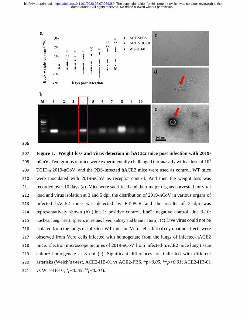
Figure 1. Weight loss and virus detection in hACE2 mice post infection with 2019-207
nCoV. Two groups of mice were experimentally challenged intranasally with a dose of 105 208
TCID50 2019-nCoV, and the PBS-infected hACE2 mice were used as control. WT mice 209
were inoculated with 2019-nCoV as receptor control. And then the weight loss was 210
recorded over 10 days (a). Mice were sacrificed and their major organs harvested for viral 211
load and virus isolation at 3 and 5 dpi, the distribution of 2019-nCoV in various organs of 212
infected hACE2 mice was detected by RT-PCR and the results of 3 dpi was 213
representatively shown (b) (line 1: positive control, line2: negative control, line 3-10: 214
trachea, lung, heart, spleen, intestine, liver, kidney and brain in turn). (c) Live virus could not be 215
isolated from the lungs of infected-WT mice on Vero cells, but (d) cytopathic effects were 216
observed from Vero cells infected with homogenate from the lungs of infected-hACE2 217
mice. Electron microscope pictures of 2019-nCoV from infected-hACE2 mice lung tissue 218
culture homogenate at 3 dpi (e). Significant differences are indicated with different 219
asterisks (Welch’s t-test, ACE2-HB-01 vs ACE2-PBS, *p<0.05, **p<0.01; ACE2-HB-01 220
vs WT-HB-01, #p<0.05, ##p<0.01). 221
author/funder. All rights reserved. No reuse allowed without permission. The copyright holder for this preprint (which was not peer-reviewed) is the. https://doi.org/10.1101/2020.02.07.939389doi: bioRxiv preprint
222
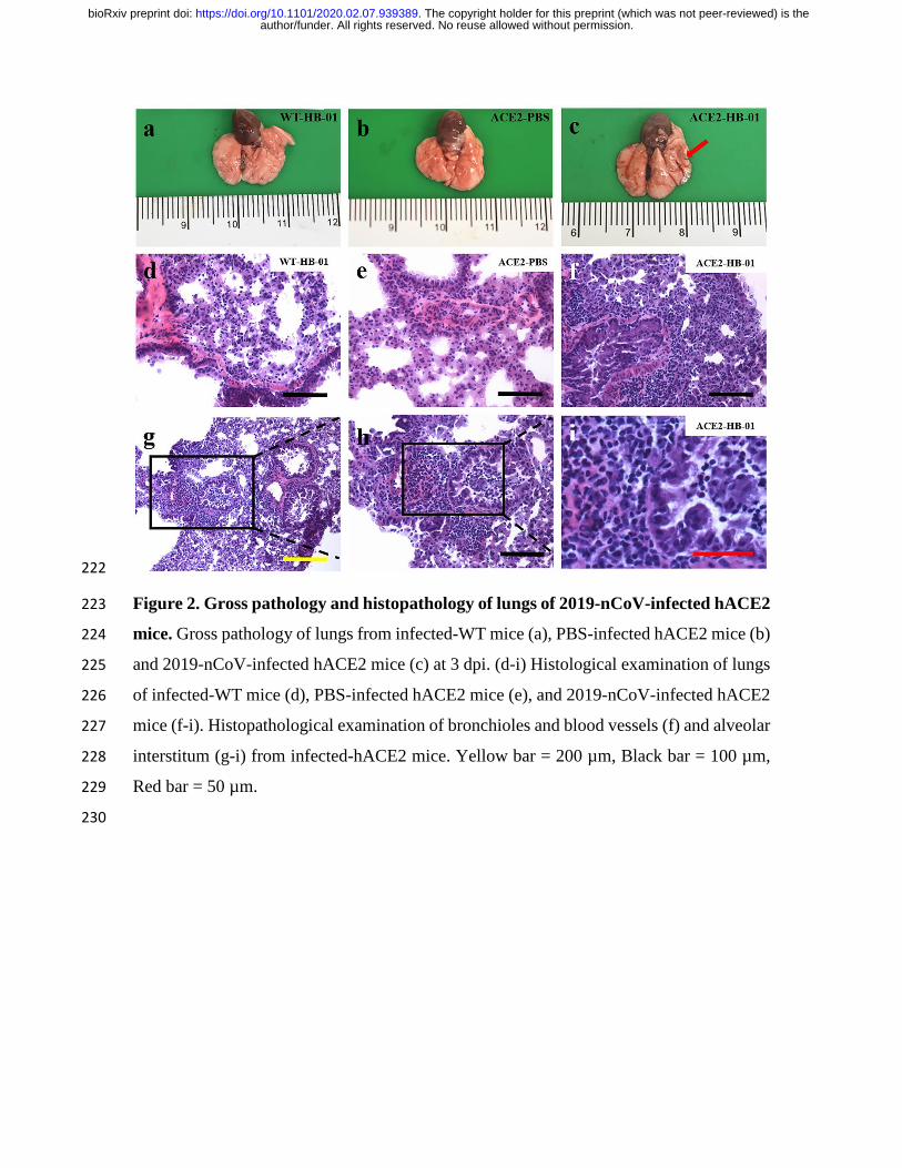
Figure 2. Gross pathology and histopathology of lungs of 2019-nCoV-infected hACE2 223
mice. Gross pathology of lungs from infected-WT mice (a), PBS-infected hACE2 mice (b) 224
and 2019-nCoV-infected hACE2 mice (c) at 3 dpi. (d-i) Histological examination of lungs 225
of infected-WT mice (d), PBS-infected hACE2 mice (e), and 2019-nCoV-infected hACE2 226
mice (f-i). Histopathological examination of bronchioles and blood vessels (f) and alveolar 227
interstitum (g-i) from infected-hACE2 mice. Yellow bar = 200 µm, Black bar = 100 µm, 228
Red bar = 50 µm. 229
230
author/funder. All rights reserved. No reuse allowed without permission. The copyright holder for this preprint (which was not peer-reviewed) is the. https://doi.org/10.1101/2020.02.07.939389doi: bioRxiv preprint

231
Figure 3. Immunofluorescence analysis of viral antigens in lungs of 2019-nCoV-232
infected hACE2 mice. (a-i): Fluorescence of sections of mice lungs after incubation with 233
DAPI or antisera of 2019-nCoV-convalescent patients respectively. The lung sections of 234
infected-WT mice (a-c), PBS-infected hACE2 mice (d-f) and infected-hACE2 mice (g-i). 235
Green arrows indicate presence of 2019-nCoV in the alveolar epithelial cells. (j-q): Co-236
localization of 2019-nCoV S protein and hACE2 receptor in hACE2 mouse lungs, the 237
sections were incubated with DAPI, a polyclonal antibody against 2019-nCoV S protein or 238
author/funder. All rights reserved. No reuse allowed without permission. The copyright holder for this preprint (which was not peer-reviewed) is the. https://doi.org/10.1101/2020.02.07.939389doi: bioRxiv preprint
human ACE2 protein respectively. The lung sections of PBS-infected hACE2 mice (j-m). 239
The lung sections of infected-hACE2 mice (n-q). The white arrows showed the viral S 240
protein (o) and hACE2 (p) respectively, the yellow arrows showed the merge of viral S 241
protein and hACE2 (q). 242
243
author/funder. All rights reserved. No reuse allowed without permission. The copyright holder for this preprint (which was not peer-reviewed) is the. https://doi.org/10.1101/2020.02.07.939389doi: bioRxiv preprint

244
Supplementary Figure 1. Identification of 7D2 antibody against 2019-nCoV S1 245
protein. The plate coated by 0.2 ug 2019-nCoV S1 protein was incubated with 7D2 246
antibody as primary antibody (1:200) and detected using HRP-conjugated goat anti-mouse 247
secondary antibody. The titer of antibody was determined using enzyme-linked 248
immunosorbent assay (ELISA) assay. 249
250
author/funder. All rights reserved. No reuse allowed without permission. The copyright holder for this preprint (which was not peer-reviewed) is the. https://doi.org/10.1101/2020.02.07.939389doi: bioRxiv preprint