
Molecular pathophysiology of fragile X-associated tremor ...
31
TITLE: Molecular pathophysiology of fragile X-associated tremor/ataxia syndrome and perspectives for drug development AUTHORS: Teresa Botta-Orfila 1,2 , Gian Gaetano Tartaglia 1,2,3 , Aubin Michalon 4 * AFFILIATIONS: 1. Fundació Centre for Genomic Regulation (CRG), Dr Aiguader 88, 08003 Barcelona, Spain 2. Universitat Pompeu Fabra (UPF), 08003 Barcelona, Spain. 3. Institució Catalana de Recerca i Estudis Avançats (ICREA), 23 Passeig Lluís Companys, 08010 Barcelona, Spain. 4. Neurimmune, Wagistrasse 13, 8952 Schlieren, Switzerland. *Corresponding author: [email protected] ABSTRACT: Fragile X-associated tremor/ataxia syndrome (FXTAS) is an inherited neurodegenerative disorder manifesting in carriers of 55 to 200 CGG repeats in the 5' untranslated region (UTR) of the fragile X mental retardation gene (FMR1). FXTAS is characterized by enhanced FMR1 transcription and the accumulation of CGG repeat-containing FMR1 mRNA in nuclear foci, while the FMRP protein expression levels remain normal or moderately low. The neuropathological hallmark in FXTAS is the presence of intranuclear, ubiquitin positive inclusions that also contain FMR1 transcript. Yet the complete protein complement of FXTAS inclusions and the molecular events that trigger neuronal death in FXTAS remain unclear. In this review we present the two most accepted toxicity mechanisms described so far, namely RNA gain-of-function and protein gain-of-function by means of repeat- associated non-AUG translation, and discuss current experimental and computational strategies to better understand FXTAS pathogenesis. Finally, we review the current perspectives for drug development with disease modifying potential for FXTAS. Keywords: FXTAS, premutation, RNA toxicity, RNA binding-protein, RAN translation, FMRpolyG,
Transcript of Molecular pathophysiology of fragile X-associated tremor ...
TITLE:
Molecular pathophysiology of fragile X-associated tremor/ataxia syndrome and
perspectives for drug development
AUTHORS:
Teresa Botta-Orfila1,2, Gian Gaetano Tartaglia1,2,3, Aubin Michalon4 *
AFFILIATIONS: 1. Fundació Centre for Genomic Regulation (CRG), Dr Aiguader 88, 08003 Barcelona, Spain
2. Universitat Pompeu Fabra (UPF), 08003 Barcelona, Spain.
3. Institució Catalana de Recerca i Estudis Avançats (ICREA), 23 Passeig Lluís Companys,
08010 Barcelona, Spain.
4. Neurimmune, Wagistrasse 13, 8952 Schlieren, Switzerland.
*Corresponding author: [email protected]
ABSTRACT: Fragile X-associated tremor/ataxia syndrome (FXTAS) is an inherited neurodegenerative
disorder manifesting in carriers of 55 to 200 CGG repeats in the 5' untranslated region (UTR)
of the fragile X mental retardation gene (FMR1). FXTAS is characterized by enhanced FMR1
transcription and the accumulation of CGG repeat-containing FMR1 mRNA in nuclear foci,
while the FMRP protein expression levels remain normal or moderately low. The
neuropathological hallmark in FXTAS is the presence of intranuclear, ubiquitin positive
inclusions that also contain FMR1 transcript. Yet the complete protein complement of
FXTAS inclusions and the molecular events that trigger neuronal death in FXTAS remain
unclear. In this review we present the two most accepted toxicity mechanisms described so
far, namely RNA gain-of-function and protein gain-of-function by means of repeat-
associated non-AUG translation, and discuss current experimental and computational
strategies to better understand FXTAS pathogenesis. Finally, we review the current
perspectives for drug development with disease modifying potential for FXTAS.
Keywords: FXTAS, premutation, RNA toxicity, RNA binding-protein, RAN translation,
FMRpolyG,
Introduction
Fragile X-associated tremor/ataxia syndrome (FXTAS) is an inherited and age-dependent
neurodegenerative disorder characterized by late-onset, progressive intention tremor and
gait ataxia (Hagerman and Hagerman, 2015). In addition to these core clinical features,
FXTAS is commonly associated with parkinsonism, autonomic dysfunction, cognitive decline,
dementia, emotional liability and peripheral neuropathy, as well as white matter
abnormalities and global brain atrophy that can be observed on magnetic resonance
imaging (Brown and Stanfield, 2015; Hagerman and Hagerman, 2015). FXTAS is linked to a
dominant mutation on the X chromosome, leading to milder clinical presentation in female
patients and higher disease penetrance at about 45% in males compared to 16% only in
females (Rodriguez-Revenga et al., 2009).
FXTAS patients are carriers of a trinucleotide CGG repeat expansion in the 5' untranslated
region (UTR) of the fragile X mental retardation gene (FMR1). The CGG repeat locus
typically contains a variable number of repeats between 3 and 45 in the general population,
and expands to a pathogenic range named premutation from 55 to 200 repeats causing
FXTAS (Cronister et al., 2008). In contrast, the full mutation above 200 repeats results in
DNA methylation and gene silencing leading to fragile X syndrome (FXS), the most frequent
genetic cause for neurodevelopmental disorder (Usdin et al., 2014). The FMR1 premutation
is also associated in women to a different clinical phenotype characterized by premature
ovarian insufficiency, as well as high frequency of thyroid disease, hypertension, seizures,
peripheral neuropathy, and fibromyalgia (Rodriguez-Revenga et al., 2009). Due to large
differences in disease manifestation, FXTAS is often misdiagnosed and many patients are
identified only after a grandchild is diagnosed with FXS (Leehey, 2009). Patient treatment is
currently limited to symptom management, with limited effects on disease progression
(Hagerman et al., 2008; Polussa et al., 2014). On this basis, understanding the molecular
mechanisms of FMR1 premutation toxicity is essential for the development of disease-
modifying therapies and improving disease prognosis.
At the molecular level, FXTAS is characterized by elevated levels of FMR1 transcription and
accumulation of the CGG repeat-containing FMR1 mRNA in nuclear foci, while the
expression levels of the encoded protein FMRP remain normal or moderately low due to a
decrease in translational efficiency (Kenneson et al., 2001; Primerano et al., 2002; Tassone
et al., 2000). As indicated by detailed analysis of transgenic mouse models and patient
samples, the increase in FMR1 transcription level is strongly correlated with the numbers of
CGG repeats, whereas FMRP expression level shows a weak, inverse correlation with repeat
number (Brouwer et al., 2008; Ludwig et al., 2014). Both may contribute to the clinical
profile of FXTAS patients, and there may be in fact a continuum of overlapping clinical and
molecular phenotypes between FXTAS and FXS, leading to the proposal of a “fragile X
spectrum disorder” (Lozano et al., 2014).
The neuropathological hallmark in FXTAS is the presence of well-delineated intranuclear,
eosinophilic, ubiquitin and αB-crystallin positive inclusions, which contain the FMR1
transcript. These inclusions are present not only in neurons and astrocytes with a broad
distribution throughout the central nervous system, but also in various peripheral tissues.
Specifically, inclusions have been observed in the cerebral cortex, basal ganglia,
hippocampus, thalamus, substantia nigra, and ependymal and choroid plexus nuclei; in the
spinal cord and autonomic ganglia; in the heart and kidney; and in the thyroid, pineal,
pituitary and testicular glands (Gokden et al., 2009; Greco et al., 2006, 2007; Hunsaker et
al., 2011). The presence of intranuclear inclusions in various structures of the central and
peripheral nervous systems is consistent with the broad range of neurological symptoms
going from dementia to dysautonomia. A second prominent neuropathological feature
consists in white matter disease, characterized by spongiosis in subcortical and deep white
matter with corresponding axonal loss and glial cell loss (Greco et al., 2006). Patients also
present a moderate to severe loss of Purkinje cells, yet the remaining Purkinje cells only
rarely present intranuclear inclusions – or only in older patients (Ariza et al., 2016).
The frequency of cells presenting intranuclear inclusions is increasing with CGG expansion
size, but it is still under debate whether these inclusions are toxic per se or if they are simply
a side product of the neutralization of toxic FMR1 transcripts (Greco et al., 2006). Attempts
to characterize the protein complement of intranuclear inclusions present in FXTAS indicates
that ubiquitinated proteins account only for a minor fraction, suggesting that the inclusions
do not result from a failure of the nuclear proteosomal system (Iwahashi et al., 2006).
Detailed histological studies found that neurons with intranuclear inclusions are not
structurally altered, whereas in astrocytes the presence of inclusions is associated with
increased size of the nucleus (Greco et al., 2002). In fact, the presence of inclusions is not
associated with the typical morphological signature indicative of progressive neuronal
death.
Why do CGG repeats become toxic upon expansion? How do they trigger neuronal death?
Even though these questions are not fully elucidated yet, significant progress has been
achieved with the identification of two different mechanisms involving RNA- and protein-
mediated toxicity, presented hereafter. The identification of these two toxic processes is
opening new perspectives for the development of innovative therapies, with the capacity to
halt and possibly reverse disease progression.
RNA-MEDIATED TOXICITY
Multiple microsatellite expansion diseases such as myotonic dystrophy type 1 (DM1) and
type 2 (DM2), spinocerebellar ataxia type 8, 10 and 31, Huntington's disease and FXTAS
have in common the presence of intranuclear RNA inclusions (Wojciechowska and
Krzyzosiak, 2011). It was first established in DM1, where a CTG repeat is present in the
3’UTR of the DMPK gene, that repeat expansion in a non-protein-coding region can lead to
RNA toxicity by a mechanism which was later found to occur in a relatively similar manner in
other repeat expansion diseases. In essence, this toxicity mechanism consists in the
sequestration of various proteins at the repeat site on the transcripts, leading to depletion
of these proteins from the common pool and to their accumulation in the form of nuclear
inclusions. Such a mechanism is taking place in FXTAS, and the identification of the proteins
sequestered in nuclear inclusions is an essential step for understanding how toxicity does
occur.
A biochemical analysis performed on aggregate-containing nuclei extracted from FXTAS
patient brain material identified more than 20 different proteins including the RNA binding
proteins hnRNP A2/B1 and MBNL1, and the chaperones HSP70, HSP27 and αB-crystallin
(Iwahashi et al., 2006). Nuclear inclusions also contained structural proteins such as lamin
A/C, GFAP and golli-MBP, as well as proteins like MBP which presence in the nucleus had
never been reported before. The presence of the splicing factor MBNL1 is particularly
interesting as it is the one protein sequestered in DM1 and DM2, which depletion from the
intracellular pool leads to splicing alterations and clinical symptoms (Ranum and Cooper,
2006). However, a role for MBNL1 in FXTAS has been excluded, because no alterations in
splicing events regulated by MBNL1 were observed in human brain material from FXTAS
patients (Sellier et al., 2010a). Another interesting case is hnRNP A2/B1, which is a DNA and
RNA binding protein involved in a large number of RNA processing mechanisms including
transcription, mRNA splicing and microRNA biogenesis. HnRNP A2/B1 plays several key
roles in neuronal functioning and reduction in its expression level contributes to the severity
of symptoms in several neurodegenerative diseases including Alzheimer's disease
(Bekenstein and Soreq, 2013). On this basis, depletion of hnRNP A2/B1 from the
cytoplasmic pool upon binding to RNA containing expanded CGG repeats was thought to
be an important factor driving toxicity in FXTAS. This hypothesis was confirmed using
transgenic Drosophila expressing expanded CGG repeats, where the overexpression of
hnRNP A2/B1 was sufficient to rescue the neurodegenerative phenotype (Sofola et al.,
2007). Furthermore, it was observed in primary neurons expressing an expanded CGG
repeat that depletion of hnRNP A2/B1 from the cytoplasmic pool is leading to alteration in
the RNA dendritic transport pathway (Muslimov et al., 2011).
Other proteins were identified which follow the same model of toxicity, where protein
binding to RNA containing expanded CGG repeats leads to depletion from the active pool
and alterations in the RNA processing machinery. Among them, the protein Sam68, officially
named KHDRBS1, is a nucleo-cytoplasmic protein involved in multiple cellular processes
including the regulation of alternative splicing. Sam68 was found to bind to expanded CGG
repeats and thereby to lose its splicing-regulatory function, which in turn leads to splicing
alterations in FXTAS patients (Sellier et al., 2010a). Interestingly in neurons, Sam68 is a key
regulator of activity-dependent splicing of neurexin 1, a cell adhesion molecule expressed at
synapses with essential function in the assembly of synapses and synaptic circuits (Iijima et
al., 2011). Sam68 is also involved in synaptodendritic post-transcriptional regulation of beta-
actin and its acute knockdown results in fewer excitatory synapses in the hippocampal
formation (Klein et al., 2013).
Another level of dysregulation resulting from protein sequestration by CGG repeat-
containing mRNAs has to do with microRNAs (miRNAs), which are essential for RNA
silencing and post-transcriptional regulation of gene expression (Bartel, 2009). Along the
miRNA processing pathway, the enzymatic complex DROSHA-DGCR8 has a key function for
the processing of pri-microRNAs into pre-miRNAs. DROSHA-DGCR8 was found to associate
specifically with CGG repeats of pathogenic size, and its sequestration within nuclear
inclusions results in reduced levels of miRNAs not only in cultured cells but also in the brain
of FXTAS patients (Sellier et al., 2013). Alterations in miRNA expression levels have also
been observed in lymphocytes collected from FXTAS patients, and in the brain of a
Drosophila model for CGG expansion (Alvarez-Mora et al., 2013; Tan et al., 2012).
Interestingly, overexpression of DGCR8 was sufficient to rescue the cellular toxicity induced
by expression of expanded CGG repeats in mouse primary neurons, indicating that
DROSHA-DGCR8 sequestration is a major liability for the cells (Sellier et al., 2013).
Moreover, DGCR8 is required for proper regulation of gene expression in neurons, and a
modest decrease in DGCR8 expression levels obtained by deletion of one Dgcr8 allele is
sufficient to induce alterations in the density, morphology and electrophysiological
properties of cortical neurons in mice (Fénelon et al., 2011). DGCR8 was also found recently
to participate in the regulation of neuronal morphogenesis (Luhur et al., 2014). Altogether,
these results suggest that DROSHA-DGCR8 sequestration and the resulting alterations in
miRNA expression may not only contribute to the late-onset neurodegenerative phenotype
in FXTAS, but also to the cognitive and psychiatric symptoms, which manifest in patients
sometimes long before the onset of neurodegenerative symptoms (Grigsby et al., 2014;
Seritan et al., 2013).
The transcriptional activator protein Pur-alpha is another protein that is sequestered in the
nucleus: Pur-alpha was shown to bind strongly to CGG repeat RNA in vitro, to be present in
intranuclear inclusions in patient brain tissue, and its overexpression in Drosophila was
sufficient to abolish the neurodegenerative phenotype caused by CGG repeats (Jin et al.,
2007). Here again, dysregulation of Pur-alpha activity upon sequestration is highly
susceptible to deteriorate neuronal activity, as indicated by the severe neurodevelopmental
phenotype observed in human carriers of mutations in Pur-alpha, including mental
retardation, encephalopathy and seizures (Hunt et al., 2014; Lalani et al., 2014).
Yet, CGG repeat expansion in the premutation range is a mutation with limited penetrance.
What are the genetic and environmental factors that determine individual susceptibility to
CGG expansion? Even though this question may be too vast to answer, especially if one
would consider the interactions between genes and environment, it may be of interest to
mention a study which identified the suppressor effect of TDP-43 on premutation toxicity in
Drosophila. This effect is related to TDP-43 capacity to interact with hnRNP A2/B1 and
prevent the alterations in RNA splicing that are triggered by hnRNP A2/B1 sequestration
(He et al., 2014). Extrapolating from that, one could speculate that individuals with high
TDP-43 expression levels would have a better resilience to CGG expansion. In contrast to
that, specific environmental factors such as exposure to neurotoxins, chemotherapy or
general anesthesia are suspected to precipitate the onset of FXTAS disease (O’Dwyer et al.,
2005; Paul et al., 2010).
The few proteins described above help to understand the RNA toxicity model, where the
sequestration of specific proteins at the CGG repeat RNA alter various intracellular
processes which may individually or synergistically lead to symptoms. Yet, this model is far
from being complete, especially with regards to possible differences between cell types and
tissues, and a more granular characterization of proteins sequestered at the CGG repeat
RNA could improve our understanding of the disease, especially at early stages, and identify
targets for pharmacological intervention.
Many techniques have been recently developed with the aim of discovering novel protein-
RNA interactions, starting from either a known protein or a known RNA. The so-called RNA-
centric approaches, where RNA is the bait, are used to identify proteins which associate with
a specific RNA Two different strategies have been developed: RBP screening within protein
libraries e.g. protein arrays (Siprashvili et al., 2012), and RNA affinity capture coupled with
mass spectrometry for protein identification, with various protocols such as CHART (Simon,
2013), MS2-BioTRAP (Tsai et al., 2011) and ChIRP (Chu et al., 2012). Complementary
experimental approaches such as RNA FISH, widely used for detecting expanded repeats in
diseases with RNA foci, can also be applied to study FXTAS RNA-protein interactions and
inclusions formation (Urbanek and Krzyzosiak, 2015).
To complement the experimental approaches which are typically work intensive,
computational methods have been developed to support the discovery of proteins involved
in protein-RNA complexes. Especially, the algorithm catRAPID is designed to predict which
proteins bind to a specific RNA sequence; it differentiates spurious interactions from those
with high affinity, and can be used to characterize associations with repeat expansion
(Agostini et al., 2013a; Bellucci et al., 2011). catRAPID was initially trained on a set of about
1000 protein-RNA complexes available in the Protein Data Bank to discriminate interacting
and non-interacting molecules using the information contained in primary structures.
Predictions of secondary structure, hydrogen bonding and van der Waals’ are combined
together to calculate the interaction propensities. The algorithm was tested against the non-
nucleic-acid-binding dataset NNBP (area under ROC curve of 0.92) (Stawiski et al., 2003),
the non-coding RNA database NPInter (area under the ROC curve of 0.88) (Wu et al., 2006),
and a number of interactions validated by RNA immunoprecipitation (RIP) and cross-linking
immunoprecipitation (CLIP) approaches (Agostini et al., 2013b; Cirillo et al., 2013, 2014;
Zanzoni et al., 2013)
Applied to the CGG repeat RNA, catRAPID predicted the sequestration of several proteins
including MBNL1 and the heterogeneous nuclear ribonucleoproteins (hnRNP) A1, A2/B1, C,
D, E, and G, whereas proteins such as FMRP, CUGBP1 and Sam68 were predicted not to
show any specific interaction with the CGG repeat RNA (figure 1A) (Cirillo et al., 2013). This
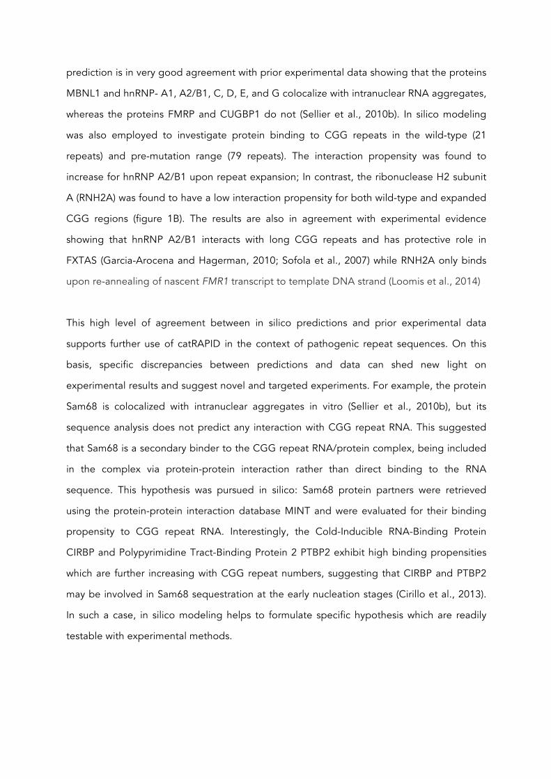
prediction is in very good agreement with prior experimental data showing that the proteins
MBNL1 and hnRNP- A1, A2/B1, C, D, E, and G colocalize with intranuclear RNA aggregates,
whereas the proteins FMRP and CUGBP1 do not (Sellier et al., 2010b). In silico modeling
was also employed to investigate protein binding to CGG repeats in the wild-type (21
repeats) and pre-mutation range (79 repeats). The interaction propensity was found to
increase for hnRNP A2/B1 upon repeat expansion; In contrast, the ribonuclease H2 subunit
A (RNH2A) was found to have a low interaction propensity for both wild-type and expanded
CGG regions (figure 1B). The results are also in agreement with experimental evidence
showing that hnRNP A2/B1 interacts with long CGG repeats and has protective role in
FXTAS (Garcia-Arocena and Hagerman, 2010; Sofola et al., 2007) while RNH2A only binds
upon re-annealing of nascent FMR1 transcript to template DNA strand (Loomis et al., 2014)
This high level of agreement between in silico predictions and prior experimental data
supports further use of catRAPID in the context of pathogenic repeat sequences. On this
basis, specific discrepancies between predictions and data can shed new light on
experimental results and suggest novel and targeted experiments. For example, the protein
Sam68 is colocalized with intranuclear aggregates in vitro (Sellier et al., 2010b), but its
sequence analysis does not predict any interaction with CGG repeat RNA. This suggested
that Sam68 is a secondary binder to the CGG repeat RNA/protein complex, being included
in the complex via protein-protein interaction rather than direct binding to the RNA
sequence. This hypothesis was pursued in silico: Sam68 protein partners were retrieved
using the protein-protein interaction database MINT and were evaluated for their binding
propensity to CGG repeat RNA. Interestingly, the Cold-Inducible RNA-Binding Protein
CIRBP and Polypyrimidine Tract-Binding Protein 2 PTBP2 exhibit high binding propensities
which are further increasing with CGG repeat numbers, suggesting that CIRBP and PTBP2
may be involved in Sam68 sequestration at the early nucleation stages (Cirillo et al., 2013).
In such a case, in silico modeling helps to formulate specific hypothesis which are readily
testable with experimental methods.
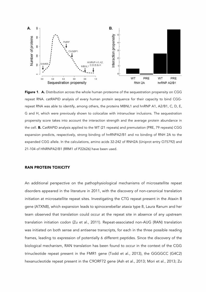
Figure 1. A. Distribution across the whole human proteome of the sequestration propensity on CGG
repeat RNA. catRAPID analysis of every human protein sequence for their capacity to bind CGG-
repeat RNA was able to identify, among others, the proteins MBNL1 and hnRNP A1, A2/B1, C, D, E,
G and H, which were previously shown to colocalize with intranuclear inclusions. The sequestration
propensity score takes into account the interaction strength and the average protein abundance in
the cell. B. CatRAPID analysis applied to the WT (21 repeats) and premutation (PRE, 79 repeats) CGG
expansion predicts, respectively, strong binding of hnRNPA2/B1 and no binding of RNH 2A to the
expanded CGG allele. In the calculations, amino acids 32-242 of RNH2A (Uniprot entry O75792) and
21-104 of HNRNPA2/B1 (RRM1 of P22626) have been used.
RAN PROTEIN TOXICITY
An additional perspective on the pathophysiological mechanisms of microsatellite repeat
disorders appeared in the literature in 2011, with the discovery of non-canonical translation
initiation at microsatellite repeat sites. Investigating the CTG repeat present in the Ataxin 8
gene (ATXN8), which expansion leads to spinocerebellar ataxia type 8, Laura Ranum and her
team observed that translation could occur at the repeat site in absence of any upstream
translation initiation codon (Zu et al., 2011). Repeat-associated non-AUG (RAN) translation
was initiated on both sense and antisense transcripts, for each in the three possible reading
frames, leading to expression of potentially 6 different peptides. Since the discovery of the
biological mechanism, RAN translation has been found to occur in the context of the CGG
trinucleotide repeat present in the FMR1 gene (Todd et al., 2013), the GGGGCC (G4C2)
hexanucleotide repeat present in the C9ORF72 gene (Ash et al., 2013; Mori et al., 2013; Zu
et al., 2013), and the CAG repeat present in the huntingtin (HTT) gene (Bañez-Coronel et al.,
2015).
FMR1 RAN translation products
The CGG repeat in the FMR1 gene is located in the 5’UTR of the transcript, and is therefore
not a target for the standard translation machinery. Using transgenic flies and human cells,
Peter Todd and co-workers described that translation can be initiated by the CGG repeat
itself in an AUG independent manner, in at least 2 of the 3 possible reading frames of the
sense transcript (Todd et al., 2013).
FMRpolyG is the main RAN translation product, which has been detected in flies, transgenic
mouse models, human cultured cells and FXTAS patient tissues. FMRpolyG consists of a
homopeptidic glycine domain, the translation product of the expansion in the glycine-
encoding frame (GGC), together with the in-frame translation product of the 5’UTR of FMR1
(Todd et al., 2013). Remarkably, FMRpolyG expression was observed in vitro not only with
repeat expansions of pathological size (over 55 repeats), but also with expansions of 30
repeats only, a length which is common in the general population and regarded as non-
pathological. Using different antibodies directed against the C-terminal 5’UTR translation
product, FMRpolyG was detected specifically in brain tissue from FXTAS patients but not
from control, demented and non-demented autopsy cases (Buijsen et al., 2014; Todd et al.,
2013). FMRpolyG was detected in multiple areas of the brain and peripheral tissues as
intranuclear aggregates which are often, but not always, colocalized with ubiquitin-positive
inclusions. FMRpolyG was also reported in tissues from patients with fragile X-associated
primary ovarian insufficiency (Buijsen et al., 2016).
Another RAN translation product was identified in vitro, in the alanine-encoding frame
(GCG). In this case, the protein was detected as a GFP fusion protein after removal of a stop
codon in the 5’UTR shortly after the CGG repeat (Todd et al., 2013). Under native
conditions, FMRpolyA protein would consist of the polyalanine domain and a short C-
terminal segment of 16 amino acids only, corresponding to the in-frame translation of the
5’UTR up until the stop codon. It is not known whether FMRpolyA expression occurs in
patients, a question which could be answered using antibodies generated specifically
against this 16 amino acid peptide.
The last potential RAN translation product on the FMR1 sense transcript is in the arginine-
encoding frame (GGC). There is no stop codon present in that frame in the 5’UTR of FMR1,
such that the theoretical FMRpolyR protein would be de facto a high molecular weight
(HMW) variant of FMRP, with an extra N-terminal part including the poly-arginine domain
and translation of the 5’UTR. RAN translation in the arginine-encoding frame was neither
detected in vitro using GFP fusion protein as detection system, nor in transgenic Drosophila
using mass spectrometry for protein identification (Todd et al., 2013), and there is no report
known to us of a HMW FMRP variant.
RAN translation was found to occur also from antisense transcripts including the CTG
repeats in the ATXN8 gene, the G4C2 repeat in C9ORF72 and the CAG repeat in the HTT
gene (Bañez-Coronel et al., 2015; Gendron et al., 2013; Zu et al., 2011). Antisense
transcription at the FMR1 locus leads to the expression of ASFMR1, a RNA molecule which
is spliced, polyadenylated and exported to the cytoplasm, which covers the repeat site and
is therefore a candidate for RAN translation (Ladd et al., 2007). Similar to FMR1 mRNA, the
expression level of ASFMR1 changes with repeat expansion, with elevated expression level
in cells bearing premutation alleles and expression silencing in cells with full mutation
alleles. Presence of RAN translation at the CCG repeat on ASFMR1 transcripts has not been
reported so far, but would lead to the expression of ASFMRpolyP (CCG), polyA (GCC) and
polyR (CGC) proteins. Knowing whether these RAN proteins are produced in FXTAS
patients will be very important for a complete understanding of the disease
pathophysiology.
RAN protein toxicity in FXTAS
The mechanisms of repeat expansion toxicity have been intensively investigated in the
context of the polyglutamine and polyalanine –associated diseases, for which expansions
occur in the middle of a protein coding region and lead to toxic gain-of-functions. A generic
mechanism emerged from these studies, where polyQ and polyA expansions lead to the
expression of misfolded proteins that are poorly degraded, accumulate, form aggregates
and interfere with specific intracellular processes, leading ultimately to cell toxicity and age-
dependent neurodegeneration. A similar mechanism could be driven by FMRpolyG and
contribute to FXTAS disease.
In vitro experiments on human cells indicated that RAN translation drives the expression of
FMRpolyG at similar levels for constructs with pathological repeat expansion sizes (>55
repeats) and non-pathological expansion size (30 repeats) (Todd et al., 2013). In contrast,
FMRpolyG-induced toxicity was observed for long FMRpolyG proteins expressed from
constructs with long CGG expansions, and not for short FMRpolyG expressed from
constructs with 30 repeats only. The intracellular expression profile also differed with repeat
numbers: long FMRpolyG formed cytoplasmic and intranuclear inclusions, whereas short
FMRpolyG expression was diffuse throughout the cytoplasm. FMRpolyG inclusions stained
positive for ubiquitin and chaperone HSP70, indicating the protein quality control system
recognized long FMRpolyG as misfolded protein and targeted it for degradation. Finally,
mutations which reduced proteasome activity worsened FMRpolyG toxicity, whereas
overexpression of chaperone proteins such as HSP70 reduced it (Oh et al., 2015).
These observations suggest that the length of the polyglycine domain is an essential
determinant of aggregate formation and toxicity, and is possibly the element linking repeat
expansion size with disease severity. Indeed, glycine is a weakly hydrophobic amino acid,
and glycine homopeptides are characterized by poor solubility in water and rapid formation
of insoluble aggregates for pure peptides as short as 9 successive glycines (Ohnishi et al.,
2006). When homopeptides are expressed in combination with a soluble protein such as
YFP, a much longer hydrophobic stretch is required to trigger aggregation. For example,
the expression of the fusion protein Ala70-YFP resulted in small and dispersed cytoplasmic
aggregates which were not observed upon expression of Ala30-YFP protein (Oma et al.,
2004). PolyQ-YFP proteins exhibited a similar length-dependent aggregation phenotype,
indicating that the size of the hydrophobic homopeptide domain is a major determinant for
protein aggregation and cellular toxicity (Oma et al., 2005, 2007).
The link between surface hydrophobicity, formation of oligomers and toxicity has been
thoroughly investigated, because protein misfolding and aggregate formation is the
hallmark of many diseases including numerous neurodegenerative conditions (Ciryam et al.,
2013; Olzscha et al., 2011). Toxicity results from a combination of surface hydrophobicity
and size, with the most toxic aggregates having high hydrophobicity and small size (Mannini
et al., 2014). Small aggregates exhibit a higher surface hydrophobicity than large ones, and
have therefore a higher capacity to interfere with various signaling processes (Campioni et
al., 2010). Small hydrophobic aggregates can, for example, penetrate membrane and cause
ionic leakage such as calcium influx, or interfere with intracellular signaling pathways. To
support the idea that macromolecular properties such as surface hydrophobicity are the
main drivers for toxicity, a genome-wide analysis was performed comparing mutations
increasing the aggregation potential of proteins are more often associated with human
diseases than neutral sequence variations (De Baets et al., 2015).
Aggregation-prone proteins are recognized by the protein folding quality control system
and targeted for degradation by the ubiquitin-proteasome system (UPS); in good
agreement, FMRpolyG was detected in the intranuclear inclusions which are positive for
ubiquitin and chaperone HSP70, suggesting that FMRpolyG itself is ubiquitinated (Oh et al.,
2015; Todd et al., 2013). Yet, FMRpolyG expression alters the activity of the UPS itself, as
revealed by the abnormal accumulation of a G76V-ubiquitin-GFP protein used as reporter
system for the monitoring of UPS activity (Oh et al., 2015). Inhibition of the UPS by
aggregation-prone protein is not a novel observation, but was already reported for alpha-
synuclein (Lindersson et al., 2004), mutant huntingtin (Díaz-Hernández et al., 2006; Ortega
et al., 2010), Abeta oligomers (Tseng et al., 2008), GFAP oligomers (Tang et al., 2010), and
the toxic conformation of prion protein (Prpsc) (Deriziotis et al., 2011; McKinnon et al.,
2015). Thus, there are common toxicity mechanisms shared by aggregation-prone proteins,
and the same intracellular toxicity mechanisms may be at play for all the RAN proteins that
form aggregates independently of the gene where the repeat expansion occurs.
Within each cell, the loss of control over the degradation of aggregation-prone proteins is
possibly the starting point of a long series of toxic insults impacting various molecular
processes. Toxicity is related to intrinsic factors such as surface hydrophobicity and structural
flexibility, as well as extrinsic factors such as intracellular localization. For example, the
accumulation of aggregation-prone peptides in the cytoplasm, but not in the nucleus, was
found to have a strong inhibitory effect on the nucleo-cytoplasmic transport of both proteins
and RNA molecules, leading to severe alterations in cytoplasmic mRNA content and protein
localization (Woerner et al., 2016). Mitochondrial dysfunction, altered calcium regulation and
altered zinc transport were also described in FXTAS disease (Kaplan et al., 2012; Napoli et
al., 2011; Ross-Inta et al., 2010). Such dysfunctions could happen as a direct consequence of
the expression of hydrophobic and aggregation-prone peptides, which could form pores
and disrupt membrane integrity, or as a downstream effect of nucleo-cytoplasmic transport
inhibition and its impact on protein expression. Mitochondrial dysfunction in FXTAS has
been established using primary fibroblasts obtained from FXTAS patients and non-
symptomatic premutation carriers. Interestingly, the specific deficits in the oxidative
phosphorylation pathway observed in FXTAS patients were also observed in aged and
young premutation carriers, indicating that mitochondrial dysfunction may precede the
onset of symptoms (Napoli et al., 2016; Ross-Inta et al., 2010). Furthermore, specific
measurements such as ATP-linked oxygen uptake, coupling, citrate synthase activity, and
mitochondrial network organization were correlated with the number of CGG repeats,
suggesting that the measurement of specific mitochondrial parameters could serve as
biomarker with potential prognosis value (Napoli et al., 2016).
Mechanism of RAN translation in FXTAS
The exact molecular mechanism of RAN translation is far from being fully elucidated.
Especially, it is unknown whether there is a unique mechanism valid for all repeat expansion
sites, nor if it is the same mechanism that initiates translation in different reading frames at
one expansion site. In the context of the CGG expansion in the FMR1 gene, RAN translation
in the glycine-coding frame was shown to occur in mammalian cells for expansions as short
as 30 repeats, the modal size in the human population. RAN translation level was not
increased, rather decreased, with longer expansions of 50 and 88 repeats (Todd et al.,
2013), suggesting that the CGG repeat is not inducing translation on its own but in
conjunction with other elements in close proximity. Indeed, RAN translation at this site in
the glycine frame is depending on the presence of pseudo AUG codons which are located -
11, -22 and -38 bp 5’ to the expansion. One pseudo AUG codon was sufficient for RAN
translation initiation, but translation level was stronger when at least 2 were present (Todd et
al., 2013). The experimental observations were different for RAN translation in the alanine
frame, which was detected from constructs with 88 repeats but not 30, and did not require
translation initiation upstream of the repeat. It suggests that two different mechanisms
support RAN translation in the glycine and alanine frames.
How could translation initiate at the CGG repeat in the 5’UTR of FMR1 gene? There are at
least 3 possible hypotheses, and so far limited experimental data to support one or the
other.
The first hypothesis involves standard cap-dependent recruitment of translation initiation
factors and ribosomal subunits for the formation of the pre-initiation complex. CGG repeats
are known to form hairpins and under specific conditions G-quadruplex structures, which are
very stable 2 and 3 dimensional structures, respectively, that are difficult to unfold (Kiliszek
et al., 2011; Kumar et al., 2011). They form a physical obstacle for the pre-initiation complex
forcing the scanning ribosome to pause (Khateb et al., 2007), allowing time for non-
canonical translation initiation at nearby pseudo-AUG codons (Kochetov et al., 2013).
Whereas this model seems plausible for RAN translation initiation in the glycine frame,
which is depending on the presence of pseudo-AUG codons just before the repeats, it
would not explain initiation in the alanine-coding frame.
A second hypothesis involves an internal ribosome entry site (IRES) located in the 5’UTR of
FMR1. IRES are not conserved sequences, but rather dense RNA tertiary structures that have
the capacity to interact with ribosomal proteins and initiate translation (Plank and Kieft,
2012). There are multiple families of IRES which share limited homology, and it has not been
possible yet to find universal rules to describe IRES structure and mechanism (Lozano and
Martínez-Salas, 2015). The presence of an IRES in the 5’UTR of FMR1 was characterized
using a standard mapping approach, where various segments of the 5’UTR were evaluated
for their capacity to induce translation of a reporter gene in a bicistronic construct. This
approach identified a 21 nt pyrimidine-rich region that is essential for cap-independent
translation initiation, and is located ~100 nt before the CGG expansion (-232 to -212 before
the ATG) (Chiang et al., 2001). The result was confirmed in another study which also
identified that the CGG repeats contribute to the IRES (repeat ablation reduced IRES activity
by 50%), that the CGG repeats and flanking sequences are themselves IRES elements (able
to induce cap-independent translation in absence of the pyrimidine-rich region), and that
IRES-dependent FMRP expression is responsive to various types of cell stimulation (Dobson
et al., 2008). There are multiple families of IRES, which differ among other things by their
capacity to initiate translation in absence of AUG codon. The IRES structure including the
CGG repeats, the flanking sequences and the pyrimidine-rich region could therefore have
the capacity to trigger expression of both FMRpolyG and polyA peptides.
Finally, a third hypothesis involves the action of a cryptic promoter located within the 5’UTR
of FMR1. The presence of a cryptic promoter was identified in the context of experiments
performed to characterize the IRES element in FMR1 leader. Dobson and his colleagues
observed that, in presence of the 5’UTR of FMR1, about 15% of the expression of a reporter
gene persisted after excision of the promoter from the vector, whereas expression was
absent in presence of unrelated 5’UTRs (Dobson et al., 2008). FMR1 cryptic promoter could
be responsible for the expression of a yet unidentified transcripts leading to RAN
translation.
THERAPEUTIC PERSPECTIVES
Genetic diseases such as FXTAS were perceived until recently as untreatable medical
conditions, and still now pharmacological care is limited to symptom management with
limited effects on disease progression (Hagerman et al., 2008; Polussa et al., 2014). This
perspective is changing with the emergence of new therapeutic modalities progressing at
rapid space from bench to bedside, including gene silencing and gene editing
technologies, a new class of small molecules to prevent RNA-mediated toxicity, and
proteasome activation approaches.
The use of gene silencing for drug development has long lived on promises, but this
technology has finally progressed to the point where the first drugs have reached the
market and many others are in clinical development. Gene silencing was initially based on
DNA or RNA oligonucleotides, which suffered from rapid degradation and limited cell
penetration in vivo, but these limitations have been circumvented with the development of
modified nucleotide analogues and advanced vectorization methods. This technology is
now being developed not only for peripheral diseases but also for neurodegenerative
disorders, with a clinical trial for Huntington’s disease (HD) initiated in 2015 leading the trail
in this field (trial identifier: NCT02519036). This clinical trial builds upon multiple
independent studies showing that a reduction in the expression level of mutant Huntingtin
protein (HTT) in the brain helped the elimination of intracellular protein aggregates and led
to significant cognitive and locomotor improvements (Aronin and DiFiglia, 2014). In January
2016, the FDA has granted orphan drug designation for the HTTRx drug, indicating strong
belief in its capacity to show efficacy in patients.
In the context of FXTAS, targeted degradation of FMR1 mRNA would have the capacity to
prevent both the RNA-mediated and the RAN protein-mediated toxicity, but the absence of
FMRP is known to cause fragile x syndrome (FXS) so this approach may not be beneficial for
the patients. This notwithstanding, a narrow therapeutic window may exist for precise gene
expression control in FXTAS: a modest and intermittent reduction in FMR1 mRNA levels
would help the cell to eliminate toxic RNA and RAN protein aggregates while preserving a
level of FMRP sufficient for normal neuron activity. In support of this idea, it was observed in
the context of Huntington’s disease that a short, 2 weeks treatment with antisense
oligonucleotides triggered long lasting therapeutic benefits, with improvement of
locomotor function and absence of polyQ aggregates persisting for at least 9 months after
the end of treatment (Kordasiewicz et al., 2012). As concerns FMRP levels, a detailed
analysis of methylation mosaicism in FXS patients indicated that residual FMRP expression
was associated with less severe forms of the disease (Pretto et al., 2014), suggesting that a
moderate and transient reduction in FMRP level may be well tolerated.
A more direct but still futuristic approach consists in genome editing and removal of the
CGG repeat expansion. The fundamentals of such an approach were established in vitro
using embryonic stem cells (ESCs) and induced pluripotent cells (iPSCs) derived from FXS
patients, in combination with the genome editing tool clustered regularly interspaced short
palindromic repeat (CRISPR)/ CRISPR-associated 9 (Cas9). In brief, the introduction of a
targeted DNA double strand cleavage a few nucleotides upstream the CGG repeat site in
FMR1 triggered deletions of variable sizes leading to reduction or complete ablation of the
CGG repeats, and reactivation of the expression of FMR1 gene (Park et al., 2015).
A different line of research is targeting specifically the deleterious effects of nuclear protein
sequestration at CGG expansion sites. A new class of small molecules is being developed,
which are characterized by their high affinity for CGG hairpins and their capacity to act as
chaperones and reduce protein sequestration. These compounds were selected on the
basis of their capacity to inhibit DGCR8 binding to CGG hairpins in vitro (Disney et al.,
2012). Tested in a cellular assay, they triggered a dose-dependent reduction in CGG
positive intranuclear inclusions, and prevented the mRNA splicing defects associated with
Sam68 sequestration at CGG hairpins (Disney et al., 2012; Tran et al., 2014). The
development of this new class of compounds is at an early stage with no information
available yet regarding in vivo activity; nevertheless, it would be very interesting to know if
these molecules are able to prevent RAN translation and FMRpolyG-mediated toxicity.
Similarly to many other toxic aggregation-prone proteins, FMRpolyG is targeted for
degradation by the UPS and is simultaneously an inhibitor of the UPS. This leads to
intracellular accumulation of FMRpolyG and toxicity, and suggests that increased
proteasome activity could prevent this toxic process. Protein homeostasis is maintained by
the coordinated activity of stress response pathways which are highly conserved in the
evolution and regulate protein synthesis, folding, trafficking, and degradation. Especially the
heat shock response (HSR) is rapidly induced by environmental and physiological stress
conditions, and is under control of the master regulator heat shock transcription factor 1
(HSF1). HSF1 is itself maintained in a monomeric inactive state by chaperone proteins such
as HSP90 and HSP70.
The antibiotic geldanamycin and its derivative 17-AAG (also called tanespimycin) are HSP90
inhibitors that have been tested in various cellular, insect and mammalian models of protein
aggregation diseases. By promoting the release of HSF1 from its complex with HSP90,
geldanamycin and 17-AAG trigger expression of chaperone molecules able to bind
misfolded proteins, neutralize their toxicity and promote their degradation. This was
observed in a mouse model of the polyQ expansion disease spino bulbar muscular atrophy
(SBMA), which upon treatment with 17-AAG or its oral form 17-DMAG presented reduced
motorneuron degeneration and significant improvements in locomotor function (Tokui et al.,
2009; Waza et al., 2005). Similarly, treatment with 17-AAG in a mouse model of the polyA
expansion disease oculopharyngeal muscular dystrophy (OPMD) promoted degradation of
the toxic protein by the UPS, reduced aggregate formation and increased in vitro and in
vivo cell survival (Shi et al., 2015). Similar results have also been obtained in the field of
amyloid diseases, supporting the idea that pharmacological UPR activation has a strong
therapeutic potential for aggregating protein diseases (Ryno et al., 2013), and could be on
this basis very useful against FMRpolyG toxicity. There are, however, some difficulties
related to long term toxicity of UPR activation and cellular adaptation leading to treatment
resistance (Labbadia et al., 2011). Targeting a specific process in the protein degradation
pathway such as selective inhibition of the deubiquitinating enzyme USP14 may prove more
translatable to patients (McKinnon et al., 2015).
Conclusion
At least two different pathogenic mechanisms have been identified for the toxicity of
neurodegenerative diseases related to microsatellite expansions, including protein gain-of-
function as in the case of Huntington’s disease and RNA gain-of-function as initially
described for myotonic dystrophy or FXTAS. The RNA gain-of-function hypothesis consists
in the sequestration of RNA-binding proteins by the mutant mRNA, leading to depletion of
these proteins from the pool required for normal gene expression and mRNA processing.
RNA-mediated sequestration of specific nuclear factors was shown to be intrinsically toxic, in
that overexpression of these factors could compensate sequestration and prevent toxicity.
Protein gain-of-function was not considered for repeat expansion located in 5’ and 3’UTRs
such as FXTAS, until discovery of the non-conventional RAN translation mechanism which
leads to the expression of aggregation-prone polypeptides with demonstrated toxicity.
These two mechanisms are not mutually exclusive and can certainly occur at the same time
within a cell. On this basis, FXTAS pathophysiology would be best described with a two-hit
model, where protein sequestration by mutant transcripts creates a liability for RAN protein
toxicity.
Acting upon one of these two mechanisms may be sufficient to reduce the amount of
molecular dysfunction to a level that is sustainable for the cells and prevent neurotoxicity.
This being said, there is no easy drug target identified so far for the treatment of FXTAS. By
targeting the root cause of the disease, genome editing would prevent both RNA-mediated
and RAN-protein-mediated toxicity, but in spite of tremendous progress in this field in the
recent years, there is no technology available yet for repeat removal in vivo in non-dividing
cells like neurons. In contrast to that, gene silencing technology has already reached
patients for the treatment of specific conditions, and is in clinical development for
Huntington’s disease. This technology could be easily adapted for use in FXTAS patients;
however, there is a fundamental antagonism between promoting FMR1 transcript
degradation for the treatment of FXTAS and preserving FMRP expression for cognitive
function. Finally, the use of drugs developed for their anti-tumoral activity such as 17-AAG,
which has already been tested in a phase 2 clinical trial against breast cancer (Modi et al.,
2011) may be the most pragmatic way forward, targeting specifically RAN protein toxicity.

In this context, more fundamental research is still needed to increase our understanding of
the molecular pathophysiology of FXTAS and other neurodegenerative conditions. The
emergence of in silico prediction tools such as catRAPID is paving the way toward a new
form of research in biology, where computer simulation is expanding research capacity and
throughput, and helping deciphering the complexity of biological processes.

Acknowledgements
Our research received funding from the European Union Seventh Framework Programme
(FP7/2007-2013), through the European Research Council, under grant agreement
RIBOMYLOME_309545 (Gian Gaetano Tartaglia), and from the Fundació La Marató de TV3
(20142731). We also acknowledge support from the Spanish Ministry of Economy and
Competitiveness (BFU2011-26206 and BFU2014-55054-P) and ‘Centro de Excelencia Severo
Ochoa 2013–2017’ (SEV-2012-0208).
CONFLICT OF INTEREST
The authors declare that they have no conflict of interest.
REFERENCES: www.zotero.org Agostini, F., Zanzoni, A., Klus, P., Marchese, D., Cirillo, D., and Tartaglia, G.G. (2013a). catRAPID omics: a web server for large-scale prediction of protein-RNA interactions. Bioinforma. Oxf. Engl.
Agostini, F., Cirillo, D., Bolognesi, B., and Tartaglia, G.G. (2013b). X-inactivation: quantitative predictions of protein interactions in the Xist network. Nucleic Acids Res. 41, e31.
Alvarez-Mora, M.I., Rodriguez-Revenga, L., Madrigal, I., Torres-Silva, F., Mateu-Huertas, E., Lizano, E., Friedländer, M.R., Martí, E., Estivill, X., and Milà, M. (2013). MicroRNA expression profiling in blood from fragile X-associated tremor/ataxia syndrome patients. Genes Brain Behav. 12, 595–603.
Ariza, J., Rogers, H., Monterrubio, A., Reyes-Miranda, A., Hagerman, P.J., and Martínez-Cerdeño, V. (2016). A Majority of FXTAS Cases Present with Intranuclear Inclusions Within Purkinje Cells. Cerebellum Lond. Engl.
Aronin, N., and DiFiglia, M. (2014). Huntingtin-lowering strategies in Huntington’s disease: antisense oligonucleotides, small RNAs, and gene editing. Mov. Disord. Off. J. Mov. Disord. Soc. 29, 1455–1461.
Ash, P.E.A., Bieniek, K.F., Gendron, T.F., Caulfield, T., Lin, W.-L., Dejesus-Hernandez, M., van Blitterswijk, M.M., Jansen-West, K., Paul, J.W., Rademakers, R., et al. (2013). Unconventional translation of C9ORF72 GGGGCC expansion generates insoluble polypeptides specific to c9FTD/ALS. Neuron 77, 639–646.
Bañez-Coronel, M., Ayhan, F., Tarabochia, A.D., Zu, T., Perez, B.A., Tusi, S.K., Pletnikova, O., Borchelt, D.R., Ross, C.A., Margolis, R.L., et al. (2015). RAN Translation in Huntington Disease. Neuron 88, 667–677.
Bartel, D.P. (2009). MicroRNAs: target recognition and regulatory functions. Cell 136, 215–233.
Bekenstein, U., and Soreq, H. (2013). Heterogeneous nuclear ribonucleoprotein A1 in health and neurodegenerative disease: from structural insights to post-transcriptional regulatory roles. Mol. Cell. Neurosci. 56, 436–446.
Bellucci, M., Agostini, F., Masin, M., and Tartaglia, G.G. (2011). Predicting protein associations with long noncoding RNAs. Nat. Methods 8, 444–445.
Brouwer, J.R., Huizer, K., Severijnen, L.-A., Hukema, R.K., Berman, R.F., Oostra, B.A., and Willemsen, R. (2008). CGG-repeat length and neuropathological and molecular correlates in a mouse model for fragile X-associated tremor/ataxia syndrome. J. Neurochem. 107, 1671–1682.
Brown, S.S.G., and Stanfield, A.C. (2015). Fragile X premutation carriers: A systematic review of neuroimaging findings. J. Neurol. Sci. 352, 19–28.
Buijsen, R.A.M., Sellier, C., Severijnen, L.-A.W.F.M., Oulad-Abdelghani, M., Verhagen, R.F.M., Berman, R.F., Charlet-Berguerand, N., Willemsen, R., and Hukema, R.K. (2014). FMRpolyG-positive inclusions in CNS and non-CNS organs of a fragile X premutation carrier with fragile X-associated tremor/ataxia syndrome. Acta Neuropathol. Commun. 2, 162.
Buijsen, R. a. M., Visser, J.A., Kramer, P., Severijnen, E. a. W.F.M., Gearing, M., Charlet-Berguerand, N., Sherman, S.L., Berman, R.F., Willemsen, R., and Hukema, R.K. (2016). Presence of inclusions positive for polyglycine containing protein, FMRpolyG, indicates that repeat-associated non-AUG translation plays a role in fragile X-associated primary ovarian insufficiency. Hum. Reprod. Oxf. Engl. 31, 158–168.
Campioni, S., Mannini, B., Zampagni, M., Pensalfini, A., Parrini, C., Evangelisti, E., Relini, A., Stefani, M., Dobson, C.M., Cecchi, C., et al. (2010). A causative link between the structure of aberrant protein oligomers and their toxicity. Nat. Chem. Biol. 6, 140–147.
Chiang, P.W., Carpenter, L.E., and Hagerman, P.J. (2001). The 5’-untranslated region of the FMR1 message facilitates translation by internal ribosome entry. J. Biol. Chem. 276, 37916–37921.
Chu, C., Quinn, J., and Chang, H.Y. (2012). Chromatin isolation by RNA purification (ChIRP). J. Vis. Exp. JoVE.
Cirillo, D., Agostini, F., Klus, P., Marchese, D., Rodriguez, S., Bolognesi, B., and Tartaglia, G.G. (2013). Neurodegenerative diseases: Quantitative predictions of protein-RNA interactions. RNA N. Y. N 19, 129–140.
Cirillo, D., Marchese, D., Agostini, F., Livi, C.M., Botta-Orfila, T., and Tartaglia, G.G. (2014). Constitutive patterns of gene expression regulated by RNA-binding proteins. Genome Biol. 15, R13.
Ciryam, P., Tartaglia, G.G., Morimoto, R.I., Dobson, C.M., and Vendruscolo, M. (2013). Widespread aggregation and neurodegenerative diseases are associated with supersaturated proteins. Cell Rep. 5, 781–790.
Cronister, A., Teicher, J., Rohlfs, E.M., Donnenfeld, A., and Hallam, S. (2008). Prevalence and instability of fragile X alleles: implications for offering fragile X prenatal diagnosis. Obstet. Gynecol. 111, 596–601.
De Baets, G., Van Doorn, L., Rousseau, F., and Schymkowitz, J. (2015). Increased Aggregation Is More Frequently Associated to Human Disease-Associated Mutations Than to Neutral Polymorphisms. PLoS Comput. Biol. 11, e1004374.
Deriziotis, P., André, R., Smith, D.M., Goold, R., Kinghorn, K.J., Kristiansen, M., Nathan, J.A., Rosenzweig, R., Krutauz, D., Glickman, M.H., et al. (2011). Misfolded PrP impairs the UPS by interaction with the 20S proteasome and inhibition of substrate entry. EMBO J. 30, 3065–3077.
Díaz-Hernández, M., Valera, A.G., Morán, M.A., Gómez-Ramos, P., Alvarez-Castelao, B., Castaño, J.G., Hernández, F., and Lucas, J.J. (2006). Inhibition of 26S proteasome activity by huntingtin filaments but not inclusion bodies isolated from mouse and human brain. J. Neurochem. 98, 1585–1596.
Disney, M.D., Liu, B., Yang, W.-Y., Sellier, C., Tran, T., Charlet-Berguerand, N., and Childs-Disney, J.L. (2012). A small molecule that targets r(CGG)(exp) and improves defects in fragile X-associated tremor ataxia syndrome. ACS Chem. Biol. 7, 1711–1718.
Dobson, T., Kube, E., Timmerman, S., and Krushel, L.A. (2008). Identifying intrinsic and extrinsic determinants that regulate internal initiation of translation mediated by the FMR1 5’ leader. BMC Mol. Biol. 9, 89.
Fénelon, K., Mukai, J., Xu, B., Hsu, P.-K., Drew, L.J., Karayiorgou, M., Fischbach, G.D., Macdermott, A.B., and Gogos, J.A. (2011). Deficiency of Dgcr8, a gene disrupted by the 22q11.2 microdeletion, results in altered short-term plasticity in the prefrontal cortex. Proc. Natl. Acad. Sci. U. S. A. 108, 4447–4452.
Garcia-Arocena, D., and Hagerman, P.J. (2010). Advances in understanding the molecular basis of FXTAS. Hum. Mol. Genet. 19, R83–R89.
Gendron, T.F., Bieniek, K.F., Zhang, Y.-J., Jansen-West, K., Ash, P.E.A., Caulfield, T., Daughrity, L., Dunmore, J.H., Castanedes-Casey, M., Chew, J., et al. (2013). Antisense transcripts of the expanded C9ORF72 hexanucleotide repeat form nuclear RNA foci and undergo repeat-associated non-ATG translation in c9FTD/ALS. Acta Neuropathol. (Berl.) 126, 829–844.
Gokden, M., Al-Hinti, J.T., and Harik, S.I. (2009). Peripheral nervous system pathology in fragile X tremor/ataxia syndrome (FXTAS). Neuropathol. Off. J. Jpn. Soc. Neuropathol. 29, 280–284.
Greco, C.M., Hagerman, R.J., Tassone, F., Chudley, A.E., Del Bigio, M.R., Jacquemont, S., Leehey, M., and Hagerman, P.J. (2002). Neuronal intranuclear inclusions in a new cerebellar tremor/ataxia syndrome among fragile X carriers. Brain J. Neurol. 125, 1760–1771.
Greco, C.M., Berman, R.F., Martin, R.M., Tassone, F., Schwartz, P.H., Chang, A., Trapp, B.D., Iwahashi, C., Brunberg, J., Grigsby, J., et al. (2006). Neuropathology of fragile X-associated tremor/ataxia syndrome (FXTAS). Brain J. Neurol. 129, 243–255.
Greco, C.M., Soontrapornchai, K., Wirojanan, J., Gould, J.E., Hagerman, P.J., and Hagerman, R.J. (2007). Testicular and pituitary inclusion formation in fragile X associated tremor/ataxia syndrome. J. Urol. 177, 1434–1437.
Grigsby, J., Cornish, K., Hocking, D., Kraan, C., Olichney, J.M., Rivera, S.M., Schneider, A., Sherman, S., Wang, J.Y., and Yang, J.-C. (2014). The cognitive neuropsychological phenotype of carriers of the FMR1 premutation. J. Neurodev. Disord. 6, 28.
Hagerman, P.J., and Hagerman, R.J. (2015). Fragile X-associated tremor/ataxia syndrome. Ann. N. Y. Acad. Sci. 1338, 58–70.
Hagerman, R.J., Hall, D.A., Coffey, S., Leehey, M., Bourgeois, J., Gould, J., Zhang, L., Seritan, A., Berry-Kravis, E., Olichney, J., et al. (2008). Treatment of fragile X-associated tremor ataxia syndrome (FXTAS) and related neurological problems. Clin. Interv. Aging 3, 251–262.
He, F., Krans, A., Freibaum, B.D., Taylor, J.P., and Todd, P.K. (2014). TDP-43 suppresses CGG repeat-induced neurotoxicity through interactions with HnRNP A2/B1. Hum. Mol. Genet. 23, 5036–5051.
Hunsaker, M.R., Greco, C.M., Spath, M.A., Smits, A.P.T., Navarro, C.S., Tassone, F., Kros, J.M., Severijnen, L.-A., Berry-Kravis, E.M., Berman, R.F., et al. (2011). Widespread non-central nervous system organ pathology in fragile X premutation carriers with fragile X-associated tremor/ataxia syndrome and CGG knock-in mice. Acta Neuropathol. (Berl.) 122, 467–479.
Hunt, D., Leventer, R.J., Simons, C., Taft, R., Swoboda, K.J., Gawne-Cain, M., DDD study, Magee, A.C., Turnpenny, P.D., and Baralle, D. (2014). Whole exome sequencing in family trios reveals de novo mutations in PURA as a cause of severe neurodevelopmental delay and learning disability. J. Med. Genet. 51, 806–813.
Iijima, T., Wu, K., Witte, H., Hanno-Iijima, Y., Glatter, T., Richard, S., and Scheiffele, P. (2011). SAM68 regulates neuronal activity-dependent alternative splicing of neurexin-1. Cell 147, 1601–1614.
Iwahashi, C.K., Yasui, D.H., An, H.-J., Greco, C.M., Tassone, F., Nannen, K., Babineau, B., Lebrilla, C.B., Hagerman, R.J., and Hagerman, P.J. (2006). Protein composition of the intranuclear inclusions of FXTAS. Brain J. Neurol. 129, 256–271.
Jin, P., Duan, R., Qurashi, A., Qin, Y., Tian, D., Rosser, T.C., Liu, H., Feng, Y., and Warren, S.T. (2007a). Pur alpha binds to rCGG repeats and modulates repeat-mediated neurodegeneration in a Drosophila model of fragile X tremor/ataxia syndrome. Neuron 55, 556–564.
Jin, P., Duan, R., Qurashi, A., Qin, Y., Tian, D., Rosser, T.C., Liu, H., Feng, Y., and Warren, S.T. (2007b). Pur α binds to rCGG repeats and modulates repeat-mediated neurodegeneration in a Drosophila model of Fragile X Tremor/Ataxia Syndrome. Neuron 55, 556–564.
Kaplan, E.S., Cao, Z., Hulsizer, S., Tassone, F., Berman, R.F., Hagerman, P.J., and Pessah, I.N. (2012). Early mitochondrial abnormalities in hippocampal neurons cultured from Fmr1 pre-mutation mouse model. J. Neurochem. 123, 613–621.
Kenneson, A., Zhang, F., Hagedorn, C.H., and Warren, S.T. (2001). Reduced FMRP and increased FMR1 transcription is proportionally associated with CGG repeat number in intermediate-length and premutation carriers. Hum. Mol. Genet. 10, 1449–1454.
Khateb, S., Weisman-Shomer, P., Hershco-Shani, I., Ludwig, A.L., and Fry, M. (2007). The tetraplex (CGG)n destabilizing proteins hnRNP A2 and CBF-A enhance the in vivo translation of fragile X premutation mRNA. Nucleic Acids Res. 35, 5775–5788.
Kiliszek, A., Kierzek, R., Krzyzosiak, W.J., and Rypniewski, W. (2011). Crystal structures of CGG RNA repeats with implications for fragile X-associated tremor ataxia syndrome. Nucleic Acids Res. 39, 7308–7315.
Klein, M.E., Younts, T.J., Castillo, P.E., and Jordan, B.A. (2013). RNA-binding protein Sam68 controls synapse number and local β-actin mRNA metabolism in dendrites. Proc. Natl. Acad. Sci. U. S. A. 110, 3125–3130.
Kochetov, A.V., Prayaga, P.D., Volkova, O.A., and Sankararamakrishnan, R. (2013). Hidden coding potential of eukaryotic genomes: nonAUG started ORFs. J. Biomol. Struct. Dyn. 31, 103–114.
Kordasiewicz, H.B., Stanek, L.M., Wancewicz, E.V., Mazur, C., McAlonis, M.M., Pytel, K.A., Artates, J.W., Weiss, A., Cheng, S.H., Shihabuddin, L.S., et al. (2012). Sustained therapeutic reversal of Huntington’s disease by transient repression of huntingtin synthesis. Neuron 74, 1031–1044.
Kumar, A., Fang, P., Park, H., Guo, M., Nettles, K.W., and Disney, M.D. (2011). A crystal structure of a model of the repeating r(CGG) transcript found in fragile X syndrome. Chembiochem Eur. J. Chem. Biol. 12, 2140–2142.
Labbadia, J., Cunliffe, H., Weiss, A., Katsyuba, E., Sathasivam, K., Seredenina, T., Woodman, B., Moussaoui, S., Frentzel, S., Luthi-Carter, R., et al. (2011). Altered chromatin architecture underlies progressive impairment of the heat shock response in mouse models of Huntington disease. J. Clin. Invest. 121, 3306–3319.
Ladd, P.D., Smith, L.E., Rabaia, N.A., Moore, J.M., Georges, S.A., Hansen, R.S., Hagerman, R.J., Tassone, F., Tapscott, S.J., and Filippova, G.N. (2007). An antisense transcript spanning the CGG repeat region of FMR1 is upregulated in premutation carriers but silenced in full mutation individuals. Hum. Mol. Genet. 16, 3174–3187.
Lalani, S.R., Zhang, J., Schaaf, C.P., Brown, C.W., Magoulas, P., Tsai, A.C.-H., El-Gharbawy, A., Wierenga, K.J., Bartholomew, D., Fong, C.-T., et al. (2014). Mutations in PURA cause profound neonatal hypotonia, seizures, and encephalopathy in 5q31.3 microdeletion syndrome. Am. J. Hum. Genet. 95, 579–583.
Leehey, M.A. (2009). Fragile X-associated tremor/ataxia syndrome: clinical phenotype, diagnosis, and treatment. J. Investig. Med. Off. Publ. Am. Fed. Clin. Res. 57, 830–836.
Lindersson, E., Beedholm, R., Højrup, P., Moos, T., Gai, W., Hendil, K.B., and Jensen, P.H. (2004). Proteasomal inhibition by alpha-synuclein filaments and oligomers. J. Biol. Chem. 279, 12924–12934.
Loomis, E.W., Sanz, L.A., Chédin, F., and Hagerman, P.J. (2014). Transcription-associated R-loop formation across the human FMR1 CGG-repeat region. PLoS Genet. 10, e1004294.
Lozano, G., and Martínez-Salas, E. (2015). Structural insights into viral IRES-dependent translation mechanisms. Curr. Opin. Virol. 12, 113–120.
Lozano, R., Rosero, C.A., and Hagerman, R.J. (2014). Fragile X spectrum disorders. Intractable Rare Dis. Res. 3, 134–146.
Ludwig, A.L., Espinal, G.M., Pretto, D.I., Jamal, A.L., Arque, G., Tassone, F., Berman, R.F., and Hagerman, P.J. (2014). CNS expression of murine fragile X protein (FMRP) as a function of CGG-repeat size. Hum. Mol. Genet. 23, 3228–3238.
Luhur, A., Chawla, G., Wu, Y.-C., Li, J., and Sokol, N.S. (2014). Drosha-independent DGCR8/Pasha pathway regulates neuronal morphogenesis. Proc. Natl. Acad. Sci. U. S. A. 111, 1421–1426.
Mannini, B., Mulvihill, E., Sgromo, C., Cascella, R., Khodarahmi, R., Ramazzotti, M., Dobson, C.M., Cecchi, C., and Chiti, F. (2014). Toxicity of protein oligomers is rationalized by a function combining size and surface hydrophobicity. ACS Chem. Biol. 9, 2309–2317.
McKinnon, C., Goold, R., Andre, R., Devoy, A., Ortega, Z., Moonga, J., Linehan, J.M., Brandner, S., Lucas, J.J., Collinge, J., et al. (2015). Prion-mediated neurodegeneration is associated with early impairment of the ubiquitin-proteasome system. Acta Neuropathol. (Berl.).
Modi, S., Stopeck, A., Linden, H., Solit, D., Chandarlapaty, S., Rosen, N., D’Andrea, G., Dickler, M., Moynahan, M.E., Sugarman, S., et al. (2011). HSP90 inhibition is effective in breast cancer: a phase II trial of tanespimycin (17-AAG) plus trastuzumab in patients with HER2-positive metastatic breast cancer progressing on trastuzumab. Clin. Cancer Res. Off. J. Am. Assoc. Cancer Res. 17, 5132–5139.
Mori, K., Weng, S.-M., Arzberger, T., May, S., Rentzsch, K., Kremmer, E., Schmid, B., Kretzschmar, H.A., Cruts, M., Van Broeckhoven, C., et al. (2013). The C9orf72 GGGGCC repeat is translated into aggregating dipeptide-repeat proteins in FTLD/ALS. Science 339, 1335–1338.
Muslimov, I.A., Patel, M.V., Rose, A., and Tiedge, H. (2011). Spatial code recognition in neuronal RNA targeting: role of RNA-hnRNP A2 interactions. J. Cell Biol. 194, 441–457.
Napoli, E., Ross-Inta, C., Wong, S., Omanska-Klusek, A., Barrow, C., Iwahashi, C., Garcia-Arocena, D., Sakaguchi, D., Berry-Kravis, E., Hagerman, R., et al. (2011). Altered zinc transport disrupts mitochondrial protein processing/import in fragile X-associated tremor/ataxia syndrome. Hum. Mol. Genet. 20, 3079–3092.
O’Dwyer, J.P., Clabby, C., Crown, J., Barton, D.E., and Hutchinson, M. (2005). Fragile X-associated tremor/ataxia syndrome presenting in a woman after chemotherapy. Neurology 65, 331–332.
Oh, S.Y., He, F., Krans, A., Frazer, M., Taylor, J.P., Paulson, H.L., and Todd, P.K. (2015). RAN translation at CGG repeats induces ubiquitin proteasome system impairment in models of fragile X-associated tremor ataxia syndrome. Hum. Mol. Genet. 24, 4317–4326.
Ohnishi, S., Kamikubo, H., Onitsuka, M., Kataoka, M., and Shortle, D. (2006). Conformational preference of polyglycine in solution to elongated structure. J. Am. Chem. Soc. 128, 16338–16344.
Olzscha, H., Schermann, S.M., Woerner, A.C., Pinkert, S., Hecht, M.H., Tartaglia, G.G., Vendruscolo, M., Hayer-Hartl, M., Hartl, F.U., and Vabulas, R.M. (2011). Amyloid-like Aggregates Sequester Numerous Metastable Proteins with Essential Cellular Functions. Cell 144, 67–78.
Oma, Y., Kino, Y., Sasagawa, N., and Ishiura, S. (2004). Intracellular localization of homopolymeric amino acid-containing proteins expressed in mammalian cells. J. Biol. Chem. 279, 21217–21222.
Oma, Y., Kino, Y., Sasagawa, N., and Ishiura, S. (2005). Comparative analysis of the cytotoxicity of homopolymeric amino acids. Biochim. Biophys. Acta 1748, 174–179.
Oma, Y., Kino, Y., Toriumi, K., Sasagawa, N., and Ishiura, S. (2007). Interactions between homopolymeric amino acids (HPAAs). Protein Sci. Publ. Protein Soc. 16, 2195–2204.
Ortega, Z., Díaz-Hernández, M., Maynard, C.J., Hernández, F., Dantuma, N.P., and Lucas, J.J. (2010). Acute polyglutamine expression in inducible mouse model unravels ubiquitin/proteasome system impairment and permanent recovery attributable to aggregate formation. J. Neurosci. Off. J. Soc. Neurosci. 30, 3675–3688.
Park, C.-Y., Halevy, T., Lee, D.R., Sung, J.J., Lee, J.S., Yanuka, O., Benvenisty, N., and Kim, D.-W. (2015). Reversion of FMR1 Methylation and Silencing by Editing the Triplet Repeats in Fragile X iPSC-Derived Neurons. Cell Rep. 13, 234–241.
Paul, R., Pessah, I.N., Gane, L., Ono, M., Hagerman, P.J., Brunberg, J.A., Tassone, F., Bourgeois, J.A., Adams, P.E., Nguyen, D.V., et al. (2010). Early onset of neurological symptoms in fragile X premutation carriers exposed to neurotoxins. Neurotoxicology 31, 399–402.
Plank, T.-D.M., and Kieft, J.S. (2012). The structures of nonprotein-coding RNAs that drive internal ribosome entry site function. Wiley Interdiscip. Rev. RNA 3, 195–212.
Polussa, J., Schneider, A., and Hagerman, R. (2014). Molecular Advances Leading to Treatment Implications for Fragile X Premutation Carriers. Brain Disord. Ther. 3.
Pretto, D., Yrigollen, C.M., Tang, H.-T., Williamson, J., Espinal, G., Iwahashi, C.K., Durbin-Johnson, B., Hagerman, R.J., Hagerman, P.J., and Tassone, F. (2014). Clinical and molecular implications of mosaicism in FMR1 full mutations. Front. Genet. 5, 318.
Primerano, B., Tassone, F., Hagerman, R.J., Hagerman, P., Amaldi, F., and Bagni, C. (2002). Reduced FMR1 mRNA translation efficiency in fragile X patients with premutations. RNA N. Y. N 8, 1482–1488.
Ranum, L.P.W., and Cooper, T.A. (2006). RNA-mediated neuromuscular disorders. Annu. Rev. Neurosci. 29, 259–277.
Rodriguez-Revenga, L., Madrigal, I., Pagonabarraga, J., Xunclà, M., Badenas, C., Kulisevsky, J., Gomez, B., and Milà, M. (2009). Penetrance of FMR1 premutation associated pathologies in fragile X syndrome families. Eur. J. Hum. Genet. EJHG 17, 1359–1362.
Ross-Inta, C., Omanska-Klusek, A., Wong, S., Barrow, C., Garcia-Arocena, D., Iwahashi, C., Berry-Kravis, E., Hagerman, R.J., Hagerman, P.J., and Giulivi, C. (2010). Evidence of mitochondrial dysfunction in fragile X-associated tremor/ataxia syndrome. Biochem. J. 429, 545–552.
Ryno, L.M., Wiseman, R.L., and Kelly, J.W. (2013). Targeting unfolded protein response signaling pathways to ameliorate protein misfolding diseases. Curr. Opin. Chem. Biol. 17, 346–352.
Sellier, C., Rau, F., Liu, Y., Tassone, F., Hukema, R.K., Gattoni, R., Schneider, A., Richard, S., Willemsen, R., Elliott, D.J., et al. (2010a). Sam68 sequestration and partial loss of function are associated with splicing alterations in FXTAS patients. EMBO J. 29, 1248–1261.
Sellier, C., Rau, F., Liu, Y., Tassone, F., Hukema, R.K., Gattoni, R., Schneider, A., Richard, S., Willemsen, R., Elliott, D.J., et al. (2010b). Sam68 sequestration and partial loss of function are associated with splicing alterations in FXTAS patients. EMBO J. 29, 1248–1261.
Sellier, C., Freyermuth, F., Tabet, R., Tran, T., He, F., Ruffenach, F., Alunni, V., Moine, H., Thibault, C., Page, A., et al. (2013). Sequestration of DROSHA and DGCR8 by expanded CGG RNA repeats alters microRNA processing in fragile X-associated tremor/ataxia syndrome. Cell Rep. 3, 869–880.
Seritan, A.L., Ortigas, M., Seritan, S., Bourgeois, J.A., and Hagerman, R.J. (2013). PSYCHIATRIC DISORDERS ASSOCIATED WITH FXTAS. Curr. Psychiatry Rev. 9, 59–64.
Shi, C., Huang, X., Zhang, B., Zhu, D., Luo, H., Lu, Q., Xiong, W.-C., Mei, L., and Luo, S. (2015). The Inhibition of Heat Shock Protein 90 Facilitates the Degradation of Poly-Alanine Expanded Poly (A) Binding Protein Nuclear 1 via the Carboxyl Terminus of Heat Shock Protein 70-Interacting Protein. PloS One 10, e0138936.
Simon, M.D. (2013). Capture hybridization analysis of RNA targets (CHART). Curr. Protoc. Mol. Biol. Ed. Frederick M Ausubel Al Chapter 21, Unit 21.25.
Siprashvili, Z., Webster, D.E., Kretz, M., Johnston, D., Rinn, J.L., Chang, H.Y., and Khavari, P.A. (2012). Identification of proteins binding coding and non-coding human RNAs using protein microarrays. BMC Genomics 13, 633.
Sofola, O.A., Jin, P., Qin, Y., Duan, R., Liu, H., de Haro, M., Nelson, D.L., and Botas, J. (2007). RNA-binding proteins hnRNP A2/B1 and CUGBP1 suppress fragile X CGG premutation repeat-induced neurodegeneration in a Drosophila model of FXTAS. Neuron 55, 565–571.
Stawiski, E.W., Gregoret, L.M., and Mandel-Gutfreund, Y. (2003). Annotating nucleic acid-binding function based on protein structure. J. Mol. Biol. 326, 1065–1079.
Tan, H., Poidevin, M., Li, H., Chen, D., and Jin, P. (2012). MicroRNA-277 modulates the neurodegeneration caused by Fragile X premutation rCGG repeats. PLoS Genet. 8, e1002681.
Tang, G., Perng, M.D., Wilk, S., Quinlan, R., and Goldman, J.E. (2010). Oligomers of mutant glial fibrillary acidic protein (GFAP) Inhibit the proteasome system in alexander disease astrocytes, and the small heat shock protein alphaB-crystallin reverses the inhibition. J. Biol. Chem. 285, 10527–10537.
Tassone, F., Hagerman, R.J., Taylor, A.K., Mills, J.B., Harris, S.W., Gane, L.W., and Hagerman, P.J. (2000). Clinical involvement and protein expression in individuals with the FMR1 premutation. Am. J. Med. Genet. 91, 144–152.
Todd, P.K., Oh, S.Y., Krans, A., He, F., Sellier, C., Frazer, M., Renoux, A.J., Chen, K., Scaglione, K.M., Basrur, V., et al. (2013). CGG repeat-associated translation mediates neurodegeneration in fragile X tremor ataxia syndrome. Neuron 78, 440–455.
Tokui, K., Adachi, H., Waza, M., Katsuno, M., Minamiyama, M., Doi, H., Tanaka, K., Hamazaki, J., Murata, S., Tanaka, F., et al. (2009). 17-DMAG ameliorates polyglutamine-mediated motor neuron degeneration through well-preserved proteasome function in an SBMA model mouse. Hum. Mol. Genet. 18, 898–910.
Tran, T., Childs-Disney, J.L., Liu, B., Guan, L., Rzuczek, S., and Disney, M.D. (2014). Targeting the r(CGG) repeats that cause FXTAS with modularly assembled small molecules and oligonucleotides. ACS Chem. Biol. 9, 904–912.
Tsai, B.P., Wang, X., Huang, L., and Waterman, M.L. (2011). Quantitative profiling of in vivo-assembled RNA-protein complexes using a novel integrated proteomic approach. Mol. Cell. Proteomics MCP 10, M110.007385.
Tseng, B.P., Green, K.N., Chan, J.L., Blurton-Jones, M., and LaFerla, F.M. (2008). Abeta inhibits the proteasome and enhances amyloid and tau accumulation. Neurobiol. Aging 29, 1607–1618.
Urbanek, M.O., and Krzyzosiak, W.J. (2015). RNA FISH for detecting expanded repeats in human diseases. Methods San Diego Calif.
Usdin, K., Hayward, B.E., Kumari, D., Lokanga, R.A., Sciascia, N., and Zhao, X.-N. (2014). Repeat-mediated genetic and epigenetic changes at the FMR1 locus in the Fragile X-related disorders. Front. Genet. 5, 226.
Waza, M., Adachi, H., Katsuno, M., Minamiyama, M., Sang, C., Tanaka, F., Inukai, A., Doyu, M., and Sobue, G. (2005). 17-AAG, an Hsp90 inhibitor, ameliorates polyglutamine-mediated motor neuron degeneration. Nat. Med. 11, 1088–1095.
Woerner, A.C., Frottin, F., Hornburg, D., Feng, L.R., Meissner, F., Patra, M., Tatzelt, J., Mann, M., Winklhofer, K.F., Hartl, F.U., et al. (2016). Cytoplasmic protein aggregates interfere with nucleocytoplasmic transport of protein and RNA. Science 351, 173–176.
Wojciechowska, M., and Krzyzosiak, W.J. (2011). Cellular toxicity of expanded RNA repeats: focus on RNA foci. Hum. Mol. Genet. 20, 3811–3821.
Wu, T., Wang, J., Liu, C., Zhang, Y., Shi, B., Zhu, X., Zhang, Z., Skogerbø, G., Chen, L., Lu, H., et al. (2006). NPInter: the noncoding RNAs and protein related biomacromolecules interaction database. Nucleic Acids Res. 34, D150–D152.
Zanzoni, A., Marchese, D., Agostini, F., Bolognesi, B., Cirillo, D., Botta-Orfila, M., Livi, C.M., Rodriguez-Mulero, S., and Tartaglia, G.G. (2013). Principles of self-organization in biological pathways: a hypothesis on the autogenous association of alpha-synuclein. Nucleic Acids Res. gkt794.
Zu, T., Gibbens, B., Doty, N.S., Gomes-Pereira, M., Huguet, A., Stone, M.D., Margolis, J., Peterson, M., Markowski, T.W., Ingram, M.A.C., et al. (2011). Non-ATG-initiated translation directed by microsatellite expansions. Proc. Natl. Acad. Sci. U. S. A. 108, 260–265.
Zu, T., Liu, Y., Bañez-Coronel, M., Reid, T., Pletnikova, O., Lewis, J., Miller, T.M., Harms, M.B., Falchook, A.E., Subramony, S.H., et al. (2013). RAN proteins and RNA foci from antisense transcripts in C9ORF72 ALS and frontotemporal dementia. Proc. Natl. Acad. Sci. U. S. A. 110, E4968–E4977.


![Actividad científica de la Corporació Sanitària Parc …...10. Artigas Pallares J, Brun Gasca C. [Medical treatment of fragile X syndrome]. Rev Neurol 2001 Oct 1; 33 Suppl 1: S41-S50.](https://static.fdocuments.es/doc/165x107/5f1287217683e468dd617956/actividad-cientfica-de-la-corporaci-sanitria-parc-10-artigas-pallares.jpg)
